total-body PET imaging
Endometriosis is a debilitating condition where cells similar to those that line the womb grow in other parts of the body, causing complications and impacting quality of life. It is often characterised by severe pain, gastrointestinal and urinary symptoms, and is also the leading cause of unexplained infertility.

Endometriosis affects one in ten women and girls, but at present, it is incredibly difficult to diagnose, sometimes taking up to nine years from the first GP visit.
How is endometriosis currently diagnosed?
Endometriosis presents differently from person to person. While ultrasound and MRI scans can detect some forms of the disease, up to 80% of patients have a type of endometriosis that, at present, can only be definitively diagnosed through surgery. Furthermore, around half of patients who undergo this invasive diagnostic procedure are not found to have the condition. This underlines the need for improved understanding of endometriosis and new, non-invasive diagnostic methods.
Total-body PET and endometriosis
Conventional Positron Emission Tomography (PET) has not traditionally been used for diagnosing endometriosis because standard radiotracers* do not reliably detect endometriotic lesions.
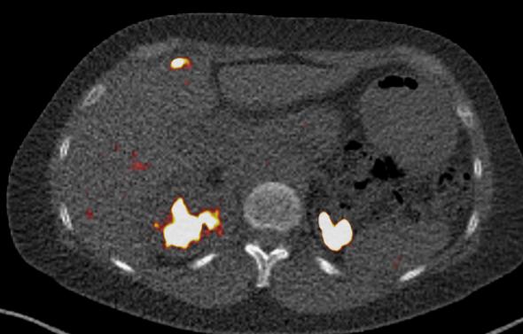
However, total-body PET scanners, which are up to 40 times more sensitive than conventional PET, allow endometriosis lesions to be visualised with greater resolution at a lower radiation dose. This enables the safer use of novel, disease-aligned radiotracers that can improve diagnostic accuracy.
Endometriosis is a highly heterogeneous disease, and its lesions involve distinct disease processes (i.e., bleeding and scarring), so different radiotracers are needed for accurate visualisation.
Advancing endometriosis research
At the Royal Infirmary of Edinburgh, a team led by Dr Lucy Whitaker at the University of Edinburgh is using a National PET Imaging Platform (NPIP) total-body PET scanner to advance research in this area.
The study, PET-Endo, is evaluating whether two novel radiotracers are effective when used with total-body PET imaging to uncover how bleeding and scarring processes vary across different types of endometriosis, throughout the menstrual cycle, and with different hormonal treatments. .
The PET-Endo study is investigating whether GP1 (a tracer for blood clots) and FAPI (a tracer for scar tissue) can better illuminate the distinct biological processes that make up endometriosis lesions.
If successful, this study is the first step in how total-body PET scanning could improve our understanding of endometriosis and uncover new targets for novel
treatments. It could also offer a faster, less invasive diagnostic pathway that could reduce unnecessary surgeries, improving care for patients with suspected endometriosis.
“The potential for total-body PET CT in endometriosis is significant. We urgently need effective non-invasive methods to reduce the diagnostic delay in endometriosis, which is now averages nearly a decade from first seeking help. The reduced radiation doses mean we can meaningfully use total-body PET imaging in reproductive-aged women and it might have particular use in identifying variants of endometriosis that are even more diagnostically challenging, such thoracic endometriosis.
It also has potential to allow us to unpick the natural history of the disease, understand how certain medical treatments work, and could have potential in the future to predict which treatments might be the most effective.”

Dr Lucy Whitaker, Senior Clinical Research Fellow and Honorary Consultant Gynaecologist, The University of Edinburgh.
Find out more about Dr Lucy Whitaker’s PET-Endo study here.
The University of Edinburgh
Figure 1. A total-body PET image with uptake in a region of diaphragmatic endometriosis.