Neuroscience
Case Study | Total-Body PET NPIP-CS004
The University of Edinburgh
Advancing stroke diagnosis with total-body PET imaging Stroke is one of the leading causes of death and disability worldwide. A stroke occurs when blood flow to the brain is suddenly blocked or reduced, most commonly as a result of a blood clot. Those who survive a first stroke face a significantly heightened risk of recurrence, making the identification of its underlying cause a critical priority. However, in over one-third of patients, the cause of stroke remains unknown despite extensive investigation.
How is stroke currently diagnosed? Total-body PET scanners offer a new approach to stroke diagnosis. They are 40x more sensitive than conventional PET scanners and are capable of imaging the whole body in a single scan. This enables clinicians to image active blood clots throughout the body, offering a comprehensive assessment that is not possible with current diagnostic techniques.
Advancing stroke research Researchers at the University of Edinburgh are using the National PET Imaging Platform (NPIP) to investigate how total-body PET can improve stroke diagnosis1. In the TORPIS study*, 100 patients with acute stroke underwent total-body PET imaging in addition to standard investigations. The study identified several previously unrecognised causes of stroke, and also reclassified the underlying cause of stroke in approximately one in four patients.
“With total-body PET, we’re moving from an inference-based approach to understanding the underlying mechanism of disease. If you’ve got a clot, where did it come from? The answer to that question can shape treatment and help clinicians make more informed decisions. By providing a wholebody view of the biological processes that contribute to disease, total-body PET is improving our understanding of complex conditions such as stroke and opening up new opportunities for better and more personalised care.” Professor David Newby, British Heart Foundation Duke of Edinburgh Chair of Cardiology, Institute for Neuroscience and Cardiovascular Research, University of Edinburgh
The TORPIS study uses the novel radiotracer [18F]-GP1,
a
radioactive
designed
to
detect
imaging
active
blood
agent clots.
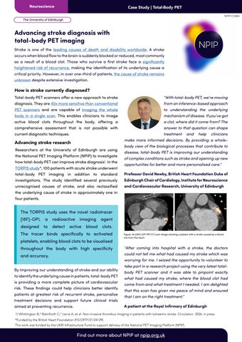
The tracer binds specifically to activated platelets, enabling blood clots to be visualised throughout the body with high specificity and accuracy. By improving our understanding of stroke and our ability to identify the underlying cause in patients, total-body PET is providing a more complete picture of cardiovascular risk. These findings could help clinicians better identify patients at greatest risk of recurrent stroke, personalise treatment decisions and support future clinical trials aimed at preventing recurrence.
Figure. An [18F]-GP1 PET/CT scan image showing a patient with a stroke caused by a blood clot from the heart.
“After coming into hospital with a stroke, the doctors could not tell me what had caused my stroke which was worrying for me. I seized the opportunity to volunteer to take part in a research project using the very latest totalbody PET scanner and it was able to pinpoint exactly what had caused my stroke, where the blood clot had come from and what treatment I needed. I am delighted that this scan has given me peace of mind and ensured that I am on the right treatment.” A patient at the Royal Infirmary of Edinburgh
1) Whittington B,* Balmforth C,* Levra A, et al. Non-invasive thrombus imaging in patients with ischaemic stroke. Circulation. 2026; in press. *Funded by the British Heart Foundation (FS/CRTF/21/24129). This work was funded by the UKRI Infrastructure Fund to support delivery of the National PET Imaging Platform (NPIP).
Find out more about NPIP at npip.org.uk