Development of a human reactive astrocyte platform for drug discovery
Introduction
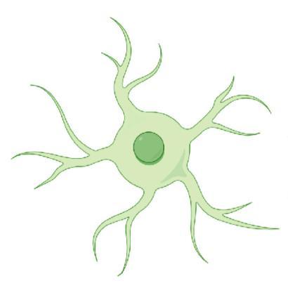
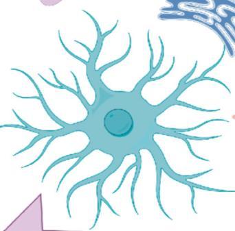
Astrocytes are one of the key glial types of the central nervous system (CNS) and play a critical role in maintaining brain homeostasis Following CNS injury or in disease, astrocytes undergo a transformation to become ‘reactive’ and change their morphology and molecular profile, adopting different phenotypic states to respond to pathological insults Reactive astrocytes participate in neuroinflammatory processes occurring in neurodegenerative diseases through secretion of neuro- and immune-modulatory factors Recently, an in vitro reactive astrocyte model was developed using a stimulation cocktail of TNF-α, IL-1α, and Complement 1q (‘TIC’), which drives a neurotoxic state ¹ TIC-induced astrocytes have been shown to secrete growth factors, chemokines and pro-inflammatory cytokines, as well as neurotoxic lipids ²
Multiparametric








LC-MS to identify changes in the lipidome of reactive astrocytes
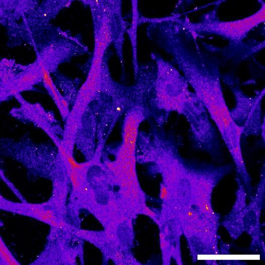
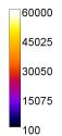
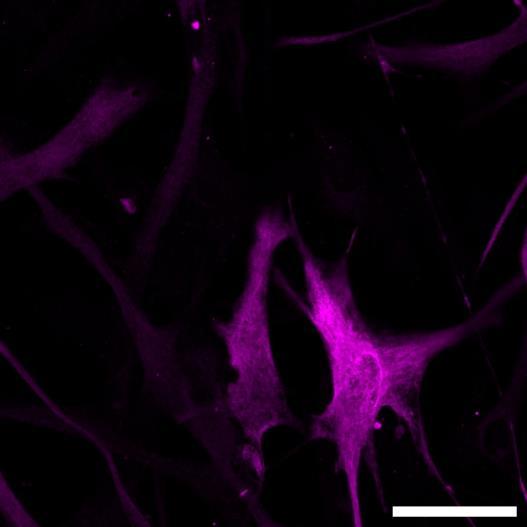
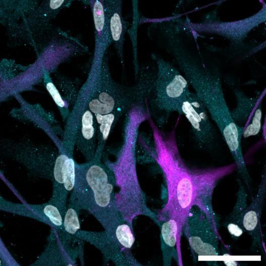
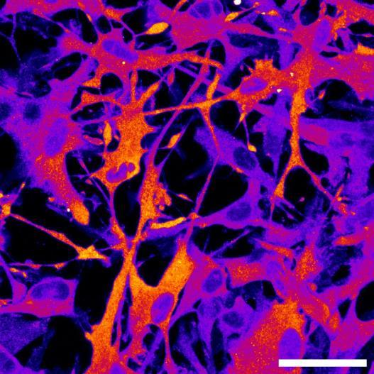
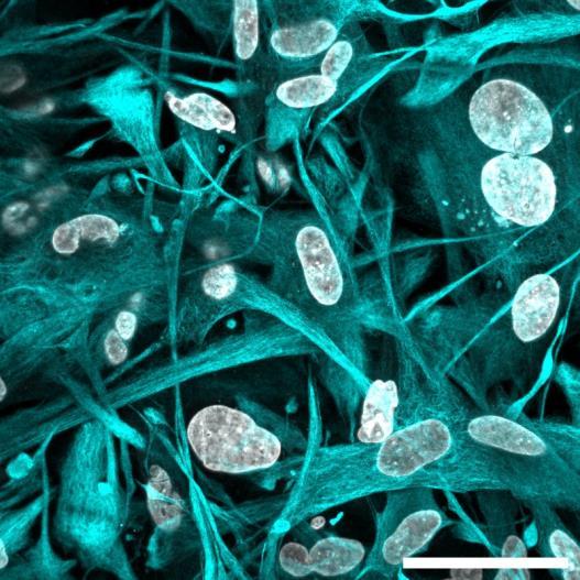
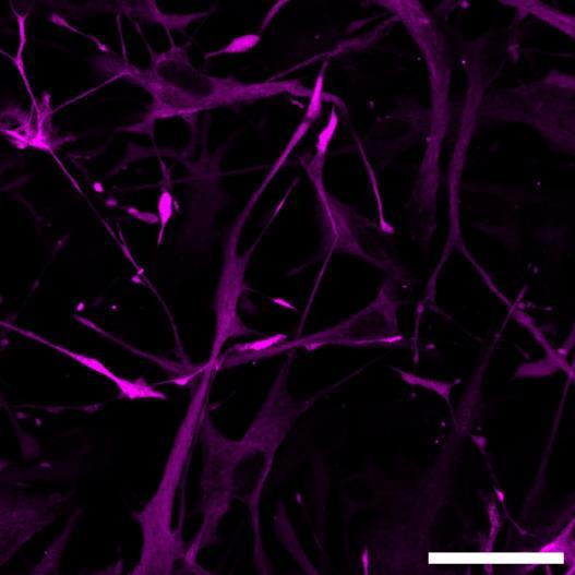
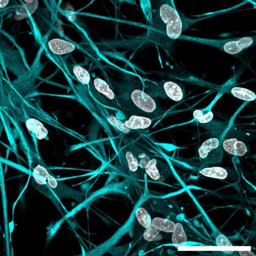
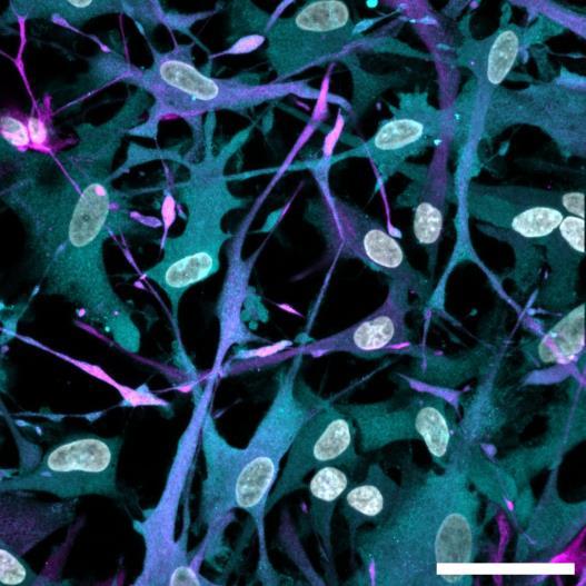
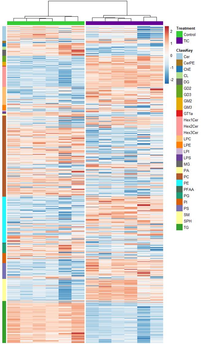
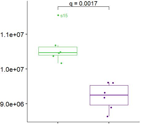
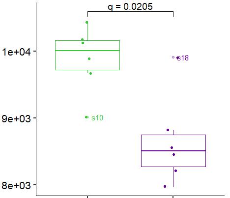
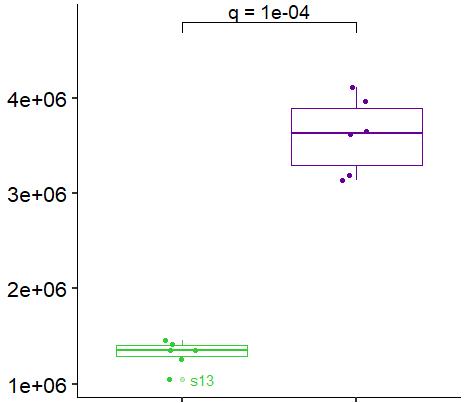
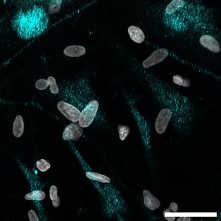
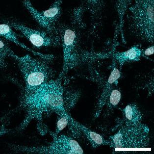
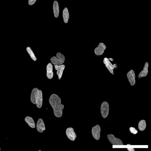
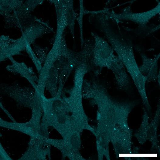
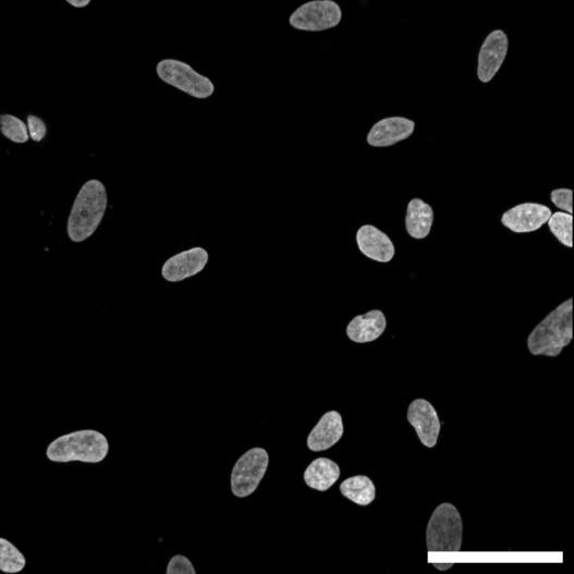
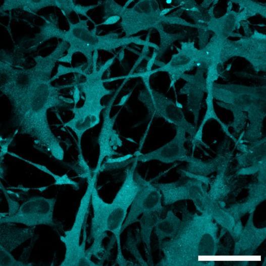
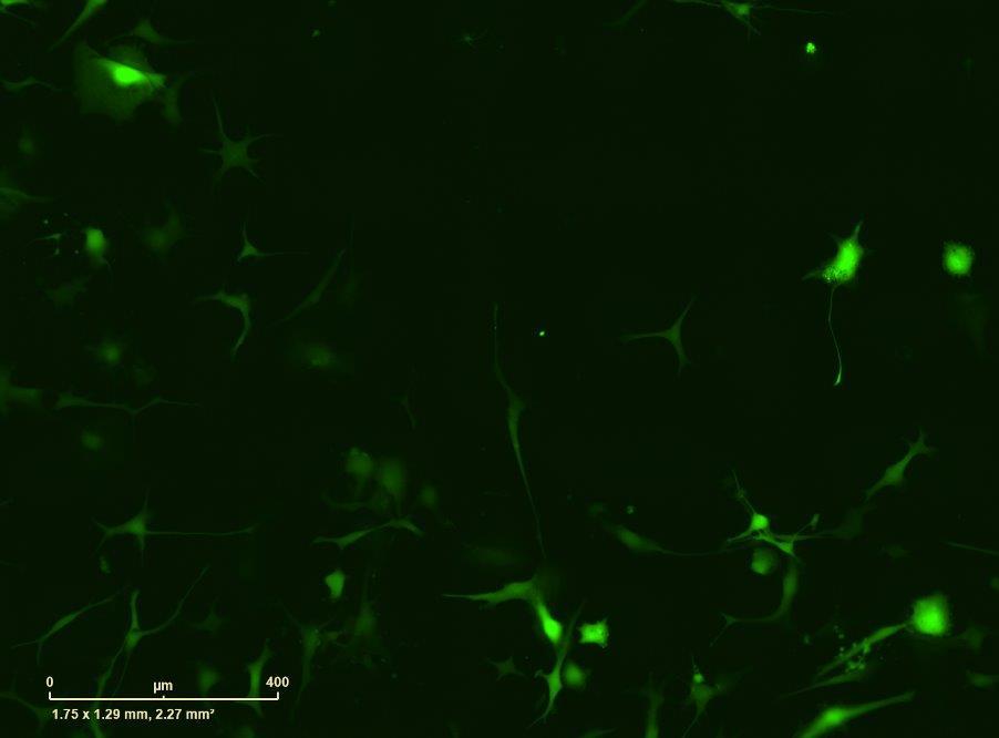
Fig 2 hiPSC-derived astrocytes upregulate GBP2, release of pro-inflammatory cytokines and growth factors and NFĸB signalling in response to TIC A) Representative images of hiPSC-derived astrocytes treated with vehicle or TIC and labelled for the nucleus (DAPI, grey), Guanylate-binding protein 2 (GBP2 cyan or FIRE) and the astrocytic marker glial fibrillary acidic protein (GFAP, magenta) and imaged using a Zeiss LSM 880 confocal microscope A dynamic look up table was used for GBP2 to highlight differences in intensities Scale bar is 200µm B) Quantification of the median GBP2 intensities in vehicle and TIC stimulated cells Confocal images were acquired with an Opera Phenix High Content Screening System (PerkinElmer) using 40x water immersion lens and image analysis was performed using Harmony 5 2 software N=3 C) Representative image of hiPSCderived astrocytes indicate a change in morphology for GFAP (cyan) under TIC conditions Scale bar is 50µm D) Interleukin-6 (IL-6) and CXCL10 levels are increased in the supernatant of TIC stimulated astrocytes (ELISA) N=3 E) Luminex data shows that chitinase 3like 1, interferon beta (IFN-β), C-C Motif Chemokine Ligand 2 (CCL2), C-C Motif Chemokine Ligand 5 (CCL5), granulocyte-macrophage colony-stimulating factor (GM-CSF), vascular cell adhesion molecule 1 (VCAM-1), complement component 3 (C3) and IL-6 are increased in the supernatant of TIC stimulated astrocytes N=3 F) Representative image of hiPSC-derived astrocytes labelled with DAPI (grey) and C3 (cyan) antibodies in control and TIC conditions Scale bar is 50µm G) Use of a lentiviral NFkB-GFP-Luciferase reporter in astrocyte reactivity model TIC induces an increase in NFĸB reporter activation Images were acquired and analysed using an Incucyte S3 Luminescence counts (ONE-Glo) are shown at 24 hours for control and TIC treated cells Statistical analyses were performed using a Student’s t test P-values are indicated where P<0 05
Results and conclusion
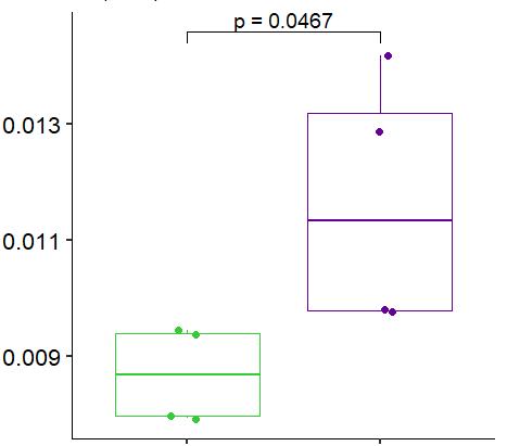
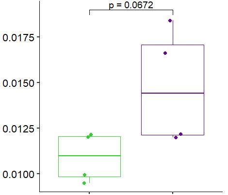
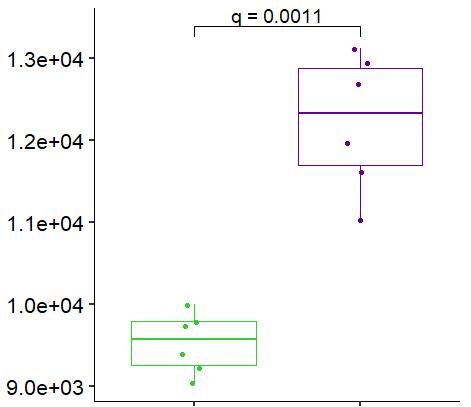
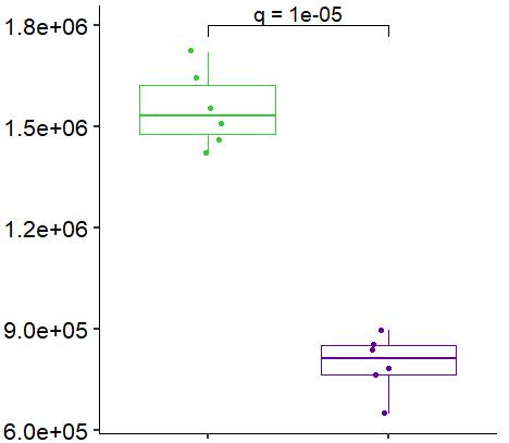
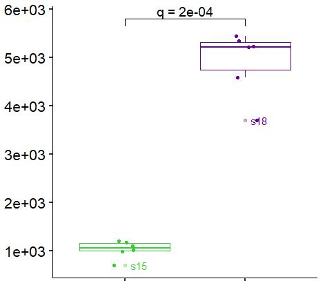
Fig. 3. hiPSC-derived astrocytes show perturbation to lipid metabolism upon TIC stimulation A) Heatmap shows data of 528 significantly perturbed (q<0 05 TIC vs Control) confidently annotated lipids measured in either positive or negative ionisation mode Redundancy was removed by selection of a single representative feature per lipid group (sum fatty acyl chains) B) Boxplots of representative PC, LPC, CL, TG, SM and Hex1Cer species N=6 samples ; P-values were calculated by t-test C) Long-chain fatty acids (FAs), palmitic (left) and stearic acid (right), are upregulated in the supernatant of TIC stimulated astrocytes N=4 samples ; P-values were calculated by t-test D+E) Summary of changes to lipid metabolism in reactive astrocytes Abbreviations: Ceramides (Cer), Ceramide phosphoethanolamine (CerPE), Cholesterol (CL), Cholesterol ester (ChE), Diacylglycerol (DG or DAG), disialoganglioside 2-3 (GD2-3), Ganglioside 2-3 (GM2-3), Trisialoganglioside 1a (GT1a), Hexosylceramide 1-3 (Hexcer1-3), Lysophosphatidylcholine (LPC), Lysophosphatidylethanolamine (LPE), Lysophosphatidylinositol (LPI), Lysophosphatidylserine (LPS), Monoacylglycerol (MG), Phosphatidic acid (PA), Phosphatidylcholine (PC), Phosphatidylethanolamine (PE), Arachnoid acid (AA), Phosphatidylglycerol (PG), Phosphatidylinositol (PI), Phosphatidylserine (PS), Sphingomyelin (SM), Sphingosine (SPH), Triglyceride (TG or
(CDP)
and growth factors and changes in morphological features including GBP2 expression In addition, using mass spectrometry we show that the lipid profile is significantly altered in TIC-stimulated astrocytes, including the secretion of saturated free fatty acids Together these innovative tools provide a platform to study reactive astrocytes, and their contribution to neuroinflammation, and to assist with development of novel therapeutics to target astrogliosis in disease