Biomarker Profiling of Radiation and DDR Inhibitor Combinations in LNCaPXenografts
Kerry Shea, Anita Liu, Prabhakar Panday, Chiara Da Pieve, Niaz Najafi, Juliana Maynard, Benedetta Arno, TJ Allen, Fiona Yau, Chris Thornton, Liviu Lucaciu, Andrzej Rutkowski, Mikaela Griffiths, Bruno Bellina, Tara Bowen, Gayle Marshall
INTRODUCTION
Ionising radiation induces DNA damage primarily through the formation of single and double strand breaks which, if unrepaired, can lead to cell death These breaks activate the DNA damage response (DDR) which is a network of cellular pathways that detect and repair any DNA damage and includes the phosphorylation of proteins like H2AX and DNA-PK When damage is extensive or incorrectly repaired, this can result in genomic instability, apoptosis, or senescence In drug discovery, this mechanism can be exploited by combining targeted delivery of ionising radiation to cancer cells with inhibition of key DDR proteins, thereby preventing repair and promoting tumour cell death
Biomarkers of radiosensitivity and DNA damage are therefore critical for both preclinical and clinical evaluation of novel combination therapies
Here, we describe the optimisation of a panel of biomarker assays in an LNCaP xenograft model to assess the effects of ionising radiation, DNA-PK inhibition (AZD7648), and their combination Using multimodal imaging alongside ex vivo analyses, including γH2AX in tumour tissue and circulating nucleated blood cells, circulating tumour DNA (ctDNA) fragmentation, and plasma metabolomics, we show that DNA-PK inhibition enhances and prolongs radiation-induced DNA damage
Although this study utilised external beam ionising radiation, which delivers a localised high dose over a short period, the assays developed are equally applicable to radioligand therapy, where radiation is delivered at lower dose rates over extended periods via tumour-targeting biomarkers such as PSMA
Despite differences in delivery and kinetics, both modalities activate DDR pathways, including γH2AX signalling, and offer opportunities for combination with DDR inhibitors
Together, these findings highlight the translational potential of DDR inhibitor combinations with both external beam radiotherapy and radioligand therapy and support the integration of biomarker strategies to inform therapeutic development and clinical application
IN VIVO IMAGINGAND SAMPLING γH2AX PROFILING
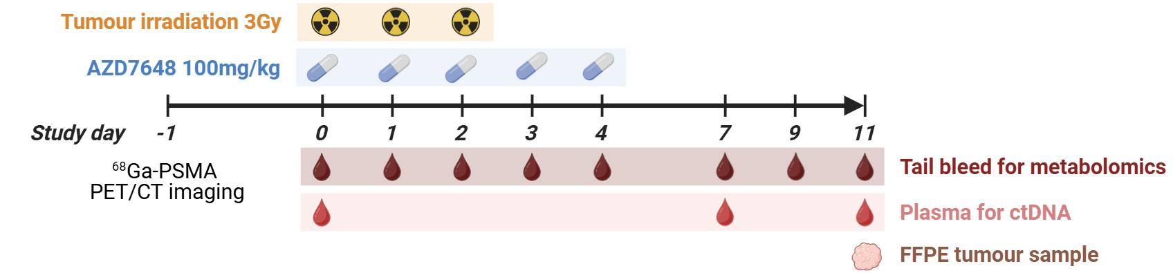
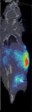
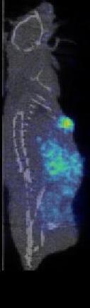
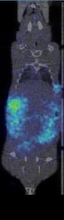
LNCaP xenografts were established through the subcutaneous implantation of cells into the rear flank of immunocompromised nude mice where tumour volume was monitored until reaching a minimal size 68Ga-labelled PSMA-617 was used to image PSMA-expressing LNCaP tumours in vivo on Study Day -1 via a 20min static PET scan followed by a CT scan Animals were assigned to one of 4 groups: 1 Untreated; 2 Radiotherapy Alone (Fractionated dose of 3 times 3Gy over 3 days); 3) DNA-PKi Alone (100mg/kg AZD7648 PO 1-2 hours prior to Radiation- Daily over 5 days); 4) Radiotherapy + DNA-PKi
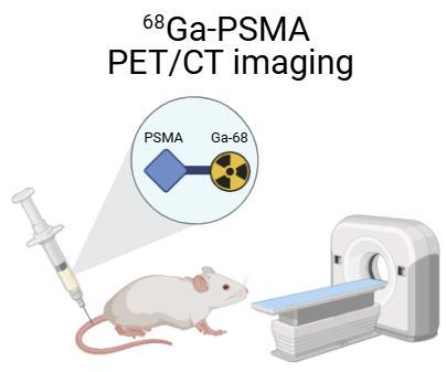

T T Representative PET/ CT images of LNCaP tumours.
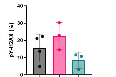
• This assay was validated using whole blood from 3 groups of LNCaP tumour bearing mice with 3-4 animals per group.
• Vehicle treated
• Single 3Gy dose of ionising radiation (IR)
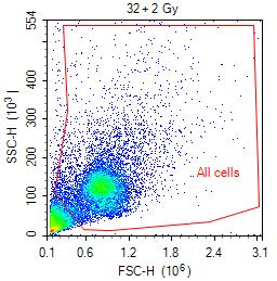
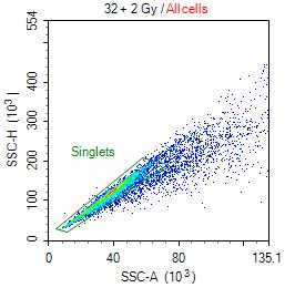
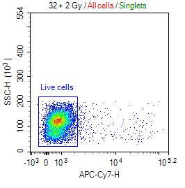
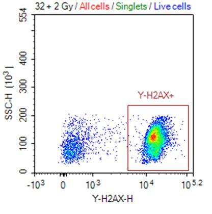
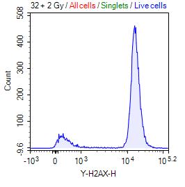
• Single dose of 100mg/kg AZD7648 1 hour prior to 3Gy IR. Whole blood was collected 1–2 hours post IR, red blood cells lysed, a viability stain applied to distinguish live and dead cells followed by fixation in PFA.
• Detection of γH2AX by intracellular flow cytometry demonstrated an increase in γH2AX phosphorylation following IR, which was reduced upon DNA-PK inhibition.
Tumour Tissue
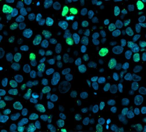
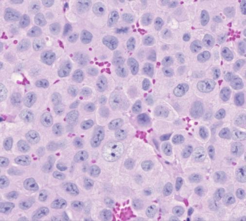
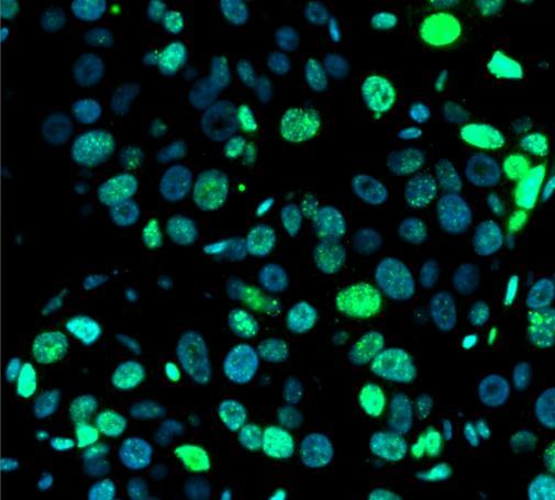
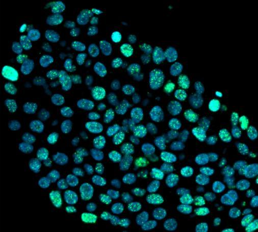
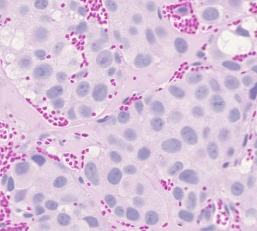
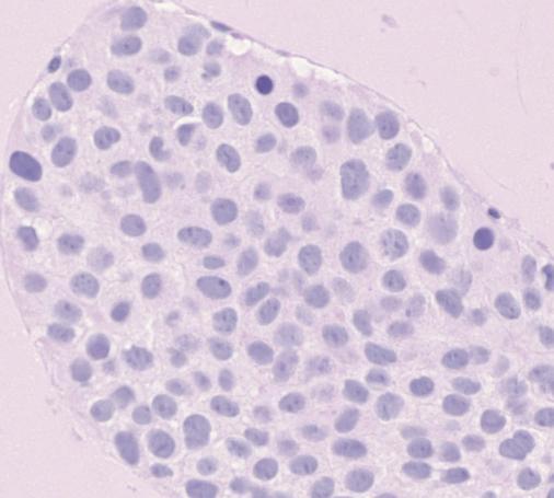
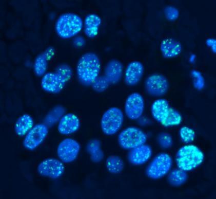
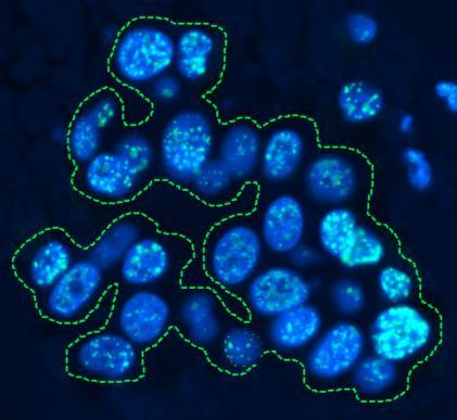
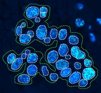
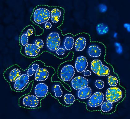
• An immunofluorescence assay was optimised to detect and localise γH2AX in LNCaP xenograft tumour tissue, using the same cohort of LNCaP tumour-bearing mice as in the whole blood assay.
• The tissue was processed into FFPE blocks, sectioned, and incubated with a primary antibody targeting γH2AX (Ser139), followed by a fluorescently labelled secondary antibody for visualisation.
• Quantification of γH2AX staining was performed using Visiopharm software, measuring the percentage area of γH2AX within the nuclei of tumour-enriched regions as well as across the entire tumour area.
• Both metrics produced consistent results and mirrored those observed in the whole blood assay, showing an increase in γH2AX phosphorylation following IR that was again reduced with DNA-PK inhibition.

CIRCULATING TUMOUR DNAASSAY
• Digital PCR assays that measure short (~50bp) and long (~150bp) DNA fragments from 0.5μL of plasma were developed with DNA derived from either human or mouse species.
• The ratio of the short fragments to the long fragments can be used as a proxy for the extent of DNA fragmentation which is indicative of radiation induced DNA double strand breaks.
• Using DNase digestion of circulating free DNA (cfDNA) we have demonstrated that the assay can detect DNA fragmentation from the 2 species. The human and mouse assays showed no cross-reactivity (data not shown).

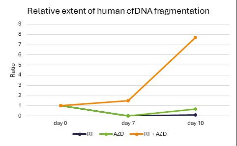
Longitudinal plasma samples were assayed for the fragmentation of xenograft derived human DNA cfDNA using the digital PCR assays described. The fragmentation of human DNA was determined as a ratio to total mouse DNA fragmentation and normalised to the cohort of untreated animals. There was a marked increase in the fragmentation of human cfDNA relative to mouse cfDNA in the IR + DNA-PKi combination treatment group, but not in groups receiving only IR or DNA-PKi monotherapy.
WHOLE BLOOD METABOLOMICSASSAY
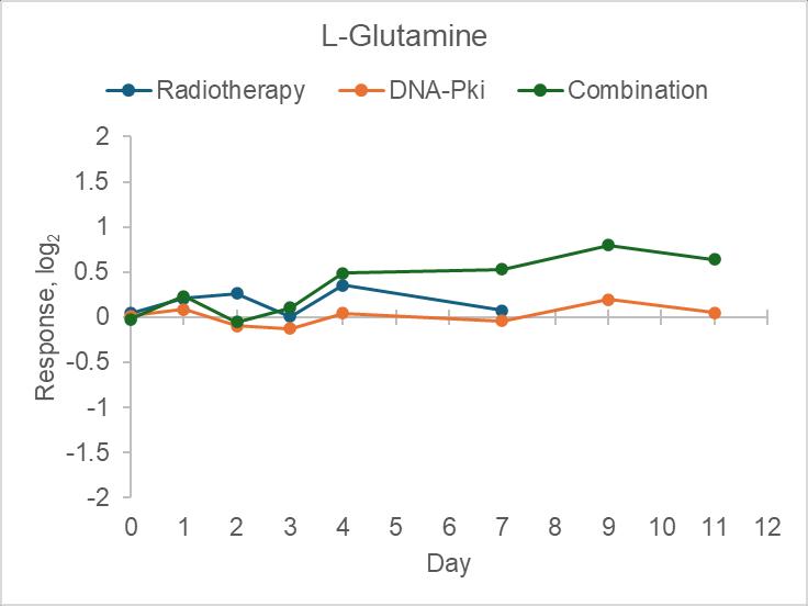
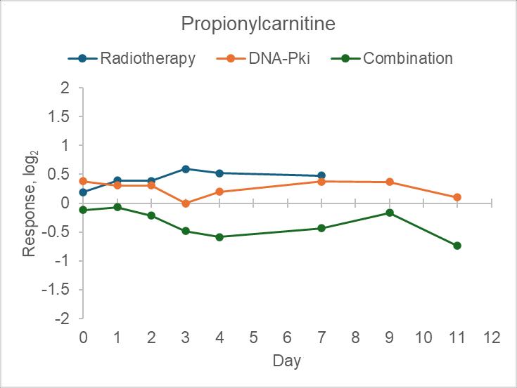
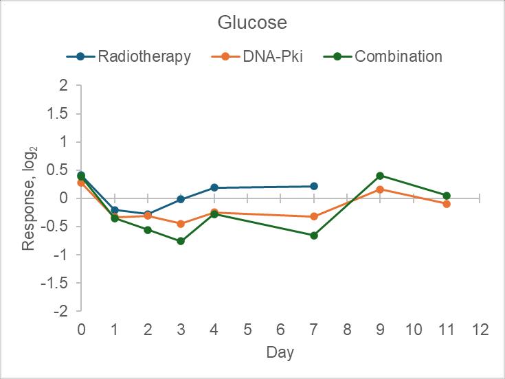
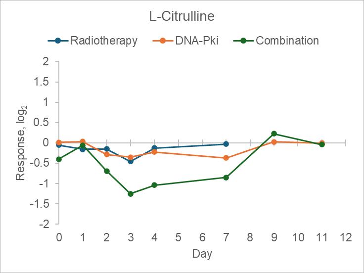
• Whole blood microsampling for mass spectrometry analysis used 5µL tail vein whole blood for simultaneous measurement of metabolomics and pharmacokinetics
• AZD6748 was simultaneously measured from the same sample injections, enabling tracing of pharmacokinetic profiles over the whole study duration (data not shown)
• Profiling of >2000 small molecule metabolites and polar lipids allows longitudinal tracking of global health status/toxicity and treatment-induced effects over the whole study duration