Savings End Sept. 9 Register Now Where All of Ophthalmology Meets® aao.org/register
Four Days. One Unmissable Meeting.
“This is my favorite meeting of the year — by far. It’s something I make sure not to miss.”
Erica Alvarez-Gonzalez, MD
Programming* begins Friday morning, including Subspecialty Day, AAOE®, and AAOP™. Starting Saturday, explore the Expo and enjoy Saturday night’s Illuminate* celebration. Stay through Monday’s Cataract Spotlight to experience all that AAO 2026 has to offer, Oct. 9–12, in New Orleans
The full program is now available in the Meeting Guide. Plan your schedule at aao.org/mtg-guide.
*Select programs and events require tickets or separate registration.
Left: Sila Bal, MD | YO Committee Chair and Global Education and Outreach Committee Member
Right: Erica Alvarez-Gonzalez, MD | YO Advocacy and Patient Education Committee Member
But Retina Rumble , this issue’s theme, isn’t about a fight. It’s more about laying experts’ cards on the table, unpacking the evidence (even the egos!) and coming up with solutions—with real-world impact—behind today’s most contested retinal decisions.
This issue’s Cover Story—The Invisible Laser (page 24), explores the ongoing “controversy” regarding the effectiveness of subthreshold micropulse laser (SML), particularly for diabetic macular edema (DME). SML’s premise is its invisibility—it treats the retina without leaving a visible burn, scar or mark versus the conventional photocoagulation. The debate centers on whether SML is a “breakthrough hiding in plain sight or a beautifully engineered placebo.”
Under the Posterior Segment section, the stubborn proliferative vitreoretinopathy (PVR) is in the spotlight. To Inject or Not to Inject? (page 8) summarizes the ongoing debate over whether methotrexate (MTX) should be routinely injected to prevent PVR, a major cause of recurrent surgical failure in retinal detachment repair and any surgeon’s nightmare. The debate concluded that while MTX holds biological promise, it remains in “experimental status”.
Casting Shadows (page 13) explores the ongoing professional debate over when pars plana vitrectomy (PPV) is justified for vision-degrading myodesopsia (VDM, or floaters), centering on the tension between a patient’s profound subjective distress and the measurable, yet sometimes minor-appearing, vitreous opacity. The consensus path forward from our contributors is a narrow middle ground...
Under Innovation, artificial intelligence (AI) is at it again, front and center. The Surgical Copilot (page 20) discusses the growing integration of AI into vitreoretinal surgery, transitioning it from a diagnostic tool to an active surgical assistant. While AI makes the operating room “computationally aware,” the discussion also highlights critical challenges, including concerns over patient trust and the algorithm’s vulnerability to bias. However, the bottomline is that for now, the human surgeon remains firmly in control.
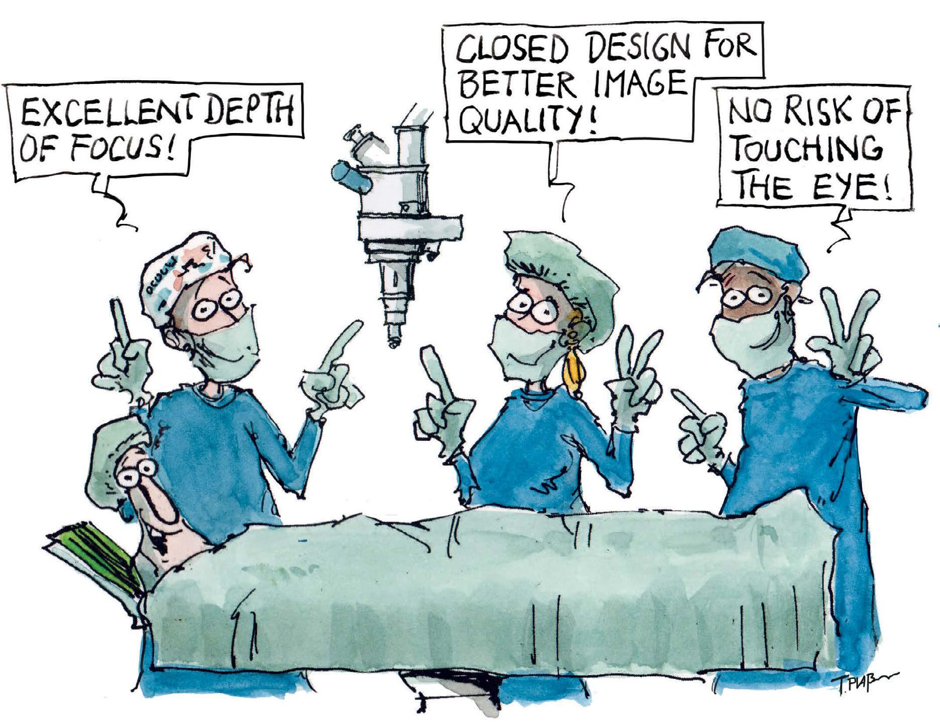
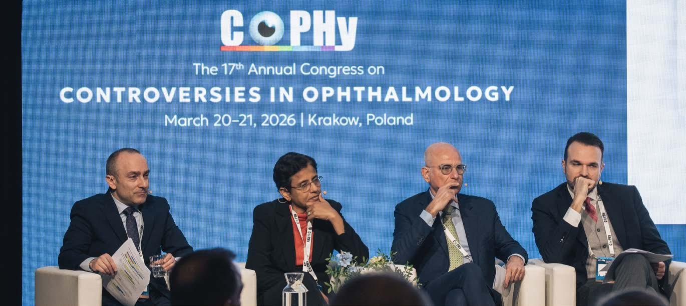
Heads Up, Seven Up (page 22) summarizes a contentious debate at COPHy 2026 earlier this year on whether heads-up display (HUD) vitreoretinal surgery has significantly improved patient outcomes. While some experts agree that HUD technology offers genuinely superior image quality, improved ergonomics for surgeons by reducing neck strain, and a distinct advantage in surgical education for trainees, they noted that the data on clinical outcomes remains “stubbornly inconclusive”. Read to find out more.
In the Enlightenment section, The Art of the Flap (page 34) discusses the limitations of standard macular hole surgery. Our esteemed contributors noted that while internal limiting membrane (ILM) peeling often achieves anatomical closure, it frequently fails to restore the foveal layers necessary for sharp vision. The inverted ILM flap technique was proposed as an improvement, especially for large, myopic, chronic and refractory holes—thereby significantly improving closure rates and postoperative
visual acuity compared to standard peeling—with high success rates in difficult cases and continued visual improvement even 10 years post-surgery.
On the other hand, The Membrane Wars (page 32) discusses the ongoing controversy among retina surgeons regarding whether the ILM should be peeled after removing the epiretinal membrane (ERM) during surgery for symptomatic macular puckers. While evidence presented at COPHy 2026 suggests ILM peeling reduces reoperation rates, meta-analyses and preferred practice reports have found it does not consistently improve final visual outcomes, leading many surgeons to base the decision on individual patient factors and surgical judgment.
Last but not least, this issue’s Women in Ophthalmology (Beyond a Family Name, page 30) features Dr. Audina Berrocal, a distinguished pediatric retina expert who moved beyond her family’s medical legacy to forge her own unique path in ophthalmology. Today she has become a global leader in her field. While she has pioneered innovative therapies, her enduring legacy is not only defined by that, but also by the profound human impact of her work—seen in both the children whose sight she has preserved and the many surgeons she has trained.
Thank you for reading, and we hope that you find this issue valuable to your practice!
Sincerely,
Gloria D. Gamat Chief Editor | PIE, CAKE and COOKIE Head, Editorial Department | Media MICE
When floaters justify the knife 08 10 13 18
To Inject or Not to Inject
From Trial Evidence to Clinical Confidence with Aflibercept 8mg The methotrexate debate in proliferative vitreoretinal disease
The data says you can extend. The question is: will you?
Shadows
Surgical Copilot Heads Up, Seven Up
MEET THE TEAM AT
APVRS | Aug 28–30, 26 | Booth 40/41
EURETINA | Oct 1–4, 26 | Booth C55
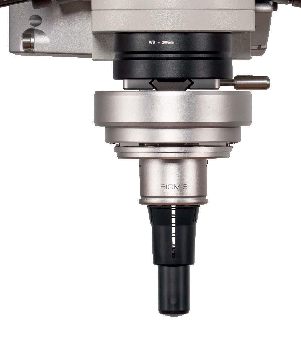
Evolved from the Original. Built for the Future.
The all-new BIOM® 6 with its innovative closed design –crafted to improve precision, elevate performance, and meet the demands of advanced vitreoretinal surgery.
Optimized Depth of View
Minimized Light Disturbance
Improved Surgical Control
ADVISORY BOARD MEMBERS
DR. ALAY S. BANKER
Banker’s Retina Clinic and Laser Centre Ahmedabad, India alay.banker@gmail.com
PROF. GEMMY CHEUNG
Singapore National Eye Centre Singapore gemmy.cheung.c.m@singhealth.com.sg
DR. ARSHAD KHANANI
Sierra Eye Associates; University of Nevada, Reno School of Medicine Nevada, USA arshad.khanani@gmail.com
THE METHOTREXATE DEBATE IN PROLIFERATIVE VITREORETINAL DISEASE
In retina surgery, few enemies return as stubbornly as PVR. MTX may be changing the conversation, but not everyone is ready to call it a breakthrough.
By Tan Sher Lynn
Despite major advances in retinal detachment surgery, proliferative vitreoretinopathy (PVR) remains one of the field’s most stubborn adversaries. Characterized by fibrocellular membrane growth that can re-detach the retina after an otherwise successful repair, PVR continues to drive surgical failure and recurrent detachment worldwide…and surgeons are still searching for a reliable way to stop it.
Methotrexate (MTX) has emerged in recent years as a promising adjunctive therapy, but enthusiasm has been tempered by uncertainty. At the 17th Annual Congress on Controversies in Ophthalmology (COPHy 2026), Polish retinal specialists Prof. Zofia Anna Nawrocka and Prof. Jerzy Mackiewicz squared off over a deceptively simple question: should MTX be routinely injected to prevent PVR, or does the evidence remain too thin to justify widespread adoption?
THE CASE FOR
Prof. Nawrocka opened by framing the stakes. PVR accounts for up to 50% of re-detachments in unsuccessful surgeries, and patients with advanced stage C disease face particularly grim odds: recurrent detachment, irreversible vision loss and surgical options that are themselves far from benign. She cautioned that retinotomy is “not free of challenges,” with postoperative hypotony a significant concern.
Against this backdrop, she argued that MTX offers a biologically rational way to interrupt the fibroproliferative cascade underpinning PVR.
The condition begins when retinal pigment epithelial (RPE) cells migrate into the vitreous cavity and undergo epithelial–mesenchymal transition, while inflammatory mediators recruit macrophages and trigger oxidative stress and growth factor activation.
Methotrexate targets this process on two fronts. “[It] inhibits DNA synthesis of actively dividing cells,” Prof. Nawrocka explained, adding that it also “modulates pro-inflammatory cytokines.” Combined with its long track record in ocular lymphoma and its low cost, this dual mechanism makes MTX an attractive adjunct.
Critically, she argued that dosing duration sets MTX apart from agents that have failed before it. Many previously investigated compounds were administered just once during surgery— a strategy she found hard to defend given the disease’s timeline. “PVR develops in the first four months,” she said. “Using a drug only once makes really no sense.”
The GUARD study protocol addressed this directly, employing eight weekly intravitreal injections followed by fortnightly injections through week 16— a schedule designed to match the drug’s
short intraocular half-life and sustain suppression throughout the critical postoperative window.1
“The GUARD study found that epiretinal membranes are observed less frequently in methotrexate-treated eyes compared to routine care,” Prof. Nawrocka noted.
Her own real-world data added weight to the argument. In a series of 34 eyes with severe stage C PVR treated with MTX for four months post-surgery, visual acuity gains were limited by disease severity, but anatomically, the results were striking. “For me, the most important data…is that in none of the eyes I observed hypotony,” she said.
She also described cases in which persistent proliferative membranes gradually regressed with serial injections, sparing patients from further surgery. “With the following methotrexate injections, the membrane shortens and finally disappears,” she noted.
Corneal toxicity, she acknowledged, is a real concern, but one that typically resolves after a brief treatment pause.
Her bottom line was unambiguous: “Methotrexate administered intravitreally seems to improve anatomical success.”
THE CASE AGAINST
Prof. Mackiewicz did not dispute the biological rationale. “The mechanism of action of methotrexate differs from classical drugs, making it theoretically attractive for PVR prevention,” he said.
The trouble is what comes after the theory. “The most important objection is the lack of convincing evidence from large, highquality clinical trials that methotrexate actually prevents PVR.”
He pointed to a recent meta-analysis evaluating the available data, which identified only three randomized controlled trials and five non-randomized studies involving just over 400 eyes. 2 The findings showed “a trend toward lower
recurrence rate,” but crucially, “there was no statistical significance.”
Visual outcomes told the same story. 2 “There was also no significant bestcorrected visual acuity difference between methotrexate and control groups,” he said.
Beyond the numbers, the heterogeneity of existing protocols makes meaningful synthesis difficult. Dosing regimens range from a single intraoperative low-dose injection mixed with silicone oil to the intensive 13-injection GUARD schedule. “We have…many different protocols,” he noted—a diversity that complicates any attempt to draw firm conclusions.
Prof. Mackiewicz also raised methodological concerns about the GUARD study specifically. Originally designed to randomize patients with recurrent PVR to MTX or placebo, the protocol was modified after investigators grew reluctant to withhold treatment from the control group given concerns about prognosis. All patients ultimately received MTX, and outcomes were compared against a historical control cohort1, a design that introduces selection and temporal bias.
“This modification of protocols, although ethical and understandable, weakens the scientific strength of evidence,” he said. “Comparison with historical data does not replace true randomization.”
He added that the available data remain preliminary: the full peer-reviewed Phase III publication has not yet been released, meaning the evidence base still rests largely on interim analyses and conference reports.
PRACTICAL AND SAFETY CONSIDERATIONS
Safety and logistics compound the evidentiary concerns. MTX’s intravitreal half-life of approximately six to eight hours demands repeated injections sustained over months—a regimen that may be prohibitive for older patients or those far from specialist centers.
Each additional injection also enhances procedural risk. “In this scheme with many injections, the risk is accumulated,” Prof. Mackiewicz warned, citing endophthalmitis, hemorrhage and iatrogenic retinal injury as real considerations.
His conclusion was measured rather than dismissive. “We really need large, rigorous randomized controlled studies,” he said, placing MTX firmly in the “experimental status” category for now.
The audience appeared to agree: few surgeons reported using it routinely in practice.
A DEBATE STILL UNRESOLVED
The debate ended, as good debates do, without a verdict. MTX has biology on its side and some encouraging early signals, but as Prof. Mackiewicz put it, promise alone is not enough to rewrite the standard of care. The field is watching, and the jury remains out.
Editor’s Note The 17th Annual Congress on Controversies in Ophthalmology (COPHy 2026) was held on March 20–21 in Krakow, Poland. Reporting for this story took place during the event.
REFERENCES
1. Aldeyra Therapeutics, Inc . Top-Line Results from Part 1 of the Phase 3 GUARD Trial of ADX-2191 in Proliferative Vitreoretinopathy. October 6, 2022. Available at: https:// ir.aldeyra.com/static-files/cde1aa20d22a-4f38-a9a5-523b803aca40. Accessed on May 20, 2026.
2. Kocak I, Sayin N, Bayramoglu SE, Thalib L. Effect of methotrexate in preventing retinal detachment after proliferative vitreoretinopathy surgery: A systematic review and meta-analysis. Surv Ophthalmol. 2026;71(2): 572-581.
THE DATA SAYS YOU CAN EXTEND. THE QUESTION IS: WILL YOU?
At COPHy 2026, aflibercept 8 mg sparked a lively rethink of just how far retina specialists should go.
Sponsored by Bayer
At the 17th Annual Congress on Controversies in Ophthalmology (COPHy 2026), debates are very much part of the program. But every now and then, the conversation shifts from if something works to how far clinicians are willing to take it.
In a Bayer-sponsored symposium, the spotlight was firmly on aflibercept 8 mg and how to use it in real-world practice. Extend earlier? Switch more decisively? Push intervals further than comfort traditionally allows?
Across debates, case studies and compelling real-world examples, a panel of retina heavyweights unpacked what higher-dose anti-vascular endothelial growth factor (VEGF) means in 2026: rethinking treatment ceilings, challenging cautious habits and, perhaps most importantly, building the confidence to act on what the data is already telling us.
RVO GETS THE GREEN LIGHT
Dr. Marco Lupidi (Italy) kicked things off with fresh data from the QUASAR study on retinal vein occlusion (RVO).1
The Phase III trial compared aflibercept 8 mg (with three loading doses) against aflibercept 2 mg monthly through week 32, before both arms transitioned into treat-and-extend.
By week 36, the 8 mg arm had already ticked the non-inferiority box. By week 64, patients were achieving gains of more than three lines of vision, with roughly 8.5 injections versus 12 in the 2 mg arm.1
“This is a major event,” Dr. Lupidi noted. “Coming down from five to three loading doses while maintaining outcomes—that simplifies everything.”
And simplify it does. By week 64, nearly 40% of patients on aflibercept 8 mg reached q20 intervals, while around 56% achieved intervals of ≥ q16.1
Dr. Lasse Jørgensen Cehofski (Denmark) added a real-world twist: switching his entire RVO cohort to aflibercept 8 mg overnight cut the proportion of patients on short intervals by more than half. “You have to be very proactive when you extend,” he advised. “Patients you couldn’t extend before—suddenly you can extend them for 12 weeks.”
EARLY DRYING, DURABILITY… OR JUST KEEPING IT SIMPLE?
Prof. Sobha Sivaprasad (United Kingdom) stepped up first, arguing for early drying. She highlighted that aflibercept 8 mg delivers a four-fold higher molar dose, with 34% lower ocular clearance and an extra six weeks of VEGF suppression compared to aflibercept 2 mg. 2
“This tells us why rapid drying matters,” she said. “It’s almost a biomarker for how to extend patients.”
To prove her point, she shared a case: a 91-year-old with retinal angiomatous proliferation (RAP) lesions who had endured 10 faricimab injections with
persistent subretinal fluid. Upon switching to aflibercept 8 mg, the patient achieved complete drying by the third injection.
Taking the mic, Prof. Anat Loewenstein (Israel) made the case for extended durability with a story that had everyone leaning in.
A 50-year-old man with choroidal neovascularization (CNV), whose father had suffered a vasovagal seizure during retinal treatment, experienced a major seizure during his very first aflibercept 2 mg injection. Subsequent treatments required a full hospital theater setup, anesthesiologist included. After switching to aflibercept 8 mg, he remained fluid-free at 24 weeks after just two injections.
She backed it up with long-term data: at 156 weeks, 62% of neovascular agerelated macular degeneration (nAMD) patients reached ≥ 4-month intervals, 43% reached ≥5 months, and 24% stretched to ≥6 months.3
Then came Prof. Paolo Lanzetta (Italy), championing simple, flexible dosing. “What I like from PULSAR and PHOTON is that they’re very close to daily practice,” he said.
In his playbook, treatment-naive patients can extend straight to q16 after three loading doses, with stepwise extension up to q24 possible per recent label
updates. Previously treated patients with suboptimal response? Three loading doses, then into treat-and-extend.
He also slipped in a sustainability angle: switching from aflibercept 2 mg to 8 mg cut carbon emissions by 35% over two years in his group’s data.4
TREATMENT-NAIVE VS. PREVIOUSLY TREATED
If the first debate was about how to use aflibercept 8 mg, this one tackled the inevitable follow-up: who benefits most?
Dr. Lupidi opened with the treatmentnaive group, showcasing a diabetic macular edema (DME) patient. After three loading doses and progressive interval extension, the patient ultimately reached a q24 treatment interval. By day 90, he had complete fluid resolution alongside a diabetic retinopathy improvement from level 47 to 35. A clean, textbook response.
On the other side, Prof. Sivaprasad made a compelling case for previously treated patients. She presented a patient with a large pigment epithelial detachment (PED) who had already undergone seven aflibercept 2 mg injections, with stubborn exudation still hanging around. After the switch to aflibercept 8 mg, the PED height dropped dramatically and the fluid finally cleared.
Then Prof. Lanzetta took a diplomatic stance: why choose? Drawing from SPECTRUM, a Phase IV real-world study with around 4,000 patients currently enrolled across multiple countries, he presented early findings from the first approximately 100 treatment-naive and 100 previously treated nAMD patients. At week 8, treatment-naive patients delivered the expected visual gains, while previously treated patients stayed stable with improvements in central retinal thickness. At this early timepoint, no cases of intraocular inflammation were reported in either cohort.5
THE FUTURE BEYOND ANTI-VEGF
Prof. Lanzetta closed with a forwardlooking sweep of what’s coming next. Bayer’s pipeline is clearly casting a wide net across the retinal disease spectrum.
MACUSTAR is working to validate new clinical endpoints for intermediate AMD trials, anchored by a six-year, multicenter European cohort of more than 600 intermediate AMD patients. 6-7 Meanwhile, CHART is digging into biomarkers that could help predict progression in non-proliferative diabetic retinopathy. 8-9
On the collaboration front, BRIDGE is doing exactly what its name promises, bringing industry and academia together to support more data-driven conversations with regulators. 10 And then there’s RetinAI Discovery, Bayer’s push into digital and AI-powered tools to sharpen how clinicians diagnose and manage retinal disease.11
Finally, for those watching the regenerative space, CLARICO is one to keep an eye on. This Phase I/IIa first-in-human study is evaluating OpCT-001, an induced pluripotent stem cell (iPSC)-derived photoreceptor precursor therapy, for adults with primary photoreceptor diseases, including retinitis pigmentosa, Usher syndrome and cone-rod dystrophy.12
PRACTICAL IMPLEMENTATION
Theory is great, but sooner or later, every retina specialist wants to know: how does this actually play out in the clinic on a busy Tuesday afternoon?
When Prof. Loewenstein put that exact question to the panel, Prof. Lanzetta kept it refreshingly straightforward. Start with three loading doses, then reassess. He noted growing confidence in extending treatment-naive patients to at least 12 weeks post-loading, while previously
treated patients with suboptimal response still go through a full loading phase before entering treat-and-extend.
Dr. Cehofski, meanwhile, was firmly in the “push it further” camp. “We’re too cautious. If the macula is dry, extend to 8 weeks,” he said. “I have approximately 300 eyes that were treatment-naive in my clinic that were initiated on aflibercept 8 mg. My experience is that we can push intervals much further than we think.”
He also underscored the importance of staying the course after loading.
“Approximately half of my patients gained ten letters or more after loading, but macular edema is almost always recurrent despite higher doses,” he shared. “Don’t pause treatment. You’ll lose your long intervals.”
Rounding it out, Prof. Sivaprasad shared her approach to identifying candidates for a switch from 2 mg to 8 mg:
• Stable patients on four-weekly intervals who might benefit most from extended intervals
REFERENCES
• Those with persistent fluid despite treatment, and
• Patients with large PEDs or fibrosis
WHY
CONFIDENCE MATTERS
Because at some point, it’s not about what the data says. It’s about what clinicians are willing to do with it. The evidence behind aflibercept 8 mg is solid. The
permission to extend more boldly, practical strategies for switching established patients, and a bit of reassurance that realworld OCTs would behave like the trial datasets promised.
That’s what confidence really looks like. Not just knowing the evidence, but trusting it enough to act on it.
label offers room to move. The delivery system? Streamlined enough to keep clinic flow intact.
What clinicians weren’t short on was data. What they’re looking for was a nudge:
Editor’s Note
The 17th Annual Congress on Controversies in Ophthalmology (COPHy 2026) was held on March 20–21 in Krakow, Poland. Reporting for this story took place during the event.
PP-EYL_8mg-ALL-0471-1 | May 2026
1. Chaudhary V , et al. Aflibercept 8 mg in macular edema following retinal vein occlusion: Week 64 results from the QUASAR trial. Presented at the Angiogenesis Meeting 2026, Virtual Edition, February 7, 2026.
2. EMA. Assessment report: Eylea. Available at: https://www.ema.europa.eu/en/documents/variation-report/eylea-h-c-2392-x-84-g-eparassessment-report-variation_en.pdf
3. Lai TYY, Wong DT, Wong TY, et al. PULSAR Extension: Clinical improvements maintained over 156 weeks with aflibercept 8 mg in patients with neovascular age-related macular degeneration. Invest Ophthalmol Vis Sci. 2025;66(8):438.
4. Veritti D, Misciagna M, Lanzetta P, et al. Reducing the carbon footprint of neovascular age-related macular degeneration therapy: Environmental impact of transitioning to aflibercept 8mg. J Ophthalmol. 2026 Feb 23:2026:9131955.
5. Bailey C, Lange C, Chaudhary V, et al.; SPECTRUM study investigators. SPECTRUM: early clinical experience from the first global real-world study of aflibercept 8 mg in patients with neovascular age-related macular degeneration. Eye (Lond). 2026;40(5):735-738.
6. Finger RP, Schmitz-Valckenberg S, Schmid M, et al; on behalf of the MACUSTAR consortium. MACUSTAR: Development and Clinical Validation of Functional, Structural, and Patient-Reported Endpoints in Intermediate Age-Related Macular Degeneration. Ophthalmologica. 2019;241(2):61-72.
7. Terheyden JH, Dunbar HMP, Schmitz-Valckenberg S, et al. Validating candidate endpoints for intermediate age-related macular degeneration trials in a multi-centre setting—lessons from the MACUSTAR study. Eye. 2025;39:1031-1039.
8. Characterization of Retinal Disease Progression in Eyes With Non-Proliferative Diabetic Retinopathy (NPDR) in Diabetes Type 2 Using Non-invasive Procedures (CHART). ClinicalTrials.gov. Identifier: NCT04636307. Accessed March 30, 2026.
9. Marques IP, Reste-Ferreira D, Santos T, et al. One-Year Progression of Capillary Nonperfusion in Nonproliferative Diabetic Retinopathy Using Noninvasive Imaging: The CHART Study. Trans Vis Sci Tech. 2026;15(1):26.
10. BRIDGE Study Group. BRIDGE: A multi-stakeholder workstream focused on assessing new clinical endpoints in ophthalmology clinical trials [Building Research Innovations and Developing Global Endpoints]. Eye (Lond). 2025;39(12):2337-2339.
11. Bartolomeo N, Schuetz YP, Nascimbeni AC, et al. Early Outcome Analysis of Intravitreal Aflibercept 8 mg Treatment in Naïve Patients with Neovascular Age-Related Macular Degeneration Using Artificial Intelligence. Ophthalmol Ther. 2025;14(11): 2819-2829.
12. A Study to Investigate the Safety of OpCT-001 in Adults Who Have Primary Photoreceptor Disease (CLARICO) (CLARICO). ClinicalTrials.gov. Identifier: NCT06789445. Accessed March 30, 2026.
WHEN FLOATERS JUSTIFY THE KNIFE
A tiny opacity can cast a disproportionate shadow. As pars plana vitrectomy for myodesopsia gains evidence and acceptance, retina specialists are still wrestling with the harder question of when the shadow justifies the knife.
By Kendra Bruning
A vitreous opacity casts a shadow on the retina. Its extent depends on the opacity’s diameter, its distance from the retina and the eye’s optical geometry, including pupil size.1 That sounds neat enough on paper. In the clinic, it’s anything but.
WHEN SYMPTOMS OUTGROW THE SLIT LAMP
A floater may look minor through the slit lamp but enormous to the patient trying to read, drive or work on a bright screen. That mismatch sits at the heart of vision-degrading myodesopsia (VDM), a condition too disruptive for many patients to ignore, yet too benign-appearing for many surgeons to touch without hesitation. Earlier utility-value data suggest that some patients with symptomatic floaters would trade roughly 10% of remaining life years to be free of the disturbance. 2
At COPHy 2026, Prof. Francesco Boscia (Italy) and Dr. William Mieler (United States) approached that problem from opposite sides. Their disagreement was not over whether floaters matter, but how much risk an otherwise healthy eye should bear.
FLOATERS AS DISEASE, NOT NUISANCE
For Prof. Boscia, measurable vitreous abnormalities move VDM firmly out of the nuisance category. “Floaters should be viewed as a disease—as cataract, as epiretinal membranes—especially when there are measurable alterations in vitreous structures,” he argued.
VDM often behaves like a quality-of-vision problem rather than a visual-acuity problem. Patients may read the chart while still struggling with contrast, glare and visual interference.
Myopia adds another layer of mischief. In myopic eyes, fibrous vitreous changes can keep opacities within the visual axis, making myopic vitreopathy a key driver of VDM in younger populations.3 Eyes that have undergone limited vitrectomy without posterior vitreous detachment (PVD) induction show recurrent central floaters in 14.1% of cases, driven largely by subsequent new-onset PVD.4
For Prof. Boscia, the answer is to objectify the complaint with wide-angle retinal photography, quantitative B-scan ultrasound, optical coherence tomography (OCT) and infrared spectraldomain OCT video. “What you appreciate as a clinician is by far less than what the patient perceives,” he noted, “because the interference is very poorly appreciated by the surgeon.”
THE EVIDENCE THAT CUTS BOTH WAYS
The case for PPV has grown harder to dismiss. A 2022 metaanalysis of 18 studies encompassing 2,077 eyes reported at least 90% patient satisfaction or symptom relief after PPV for primary symptomatic floaters, with significant pooled improvement in contrast sensitivity.5 An EVRS survey of 581 eyes reported 86.3% complete symptom resolution and 92% overall satisfaction. 6 Cost-
utility analysis for limited vitrectomy also compares favorably with established ophthalmic procedures, at $1,574 per qualityadjusted life year (QALY) against $2,262 for cataract surgery and $45,304 for retinal detachment repair.7
Nd:YAG laser vitreolysis has yielded more variable outcomes. A single-center randomized trial found that YAG objectively improved Weiss ring appearance in 94% of eyes, but subjectively improved Weiss ring-related symptoms in only 54%. 8 A Cochrane review found no studies directly comparing Nd:YAG laser vitreolysis with PPV and concluded that it remains unclear which approach is more effective or safer.9
THE RISKS THAT REFUSE TO SHRINK TO ZERO
Dr. Mieler accepts that symptomatic floaters can cause real distress, but his concern is what an elective procedure introduces into a structurally healthy eye.
Smaller-gauge surgery has lowered the temperature of the debate, but it hasn’t removed the risk. One classic series documented retinal detachment in 10.9% of cases and permanent visual loss from serious complications in 5.6%.10
Contemporary pooled data are less alarming, but the risk is still there. The 2022 meta-analysis reported postoperative retinal detachment in 1.54%, endophthalmitis in 0.18%, retinal tears or breaks in 2.92% and cataract in 31.7%5 —a figure of particular concern in phakic patients.
Are we willing to accept a retinal tear rate of five to 10%, a detachment rate of 2%, a cataract rate of 50%? Is that safe? Is it effective?
“Are we willing to accept a retinal tear rate of five to 10%, a detachment rate of 2%, a cataract rate of 50%? Is that safe? Is it effective?” Dr. Mieler challenged.
Those numbers are harder to stomach when a patient starts with good vision and the surgery is entirely elective. In an AAO IRIS Registry analysis, of 50,836 eyes that underwent vitrectomy for floaters, nearly 18,000 were otherwise completely healthy. Within a single year, 3.7% of those patients had to return to the OR for a non-cataract procedure, and 2.6% required retinal detachment repair.11
For Dr. Mieler, when a surgery is elective, there is zero margin for error. A single procedure can trigger a domino effect that lands the patient right back on the table.
THE MISSING SELECTION THRESHOLD
We know VDM exists, and we know PPV can relieve it. The harder challenge is identifying which patients are genuinely worth the operative risk. Dr. Mieler described two secondhand cases in which patients advised against floater surgery later died by suicide. The anecdote does not establish causation, but it is a grim reminder that distress, expectations and decision-making context cannot be treated as side issues.
Dr. Jordi Monés (Spain) warned that a patient saying, “I cannot live with this,” should raise a red flag, though that caution cuts both ways. Severe distress warrants careful assessment, but a measurable optical disturbance should not be dismissed simply because the emotional burden appears larger than the slit-lamp finding.
Without a formal psychological screening protocol, clinical determination remains highly subjective, relying on multi-angle imaging to objectify vitreous opacities alongside intensive patient discussions to determine whether the physical shadows match the reported functional toll.
The Vitreous Floaters Functional Questionnaire (VFFQ-23) may help standardize that assessment. The validated patientreported outcome correlates with vitreous density on quantitative ultrasonography and contrast sensitivity12 , and for Prof. Boscia, it belongs alongside imaging and functional testing rather than replacing them.
WHERE THE SHADOW LANDS
For now, the path forward is neither reflexive reassurance nor routine vitrectomy. It occupies a narrower middle ground, where clinicians must exclude other causes, document the opacity, measure visual function, understand the patient’s expectations and counsel with unusual care.
The shadow on the retina can be photographed, measured and scored with increasing sophistication. PPV can remove the opacity—but not every ambiguity around patient selection, surgical risk and expectation management. The shadow on the retina may be small. The shadow over the decision to operate remains far larger.
Editor’s Note
The 17th Annual Congress on Controversies in Ophthalmology (COPHy 2026) was held on March 20–21 in Krakow, Poland. Reporting for this story took place during the event.
REFERENCES
1. Serpetopoulos CN, Korakitis RA. An optical explanation of the entoptic phenomenon of ‘clouds’ in posterior vitreous detachment. Ophthalmic Physiol Opt. 1998;18(5):446-51.
2. Wagle AM, Lim WY, Yap TP, et al. Utility values associated with vitreous floaters. Am J Ophthalmol. 2011;152(1):60-5.e1.
3. Albrecht M, Auffarth GU, Friedrich M, et al. Vision degrading myodesopsia from vitreous floaters in the young: An important aspect of myopia. Surv Ophthalmol. 2025;70(2):265-82.
4. Boneva SK, Nguyen JH, Gui W, et al. Recurrent floaters following limited vitrectomy for vision degrading myodesopsia. Retina. 2023;43(7):1114-21.
5. Dysager DD, Koren SF, Grauslund J, et al. Efficacy and safety of pars plana vitrectomy for primary symptomatic floaters: A systematic review with meta-analyses. Ophthalmol Ther. 2022;11(6):2225-42.
6. Zeydanli EO, Parolini B, Ozdek S, et al. Management of vitreous floaters: An international survey, the European VitreoRetinal Society Floaters study report. Eye (Lond). 2020;34(5):825-34.
7. Rostami B, Nguyen-Cuu J, Brown G, et al. Cost-effectiveness of limited vitrectomy for vision-degrading myodesopsia. Am J Ophthalmol. 2019;204:1-6.
8. Shah CP, Heier JS. YAG laser vitreolysis vs sham YAG vitreolysis for symptomatic vitreous floaters: A randomized clinical trial. JAMA Ophthalmol. 2017;135(9):918-23.
9. Kokavec J, Wu Z, Sherwin JC, et al. Nd:YAG laser vitreolysis versus pars plana vitrectomy for vitreous floaters. Cochrane Database Syst Rev. 2017;2017(6):CD011676.
10. de Nie KF, Crama N, Tilanus MAD, et al. Pars plana vitrectomy for disturbing primary vitreous floaters: Clinical outcome and patient satisfaction. Graefes Arch Clin Exp Ophthalmol. 2013;251(5):1373-82.
11. Rubino SM, Parke DW 3rd, Lum F. Return to the operating room after vitrectomy for vitreous opacities: Intelligent Research in Sight Registry analysis. Ophthalmol Retina. 2021;5(1):4-8.
12. Nguyen JH, Boneva SK, Nguyen-Cuu J, et al. Vitreous floaters functional questionnaire for vision-degrading myodesopsia from vitreous floaters. JAMA Ophthalmol. 2025;143(11):925-31.
THE CARROT-VISION MYTH
WAS ACTUALLY A WWII SPY OPERATION
BEHIND EVERY PERSISTENT HEALTH MYTH, THERE’S A STORY. THIS ONE INVOLVES SPIES, RADAR AND A LOT OF CARROTS.
By Michaela Perez
“Eat your carrots. They’ll help you see in the dark.” It’s one of the most confidently repeated pieces of health advice in history, passed from parents to children at dinner tables across generations. Patients still walk into eye clinics repeating it as fact.
Yet it didn’t come from a clinical trial or a research laboratory. It came from a government trying to protect a military secret.¹ More than 80 years later, the myth has never fully been corrected.
THE COVER-UP THAT STARTED IT ALL
During World War II, the British Royal Air Force (RAF) gained a decisive advantage with airborne radar technology, allowing pilots to detect incoming German aircraft before they became visible and dramatically improving nighttime interceptions.¹ But radar was a closely guarded secret.
To conceal the technology from enemy intelligence, British officials promoted a different explanation for the RAF’s success: carrots.¹ The story became attached to RAF fighter ace John Cunningham, nicknamed “Cat’s Eyes,” who was credited with extraordinary nighttime accuracy against German bombers.¹ The government attributed his performance to diet rather than to the radar equipment guiding him. Whether the deception misled German tacticians remains debated, but its effect on the British public was immediate and far-reaching.¹
The campaign quickly expanded beyond a simple cover story. Posters, BBC radio broadcasts and a cartoon mascot named Doctor Carrot encouraged the British public to eat more carrots. So aggressively,
in fact, that by 1942 the nation had accumulated a 100,000-ton surplus.¹ A direct consequence of a story that was never meant to inform.
THE KERNEL OF TRUTH
Unlike many myths, this one was not entirely invented.
Carrots contain beta-carotene, which the body converts into vitamin A, a nutrient that plays an essential role in the visual cycle, helping photoreceptors convert
Vitamin A restores retinal function where a deficiency exists. It does not strengthen eyesight or slow visual decline in individuals who already have adequate levels.1,2
As John Stolarczyk, curator of the virtual World Carrot Museum, observed, the message that carrots are good for your eyes gradually became distorted into the belief that they actively improve eyesight.¹
THE TAKEAWAY
Good storytelling, it turns out, has a longer shelf life than good science. And in this case, a wartime cover story designed to protect a military secret became one of the most successful health messages ever created.
The connection between vitamin A and vision is real. The belief that carrots can sharpen healthy eyesight is not. More than 80 years on, eye care practitioners remain on the front line of helping patients understand the difference.
Editor’s Note
light into signals processed by the brain.² Without adequate vitamin A, night blindness can develop, and prolonged deficiency can eventually damage retina cells.² These risks are highest among young children and pregnant women in settings where access to nutrient-rich food is limited.¹
Leading ophthalmologists note that vitamin A in normal, recommended quantities supports retinal function, night vision and dry eye management—all of which deteriorate with age.³ Here, though, is precisely where the science and the myth part ways.
A version of this article was first published on MediaMICE.com.
REFERENCES
1. Smith KA, Solly M. Carrots can’t help you see in the dark: Here’s how a World War II propaganda campaign popularized the myth. Smithsonian Magazine. Updated April 2, 2025. Accessed June 9, 2026.
2. Sajovic J, Meglič A, Glavač D, Markelj Š, Hawlina M, Fakin A. The role of vitamin A in retinal diseases. Int J Mol Sci. 2022;23(3):1014.
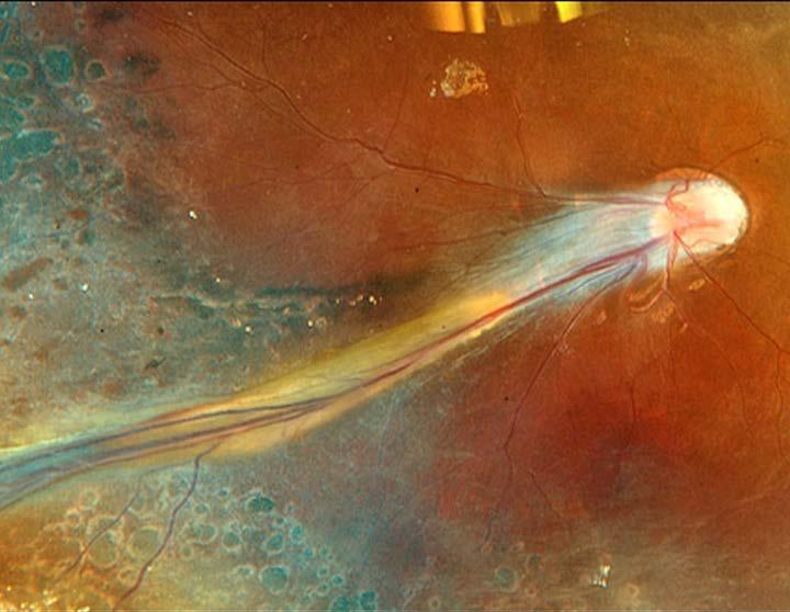
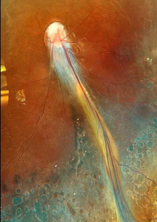
A comet's tail is the closest thing to nothing anything can be.
DR. JOÃO PEDRO MARQUES
CONTRIBUTOR
Dr. João Pedro Marques @retinaldystrophies
GA has officially entered its “now what?” era. In Manila, the newly formed Asian GA Expert Committee convened retinal leaders to tackle under-recognition, fast progressors, and what evidence-based GA care across Asia-Pacific should look like next.
By Tan Sher Lynn
A new chapter in geographic atrophy (GA) care began in the AsiaPacific on December 11, 2025, as regional experts gathered in Manila, the Philippines, for the inaugural meeting of the Asian GA Expert Committee.
For a disease space that has long been defined by clinical frustration, it was a decidedly welcome step forward for a condition that, until recently, had no effective treatment options.
Chaired by Prof. Gemmy Cheung of the Singapore National Eye Centre, the committee brought together leading retinal specialists from Australia, Japan, Korea, Singapore and Taiwan.
A COMMITTEE WITH A CLEAR MISSION
In line with the vision of becoming a trusted authority in the Asia-Pacific region, the committee is committed to advancing leadership through evidence-based education and consensusdriven guidance, focusing on early detection, optimal management and patient-centered care for GA.
Speaking about the significance of forming the committee, Prof. Cheung described the current moment as a turning point in retinal medicine. “This is a really exciting time for us in the retinal space,” she said. “If I look back personally at my career, this is probably the third time that we have seen a disease that was previously untreatable now coming into having novel treatment for the first time.”
She emphasized that GA, long regarded as an inevitable cause of irreversible vision loss, is now entering a new era of clinical relevance.
GA ISN’T A ONE DISEASE STORY; IT’S MANY
A central theme of the committee’s discussion was the complexity and heterogeneity of GA. According to Prof. Cheung, “There can be huge differences in how quickly the disease progresses. In some patients, the disease may be mild and never reach a stage where it impacts vision significantly. In others, it can progress very rapidly.”
This variability highlights the need for individualized patient assessment and a better understanding of each aspect of the disease.
GA HIDING IN PLAIN SIGHT
Another major concern raised was the under-recognition of GA, particularly among elderly populations. “Geographic atrophy is currently very much overlooked in most parts of the world because there has been no effective treatment until very recently,” she noted.
EPIDEMIOLOGY
The committee also discussed emerging epidemiological data suggesting that GA prevalence and progression patterns may differ across populations.
While Asian patients appear, on average, to experience slower lesion growth than Western counterparts, Prof. Cheung cautioned against overgeneralization. “Within Asian populations, there are still fast progressors—about 10% to 20% may grow at a rate of 2 mm² per year or more. Phenotypes associated with faster progression include strong drusen background, large baseline lesion size, and multifocal disease. These predictors are common across both Asian and non-Asian populations.”
Recognizing these higher-risk phenotypes will be essential for prioritizing monitoring and intervention.
WHAT COMES NEXT
Looking ahead, the Asian GA Expert Committee aims to function as a collaborative platform for research, education and clinical guidance across the region. “This is just the beginning,” Prof. Cheung said. “We hope to generate new data, answer unmet clinical questions, disseminate knowledge and guide clinical practice to ultimately benefit patients.”
Her closing message to the next generation of ophthalmologists captured the spirit of the meeting. “Look out for geographic atrophy. This is a new era, and it is only going to grow.”
Scan here to watch the full video: Finding GA Patients Faster | Prof. Gemmy Cheung on the New Era of Geographic Atrophy
Editor’s Note
A version of this article was first published on MediaMICE.com.
Many patients, she added, are not even presenting to clinics and are “quietly suffering in the community.” Hence, “one very urgent unmet need is simply to recognize these patients and bring them into care.”
AI AND MODERN RETINA SURGERY
AI has already learned how to read retinal scans. Now, it’s beginning to enter the OR, predicting outcomes, tracking instruments and reshaping what surgical assistance may look like in vitreoretinal care.
By Marhiel Garrote
The operating room depends on a choreography of vision, timing and instinct. In vitreoretinal surgery, much of the work rests on the physician’s ability to interpret subtle visual cues in real time—a membrane edge, a hidden retinal tear, the moment traction shifts direction.
Artificial intelligence (AI) has already transformed how ophthalmologists interpret images outside the OR. It can flag diabetic retinopathy, segment optical coherence tomography (OCT) scans and identify pathology faster than many clinicians thought possible just a decade ago.¹
But surgery presents an altogether different challenge. Unlike diagnostic AI, surgical AI must contend with motion, uncertainty and unpredictability, processing shifting anatomy, moving instruments and dynamic tissue behavior while a human surgeon operates inside a living eye.
That tension sat at the center of Prof. Stanislao Rizzo’s (Italy) keynote lecture during the 17th Annual Congress on Controversies in Ophthalmology (COPHy 2026), where he
explored how AI is beginning to move from passive observer to active surgical assistant in vitreoretinal practice.
FROM DIAGNOSIS TO PROGNOSIS
For years, ophthalmic AI has been framed primarily as a diagnostic tool: feed it an OCT scan or fundus image and it helps classify pathology. But research referenced during Prof. Rizzo’s lecture suggests a broader transition is underway.
One study explored machine learning models designed to predict postoperative visual outcomes using OCT biomarkers and clinical variables.² Rather than simply identifying disease, these systems attempt to forecast recovery potential after surgery—which, in vitreoretinal practice, is often where the uncertainty lives.
AI trained on large datasets may eventually identify structural patterns that correlate with functional recovery more reliably than human interpretation alone, with ellipsoid zone disruption, retinal thickness changes and subtle microstructural alterations becoming quantifiable predictive markers.²
Prof. Rizzo demonstrated this during the lecture using a Naïve Bayes model that analyzed OCT images and estimated visual recovery probability within seconds.² “This image will have an 80% chance of recovery of visual acuity,” he described.
AI-assisted prognosis introduces a subtle but consequential shift in clinical workflow, turning the question from, “What disease is this?” to “What is likely to happen next?”
AI SCRUBS IN
If predictive AI changes preoperative planning, intraoperative AI may change the surgical environment entirely.
Several studies highlighted in the keynote focused on real-time surgical guidance—augmented reality overlays, intraoperative image registration and computer vision systems—that continuously analyze the surgical field during the procedure itself.2-5 AI models can now recognize surgical phases, enhance contrast and brightness, and identify instrument location in real time.²
One study examined AI-assisted epiretinal membrane surgery, where dyes are currently used to visualize membrane borders during peeling. AI-assisted models, the research suggested, may eventually reduce the need for staining altogether.³ “Artificial intelligence paints the border of the membrane,” Prof. Rizzo explained. “No stain, no dyes—just safer and faster surgery.”
Researchers are also developing intraoperative OCT registration systems capable of synchronizing preoperative retinal maps with live surgical video—something akin to GPS navigation for the posterior segment—helping surgeons identify tissue landmarks and membrane boundaries in real time.⁵
Meanwhile, deep learning models trained to recognize surgical instruments, estimate their three-dimensional position and classify surgical phases may, in time, support robotic assistance and future autonomous surgical platforms.³
TRUST, LIABILITY AND THE LIMITS OF THE ALGORITHM
Conversations about AI in surgery tend to gravitate toward performance metrics: accuracy, efficiency, precision. But some of the most revealing moments at COPHy 2026 emerged around questions of trust and responsibility.
As AI systems grow more clinically sophisticated, patients may increasingly arrive in consultation rooms armed with algorithmgenerated opinions about their treatment. Surgical judgment, long shaped by training and experience, may soon have to co-exist with machine-generated recommendations.
The concerns extend beyond patient perception. AI systems remain vulnerable to hallucinations, bias and overconfidence. And during the discussion, it was noted that large language models
often produce overly optimistic surgical predictions compared to real-world outcomes.
Then comes the liability question: if AI guidance influences a surgical decision and the outcome falls short, who bears responsibility? The surgeon, the hospital or the developer?
There is also the matter of variability. Vitreoretinal surgery is rarely standardized. Bleeding changes visibility. Retinal mobility shifts anatomy. Tissue behaves unpredictably. Human surgeons adapt instinctively to those changes in ways machines still cannot fully replicate.
THE COMPUTATIONALLY AWARE OR
The research Prof. Rizzo referenced traces a trajectory already taking shape across ophthalmic surgery: predictive analysis, intraoperative overlays, instrument tracking and AI-guided surgical support. Fully autonomous retinal surgery may still be distant, but even partial automation could meaningfully reshape how surgeons operate.
That possibility reframes what AI actually represents in the surgical context: not a replacement but an augmentation. The surgeon remains in control, but the OR itself is becoming computationally aware.
For now, one thing holds: real surgeons with real eyes, hands and clinical judgment are still the ones holding control inside the OR. How long that remains the full picture—and under what ethical and regulatory frameworks any shift will unfold—may be an important question the field hasn’t quite finished asking.
Editor’s Note
The 17th Annual Congress on Controversies in Ophthalmology (COPHy 2026) was held on March 20–21 in Krakow, Poland. Reporting for this story took place during the event.
REFERENCES
1. Sheng B, Chen X, Li T, et al. An overview of artificial intelligence in diabetic retinopathy and other ocular diseases. Front Public Health. 2022;10:971943.
2. Savastano MC, Crincoli E, Rizzo S, et al. Prediction of postoperative visual acuity restoration in macula off rhegmatogenous retinal detachment using artificial intelligence. AJO International. 2024;1(9):100008.
3. Ye RZ, Iezzi R. Intraoperative augmented reality for vitreoretinal surgery using edge computing. J Pers Med. 2025;15(1):20.
4. Hwang SH, Kim YJ, Cho JB, et al. Digital image enhancement using deep learning algorithm in 3D heads-up vitreoretinal surgery. Sci Rep. 2025;15(1):18429.
5. Baldi PF, Abdelkarim S, Liu J, et al. Vitreoretinal surgical instrument tracking in three dimensions using deep learning. Transl Vis Sci Technol. 2023;12(1):20.
HAS HUD SURGERY ACTUALLY MOVED THE NEEDLE?
At COPHy 2026, two retinal heavyweights debated the promise, proof and persistent question marks behind vitreoretinal surgery’s most cinematic technology.
By Diana Truong
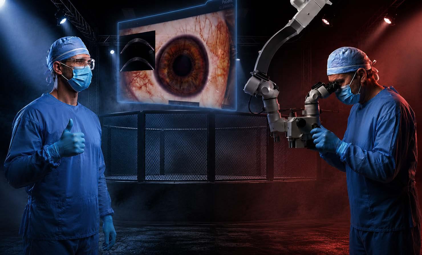
Raise your hand if you’ve felt it—that slight twinge of envy when a colleague rolls into the OR with a gleaming digital tower, fires up a cinema-sized 3D display and proceeds to peel membranes like they’re directing a blockbuster from the director’s chair.
Heads-up display (HUD) vitreoretinal surgery has that effect on people. It looks transformative. It feels transformative. But does it perform transformatively?
That was the deliciously contentious question put to two of the field’s most respected voices at the 17th Annual Congress on Controversies in Ophthalmology (COPHy 2026) in a debate that was, at times, less a clash of opinions than a shared inventory of everything we still don’t know.
Dr. Stratos Gotzaridis (Greece) took the “Yes” podium. Dr. William Mieler (United States)— who had, by his own admission, debated this very topic with Dr. Gotzaridis two years prior—lined up on “No.” What followed was a seven-point tour through the promise, the proof and the persistent gaps of one of the OR’s most talked-about technologies.
THE LITERATURE GREW UP— A LITTLE
Two years ago, Dr. Gotzaridis had nearly nothing to stand on. “At that time, the
literature…advocated that there was no difference in final visual acuity and anatomical findings,” he admitted. “So…I said, ‘You’re right. I have nothing to present.’”
This time, he arrived with more ammunition. A recent paper comparing NGENUITY®assisted surgery against standard microscopy found not-statistically-significant but directionally better visual acuity in the HUD group, along with improved cone and ganglion cell function on pattern electroretinogram.1
Another study on difficult cases— endophthalmitis included—showed better final visual acuity and superior intraoperative conditions. 2
Progress, yes. Proof, not quite.
OUTCOMES DATA REMAINS STUBBORNLY INCONCLUSIVE
Dr. Mieler, characteristically measured, walked the room through the full arc of the literature. Papers from 2019 onward, he noted, “for the most part show no difference in postoperative complications, no differences in anatomic outcomes.”
A 2023 paper did suggest a slightly lower rate of recurrent membrane formation with 3D surgery 3 —one of those encouraging breadcrumbs the field keeps chasing—but the
broader picture remains that, as Dr. Mieler put it, better visual and clinical outcomes have “in most cases, not exactly” been proven.
That’s not a damning verdict. It is, however, an honest one.
THE IMAGE IS GENUINELY BETTER
Here’s where both surgeons found common ground, and where Dr. Gotzaridis got visibly animated.
The high dynamic range processing of systems like the NGENUITY® 3D Visualization System (Alcon; Geneva, Switzerland) compresses exposures into a single optimized image, reaching up to 90 decibels of dynamic range against a conventional camera’s 40.
The practical upshot? Surgeons can boost blue dye visualization, use color contrast to identify vitreous without triamcinolone, and toggle filters to track indocyanine green or work through vitreous hemorrhage by blocking red channels.
“You don’t need a chemical pyrazinolone to assist you to remove the old vitreous,” Dr. Gotzaridis noted.
ischemic zones, laser only what’s necessary, spare the healthy retina.4
Whether this translates to measurably better outcomes remains the stubborn open question, but the toolbox is undeniably richer.
ERGONOMICS ARE REAL, BUT PERSPECTIVE MATTERS
Both surgeons acknowledged that the heads-up posture reduces neck and back strain—hardly a trivial concern in a specialty where decades are spent hunching over eyepieces.
Dr. Gotzaridis noted that “surgeons experience less back and neck pain, and they have a career of improvement in longevity and productivity.”5
Dr. Mieler, true to form, added a reality check: “Other parts of your life sometimes can play a role in terms of ergonomic discomfort as well.”
It’s a fair point. Posture in the OR matters, but so does posture at dinner, at the desk and everywhere else surgeons exist as full human beings.
TEACHING IS WHERE THE TECH SHINES BRIGHTEST
If there’s one category where the evidence is least contested, it’s education. When everyone in the room—fellows, residents, scrub nurses, observers—sees exactly what the operating surgeon sees, the quality of teaching transforms.
A study on trainees using microplatforms found that the success rate in the 3D group reached 86%, compared to 60% in the microscope group. 6 Dr. Gotzaridis attributed this to better visualization of the wide-field image and more precise coaching from the supervising surgeon.
Even Dr. Mieler, the debate’s designated skeptic, conceded that the positive impact on surgical education is “distinctly yes.”
That’s not nothing. A technology that accelerates the learning curve of the next generation has compounding value.
OPERATING TIMES HAVEN’T BUDGED
One of the early promises of HUD surgery was efficiency: slicker visualization, less fiddling, faster cases. The data, however, has not cooperated.
“Overall the answer was not statistically significantly different,” Dr. Mieler said of operating times, and some studies actually suggest procedures run slightly longer, at least until the learning curve is conquered.7
Issues like manual image inversion, more frequent focusing adjustments and occasional latency in earlier systems contributed to the drag. The good news is that most of these wrinkles smooth out with experience and improve with hardware generations.7
AI AND ROBOTICS ARE THE REAL NEXT
CHAPTER
Here’s where Dr. Gotzaridis let himself get genuinely excited, and it’s hard to blame him. A better microscope with a heads-up display, coupled with artificial intelligence, means you can map anatomical landmarks in real time, tracking instrument tips relative to the optic nerve or macula.
Dr. Gotzaridis described a prototype in which the robotic system simply stops the instrument when it approaches the retina. “It doesn’t allow you to continue,” he said.
He also pointed to helmet-inspired wearable displays on the horizon. The comparison to aviation is intentional and apt: surgeons are, in a sense, learning to fly by instruments.
Dr. Mieler closed the debate with a line that probably both men could have delivered: “3D surgery is a work in progress. It is currently not essential, but it’s great technology. It’s here to stay.”
That verdict—somewhere between endorsement and acquittal—may be the most honest thing that can be said about HUD surgery in 2026.
The screen is bigger. The image is better. The back hurts less. The outcomes? Still loading. But given where this technology is pointing—
toward AI integration, robotic assistance, and a generation of surgeons trained from day one with a cinematic view of the retina—the question may soon flip from “has it moved the needle?” to “how far will it go?”
Editor’s Note
The 17th Annual Congress on Controversies in Ophthalmology (COPHy 2026) was held on March 20–21 in Krakow, Poland. Reporting for this story took place during the event.
REFERENCES
1. Lubinski W, Charabin T, Podboraczynska-Jodko K, et al. Comparison of retinal function and macular structure after 27G pars plana vitrectomy with minimal heads-up versus standard illumination in patients with idiopathic epiretinal membranes: A pilot randomized study. Acta Ophthalmol. 2025;103(7):e456-e471.
2. Guan L, Li M, Fan W, et al. Threedimensional digital visualization system-assisted vitrectomy for infectious endophthalmitis. J Ophthalmol. 2026;2026:3447802.
3. Kim DJ, Kim DG, Park KH. Threedimensional heads-up vitrectomy versus conventional microscope vitrectomy for patients with epiretinal membrane. Retina. 2023;43(6):10101018.
4. Coppola M, La Spina C, Rabiolo A, et al. Heads-up 3D vision system for retinal detachment surgery. Int J Retina Vitreous. 2017;3:46.
5. Helayel HB, Al-Mazidi S, AlAkeely A. Can the three-dimensional heads-up display improve ergonomics, surgical performance and ophthalmology training compared to conventional microscopy? Clin Ophthalmol. 2021;15:679-686.
6. Shoshany TN, Agranat JS, Armstrong G, et al. The user experience on a 3-dimensional heads-up display for vitreoretinal surgery across all members of the health care team: A survey of medical students, residents, fellows, attending surgeons, nurses and anesthesiologists. J Vitreoretin Dis. 2020;4(6):459-466.
7. Talcott KE, Adam MK, Sioufi K, et al. Comparison of a three-dimensional heads-up display surgical platform with a standard operating microscope for macular surgery. Ophthalmol Retina. 2019;3(3):244-251.
WHEN SEEING NOTHING SPARKS A BIG DEBATE
For a laser that leaves no scar, SML has left retina with plenty of bruises. Twenty-five years on, the field can’t decide whether it’s a breakthrough hiding in plain sight or a beautifully engineered placebo.
By Michaela Perez
In retinal practice, most treatments leave a trail. An anti-vascular endothelial growth factor (VEGF) injection appears in the chart. Conventional photocoagulation leaves a scar. Even a failed intervention leaves evidence: a lesion, a reading, a change on optical coherence tomography (OCT).
Subthreshold micropulse laser (SML) leaves nothing. No burn. No scar. No visible mark. Fire the laser, and the retina looks exactly as it did before.
That invisibility is the entire premise— and, it turns out, the entire problem.
THE CLAIM
But Dr. Luttrull draws a sharp distinction. SML is a broad term covering a range of laser approaches—different wavelengths, duty cycles and protocols—applied inconsistently across centers worldwide. What he practices is more specific: subthreshold diode micropulse (SDM), using an 810 nm diode laser at 5% duty cycle, delivered at high density over large retinal areas, including the fovea.
For Dr. Luttrull, SDM is not simply a variant of SML. It’s SML done correctly. And that distinction, he argues, explains much of the confusion in the published literature.
Dr. Jeffrey Luttrull (United States) has been using SML as first-line treatment for diabetic macular edema (DME) for 25 years. More than 90% of his DME patients, he reports, never need an anti-VEGF injection at any point in their care.
He called SDM “the most useful and important therapeutic tool we have in retina and maybe in ophthalmology.”
That’s not a hedged claim. It’s a provocation. And for a field spending billions annually on anti-VEGF injections, it deserves serious consideration.
Dr. Glenn Yiu (United States), who led the PULSE trial at University California, Davis (UC Davis), reached a different conclusion. His study found SML didn’t prevent DME progression in patients with good vision.¹ “Results are inconclusive,” said Dr. Yu, “and likely to vary between individuals.”
Same laser. Same disease. Entirely different clinical realities.
AN ACCIDENTAL DISCOVERY
Dr. Luttrull’s conviction didn’t begin with a research hypothesis. It began with a budget problem.
Early in his career, he was looking for ways to make better use of a laser he already owned. “I was not trying to do anything special,” he recalled. “I basically stumbled upon the application that became SDM.”
What followed was careful attention to unexpected results—outcomes he hadn’t anticipated, in a treatment he knew was dramatically safer than anything he’d done before. “Within a short time, the clinical results were very, very good,” he said.
That accidental origin matters. Most advances in SDM have come not from academic laser research programs but from a private-practice physician watching carefully and thinking differently—an uncomfortable fact for a field that prizes randomized controlled trials above all else.
THE BIOLOGY BEHIND THE BENEFIT
To understand why Dr. Luttrull is so convinced, you have to start where most SML discussions don’t: the biology.
The mechanism of all therapeutic retinal laser treatment is identical, whether conventional or subthreshold, scarring or not. The laser creates sublethal hyperthermia in the retinal pigment epithelium (RPE). Heat shock proteins activate. Retinal function normalizes, regardless of the cause of the underlying dysfunction.
A recent narrative review in Survey of Ophthalmology described this as the “reset theory.” Heat shock protein activation effectively resets RPE cells to their default, healthy function, reducing inflammation and repairing cellular damage without structural retinal injury.²
The critical implication is straightforward: if heat shock protein activation is the mechanism, tissue destruction contributes nothing. Burned cells are dead, and dead cells cannot mount a biological response. With conventional photocoagulation, whatever therapeutic benefit occurs happens at the wound margins— in tissue heated but not destroyed. The burn was never the therapy. It was always the complication.
“Retinal laser damage by any means is directly non-therapeutic and simply
a complication of treatment to be avoided,” said Dr. Luttrull. “Only SDM does this reliably, predictably and effectively.”
SDM delivers the biological effect without the destruction, enabling high-density coverage of large retinal areas—including direct foveal treatment—all categorically off-limits with conventional lasers. “If you have even the potential for doing damage, you can’t do any of those things,” he said.
The implications extend beyond DME. Dr. Luttrull uses SDM as first-line treatment for proliferative diabetic retinopathy, a condition typically managed with panretinal photocoagulation, which carries significant risks of peripheral vision loss and night blindness.
The Survey of Ophthalmology review also confirms SML’s expanding evidence base across central serous chorioretinopathy, macular edema secondary to retinal vein occlusion and age-related macular degeneration.²
THE LONG SHADOW OF A CHRONIC DISEASE
To appreciate why SML’s promise matters so acutely, consider what DME actually
asks of patients—and, by extension, of the clinicians managing them.
DME is not a condition physicians treat and resolve. It’s a decades-long companion: chronic, progressive, demanding regular monitoring, frequent clinic visits and often monthly injections.
Anti-VEGF therapy transformed outcomes when it arrived, but it didn’t change the fundamental burden of needles, appointments, costs and the anxiety of a disease that never fully goes away. Over a lifetime, the cumulative burden— financial, psychological, logistical— is substantial.
A safe, repeatable office procedure that meaningfully reduces it without compromising vision would be, for many patients, genuinely life-changing. That’s the promise SML carries. Whether it delivers is precisely what the field cannot agree on.
WHAT THE TRIALS FOUND—AND DIDN’T
The published record is not encouraging, at least on the surface. DIAMONDS randomized DME patients to SML versus standard laser; both arms produced equivalent outcomes, and both groups lost vision letters over two years.³
PULSE tested whether SML could prevent or delay vision loss in patients with good visual acuity and found no benefit.¹
Two meta-analyses found reduced injection frequency when SML was added to anti-VEGF therapy, but disagreed on visual acuity and macular thickness outcomes.4,5
Read plainly, that’s an underwhelming record. But Dr. Luttrull argues the trials weren’t testing what they claimed to test, and that the SDM distinction explains the gap between published results and his own outcomes.
The meta-analyses, he notes, aren’t themselves the problem. “The problem is what they’re analyzing. The SML studies that they analyze are generally poor, highly variable and typically suboptimal.”
The PLACE trial,⁶ which compared SML to half-dose, “basically homeopathic” photodynamic therapy for central serous chorioretinopathy, he takes not as a ceiling but as evidence of untapped potential.
“What is remarkable is that despite the high variability in how people do SML, it often works pretty well anyway,” he said. “Like anything else, it would work even better if done correctly.”
SAFETY AS A STARTING POINT
Dr. Yiu ran a trial that found no efficacy, but he hasn’t abandoned the technology. “I think the strongest evidence is for the safety of the technology,” he said. “None have demonstrated any evidence of danger or damage to the neurosensory retina.”
For DME patients with good vision— those who, per DRCR Protocol V, could reasonably be observed without immediate anti-VEGF intervention— SML offers a meaningful middle ground. “For conditions where the alternative is to observe, I think SML is an excellent first choice prior to trying more aggressive therapy,” said Dr. Yiu.
His PULSE trial was designed for this population—Protocol V-eligible patients randomized to SML or sham laser.¹ The study found no significant difference, but Dr. Yiu is measured about what that means.
The trial was underpowered at baseline, a problem compounded when COVID-19 disrupted follow-up.¹ Treating milder disease, where some patients improve spontaneously, also makes detecting a treatment signal significantly harder.
The meta-analyses support a modest case: adding SML to anti-VEGF appears to reduce injection frequency even when visual outcomes remain comparable.4,5 The Survey of Ophthalmology review reinforces this, confirming reduced injection burden as a notable benefit while acknowledging larger studies are still needed.²
Dr. Yiu also named a perception problem directly. “Because you do not observe a physical effect—no laser burn—some physicians assume it is homeopathic.”
That assumption has kept adoption low, real-world data thin and the evidence base
inconclusive—a self-reinforcing cycle that may be obscuring genuine signal.
THE PROTOCOL PROBLEM
Both experts agree that variability in how SML is applied is a fundamental clinical issue, and the Survey of Ophthalmology review makes this plain.² Duty cycle, wavelength, spot size, power, treatment density—no two centers do it the same way. Darker eyes absorb more laser energy, compounding inconsistency further.
That variability, Dr. Luttrull argues, explains the published record. DIAMONDS used a 577 nm wavelength at 5% duty cycle³—which he describes as “a very energetic wavelength, very prone to retinal damage”—paired with titration techniques that virtually guarantee tissue injury.
If both study arms cause retinal damage, the trial wasn’t comparing a subthreshold approach against a conventional one. It was comparing two damaging techniques wearing different labels. “They thought they were doing different kinds of treatment,” he said. “And they actually weren’t.”
His critique of titration goes further. Hyperthermia in a calibration test spot conditions that tissue against further insult, but the surrounding treatment area receives no such conditioning. “When you finally get a burn to show
up in the conditioned tissue, the effect in unconditioned tissue will be vastly more intense than you anticipated,” Dr. Luttrull noted.
The Survey of Ophthalmology review corroborates this concern, noting that inconsistencies in laser parameters— wavelength, duty cycle, fluence and spot size—continue to pose challenges in standardizing protocols and make results across studies difficult to compare.²
Most specialists still approach SML through the framework of conventional photocoagulation, expecting heat spread, rationing treatment density and avoiding the fovea.
Those instincts are appropriate for a damaging laser. They are counterproductive for one that isn’t. “There is no heat spread” with correctly performed micropulse, said Dr. Luttrull. The model is wrong, and the protocols follow the model.
His prescription is education. “Once you understand how it works, you’ll understand how to do it correctly,” Dr. Luttrull said.
Dr. Yiu’s is complementary: broader adoption generating the real-world data needed to support a properly powered prospective trial. “If more physicians employ this technology, we could conduct
larger real-world retrospective studies and provide support for a larger, well-designed prospective trial,” he noted.
THE UNANSWERED QUESTION
Two experienced retinal specialists. Same technology (though, as Dr. Luttrull would insist, not always the same technique). Entirely different conclusions.
One has used SDM as first-line therapy for 25 years with extraordinary reported outcomes. The other tested SML rigorously, found no benefit and still recommends it as a reasonable first option when observation is the alternative.
Perhaps they’re each describing a genuine clinical reality, shaped by different protocols producing different results in different hands. Perhaps Dr. Luttrull has refined a technique most centers haven’t learned. Perhaps the published trials have tested SML in ways that don’t reflect SDM’s actual potential.
Or perhaps the promise is simply larger than the delivery.
What’s clear is that the field hasn’t done the work needed to find out.
As the Survey of Ophthalmology review concludes, larger, high-quality studies are still needed to fine-tune treatment parameters, improve patient
selection and clarify SML’s role alongside other therapies.²
A properly powered, multicenter, prospective trial with standardized protocol has never been done. Until it is, every physician using SML is effectively running their own small, uncontrolled study, adding to the noise without adding to the answer.
For DME patients with good vision where Protocol V supports observation, SML’s safety profile makes it a rational first step before escalating to anti-VEGF. The worstcase scenario, per current evidence, is that it does nothing.
But for a technology this contested, with stakes this high for patients managing a lifelong disease, “probably harmless” shouldn’t be where the conversation ends.
The laser fires. The tissue shows no mark. Whether that absence represents therapeutic elegance or simple absence of effect depends on who you ask, and quite possibly on how the laser was used.
The invisible laser, it seems, remains invisible in more ways than one.
REFERENCES
1. Feldman S, Basra J, Lawani A, et al. Subthreshold micropulse laser for eyes with center-involving diabetic macular edema with good visual acuity: The PULSE study, a randomized clinical trial. Ophthalmic Surg Lasers Imaging Retina. 2025;56(2):86-93.
2. Sim SY, Lim LT, Chaudhary S, et al. Update on subthreshold micropulse laser treatment for retinal diseases: A narrative review. Surv Ophthalmol. 2026 Feb 26:S0039-6257(26)00022-6. Epub ahead of print.
3. Lois N, Campbell C, Waugh N, et al. Diabetic macular edema and diode subthreshold micropulse laser: A randomized double-masked noninferiority clinical trial.. Ophthalmol. 2023;130(1):14-27.
4. Hosoya H, Ueta T, Hirasawa K, et al. Subthreshold micropulse laser combined with anti-vascular endothelial growth factor therapy for diabetic macular edema: A systematic review and meta-analysis. Graefes Arch Clin Exp Ophthalmol. 2024;262(10):3073-3083.
5. Jiang Y, He W, Qi S. Evaluating the efficacy of subthreshold micropulse laser combined with anti-VEGF drugs in the treatment of diabetic macular edema: A systematic review and meta-analysis. Front Endocrinol. 2025;16:1553311.
6. van Dijk EHC, Fauser S, Breukink MB, et al. Half-dose photodynamic therapy versus high-density subthreshold micropulse laser treatment in patients with chronic central serous chorioretinopathy: The PLACE trial. Ophthalmol. 2018;125(10):1547-1555.
SAMSUNG BIOEPIS, HARROW TEAM UP TO RELAUNCH RANIBIZUMAB BIOSIMILAR IN THE U.S.
THE FIRST OPHTHALMIC BIOSIMILAR APPROVED IN THE UNITED STATES IS MAKING A COMEBACK, AND THIS TIME, IT’S SHARING A ROOF WITH ITS SIBLING.
By Marhiel Garrote
Samsung Bioepis (Incheon, South Korea) has relaunched BYOOVIZ (ranibizumab-nuna) in the U.S. through a new commercialization partnership with Harrow (Tennessee, USA).
The move follows Biogen’s transfer of U.S. commercial rights back to Samsung Bioepis at the end of 2025 and places interchangeable biosimilars referencing both ranibizumab and aflibercept under a single ophthalmic company.
Under the new agreement, Harrow leads U.S. commercialization of both BYOOVIZ and OPUVIZ (aflibercept-yszy), while Samsung Bioepis continues to handle product development, manufacturing and regulatory activities.
BUILDING A BROADER RETINA THERAPY PORTFOLIO
BYOOVIZ was approved by the U.S. Food and Drug Administration (FDA) in 2021 as the country’s first ophthalmic biosimilar and received interchangeability designation in 2023. It is indicated for neovascular age-related macular degeneration (nAMD), macular edema following retinal vein occlusion (RVO) and myopic choroidal neovascularization (mCNV).
In a news release, Samsung Bioepis Executive VP and Global Head of Commercial Linda Choi MacDonald called the launch “an exciting new chapter” for BYOOVIZ in the U.S.
“With Harrow now leading commercialization efforts, we are reigniting our commitment to ensuring patients and retina specialists across America to have access to this quality-proven, safe and effective biosimilar option,” said MacDonald. “We believe this relaunch will ultimately help more patients with critical ophthalmic diseases to receive the vision-saving treatments they need.”
IMPROVING ACCESS TO ANTI-VEGF THERAPY
Approximately 1.5 million Americans are living with late-stage wet AMD, according to data cited by Samsung Bioepis. While anti-VEGF therapies have transformed outcomes for retinal disease, repeated intravitreal injections carry a substantial long-term financial burden for both healthcare systems and patients.1,2
Samsung Bioepis said biosimilars can help ease that burden by offering lowercost alternatives to reference biologics without clinically meaningful differences in safety or efficacy.
AVAILABILITY
Samsung Bioepis and Harrow have not yet announced an official relaunch date or rollout timeline for BYOOVIZ. For now, the announcement marks the start of a new commercial chapter for the biosimilar. Its clinical impact will hinge on how quickly it reaches retina specialists and their patients.
Editor’s Note
A version of this article was first published on MediaMICE.com.
REFERENCES
1. CDC. VEHSS Modeled Estimates: Age-Related Macular Degeneration (AMD). May 15, 2025. Available at: https://www. cdc.gov/vision-health-data/prevalence-estimates/amd-prevalence. html Accessed on July 2, 2026.
2. Saundankar V, Borns M, Broderick K, et al. Annual prevalence of geographic atrophy and wet age-related macular degeneration among Medicare Advantage enrollees in a US health plan. J Manag Care Spec Pharm. 2025;31(1):88-94.
DR. AUDINA BERROCAL’S JOURNEY TO THE FOREFRONT OF OPHTHALMOLOGY
Born into ophthalmology royalty, Dr. Audina Berrocal could have coasted on a celebrated family name. Instead, she built something entirely her own, becoming one of the world’s foremost pediatric retina experts.
By Chow Ee-Tan
Few surgeons spend their days operating on patients who weigh less than a kilogram. For Dr. Audina Berrocal (United States), some of the most consequential battles against blindness begin in neonatal intensive care units, where premature infants face life-altering retinal conditions before they have even opened their eyes to the world.
Over three decades, her expertise in retinopathy of prematurity (ROP), pediatric vitreoretinal surgery and inherited retinal disorders has earned international recognition, but the significance of her work is ultimately human. Every successful intervention means a child may one day recognize a parent’s face, read a book independently or experience the world through sight.
FROM POLI-SCI TO THE OR Medicine was part of Dr. Berrocal’s world long before she became a physician. Her father, Dr. José Berrocal, is widely regarded as the first retina specialist in Puerto Rico and the Caribbean, and her sister, Dr. María Berrocal, went on to become a distinguished retina specialist in her own right.1
Given such a pedigree, a career in medicine might seem inevitable. It was not. In an oral history interview with the American Society of Retina Specialists (ASRS), Dr. Berrocal revealed that her first academic passion was political science—she was drawn to people, leadership and the power of ideas to shape society. Medicine came later, eventually crystallizing into a calling that merged intellectual rigor with tangible human impact.1
Even then, a subtler challenge remained: carving out her own identity in a field where the Berrocal name already carried considerable weight. She addressed that by gravitating toward pediatric retina, one of ophthalmology’s most demanding subspecialties, where science, surgery and compassion converge in ways few disciplines can match.1
PIONEERING AT THE FRONTIER
A defining characteristic of Dr. Berrocal’s career has been her appetite for innovation when it offers better outcomes. She has helped pioneer the use of anti-vascular endothelial growth factory (VEGF) therapy in children and was among the earliest adopters of retinal gene therapy in pediatric patients— breakthroughs that transformed the outlook for inherited retinal diseases once considered untreatable. 2
As genetic medicine continues to evolve, she remains a leading voice shaping the future of the field.
THE MENTOR WHO MULTIPLIES
Ask Dr. Berrocal about her proudest accomplishments and she is just as likely to speak about people as procedures. As a fellowship director and educator, she has trained retina specialists now practicing across the United States and around the world.3
She has been candid about what gives her the deepest satisfaction. In an interview with The Ophthalmologist, she reflected “having developed a pediatric retina surgical practice and being able to mentor young surgeons in this field.” She added, “There is nothing better than to see someone I trained want to follow in my footsteps.”3
The sentiment is consistent with a broader conviction: that investing in people is every bit as important as advancing science.
OPENING DOORS
That conviction extends to her advocacy for women in ophthalmology. Dr. Berrocal has long recognized that professional success depends heavily on access to guidance, opportunity and supportive networks—and she has worked systematically to expand all three.
She served as co-editor of Women in Ophthalmology: A Comprehensive Guide for Career and Life, a landmark publication addressing mentorship, leadership, work-life integration and professional development.
When named to The Ophthalmologist Power List, she used the platform characteristically—not to celebrate herself, but to spotlight her peers. “We have worked very, very hard,” she said. “It is time to see us come together and be recognized for our hard work and brilliance.” 3
She has also pushed colleagues to engage with aspiring physicians well before residency, with a challenge that is simple and direct: “Be that person who can change someone’s life.” 3
A LEGACY BUILT
International recognition and numerous honors have followed, and Dr. Berrocal is consistently acknowledged among the most influential figures in global retinal practice.
Yet awards tell only part of the story.
Her legacy lives in the premature infant whose vision was preserved, the young surgeon who found confidence through her guidance, and the patients whose futures brightened because someone refused to accept the prevailing limits of care.
She took an extraordinary inheritance and transformed it into something distinctly her own—a prime example that true legacy is earned, one patient and one protégé at a time.
REFERENCES
1. American Society of Retina Specialists. Leaders & Legends: An Interview with Audina M. Berrocal, MD, FASRS. Retina History Project. Accessed on June 8, 2026.
2. Bercuson M. Nine Bascom Palmer doctors named to Power List honoring top women in ophthalmology. University of Miami Miller School of Medicine. Accessed on June 8, 2026.
3. Power List 2021: Audina M. Berrocal. The Ophthalmologist. Accessed on June 8, 2026.
ERM, ILM AND WHERE TO STOP
One membrane is the problem. The other may be the solution…or an unnecessary casualty. Retina surgeons remain divided on where the peel should end.
By Hazlin Hassan
For decades, pars plana vitrectomy (PPV) with epiretinal membrane (ERM) peeling has been the standard surgical treatment for symptomatic macular puckers, reliably improving vision and reducing retinal distortion.
Yet one question continues to divide even the most experienced vitreoretinal surgeons: once the ERM is removed, should the internal limiting membrane (ILM) come off too?
At the 17th Annual Congress on Controversies in Ophthalmology (COPHy 2026), two senior surgeons made the case for each side in a debate that perfectly captured modern retina surgery’s balancing act between anatomical perfection and surgical restraint.
On one side stood Prof. Stanislao Rizzo (Italy), arguing that ILM peeling is essential to minimize recurrence and fully relieve tractional forces. Opposing him was Dr. William Mieler (United States), who maintained that the additional peel may expose patients to unnecessary retinal trauma without delivering meaningful visual benefit.
THE MEMBRANE BENEATH THE MEMBRANE ERMs are relatively common fibrocellular proliferations that develop on the inner retinal surface above the ILM. They can wrinkle the macula, distort foveal architecture and impair vision. On optical coherence tomography (OCT), ERMs typically appear as hyperreflective membranes associated with retinal thickening and tractional distortion.
Most patients remain stable, but some progress to blurred vision, metamorphopsia and significant visual dysfunction. Surgical intervention—typically small-gauge PPV with membrane peeling—has evolved considerably since Dr. Robert Machemer’s pioneering work in the 1970s, with one study reporting visual improvement in around 80% of patients.1
But where ERM peeling ends, controversy begins.
The ILM is the retina’s basement membrane, composed primarily of type IV collagen, laminin and fibronectin. It also serves as the attachment point for Müller cell footplates. Removing
it theoretically eliminates the scaffold that allows residual proliferative cells to regrow—and that logic forms the foundation of the pro-ILM argument.
THE CASE FOR GOING DEEPER
Prof. Rizzo put it plainly, saying, “ILM peeling is performed in order to increase macula elasticity, completely release tractions and remove scaffold for cellular re-proliferation.”
Recurrence is the central concern. Residual cellular elements— including glial cells and hyalocytes—may contribute to postsurgical epiretinal proliferation. Evidence presented during the debate suggested that additional ILM peeling lowers ERM recurrence rates in approximately two-thirds of published studies. 2
THE COST OF PEELING DEEPER
But does that reduction justify additional manipulation of the retina’s innermost structural layer?
“Removal of the ILM does not appear to improve visual outcomes,” Dr. Mieler stated. “Long-term consequences of ILM removal are not fully known.”
He also reminded the audience that the mere presence of an ERM is not itself an indication for surgery. The decision depends on the patient’s visual needs and possible comorbidities.
The concern is that ILM peeling introduces another layer of potential trauma. Reported complications include retinal hemorrhages, paracentral scotomas, dissociated optic nerve fiber layer changes, rare eccentric macular holes, and potential dye toxicity from agents such as indocyanine green or brilliant blue G.3
For critics, this raises the uncomfortable question of whether surgeons are trading microscopic retinal injury for recurrence prevention that may never become clinically significant.
THE CASE FOR KNOWING WHEN TO STOP
The evidence, on balance, is equivocal. Meta-analyses found that both techniques improve vision and reduce central macular thickness, but concluded that further comparative data are needed.4,5
The American Academy of Ophthalmology Preferred Practice Pattern 2019 report assessed ten studies and similarly found that ILM peeling reduces recurrence without clearly improving final visual outcomes, while potentially altering inner retinal architecture—including apparent retinal thinning on OCT. 6
Large database studies add further nuance. Rayess and colleagues analyzed more than 10,000 patients and found lower reoperation rates with ILM peeling—0.88% versus 1.48% at one year—but no difference in retinal detachment rates or visual outcomes.7
“Why subject your patients to complications, to possibly lessen the miniscule risk of insignificant recurrent ERM formation?” asked Dr. Mieler.
For surgeons on the other side, however, recurrence itself represents incomplete surgery. Even where initial visual outcomes are comparable, eliminating the substrate for reproliferation may offer greater long-term anatomical stability.
SO WHERE SHOULD SURGEONS STOP?
Ironically, modern surgery may already blur the distinction. Both surgeons acknowledged that the ILM is unintentionally removed in 40% to 55% of cases during ERM peeling anyway. 8
For many surgeons, the decision ultimately comes down to individual patient factors, surgical judgement and how much additional intervention is considered necessary once the membrane has been removed.
Editor’s Note
The 17th Annual Congress on Controversies in Ophthalmology (COPHy 2026) was held on March 20–21 in Krakow, Poland. Reporting for this story took place during the event.
REFERENCES
1. García-Fernández M, Navarro JC, Castaño CG, et al. Epiretinal membrane surgery: Anatomic and functional outcomes. Arch Soc Esp Oftalmol. 2013 Apr;88(4):139-44.
2. Zhang B, Dong X, Sun Y. Effect of internal limiting membrane peeling for idiopathic epiretinal membrane. Heliyon. 2023;9(3):e14079.
3. Asencio-Duran M, Manzano-Muñoz B, Vallejo-García JL, et al. Complications of macular peeling. J Ophthalmol. 2015;2015:467814.
4. Christodoulou E, Batsos G, Galanis P, et al. Vitrectomy for the removal of idiopathic epiretinal membrane with or without internal limiting membrane peeling: A meta-analysis. Ther Adv Ophthalmol. 2020;12:2515841420927133.
5. Huang Q, Li J. With or without internal limiting membrane peeling during idiopathic epiretinal membrane surgery: A metaanalysis. PLoS One. 2021;16(1):e0245459.
6. American Academy of Ophthalmology. Idiopathic Macular Hole Preferred Practice Pattern. Elsevier. 2019. Available at: https://www.aao.org/Assets/f5913890-ec6a-40b1-ac77c6f6ad22b779/637163521770000000/idiopathic-macular-holeppp-2019-pdf. Accessed on May 20, 2026.
7. Rayess N, Vail D, Mruthyunjaya P. Rates of reoperation in 10,114 patients with epiretinal membranes treated by vitrectomy with or without inner limiting membrane peeling. Ophthalmol Retina. 2021;5(7):664-669.
8. Kim BH, Yoon CK, Bae K, et al. Influence of internal limiting membrane peeling during idiopathic epiretinal membrane removal: A randomized controlled trial. Sci Rep. 2025;15:17499.
BEYOND CLOSURE, REBUILDING THE FOVEA
A closed macular hole is not always a restored fovea. For large, myopic, chronic and refractory holes, the inverted ILM flap turns tissue once peeled away into a scaffold for repair.
By Kendra Bruning
On optical coherence tomography (OCT), a macular hole can appear closed even when the fovea hasn’t truly recovered. The edges flatten and the defect may look resolved, but the underlying layers responsible for sharp vision may not follow. What registers as surgical success can leave functional recovery unfinished.
Conservators restoring damaged frescoes face the same problem. Filling a crack satisfies a quick inspection, but without a stable ground layer, the original surface cannot re-adhere and the work conceals the damage rather than repairs it.
Prof. Jerzy Nawrocki (Poland) has spent 20 years thinking about difficult macular holes in those terms. For him, closing the defect is where surgery begins, and rebuilding the fovea is where it should end.
WHEN CLOSURE FALLS SHORT
Macular hole surgery was transformed by internal limiting membrane (ILM) peeling, which improved closure rates markedly over earlier techniques.1 Type 2 closures and flat-open configurations were the exception, cases in which the hole edges flattened without full foveal reconstruction, looking better on OCT than they performed in the clinic.
In 2005, Prof. Nawrocki was operating on a macular hole complicated by tractional splitting of the inner and outer retinal layers. He peeled the ILM and removed whitish bridging tissue, which he later suspected could have closed the hole. The case ended in a flat-open outcome, and the question it planted was whether the tissue being removed was the very material the fovea needed to heal.
THE TISSUE THAT STAYED BEHIND
Prof. Nawrocki preserved the ILM at the hole margin, folded it over the defect and secured it with fluid-air exchange—the inverted ILM flap. 2 Trimming was the first thing he abandoned: it increased the risk of losing the flap without adding benefit. By 2011, a temporal-only approach had replaced the 360° peel.3
Experimental work suggests the ILM can function as a scaffold for Müller cell proliferation, with neurotrophic factors contributing to repair. 4 A comparative study found better photoreceptor recovery and final visual acuity with the inverted flap than with ILM insertion.5
“If you insert the ILM into the macular hole, you close the macular hole as well, but you do not allow ellipsoid zone and
external limiting membrane regeneration,” said Prof. Nawrocki. “It happens only if ILM is put on top of the retina.”
WHERE THE FLAP EARNS ITS KEEP
In large macular holes, a 620-eye series found closure rates of 78.75% with ILM peeling and 91.93% with the inverted flap, with significantly better postoperative visual acuity in the flap group. 6
In high myopia, one study found 61% closure with complete ILM removal versus 94% with the inverted flap—22 times higher adds of anatomic success regardless of hole diameter.7 For myopic holes complicated by retinal detachment, the inverted flap achieved 90% closure and reattachment against 33% closure and 50% reattachment with peeling. 8
Holes associated with proliferative diabetic retinopathy were larger with consistently detached margins, yet 21 of 25 closed after one surgery and all closed after a second.9
Among Prof. Nawrocki’s cases, one patient who declined surgery for 10 years improved from 20/400 to 20/25, a 20-year laser trauma hole eventually closed, and a post-traumatic hole of 25 years improved from 20/200 to 20/60. A large individual participant data study confirmed that symptom duration predicts poor outcomes10 , but these cases suggest duration alone does not make repair impossible.
PLAN B, PLAN C
Peeling only from the temporal side preserves the nasal ILM should the initial flap fail. “If this does not work…you can peel ILM from the nasal part and cover the macular hole—a rescue surgery,” Prof. Nawrocki said.
In a published reoperation series, 89% of failed cases closed after a second surgery and 100% after a third, with visual acuity improving in 87.5% of cases.11 Across approximately 830 cases, Prof. Nawrocki reported a 3.8% reoperation rate.
WHERE REPAIR KEEPS WORKING
A restored fresco does not finish settling on the day the conservator steps back. The repaired surface keeps adhering, layer by layer. The inverted ILM flap appears to follow a similar rhythm.
In a 10-year follow-up of 99 eyes, the proportion achieving 20/40 or better rose from 44.5% at year one to 70.7% at year 10. Eyes reaching better than 20/25 increased from 12% to 42.4%, and no macular hole reopened.12 The strongest predictors of visual gain were a continuous interdigitation zone and ellipsoid zone before surgery.
FROM RELEASE TO RESTORATION
Macular hole surgery began with release: remove the traction, peel the membrane and let the retina close. The inverted ILM flap adds
a different dimension, treating the fovea as damaged tissue that may still be guided back toward architecture.
For the holes that resist standard closure, that distinction is the difference between sealing a crack and conserving the original. Filling the gap is one kind of success. Giving the retina a surface on which to rebuild is the art.
Editor’s Note
Prof. Jerzy Nawrocki delivered this keynote at the 17th Annual Congress on Controversies in Ophthalmology (COPHy 2026), held March 20–21, 2026 in Krakow, Poland. Reporting for this story took place during the event.
REFERENCES
1. Mester V, Kuhn F. Internal limiting membrane removal in the management of full-thickness macular holes. Am J Ophthalmol. 2000;129(6):769-77.
2. Michalewska Z, Michalewski J, Adelman RA, et al. Inverted internal limiting membrane flap technique for large macular holes. Ophthalmology. 2010;117(10):2018-25.
3. Michalewska Z, Michalewski J, Dulczewska-Cichecka K, et al. Temporal inverted internal limiting membrane flap technique versus classic inverted internal limiting membrane flap technique: A comparative study. Retina. 2015;35(9):1844-50.
4. Shiode Y, Morizane Y, Matoba R, et al. The role of inverted internal limiting membrane flap in macular hole closure. Invest Ophthalmol Vis Sci. 2017;58(11):4847-55.
5. Park JH, Lee SM, Park SW, et al. Comparative analysis of large macular hole surgery using an internal limiting membrane insertion versus inverted flap technique. Br J Ophthalmol. 2019;103(2):245-50.
6. Rizzo S, Tartaro R, Barca F, et al. Internal limiting membrane peeling versus inverted flap technique for treatment of fullthickness macular holes: A comparative study in a large series of patients. Retina. 2018;38 Suppl 1:S73-8.
7. Mete M, Alfano A, Guerriero M, et al. Inverted internal limiting membrane flap technique versus complete internal limiting membrane removal in myopic macular hole surgery. Retina. 2017;37(10):1923-30.
8. Matsumura T, Takamura Y, Tomomatsu T, et al. Comparison of the inverted internal limiting membrane flap technique and the internal limiting membrane peeling for macular hole with retinal detachment. PLoS One. 2016;11(10):e0165068.
9. Michalewska Z, Nawrocki J. Fovea morphology after vitrectomy in eyes with full-thickness macular hole coexisting with diabetic retinopathy. J VitreoRetinal Dis. 2021;5(1):53-9.
10. Murphy DC, Al-Zubaidy M, Lois N, et al; Macular Hole Duration Study Group. The effect of macular hole duration on surgical outcomes: An individual participant data study of randomized controlled trials. Ophthalmology. 2023;130(2):152-63.
11. Michalewska Z, Nawrocki J. Repeat surgery in failed primary vitrectomy for macular holes operated with the inverted ILM flap technique. Ophthalmic Surg Lasers Imaging Retina. 2018;49(8):611-8.
12. Nawrocka ZA, Nawrocki J. Ten-year follow-up after vitrectomy with the inverted internal limiting membrane flap technique for full-thickness macular hole. Ophthalmol Retina. 2026;:ePub ahead of print.