





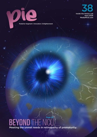
Welcome to PIE 38!
In this issue, the overall theme is Inside the Retina Lab a subject matter that, as a former research chemist, is closest to my heart…and we invite you to step right in.
This issue’s theme explores the rapid movement of knowledge from basic science benchtop to bedside, focusing on cutting-edge research, translational science and early clinical trial scoops that are defining the future of vitreoretinal care.
The research revolution in recent years is at its fastest than ever, driven by a deeper understanding of biology and the powerful integration of new technologies.
Inside this issue, we covered the future of personalized medicine in Genes Aren’t Destiny, challenging the myth of genetic certainty in inherited retinal disease.
Also, The AI Update tracks how artificial intelligence is moving beyond diabetic retinopathy screening to usher in the oculomics revolution. We also investigate a potential game-changer in drug delivery with the Topical Retina Revolution, exploring the novel idea of whether a simple eye drop can truly change the injection paradigm forever.
While innovation is critical, our commitment to clinical care remains a top priority. More so when children, especially newborn infants, are involved. Our Cover Story—Beyond the NICU, confronts head-on the unmet needs in retinopathy of prematurity (ROP), highlighting areas where immediate intervention, new thinking and the power of collaboration are vital.
In our Innovation section, Shining a Light or Just a Glimmer? takes a critical look at photobiomodulation for retinal diseases— a field holding immense promise for non-invasive treatment strategies that could revolutionize the management of conditions leading to vision loss.
Finally, we turn our lens to the realities of global practice and professional development. The Three-Injection Problem examines the harsh economics of anti-VEGF therapy in the Philippines, a compelling look at the challenges of accessibility and sustainability. As a Filipino, this is the most heart-touching piece in this issue, and I’m sure it will resonate with people from many other developing countries too.
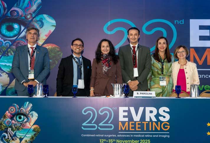
And for those dedicated to perfecting their craft, Through Her Lens—this issue’s Women in Ophthalmology feature—puts the spotlight on Dr. Gitalisa Andayani Adriono of Indonesia. In a country where geography, disease burden and late referrals stack the odds against effective retinal care, Dr. Adriono has not only spent decades sharpening her vitreoretina expertise, but is also actively training the next generation of specialists to meet this formidable challenge head-on. This perspective is complemented by our Conference Highlights from EVRS 2025 and AIOC 2026, highlighting cutting-edge surgical techniques, a worldwide view of vitreoretinal advancements, and the powerful synergy of science and culture in crafting a truly memorable conference.
This issue promises a journey through the most exciting and challenging frontiers in retina today.
We hope it inspires your practice and research!

Sincerely,
Gloria D. Gamat Chief Editor | PIE, CAKE and COOKIE Head, Editorial Department | Media MICE

Meeting the unmet needs in retinopathy of prematurity



Genes Aren’t Destiny The AI Update Inherited retinal disease and the myth of genetic certainty
From diabetic retinopathy screening to the oculomics revolution

Shining a Light or Just a Glimmer?
A critical look at photobiomodulation for retinal diseases
Topical Retina Revolution
Can a simple eye drop change the injection paradigm forever?
Links Triton2 imaging with photobiomodulation progress
Through Her Lens
Sharpening the craft of vitreoretina
From Pixels to Performance The Three-Injection Problem
When anatomy should not lead the conversation The harsh economics of anti-VEGF therapy in the Philippines
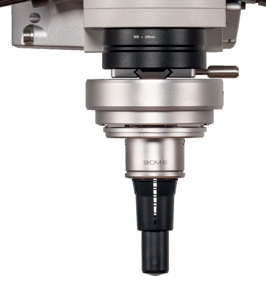

Evolved from the Original. Built for the Future.
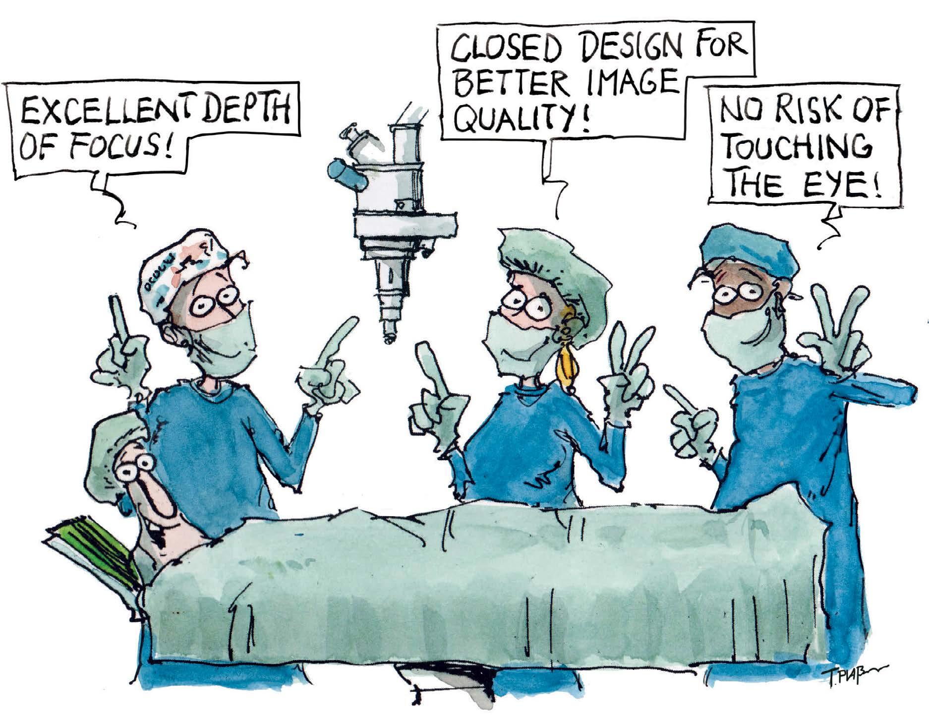
The all-new BIOM® 6 with its innovative closed design –crafted to improve precision, elevate performance, and meet the demands of advanced vitreoretinal surgery.
Optimized Depth of View
Minimized Light Disturbance
Improved Surgical Control

DR. ALAY S. BANKER
Banker’s Retina Clinic and Laser Centre Ahmedabad, India alay.banker@gmail.com
PROF. GEMMY CHEUNG
Singapore National Eye Centre (SNEC) Singapore gemmy.cheung.c.m@singhealth.com.sg


DR. ARSHAD KHANANI
Sierra Eye Associates; University of Nevada, Reno School of Medicine Nevada, USA arshad.khanani@gmail.com
DR. HUDSON NAKAMURA
Bank of Goias Eye Foundation Goiânia, Brazil hudson.nakamura@gmail.com
DR. BARBARA PAROLINI
Eyecare Clinic Milan, Italy parolinibarbara@gmail.com
DR. VEERAL SHETH
University Retina and Macula Associates; University of Illinois at Chicago, USA vsheth@gmail.com
Society Friends


We are looking for eye care professionals who can contribute articles to PIE. Interested? Let's talk! Send us an email at editor@mediamice.com.
To place an advertisement, advertorial, symposium highlight, video, email blast, or other promotion in PIE contact sales@mediamice.com

Matt Young CEO & Publisher
Gloria D. Gamat Chief Editor
Diana Truong Associate Editor
Andres Pacheco Art Director
Nhat Vo
Yura Yu Graphic Designers
Writers Chow Ee-Tan
Hazlin Hassan
Kendra Bruning
Marhiel Garrote
Michaela Perez
Dr. Sashwanthi Mohan Contributor
Matt Herman Head of Content Strategy
Hannah Nguyen COO
Travis Plage CFO
Ruchi Ranga Society Relations & Conference Manager
International Business Development
Brandon Winkeler
Robert Anderson
Sven Mehlitz
Published by


A “pathogenic” gene has long been treated as a crystal ball for IRDs. But what if the forecast isn’t so certain? Using population biobank data instead of cliniconly cohorts, one study challenges long-held assumptions about inherited retinal diseases and reshapes how we think about genetic risk, counseling and phenotype-first care.
By Dr. Sashwanthi Mohan
For years, inherited retinal degenerations (IRDs) have been held up as textbook examples of genetic determinism. Conditions such as retinitis pigmentosa, Leber congenital amaurosis and other inherited dystrophies are traditionally framed as monogenic diseases.
Find the faulty gene, predict the disease, and anticipate progressive vision loss, often culminating in blindness. Neat. Logical. And, as it turns out, not quite the full story.
A new large-scale genetic study from Mass General Brigham now challenges this long-held assumption. By analyzing population-level data rather than only affected patients, the researchers showed that genes believed to guarantee inherited blindness often did not lead to vision loss at all.
*
The findings force us to rethink how we interpret genetic results, especially in daily clinical practice.
Published in the American Journal of Human Genetics , the study examined 167
genetic variants across 33 genes classified as pathogenic for IRDs. Instead of focusing solely on patients already diagnosed with retinal degeneration, the researchers turned to two vast population biobanks: the All of Us Research Program in the United States and the UK Biobank.*

These datasets link genetic information to health records and—in the case of the UK Biobank—retinal imaging, allowing investigators to assess how often so-called disease-causing variants actually result in disease in the general population.
The findings were striking. Among nearly 318,000 participants in the All of Us cohort, 481 individuals carried IRDassociated genotypes, yet only 9.4% had a formal diagnosis of IRD. Even when broader definitions of retinal disease were applied, penetrance rose to just 28.1%.*
Similar results were seen in the UK Biobank, where only 16% to 28% of variant carriers showed any structural signs of retinal disease on imaging.* Put simply, most people carrying these variants did not have inherited blindness.
One key reason these results diverge from decades of genetic literature lies in how IRD genetics has traditionally been studied. Family-based and clinic-based cohorts dominated early research, meaning that individuals with significant vision loss were sequenced, while unaffected carriers were largely invisible.
This inevitably made variants appear highly penetrant. By including all carriers regardless of symptoms, population biobanks reveal a more complete picture. Genetic variants may increase risk, but they do not dictate inevitable visual loss.
The earlier studies were not wrong though, they were just incomplete. The new data show that even so-called “Mendelian” diseases can exhibit variable penetrance and expression, shaped by genetic modifiers, epigenetic regulation, systemic health, environmental exposures and time.
As a retina specialist, these findings echo what many of us observe in practice


but have struggled to reconcile with traditional genetic models.
Patients carrying variants labeled as pathogenic and associated with severe early-onset disease can present with minimal structural change on optical coherence tomography and retain good functional vision. Meanwhile, other patients with the very same variant may experience rapid progression to blindness.
If the gene is the same, why isn’t the disease?
This study is a reminder of the retina’s resilience.
A genetic variant may predispose, but it does not act in isolation. Modifier genes, metabolic factors, systemic health, inflammation and oxidative stress likely all influence whether photoreceptors ultimately function or fail. This has real consequences for clinical care.
The therapeutic implications are just as significant. Gene therapy has transformed the outlook for certain inherited retinal diseases, yet this study reinforces that treatment decisions must be phenotypedriven. We should
complex disease is far less distinct than once believed. Even classic Mendelian conditions exist on a spectrum shaped by interactions between genes, environment, biology and time. With its capacity for high-resolution imaging and functional testing, the retina is uniquely positioned to lead this shift in thinking.

This research does not diminish the importance of genetics in IRDs; in fact, it strengthens it. By moving beyond fixed thinking, we gain a more accurate and clinically meaningful understanding of visual loss.
For clinicians, it serves as a reminder to interpret genetic results with care. For patients and families, it offers hope grounded in real-world evidence. And for researchers, it raises a compelling question: why do some retinas remain unaffected despite proven genetic risk, and how can we help others do the same?
NguyenHo,©MediaMICEPteLtd,2026
treat patients, not mutations. Structural and functional evidence of disease should guide intervention.
Genes matter. But as this study makes clear, genes are not destiny.

Perhaps one of the most intriguing takeaways lies with the unaffected carriers themselves. Individuals who maintain good retinal structure and function despite carrying high-risk variants may hold clues to natural protective mechanisms.

Of course, genetic testing remains a cornerstone of IRD management, guiding diagnosis, prognosis, eligibility for gene therapy and family counseling. This study underscores the need for careful interpretation and thoughtful communication of results. Genetics should not be presented as fate. Patients deserve balanced explanations that reflect likelihood—not certainty—especially when penetrance is low.
As whole-genome sequencing becomes more common, retina specialists will increasingly encounter IRD-associated variants in asymptomatic individuals. A pathogenic gene label alone is insufficient without phenotype correlation. Retinal imaging, functional testing and long-term follow-up remain essential. Genetics may provide the map, but phenotype reveals the journey.
Understanding these modifier factors could open new therapeutic avenues, shifting focus from replacing non-functioning genes alone to strengthening the retina and preserving existing function. Such strategies could benefit patients across the retinal disease spectrum.
This study adds to growing evidence that the boundary between monogenic and

*Zaslavsky K, Chen L, Park C, et al. Low population penetrance of variants associated with inherited retinal degenerations. AJHG. 2026;113(1):P71-82.

Dr. Sashwanthi Mohan Specialist ophthalmologist in the Department of Ophthalmology at the Medcare Eye Centre, Dubai, United Arab Emirates. sashu23@gmail.com



FROM
SCREENING TO THE OCULOMICS REVOLUTION
There’s an AI arms race underway, and retina is the testing ground. What started as a debate about screening has escalated into predictive medicine, opensource disruption and foundation models that could outpace traditional workflows. If you think this is about DR detection, you’re already behind.
By Michaela Perez
While ophthalmologists were debating whether artificial intelligence (AI) could screen for diabetic retinopathy (DR), something bigger was quietly unfolding: the retina was becoming medicine’s most powerful diagnostic tool for conditions far beyond the eye.
When we first caught up with Assoc. Prof. Daniel Ting (Singapore) two years ago, ChatGPT had just launched and the questions were cautious: Could machines really diagnose eye disease? Would regulators approve autonomous screening?
Today, those questions miss the point entirely.
“Right now, I think the world is experiencing a very intense AI race,” Prof. Ting said when we met up with him again at the 18th Congress of the Asia-Pacific Vitreo-Retina Society (APVRS 2025, Manila). As co-director of the Artificial Intelligence in Medicine Institute (AIMI; Singapore) and a practicing retina specialist at the Singapore National Eye Centre (SNEC), he’s watching this transformation from both sides: the clinic and the cutting edge.
One of the most significant shifts has been the move from closed-source to open-source AI platforms. Early systems like ChatGPT 3.5, Gemini and Claude were proprietary black boxes. You could use them, but you couldn’t see how they worked or build on top of them.
That’s changing rapidly. “A closed-source system means that the developer would not have the chance to actually use it to build their own system,” Prof. Ting explained. “As opposed to the open-source model, when you actually release to the world, that encourages everyone to actually jump in.”
This isn’t just technical wonkery. Open-source AI accelerates innovation, brings in voices from resource-limited settings and creates accountability through transparency. When thousands of developers can examine and improve the code, the systems get better faster.
Regulators have largely embraced this direction, encouraging innovative uses of data while protecting intellectual property. “I’m quite glad to see that a lot of the regulators have taken an approach where they’re encouraging the data being used in an innovative way,” Prof. Ting noted.
The result? More data sharing, more collaboration and more rapid translation of research into clinical tools.
While the world was busy celebrating AI’s ability to detect DR, Prof. Ting and his colleagues were quietly working on something far more ambitious: using the eye to predict systemic disease before it manifests. Welcome to oculomics, the practice of using ocular imaging and biomarkers as windows into whole-body health.
The logic is elegant. The retina is the only place in the body where you can directly visualize microvasculature without cutting anything open. And since microvascular changes always precede macrovascular ones— problems start in small vessels before hitting big ones—retinal imaging offers a unique opportunity for early detection of cardiovascular disease, chronic kidney disease and other systemic conditions.
“The eye is the only organ in the entire body system that allows you to have direct visualization without needing to adopt an invasive approach,” Prof. Ting said.
Some AI systems already use retinal images to calculate coronary artery calcium scores—a task impossible for human readers. “If you ask the human to read the image and quantify the calcium scores based on the retina imaging, that is impossible. So the only way you can do it is through AI,” he said.
Imagine walking into a pharmacy for a routine eye exam and walking out with your cardiovascular risk profile, kidney function markers and metabolic health assessment—all from a single photograph. That’s where oculomics is headed, and it’s arriving faster than most people realize.
Having brilliant AI is meaningless if it never leaves the lab. This is why Prof. Ting’s role with the Community Collaborative for Ophthalmic Innovation (CCOI) matters. Led by former FDA director Dr. Malvina Eydelman (USA), CCOI exists specifically to bridge regulators, academics and industry.
“The one aim that CCOI is trying to achieve is to actually accelerate the translation of innovations into real-world settings,” Prof. Ting said.
But regulatory approval solves only half the problem. Clinical integration presents its own challenges. This is where ambient AI— systems that capture doctor-patient conversations, extract relevant


information and automatically populate electronic medical records— becomes transformative.
“As opposed to in the past where EMR [electronic medical records] was causing a lot of burnout from physicians and the physician had to keep looking at the computer, now the doctors can actually talk to the patients while having face-to-face contact,” Prof. Ting explained.
It sounds simple, but the implications are profound. Physicians can maintain eye contact, build rapport, and focus on healing rather than documentation. The human elements of medicine—connection, empathy, trust—survive even as AI eliminates the administrative drudgery that’s been crushing healthcare workers for decades.
If AI is embedded in every diagnostic device, surgical tool and medical record system, can you practice modern medicine without understanding how it works? Prof. Ting’s answer is unequivocal: no. “Whether you like it or not, AI is already here,” he said. “But how are you gonna use AI? That is the big question.”
His analogy is instructive: you don’t need to be a car engineer to drive, but once your hands are on the wheel, you’d better understand safe operation. “Every single thing we do in healthcare touches patient safety. I think 100% of medical students, residents and fellows will need to at least know the basic concepts of AI so that they can use it safely.”
This extends beyond clinical training to reshape hiring and team building across the industry. Companies that can hire two people with AI expertise might accomplish what previously required 10 or 20. Those who embrace AI will gain efficiency, and they’ll also operate on an entirely different competitive plane.
The industry is taking note. Major acquisitions of imaging-based AI companies signal that the field is consolidating rapidly. The next year will reveal whether these integrations spark genuine innovation or merely consolidate market share.
Prof. Ting’s team is developing multi-disease, multi-imaging foundation models—universal diagnostic systems that work across medical specialties, democratizing specialist-level skills globally.
They’re also building visual language models that don’t just detect conditions but generate human-like diagnostic reports. And through a co-founded company, they’re creating sovereign agentic AI that runs locally without internet connectivity or graphic processing unit (GPU) support.
“This model doesn’t really need GPU support. It runs in a very lightweight central processing unit (CPU)-based system,” Prof. Ting explained. “So it actually democratizes AI use in many different countries.”
That matters. If AI requires expensive hardware and constant connectivity, it remains accessible only to wealthy institutions. But lightweight, locally-run systems can go anywhere, bringing world-class diagnostics to the places that need them most.
What emerges from Prof. Ting’s work is a profound reframing: the retina isn’t just a subspecialty anymore. It’s a diagnostic portal into systemic health, a predictive tool for disease prevention and a proving ground for AI that will eventually transform all of medicine.
The regulatory frameworks are evolving in real time. Clinical workflows are being redesigned. Training paradigms are being rewritten. And the industry is betting heavily that this transformation is permanent.
For retina specialists, the message is that you’re not just treating the posterior segment. You’re at the forefront of medicine’s AI revolution. The question isn’t whether you’ll participate—AI is already in your imaging devices, surgical tools and EMR systems. The question is whether you’ll understand the systems making clinical decisions alongside you, or trust them blindly.
Prof. Ting’s advice? Learn how the engine works. Because your patients’ sight depends on it.

Editor’s Note
The insights provided by Prof. Daniel Ting in this article are based on MICE TV’s report of the 18th Congress of the Asia-Pacific Vitreo-Retina Society (APVRS 2025), held December 12–14 in Manila, Philippines.

Assoc. Prof. Daniel Ting Director of Singapore Health Service AI Office, Singapore National Eye Centre (SNEC) Chief Data and Digital Officer, and Head of AI and Digital Innovation at Singapore Eye Research Institute (SERI). daniel.ting.s.w@singhealth.com.sg


A Topcon Healthcare-sponsored lunch symposium at APAO 2026 in Hong Kong saw experts from Singapore and Italy spotlight multimodal imaging workflows with the DRI OCT Triton2 and 12-month photobiomodulation data in intermediate dry age-related macular degeneration.
Sponsored by Topcon Healthcare
Retinal care is becoming more precise, not simpler. Clinics generate vast amounts of optical coherence tomography (OCT) and OCT angiography (OCTA) data, yet the bottleneck remains human. The human related limitations remain the challenge. Success still depends on three variables: clean acquisition, efficient interpretation and the discipline to ignore misleading artifacts.
The margin for error is particularly narrow in intermediate dry age-related macular degeneration (AMD), where disease progression can occur while visual acuity appears deceptively stable. This raises the bar for imaging that classifies disease accurately and tracks subtle change, as well as therapies that improve outcomes without damaging surrounding tissue.
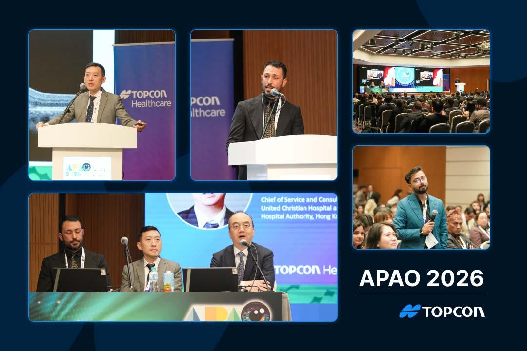
At the 41st Asia-Pacific Academy of Ophthalmology Congress (APAO 2026) in Hong Kong, Topcon Healthcare’s (Tokyo, Japan) lunch symposium Retinal Care Today: Imaging Precision to Therapeutic Progress addressed both sides of that challenge.
Prof. Kenneth Li (Hong Kong) opened with “exciting developments… transforming how we diagnose and manage retinal diseases.” Assoc. Prof. Kelvin Teo (Singapore) focused on multimodal imaging workflows, while Dr. Claudio Iovino (Italy) presented 12-month outcomes of photobiomodulation (PBM) therapy in intermediate dry AMD.
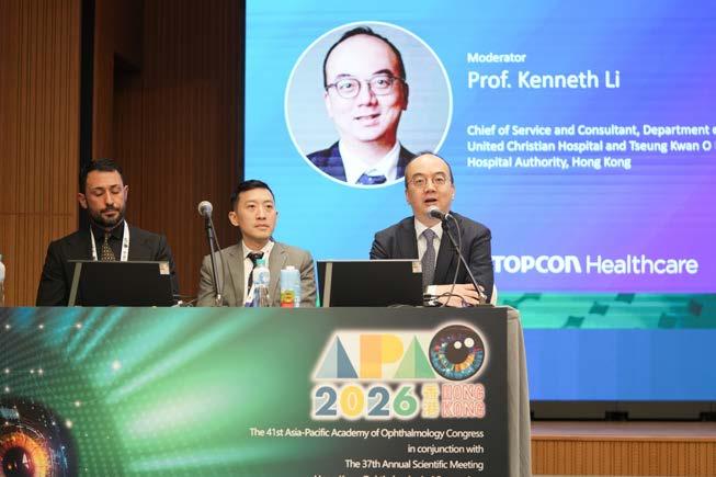
Prof. Teo’s argument was not that artificial intelligence (AI) will rescue weak inputs. It was that clinics still win or lose on the basics.

“Artificial intelligence is only as good as the source imagery we provide,” he said, stressing that advanced algorithms only help after acquisition quality has done the heavy lifting.1 In early Triton2 use, he pointed to fewer failed images, smoother multimodal correlation and greater confidence in what a single imaging session can justify clinically.
Small-pupil reliability and workflow gains. Prof. Teo focused on color fundus imaging through small pupils, a daily clinic constraint that rarely shows up in clinical trial slide decks. He presented a slit-scanning approach in which slit light is passed through the pupil and composited via a rolling-shutter mechanism, effectively minimizing glare and shadow and ensuring stable, high-quality imaging.
“The slit-scanning method essentially sends slit light to a small pupil and composites the color fundus image, reducing glare and achieving excellent quality color photographs regardless of pupil size,” he said. In his own clinic use, that translated into reliability. “I have not missed a single color fundus image in several months, regardless of how small the pupil.”
He tied that reliability to how clinicians actually read images. Rather than treating fundus photography, infrared reflectance, OCT and OCTA as separate stops, he described IMAGEnet 7 as a singlescreen review environment that displays multimodal data together so findings can be checked quickly across layers. That meant correlating clinical appearance and OCT structure in one view, without bouncing between stations or screens. It also reduced the time spent debating whether a suspicious OCT feature represents true pathology, an imaging
artefact, or simply a mismatch between structural and en face context.
Widefield OCT and OCTA for peripheral insights. Prof. Teo also discussed the value of widefield OCT and OCTA imaging up to 21 mm, bringing peripheral retinal context into clinical decisions often based solely on posterior pole data. In practice, he said the wider view helped him pick up nonperfusion patterns that standard fields may miss, particularly in diabetic retinopathy (DR) and polypoidal choroidal vasculopathy (PCV). He also avoided overselling it as a dye replacement.
“With the 21 mm wide-field scan and denoising technology, you can already detect areas of non-perfusion quite well,” he said, adding that where fluorescein angiography is not available, widefield imaging can still support a comprehensive evaluation.
Smart Denoise efficacy data. A key limitation of OCTA is distinguishing true blood flow signals from noise and distinguishing target-layer vasculature from projection artefacts. Prof. Teo described Smart Denoise as an AI-driven method to reduce artifacts and boost contrast in structural OCT and OCTA. 2
He stressed that denoising only helps if it improves readability without creating false lesions. In a local evaluation of 145 eyes, three ophthalmologists rated image quality as improved, with quantitative metrics showing better vessel visibility and no false pathology, reporting that the underlying retinal structure remained unchanged. Cleaner slabs, he argued, reduce noise-driven overinterpretation and make true vascular patterns easier to trust.
Prof. Teo then shifted the focus from technical features to clinical impact, using case-based examples to demonstrate how multimodal image overlays can improve interpretability and clinical
decision - making by overcoming the challenges associated with reliance on multiple standalone modalities.1
In Type 1 macular neovascularization (MNV), he described overlaying OCTA on color fundus photography and adjusting transparency to localize the neovascular network against visible landmarks. 1 The same approach also helped avoid misclassifying the case as AMD. Although intraretinal fluid and a double-layer sign suggested wet AMD, the overlay showed the vascular network corresponded with previous laser scars from branch retinal vein occlusion (BRVO), supporting secondary choroidal neovascularization rather than AMD.1
He applied a similar “classify before you treat” approach to geographic atrophy (GA), using color fundus photography to define the atrophic area, followed by OCT and OCTA to exclude MNV before considering newer GA therapies.1
In hemorrhagic cases suggestive of polypoidal choroidal vasculopathy (PCV), he described correlating an orange nodule on fundus imaging with OCTA flow and a sharply peaked pigment epithelial detachment on B-scan, then monitoring the response following anti-vascular endothelial growth factor (anti-VEGF) therapy.1 For DR and diabetic macular edema (DME), he highlighted widefield assessment of peripheral nonperfusion and OCTA visualization of neovascular tufts extending toward the vitreous.
These examples supported his broader point about integration. “The Triton2 is a very nice all-in-one platform that marries together the best in acquisition technology for OCT angiography, OCT and color fundus imaging,” he said.
PBM IN INTERMEDIATE AMD
Dr. Iovino’s presentation addressed a different challenge in retinal care. Intermediate AMD is a stage where visual acuity can stay relatively good while drusen burden, oxidative stress and retinal pigment epithelium (RPE) strain continue to evolve. In this window, clinicians need
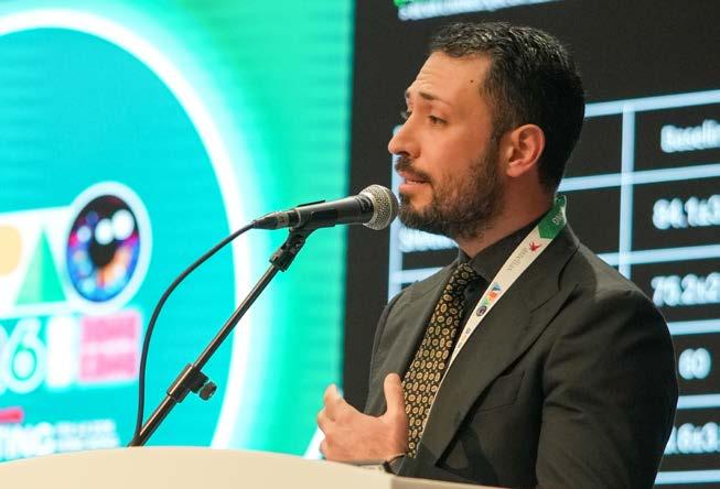
endpoints that change before vision declines, along with interventions safe enough to consider before the disease progresses to late stage.
Dr. Iovino presented PBM via eye-light® (Espansione Group, Italy) as an approach designed for earlier intervention, supporting the concept with its underlying mechanisms and 12-month clinical outcomes.
PBM fundamentals . Dr. Iovino defined PBM as low-level light therapy (LLLT) using non-coherent visible light to induce cellular responses without thermal damage. “Photobiomodulation therapy works within the spectrum of non-coherent light between 590 and 625 nanometers,” he said. “It does not cause any thermal damage, making it a very safe light source for our patients.”
He described three biological layers: improved mitochondrial metabolism and adenosine triphosphate (ATP) production, reduced oxidative stress and inflammatory cytokine signaling and modulation of cellular survival pathways to reduce apoptosis and support regeneration. The broader ophthalmic literature similarly describes PBM as influencing mitochondrial function, oxidative stress pathways and inflammation, aligning with mechanisms implicated in degenerative retinal disease.3
Trial design and protocol details. Dr. Iovino described a multicenter randomized clinical trial across six European centers involving 138 eyes with AREDS Grade 2 and 3 AMD. Key exclusions included geographic atrophy, choroidal neovascularization, pregnancy and active neurologic disorders, keeping the cohort focused on intermediate disease biology rather than late-stage endpoints.
He reported a two-cycle protocol separated by a rest period. “The treatment protocol consists of two cycles,” he said. Cycle 1 delivered two treatments per week for four weeks (eight sessions total), followed by a 6-month rest period. Cycle 2 then provided two treatments weekly for 3 weeks. Each session lasted 12 minutes, split between a yellow mask and a red mask at six minutes each. Published PBM studies in dry AMD have also used structured, multi-session protocols with short exposures, supporting feasibility for repeated cycles in clinic without invasive procedures.4
Endpoints and 12-month outcomes. Dr. Iovino’s primary endpoint was drusen volume change over time, reflecting the reality that many intermediate AMD patients start with relatively good acuity. Best-corrected visual acuity (BCVA) was tracked as a secondary endpoint.
“After 12 months, we observed statistically significant reduction in mean drusen volume in the photobiomodulation group while the sham group showed slight increase,” he said.
Functional results moved in the same direction. He reported a mean gain of 1.3 ETDRS letters in the PBM group versus a 2-letter loss in the sham group, with a statistically significant difference. He also highlighted a stratified signal in patients starting above 80 ETDRS letters, where the sham group showed decline, suggesting that waiting for vision to deteriorate may miss a window in which earlier intervention could be beneficial.
Regarding safety, he reported no devicerelated adverse events. He also noted one case of GA onset in the PBM group compared with four cases of choroidal neovascularization in the sham group, as presented.
Imaging observations. Dr. Iovino’s imaging nuance was the part most likely to stick with retina specialists who have seen drusen collapse precede bad outcomes. He described progressive drusen reabsorption without accompanying RPE atrophy,
suggesting the effect was not simply drusen disappearance with associated damage in surrounding tissue.
“The drusenoid material reabsorbed without associated retinal pigment epithelium atrophy,” he said. He tied this to RPE cell stimulation, arguing that PBM may improve phagocytic capacity to clear drusenoid material and enhance pump function, which could also explain fluid dynamics changes observed in selected cases.
He briefly referenced a chronic central serous chorioretinopathy (CSCR) case with a large serous pigment epithelium detachment that gradually flattened and showed improved retinal sensitivity on microperimetry.
Related publications from his group have described PBM in chronic CSCR settings, including a pilot study and a separate case report focused on serous pigment epithelium detachment behavior over follow-up.5,6 These reports do not replace randomized AMD trial data, but they support his broader claim that PBM’s effects may involve RPE function and fluid handling, not only drusen metrics.
The symposium’s overarching theme, as summarized by Prof. Li, was that progress in retinal care is no longer just about seeing more, but about seeing with greater certainty. By bridging the gap between “trial-perfect” conditions and the “small-pupil” realities of everyday clinical practice, technologies such as the Triton2 help address the human bottleneck at its source. When acquisition becomes a reliable and stable baseline diagnostic decision rather than a variable, the clinician’s role shifts from troubleshooting image quality to making faster, data-driven diagnostic calls.
This reliability creates the necessary foundation for emerging therapies like PBM. As Dr. Iovino’s 12-month data suggests, treating intermediate dry AMD requires a delicate balance: achieving structural improvements, such as drusen reduction, without the “biological tax” of RPE damage. 3,5,6 This is

a significant shift in the dry AMD philosophy—moving away from “watchful waiting” toward a proactive, nonthermal intervention.
Ultimately, the combination of high-fidelity multimodal imaging and targeted cellular therapy changes the clinical equation. When “clean” data becomes the standard and monitoring can separate true therapeutic signals from noise, the move toward earlier intervention becomes less a leap of faith and more a measured, evidence-based evolution in patient care.
Editor’s Note
The 41st Asia-Pacific Academy of Ophthalmology Congress (APAO 2026) was held from 5-8 February in Hong Kong. Reporting for this story took place during the event. A version of this article was first published on MediaMICE.com
1. Teo K. Increased value of swept source OCT with high quality color fundus: The new Triton 2. In: International Congress on OCT and OCT Angiography (ICOOR) 2025; 2025 Dec; Florence, Italy.
2. Topcon Healthcare. DRI OCT Triton2 SweptSource Optical Coherence Tomography Slit-Scan Fundus Camera. Brochure M000253O-1 (OUS EN 2601).
3. Tan NRX, Chan KE, Lim BXH, et al. Photobiomodulation: evidence and applications in ophthalmology. Curr Opin Ophthalmol. 2025;36(5):345-381.
4. Borrelli E, Coco G, Pellegrini M, et al. Safety, tolerability, and short-term efficacy of low-level light therapy for dry age-related macular degeneration. Ophthalmol Ther. 2024;13(11):2855-2868.
5. Iovino C, Termite AC, Boscia G, et al. Photobiomodulation therapy with low-level light in chronic central serous chorioretinopathy: a pilot study. Ophthalmol Ther. 2026;15(1): 193-203.
6. Iovino C, Damiano L, Piccirillo V, et al. Photobiomodulation therapy for serous pigment epithelium detachment in chronic central serous chorioretinopathy. Retin Cases Brief Rep. 2025;19(6):766-770.


In retina, a few letters can change everything…or very little.
Photobiomodulation’s LIGHTSITE III trial reports a modest visual acuity gain for intermediate dry AMD, but as the numbers flicker into view, experts are asking the question that always matters most: signal or statistical glow?
By Marhiel Garrote
Across retinal clinics worldwide sits a quiet but immense population of patients suffering from intermediate, dry age-related macular degeneration (AMD). These patients are not candidates for intravitreal injections like those with neovascular AMD, but what is clear is they are at risk for progressive loss of their central vision. And therein lies the therapeutic gap.
Enter photobiomodulation (PBM), a non-invasive, light-based intervention that has attracted attention following the LIGHTSITE III trial and subsequent U.S. Food and Drug Administration (FDA) authorization for select patients with dry AMD. But as emphasized by Dr. Susan Bressler (United States) during her lecture at the 18th Asia-Pacific Vitreo-retina Society Congress (APVRS 2025, Manila), the question is not simply whether a signal exists. The more important questions are how strong that signal is, how clinically
meaningful it may be and whether it justifies early adoption in practice.
WHY SHINE LIGHT ON THE RETINA?
According to Dr. Bressler, “It is the application of light—both light within the visible and the near infrared spectrum— to a specific tissue, in this case the eye, to promote a beneficial cellular effect.”
PBM uses red, yellow and nearinfrared wavelengths to stimulate the mitochondria, often described as the powerhouse of the cell. The working theory is that stimulation of mitochondrial components may stabilize cellular metabolic function and result in cytoprotection.


In the LIGHTSITE III trial, a PBM system delivered three wavelengths across nine sessions over three to five weeks, with treatment cycles repeated every four months for up to two years.1
WHAT LIGHTSITE III SHOWED
Among 100 participants (148 eyes), randomization favored PBM over sham control in a 2:1 ratio. The primary endpoint was change in best-corrected visual acuity (BCVA) at month 13.1
In dry AMD—where chronic oxidative stress and progressive cellular dysfunction are central—this mechanism appears biologically plausible. Yet as translational medicine repeatedly reminds us, plausibility is not the same as proof. Clinical trials are ultimately needed to determine whether that biological rationale translates into meaningful patient outcomes.
Results showed the following:1
• 5.4-letter average improvement in the photobiomodulation group 2
• 3-letter average improvement in the sham group
• 2.5-letter difference favoring PBM
While five letters correspond roughly to one line on the ETDRS chart, a three-line (15-letter) gain has traditionally served as a benchmark for moderate visual improvement in many clinical trials. Against that backdrop, the 2.5-letter difference, though measurable, remains modest.
Perhaps more notable is the improvement seen in the sham group. As Dr. Bressler noted, “the 3 letter on average gain in sham raises the possibility that these observed gains—in both the sham group and the photobiomodulation group—may be placebo effects or regression to the mean, or both at play.”
REGRESSION TO THE MEAN
Regression to the mean refers to the tendency of an unusually low measurement to move closer to the average upon repeat testing, even without intervention.
Dr. Bressler illustrated this with a familiar clinical scenario. A patient who typically sees 20/25 might measure 20/50 on a day complicated by fatigue or dry eye, qualify for trial enrollment and later return to 20/25. The apparent “improvement” may simply reflect normal variability.
Trial entry criteria in LIGHTSITE III allowed visual acuity as low as 20/1001— an atypical cohort compared with the broader intermediate AMD population, which generally has better baseline vision.
For comparison, the large-scale AREDS2 study—one of the landmark trials in intermediate AMD, enrolling thousands of eyes across diverse populations—reported that about 50% of eyes were between 20/25 and 20/40, and only 12% were worse than 20/40. In contrast, roughly 30% of eyes in LIGHTSITE III fell into the lower-vision range at baseline.1,2
In smaller trials with greater baseline variability, outcomes can become more sensitive to statistical artifacts.
Prespecified secondary outcomes examined functional and anatomical measures. “For the functional outcomes, they were unable to find support in any of the battery of functional outcomes that differentiated the two treatment groups,” Dr. Bressler explained.
There was also no difference in subretinal pigment epithelium (sub-RPE) drusen volume change and no difference in geographic atrophy (GA) growth at 13 months. 1 If a therapy is truly disease-modifying, structural correlates are typically expected. Their absence raises an important question: are the visual gains genuine disease effects or simply fluctuations?
Another issue raised during Dr. Bressler’s talk was conversion to neovascular AMD. In the LIGHTSITE III trial, approximately 5% of eyes in the PBM group converted to neovascular AMD compared with 2% in the sham arm.1
In absolute terms, the numbers are small. But in retinal medicine, even small imbalances matter, particularly in a disease where conversion fundamentally alters prognosis, treatment burden and visual trajectory. While the trial was not powered to definitively assess differences in conversion rates, the observation invites continued vigilance.
“Given all the things that we’ve just mentioned, it clearly warrants further study rather than early adoption of this treatment,” Dr. Bressler noted.
Her position does not dismiss PBM. Rather, it reflects a long-standing principle in medicine: regulatory clearance does not automatically translate into widespread clinical adoption.
Beyond efficacy and safety, the discussion also turned to mechanisms. The mitochondrial hypothesis is undeniably attractive. AMD involves oxidative stress, metabolic compromise and progressive cellular dysfunction. Stimulating mitochondrial activity to promote cytoprotection fits neatly into that biological narrative.
During the Q&A session, however, a cautionary perspective emerged. Dr. Neil Bressler (United States), one of the other speakers at the same APVRS 2025 symposium, remarked, “You get a result from a clinical trial, and then, sometimes, people go back and try to explain it by what’s in the translational or basic science literature.”

Fewer new GA lesions were reported in the PBM group. However, patients in the sham group were older, and no p-value adjustment was performed for multiple analyses.1 According to Dr. Bressler, these factors complicate interpretation.

The implication is subtle but important. When clinical signals are modest, there is a natural temptation to anchor them

to compelling biological explanations. But mechanisms should support data, not compensate for it.
PBM may indeed influence mitochondrial pathways. The biological plausibility is real. Yet until clinical outcomes demonstrate consistent, reproducible and structurally correlated benefit, mechanistic elegance remains an adjunct—not a substitute—for robust evidence.
BETWEEN THE PROMISE AND PROOF
Intermediate dry AMD represents one of the largest therapeutic gaps in retina care. Millions of patients sit in a gray zone: not yet candidates for anti-VEGF injections, yet clearly at risk for progression.
PBM brings several appealing elements:
• A non-invasive approach
• A biologically plausible mechanism
• A structured treatment protocol
• A statistically significant, though modest, visual signal
• Regulatory approval in selected regions
Yet key questions remain:
• Is a 2.5-letter difference sufficient to justify routine adoption?
• Are the observed gains durable beyond the 13-month mark?
• Will larger and more diverse populations replicate these findings?
• Can future studies demonstrate clear anatomical correlation with functional improvement?
• Does long-term safety remain stable across repeated treatment cycles?
Retinal science has long maintained high evidentiary standards, particularly for interventions targeting chronic, progressive disease. Large-scale trials, such as AREDS2, have shaped expectations around sample size, follow-up duration and population diversity. Against that backdrop, LIGHTSITE III may be best viewed as a signal-detection study: suggestive, intriguing, but not yet definitive.
The responsible position lies somewhere between dismissal and premature enthusiasm. PBM may indeed represent an emerging modality worthy of continued investigation. It may even prove to be an important complement in the management
of intermediate AMD. For now, photobiomodulation remains a careful glimmer—illuminating possibility while the evidence slowly gathers more light around it.
Editor’s Note
The insights provided by Dr. Susan Bressler in this article are based on her presentation at the 18th Congress of the Asia-Pacific Vitreo-Retina Society (APVRS 2025), held December 12–14 in Manila, Philippines.
1. Boyer D, Hu A, Warrow D, et al. LIGHTSITE III: 13-Month Efficacy and Safety Evaluation of Multiwavelength Photobiomodulation in Nonexudative (Dry) Age-Related Macular Degeneration Using the Lumithera Valeda Light Delivery System. Retina. 2024;44(3):487–497.
2. National Eye Institute. Age-Related Eye Disease Studies (AREDS/AREDS2). National Institutes of Health. Available at: https://www.nei.nih.gov/ eye-health-information/clinical-trials/age-relatedeye-disease-studies-aredsareds2. Accessed on February 15, 2026.

Dr. Susan Bressler
Professor of Ophthalmology at the Wilmer Eye Institute, Johns Hopkins University. She specializes in medical retinal disorders, vitreoretinal disease and retinal surgery.
s.bressler@jhmi.edu
An ophthalmologist and a pediatrician take turns at the mic, swapping verses in place of case notes as they navigate one shared challenge: retinoblastoma.
What follows is both tender and telling. A glimpse into the delicate choreography of diagnosis and treatment, where no one works solo. Through its measured lines, the poem sets the stage for something bigger than either voice alone: a quiet testament to collaboration, and to the unwavering commitment behind every effort to safeguard a child’s sight…and future.
In their own words…
Ophthalmologist
In the realm of the eye, I stand and stare, Through lenses and lights, with a gentle glare. For children with orbs, so pure and so white, I search for the shadows that shouldn’t be right.
Pediatrician
In the world of the young, I stand by your side, Guiding their health, in you I confide. Tell me, dear friend, of this thing you see, In the eyes of the young, what could it be?
Ophthalmologist
It’s retinoblastoma, a foe quite sly, Hiding and lurking in a young child’s eye. A tumor it is, malignant and rare, But with swift intervention, there’s hope in the air.
Pediatrician
Such tiny beings, with battles so grand, I admire your work, lend a helping hand. How do we combat this intruder so vile?
To ensure for the child, a future worthwhile?


Ophthalmologist
With lasers and cryo, we’ll freeze it away, Or with chemotherapy, we’ll keep it at bay. The journey’s not easy, tears may be shed, But hope is our beacon, where we’re being led.
Pediatrician
Together we stand, as guardians of sight, With knowledge and care, we’ll make it all right. For every young patient, we’ll give all we’ve got, To ensure that this battle is one that’s hard-fought.
Ophthalmologist
In unity, we’ll face this challenge head on, With love, care, and science, till the darkness is gone. For the eyes of the young, so innocent and clear, Deserve a bright future, one free from fear.
Reprinted with permission from JNV.


Inside retinal research labs, a once-dismissed idea is getting a second look, with considerably better science behind it. Could an eye drop meaningfully treat diseases that currently require a needle inside the eye?
By Hazlin Hassan
For conditions such as age-related macular degeneration (AMD) and diabetic macular edema (DME), intravitreal injections have transformed outcomes. As Dr. S. Patricia Becerra (United States), senior investigator and chief of the Section of Protein Structure and Function at the National Eye Institute (NEI) emphasized, “Their impact on vision outcomes is undeniable.”
And yet, she added, effectiveness does not eliminate burden. “These are lifelong diseases requiring repeated treatments over many years. The cumulative burden—logistical, psychological and financial—is real for both patients and clinics.”
Assoc. Prof. Dr. Ovidiu Samoilă (Romania), of the Department of Ophthalmology at Iuliu Hatieganu University of Medicine and Pharmacy, noted that “topical therapy offers a fundamentally different risk-benefit profile compared with intravitreal injection.”
“First, it eliminates the need to breach the ocular globe. Even microincisions created by fine-gauge needles carry a measurable risk of endophthalmitis, intraocular inflammation, retinal detachment and patient anxiety related to the invasive act itself,” he continued. “Avoiding trans-scleral penetration, therefore, addresses both safety and psychological burden.”
With retina clinics across Europe performing millions of injections annually, that burden stretches to capacity and specialist time.

“Non-invasiveness improves both patient adherence and healthcare system efficiency,” Dr. Samoilă noted. “Self-administration at home aligns with established eye-drop behaviors, eliminating logistical barriers such as travel, caregiver coordination and clinic wait times.”
That flexibility may be more important than it first appears. Longacting biologics and gene-based therapies can remain active with little opportunity for adjustment.
“Once administered, they can persist with limited opportunity for reversal,” Dr. Becerra noted. “In contrast, topical therapy can be adjusted or stopped immediately if safety concerns arise.” For chronic retinal disease, that controllability is strategically valuable. And in long-term retinal care, stability can be as valuable as potency.
“Intravitreal bolus injections generate high initial intraocular concentrations followed by exponential decline, leading to cyclical peaks and troughs in drug exposure,” Dr. Samoilă added. “In contrast, appropriately-designed topical delivery systems could enable more continuous tissue exposure, potentially stabilizing retinal homeostasis and reducing disease reactivation intervals.”
But for this to happen, biology must cooperate.
The eye did not evolve to welcome pharmaceuticals. “[Its] anatomy is designed to protect the retina,” Dr. Becerra explained.
Tear turnover, epithelial tight junctions, scleral structure and choroidal circulation all limit drug access. For decades, these barriers led many to conclude that topical retinal therapy was unrealistic. Findings from Dr. Becerra’s laboratory suggest, however, “a more nuanced reality—the barrier is quantitative rather than absolute.”
Small amounts of drug can reach the retina. The decisive factor is whether those small amounts are biologically meaningful. “The challenge isn’t whether the retina can be reached but whether the drug’s design maximizes the effect of limited exposure,” she said.
This reframing—viewing retinal delivery not as impossible but as an optimization challenge—marks an important conceptual shift in the field.
For Dr. Samoilă, the transconjunctival–transscleral route appears particularly promising. “The conjunctiva is more permeable
The challenge isn’t whether the retina can be reached but whether the drug’s design maximizes the effect of limited exposure.
than the cornea,” he explained. “The sclera, although thick and paucivascular, possesses a large surface area and a hydrated collagenous matrix that can allow passive diffusion toward the choroid and retina.”
The barrier, however, remains formidable. “Importantly, the biological barriers themselves have not changed. What has evolved is our capacity to engineer around them,” he continued. That engineering—increasingly sophisticated—is where the real story unfolds.
The most credible strategies now emerging combine molecular potency, pathway specificity and compatibility with ocular anatomy. Among these, peptides are attracting significant attention.
Peptides offer high specificity and reduced off-target effects, Dr. Becerra explained. They can modulate biological pathways rather than simply block a target, and they may be effective at low concentrations because activated pathways amplify biological effects.
Her team has focused on H105A, a peptide derived from pigment epithelium-derived factor (PEDF).1 Importantly, it is gene-agnostic.
“H105A does not correct a defective gene,” she said. “Instead, it modulates convergent cell-death pathways—oxidative stress, apoptotic signaling and mitochondrial dysfunction—common across genetically distinct subtypes and stress-induced injury.”
Continued on page 25

Premature infants are surviving in record numbers, but their retinas are paying the price. No longer a mystery of biology, ROP is now a systems problem hiding in plain sight. From oxygen protocols to AI screening, this deep dive explores why preventable blindness still persists, and what must change to finally outpace the “third wave.”
By Diana Truong

The paper-thin eyelids of a 28-week premature infant barely conceal the drama unfolding behind them. Inside, retinal vessels meant to nourish a developing eye can veer off course—sometimes chaotically—triggering traction, detachment and lifelong blindness. This is retinopathy of prematurity (ROP), and despite decades of progress in neonatology and ophthalmology, it remains stubbornly prevalent.
“One child blind from ROP is one child too many,” said specialist Dr. Rachelle Anzures, who is part of the ROP working group in the Philippines. Globally, the disease continues to rank among the leading causes of preventable childhood blindness, affecting at least an estimated 50,000 children each year.1
The frustrating part for clinicians is that ROP is not an unsolved mystery. Screening protocols are well established. Treatments are highly effective when delivered on time. Pathophysiology is extensively studied. And yet, gaps in the care pathway persist.
The uncomfortable question lingers in neonatal intensive care units (NICUs) and retina clinics alike: Why does a largely preventable disease continue to steal sight worldwide?
THE PARADOX OF PROGRESS
The answer begins with a paradox. ROP is, in many ways, a byproduct of success. Advances in perinatal and neonatal care mean smaller and more premature infants survive. But survival comes with risk.

One Asian country illustrates the scale of the challenge. “India has a very large population and the birth rate is extremely high. We have the most preterm babies born in the world,” said Dr. Jaydeep Walinjkar, an ROP specialist in India. “The ratio to the number of babies who survive because of the high and good NICU care is very less compared to the ROP specialists who can cater to them. That is why it’s becoming a very big burden in India.”
This dynamic has fueled what many describe as the “third wave” of the ROP epidemic. As survival improves in low- and middleincome countries—often without parallel investment in ROP infrastructure, training and referral systems—the epidemiology has shifted. Yet the underlying vulnerability remains unchanged: an incompletely vascularized retina exposed to the stresses of life outside the womb.
Timing, as retina specialists know, is everything.
Normal retinal vascularization begins at the optic nerve around 16 weeks’ gestation and progresses centrifugally, reaching the periphery near term. Premature birth interrupts this orderly process. In the extrauterine environment, fluctuating oxygen levels, systemic instability and inflammatory stressors can tip the balance toward vaso-obliteration followed by pathologic neovascularization. 2
The disease is not present at birth. It evolves, and that evolution unfolds on a tight biological clock. “ROP has a small window period for examination and treatment. If you miss that, it’s game over almost,” emphasized Dr. Anzures. “The baby is not born with ROP...The ideal [screening time] would be 20 days from birth.”
The tension lies in aligning that biological window with real-world logistics. “We still lack ROP screeners... mostly it’s in the urban areas, and in the provincial areas sometimes the baby has to travel far just to have their eyes screened,” noted Dr. Anzures.
Dr. Walinjkar sees a similar pattern in India, where traditional timelines may be too conservative for today’s NICU populations. “The traditional concept was after four weeks or one month of birth. But in my opinion, two to three weeks [is better] because we have very premature or very low birth weight babies coming in who develop ROP even before their first screening.”
That clinical intuition is supported by data. A study by Vinekar and colleagues demonstrated that screening at two to three weeks rather than the conventional four to six weeks allowed identification of severe ROP requiring treatment roughly two weeks earlier.3 In a disease where progression from early stage to treatment-requiring disease can be rapid, those two weeks matter.
Once abnormal vascularization accelerates, staging can advance from mild demarcation lines to extraretinal fibrovascular proliferation and tractional detachment. Aggressive posterior ROP, in particular, leaves little room for delay. The narrower the posterior zone, the faster the clock seems to tick. 2
Overlay this biology with workforce limitations, and the cracks widen. “The specialists are not enough to handle the burden,” said Dr. Walinjkar. Delayed referral cascades into delayed examination, delayed treatment and preventable blindness.
Even in regions where specialists are available, coordination is not guaranteed. ROP care is inherently multidisciplinary, spanning obstetrics, neonatology, nursing, ophthalmology and family education.
“Taking care of the preterm babies’ vision is a team effort,” said Dr. Anzures. “It should be a collaboration from the obstetrician to the pediatrician to the ophthalmologist to the parents and also the nurses, the NICU nurses, and also the midwives, allied health. So everybody should be in it together to save the vision of the baby.”
Breakdowns often occur at the referral interface. “Sensitization of the pediatricians, neonatologist and the associated medical support staff...is very important because they are the ones who are going to refer,” explained Dr. Walinjkar. “If they don’t refer, it’s very difficult to catch these babies at the right time.”
A systematic review by Fierson et al. underscored this point, showing that implementation of standardized screening protocols, combined with improved communication between neonatologists and ophthalmologists, significantly reduced missed or delayed examinations. 4 Protocols alone are insufficient; they must be embedded within a functioning system.
Geography compounds the challenge. In many countries, the small number of ROP-trained ophthalmologists cluster in metropolitan centers. Infants born in rural settings face longer travel times, inconsistent follow-up and higher attrition.
Screening criteria themselves are also under scrutiny. Traditional guidelines rely primarily on gestational age and birth weight, typically recommending screening for infants under 30 to 32 weeks’ gestation or weighing less than 1.5 kilograms. These thresholds were developed largely from data in highincome countries.
In settings like India, those thresholds may miss infants at risk. “We get ROP in babies who are much higher in birth weight compared to the traditional standards across the world and also the number of weeks they are born,” said Dr. Walinjkar.

According to Dr. Anzures, the Philippines responded by broadening its referral criteria with “STOP,” which stands for sepsis, blood transfusion, oxygen supplementation and prematurity with unstable course.
This expanded framework acknowledges what translational research has long suggested: ROP is not solely a function of immaturity. It is the product of a complex interplay among oxygen exposure, inflammation, transfusion practices, nutrition and systemic instability. Risk models that integrate dynamic clinical variables, rather than static birth metrics alone, may ultimately provide more precise triage.
Few areas in pediatric ophthalmology illustrate translational science as clearly as ROP.
Early interventions relied on cryotherapy, targeting the avascular peripheral retina to reduce angiogenic drive. Laser photocoagulation refined that approach, offering more controlled ablation with improved structural outcomes. The field then pivoted again with the recognition that vascular endothelial growth factor (VEGF) plays a central role in pathologic neovascularization. 2
“The two current treatments of choice, which are virtually 100% effective, are an injection into the eye of medicine which stops the blood vessels from misbehaving, and then the second form of treatment is using laser,” said ROP educator Dr. Paul Runge. “Those are readily accessible and easily administered really throughout the world, whether that be the Western world or the third world. But it’s just a matter of getting the treatment information out to the folks that need it.”
The introduction of anti-VEGF agents into the ROP armamentarium marked a turning point. Repurposed from adult retinal disease, these agents offered
particular advantages in posterior disease, especially zone I ROP. They allowed more physiologic vascularization of the peripheral retina compared to laser, at least in the short term.5
Yet, as with all translational leaps, new questions emerged. A 2018 study by Stahl and colleagues raised concerns about systemic absorption of anti-VEGF agents in premature infants and potential downstream effects on developing organs. Serum VEGF suppression after intravitreal injection prompted debate about long-term safety, optimal dosing and retreatment intervals. 6
The BEAT-ROP trial7 and subsequent studies shifted practice patterns, but dosing strategies remain heterogeneous worldwide. Ongoing research is exploring lower doses to minimize systemic exposure while preserving efficacy.
The balance between ocular benefit and systemic risk remains an active area of investigation, underscoring that translational science does not end at approval or publication; it continues in post-market vigilance and iterative refinement.
If workforce constraints are the bottleneck, technology may be the release valve. In India, community-based screening models have demonstrated that non-ophthalmic personnel, equipped with wide-field fundus cameras, can capture images for remote interpretation.
“India has experimented extensively in ROP screening at a community level. And there are very good models, especially in South India and some in North India,” said Dr. Walinjkar. “But that has to be disseminated across India. It’s very localized, and those models are not working across India.”
The Philippines has built a national ROP network that maps screening and
treatment centers, providing transparency around available services. 8 In an archipelago, telemedicine must contend with uneven internet infrastructure and geographic fragmentation, but digital coordination is steadily improving reach.
Artificial intelligence (AI) is increasingly entering the conversation. “Along with AI, it’s going to do wonders in the future if we can really make it work,” said Dr. Walinjkar. “There are models where we can predict ROP in babies who haven’t even developed it.”
A 2018 study by Brown et al. demonstrated that a deep learning algorithm could identify referral-warranted ROP with high sensitivity and specificity, suggesting that AI-assisted grading may enable taskshifting without compromising safety.9 For regions with limited specialist availability, such systems could triage cases effectively, reserving ophthalmologist time for confirmation and treatment.
At the hardware level, innovation is also reducing barriers. “The traditional concept was high-end, very sophisticated equipment with a lot of expenditure involved and highly trained personnel, but that has changed over the last 10 years,” said Dr. Walinjkar. “We have very low-cost fundus cameras, screening tools, very well-trained non-ophthalmic, nonmedical personnel who can actually go to the field and screen these ROP babies with these devices.”
Taken together, these developments suggest a gradual shift from a specialistdependent model to a networked ecosystem in which image capture, AI support and centralized expertise intersect.
Clinical innovation alone cannot close the gap. Policy must follow. In India, partnerships between government programs and non-governmental organizations have expanded geographic reach.
“The models where the government staff, the government and the NGOs have come together has given us a greater exposure or area where we can cover because NGOs have limitations,” explained Dr. Walinjkar. “If you can join hands with the government organizations, they have a wider reach across all the states in India.”
In the Philippines, the inclusion of ROP screening under the national health insurance package for prematurity represented a tangible policy win, according to Dr. Anzures. Coverage reduces financial barriers and signals institutional commitment.
The economic argument is compelling. “It’s much, much cheaper to intervene and save a child’s vision than to look after them over their lifetime,” argued Dr. Runge. “It’s really a no-brainer, and I think once we get that message across, I think people will understand and that they will allocate the resources to help us solve this problem.”
Cost-effectiveness analyses published in the American Journal of Ophthalmology have reinforced this perspective, demonstrating that universal screening for extremely preterm infants falls well within accepted cost-effectiveness thresholds.10 In purely economic terms, prevention outperforms lifelong disability support.
Even when acute disease is successfully treated, the story does not end at discharge. Long-term studies, including work published in the British Journal of Ophthalmology, show that children treated for ROP have high rates of refractive error, strabismus and significant eye problems by early school age.11
This reality reframes ROP not as an episodic NICU event but as a chronic risk state. Lifelong surveillance is often warranted, yet structured follow-up programs are inconsistent, particularly in resource-limited settings where even acute care strains capacity.
As the field looks forward, several research threads converge.
First, more precise risk stratification is needed. Current criteria based on gestational age and birth weight cast a wide net. Some infants undergo multiple stressful examinations without ever developing significant disease, while others at the margins progress unexpectedly. Biomarkers, predictive algorithms incorporating longitudinal physiologic data, and AI-driven risk models could refine targeting and reduce unnecessary interventions.
Second, oxygen management remains an area of active debate. The relationship between oxygen saturation targets, mortality, neurodevelopmental outcomes and ROP risk is complex. Large neonatal trials have informed practice, but consensus on optimal saturation ranges continues to evolve. The challenge is not merely setting targets but ensuring consistent implementation in busy NICUs with varying resource levels.
Third, the long-term systemic effects of anti-VEGF therapy demand continued surveillance. As cohorts of treated infants age, registries and longitudinal studies will be essential to detect subtle developmental

or systemic signals that may not be apparent in early childhood.
Finally, workforce expansion through standardized training programs is critical. Scaling high-quality ROP care requires reproducible curricula, competency benchmarks and mentorship networks that extend beyond urban academic centers.
Dr. Runge is engaged in precisely this effort: “One of the projects that I’m working on here is doing some projects out in remote areas where we actually physically do screening and train people in these areas how to screen and then monitor them as they are screening the babies and then be able to intervene to prevent blindness.”
These priorities are interconnected. Better risk models can reduce workload. Improved oxygen protocols can lower incidence. Robust follow-up systems can mitigate late sequelae. Training initiatives can close geographic gaps. None operate in isolation.
Despite the complexity, optimism persists. “I think this is a fixable problem,” said Dr. Runge. “It’s just so important because if a baby gets missed and we end up with

a blind child, that’s a lifetime of visual loss and that’s a person that then has to be looked after their entire lives.”
Dr. Walinjkar echoed that conviction. “If we can adopt these models in these countries—there are a lot of takeaways from a lot of studies which we have done in India and a lot of screening tools and models which can be implemented—they can save a lot of babies from going blind due to ROP.”
ROP is not a mysterious adversary. Its biology is well characterized. Its treatments are effective. Its economic burden is quantifiable.
The remaining challenge is integration. Neonatal care, ophthalmology, policy, technology and community education must align so that no infant slips through the cracks of timing, geography or system failure.
The tiny eyelids in the NICU may be paper-thin. The margin for error may be narrow. But the tools to prevent blindness are already in hand. The task now is ensuring they reach every retina at risk.

1. Gilbert C. Retinopathy of prematurity: A global perspective of the epidemics, population of babies at risk and implications for control. Early Human Dev. 2008;84(2):77-82.
2. Kaur K, Mikes BA. Retinopathy of prematurity. National Library of Medicine, NIH. June 2, 2025. Available at: https://www. ncbi.nlm.nih.gov/books/NBK562319/. Accessed on February 1, 2026.
3. Vinekar A, Jayadev C, Mangalesh S, et al. Role of tele-medicine in retinopathy of prematurity screening in rural outreach centers in India - A report of 20,214 imaging sessions in the KIDROP program. Semin in Fetal Neonatal Med. 2015;20(5):335-345.
4. Fierson WM, Chiang MF, Good W, et al. Screening examination of premature infants for retinopathy of prematurity. Pediatrics. 2018;142(6):e20183061.
5. Dogra MR, Vinekar A. Role of anti-vascular endothelial growth factor (anti-VEGF) in the treatment of retinopathy of prematurity: A narrative review in the context of middle-income countries. Pediatric Health Med Ther. 2023;14:59-69.
6. Stahl A, Lepore D, Fielder A, et al. Ranibizumab versus laser therapy for the treatment of very low birthweight infants with retinopathy of prematurity (RAINBOW): An open-label randomised controlled trial. Lancet. 2019;394(10208):1551-1559.
7. Mintz-Hittner HA, Kennedy KA, Chuang AZ, for the BEAT-ROP Cooperative Group. Efficacy of intravitreal bevacizumab for stage 3+ retinopathy of prematurity. NE J Med. 2011;364:603-615.
8. Marcos M. Philippine hospital’s digital ROP-MAS system ensures timely eye care for premature babies. Hospital Management Asia. October 16, 2025. Available at: https://www.hospitalmanagementasia.com/patient-safety/2025-philippine-hospitalsdigital-rop-mas-system-ensures-timely-eye-care-for-premature-babies. Accessed on February 1, 2026.
9. Brown JM, Campbell JP, Beers A, et al. Automated diagnosis of plus disease in retinopathy of prematurity using deep convolutional neural networks. JAMA Ophthalmol. 2018;136(7):803-810.
10. Rothschild MI, Russ R, Brennan KA, et al. The economic model of retinopathy of prematurity screening and treatment: Mexico and the United States. Am J Ophthalmol. 2016;168:110-121.
11. Larsson E, Hellstrom A, Tornqvist K, et al. Ophthalmological outcome of 6.5 years children treated for retinopathy of prematurity: A Swedish register study. Br J Ophthalmol. 2023;108(1):137-142.

CONTRIBUTORS

Dr. Rachelle Anzures
Medical and surgical retina, ROP specialist at St. Luke’s Medical Center in Manila, the Philippines.
raychmd@hotmail.com
Dr. Jaydeep Walinjkar
Senior consultant, vitreo-retina and ROP specialist at Shree Ramkrishna Netralaya, India.

Dr. Paul Runge
Retina specialist and ROP educator from the United States. He is a volunteer physician for the Children’s Clinical Hospital and Regional Medical Center in Ivano-Frankivsk, Ukraine.
jwalinjkar@shreeramkrishnanetralaya.com per123mail@gmail.com

... Continued from page 19
Peptides also offer modularity. “H105A is derived from PEDF, a multifunctional protein with neuroprotective and anti-angiogenic effects,” Dr. Becerra said. “Isolating functional fragments allows selective engagement of one activity—in this case, photoreceptor survival—without triggering all of PEDF’s effects.”
Formulation science plays an equally critical role. Prof. Ede Bodoki (Romania), from the Department of Analytical Chemistry at Iuliu Hatieganu University of Medicine and Pharmacy, approaches the challenge from a formulation perspective. “Mucoadhesive and in situ-forming hydrogels meaningfully extend ocular surface contact time,” he explained.
By thickening after instillation and interacting with conjunctival mucins, these systems reduce rapid drainage and increase the concentration gradient driving diffusion. “Nanoencapsulation platforms, including polymeric nanoparticles, lipid nanoparticles, liposomes and micelles represent a particularly compelling advance,” said Prof. Bodoki. These systems protect fragile molecules, improve solubility of hydrophobic drugs and create local reservoirs that sustain exposure.
He noted that such formulation strategies are already being explored. “Our group has generated and continues to build robust preclinical data showing that innovative lutein-based topical formulations reach intraocular tissues, including inner retinal structures, and exert measurable biological effects in several animal models,” Prof. Bodoki shared.
These efforts, driven by nanotechnology and crystal engineering approaches, have been supported by research grants from the Romanian Executive Agency for Higher Education, Research, Development and Innovation Funding (UEFISCDI), reflecting growing recognition of the urgency and potential impact of noninvasive therapeutic strategies for degenerative retinal diseases. 2
Will eye drops replace injections for conditions like wet AMD or DME? “Fully replacing intravitreal anti-VEGF [vascular endothelial growth factor] therapy would require strong clinical trial evidence demonstrating that the alternative treatment is non-inferior in terms of visual acuity outcomes,” Dr. Becerra explained. “That remains a high bar.”
In the nearer term, she envisions topical therapies as “neuroprotective adjuncts, maintenance strategies to reduce injection frequency, early intervention in degenerative disease or primary approaches in conditions driven predominantly by photoreceptor loss.”
Prof. Bodoki shares a similarly pragmatic outlook. “A more realistic and potentially transformative role for topical therapies lies in earlier-stage disease, maintenance regimens and combination strategies,” he said.
The barriers of the eye remain unchanged. What has changed is the science. Researchers are no longer asking whether a drop can reach the retina. They are asking how to make that exposure biologically meaningful.
Revolution may be too strong a word for now. But inside the lab, the needle is no longer the only idea on the table.
1. Bernardo-Colón A, Bighinati A, Parween S. et al. H105A peptide eye drops promote photoreceptor survival in murine and human models of retinal degeneration. Commun Med. 2025;5:81.
2. Lutein-loaded mucoadhesive topical drug delivery nanosystem for prevention and treatment of invalidating eye disease. LUTNANODEL. Project Summary: PN-III-P22.1-PED-2019-1288. Available at: https://sites. google.com/view/lut-nanodel/home. Accessed on March 1, 2026.
3. Samoila L, Farcasanu A, Bodoki E, et al. Noninvasive 11.7-T Magnetic Resonance Spectroscopy and Imaging Reveals Retinal Metabolic Alterations Induced by Blue Light Exposure. NMR Biomed. 2026;39(3):e70240.

Dr. S. Patricia Becerra
Senior Investigator and Chief of the Section of Protein Structure and Function in the Laboratory of Retinal Cell and Molecular Biology at the National Eye Institute (USA). becerras@nei.nih.gov
Assoc. Prof. Dr. Ovidiu Samoilă Romanian ophthalmologist, clinicianscientist and academic researcher specializing in vitreoretinal diseases, retinal degeneration and ocular therapeutics. iovidius@yahoo.com

Prof. Ede Bodoki
Professor in the Department of Analytical Chemistry at Iuliu Hațieganu University of Medicine and Pharmacy in Cluj-Napoca. bodokie@umfcluj.ro




Born in Jakarta, Indonesia, into a family deeply rooted in medicine, Dr. Gitalisa Andayani Adriono grew up immersed in academic life and clinical conversations.
Her maternal grandfather, Prof. Dr. Isak Salim, was an ophthalmologist trained in Amsterdam who later led the Department of Ophthalmology at the University of Indonesia and became the first head of PERDAMI (Indonesian Ophthalmologist Association).
Her mother followed a similar path, becoming a vitreoretina (VR) specialist and academic. On her paternal side, her grandfather was a businessman in Yogyakarta, while her grandmother was a homemaker—an upbringing that blended academic rigor with grounded, everyday realities.
Her early years also included a formative period in Monterey, California, where her family lived while her father pursued postgraduate studies. “Being exposed to ophthalmology from an early age made the culture of the department feel familiar,” Dr. Gitalisa reflects. “It was never imposed on me, but it became a world I understood.”
After graduating from Fakultas Kedokteran Universitas Indonesia (FKUI) in 1992 and completing general practitioner training, she was assigned to work in primary health centers (puskesmas) in underdeveloped areas of Jakarta. The experience sharpened her understanding of community-based healthcare and the stark inequities in access to specialist services.
“Working at the primary care level made me acutely aware of how delayed referrals and limited resources shape outcomes,” she says. Encouraged by her husband to continue professional training, she entered ophthalmology residency at FKUI in 1999 and completed it in 2003, becoming the third generation of ophthalmologists in her family.
During residency, vitreoretina stood out as both daunting and compelling. “It is one of the most technically challenging subspecialties,” she notes. Beyond microsurgical precision, VR requires mastery of multimodal retinal imaging and complex interventions such as laser photocoagulation, scleral buckling and vitrectomy.
Unlike cataract surgery, outcomes in retinal disease can be unpredictable; even with optimal treatment, patients may continue to experience visual impairment. “That reality makes VR humbling. You learn to balance technical excellence with empathy and to set realistic expectations with patients,” she says.
Another influence was her mother’s example as a clinician respected nationally and internationally. Seeing how her mother managed and balanced medical practice and academic contribution had left a deep impression on a young Dr. Gitalisa.
Her doctoral research focused on topical indomethacin in vitrectomy patients to reduce inflammation and intraocular
In a country where geography, disease burden and late referrals stack the odds against retinal care, Dr. Gitalisa Andayani Adriono has spent decades sharpening the craft of vitreoretina, while training the next generation to meet the challenge head-on.
By Chow Ee-Tan
pressure elevation—an early attempt to link clinical outcomes with evidence-based refinement of care pathways.
After in-house VR training (2004–2006), she pursued a clinical fellowship at Tan Tock Seng Hospital in Singapore in 2009 and participated in Orbis Eye Hospital workshops in Jakarta. These experiences refined her surgical judgement and reinforced the scientific foundations of VR practice.
In 2008, she joined the Vitreoretina Division at FKUI and has practiced medical and surgical retina at Jakarta Eye Center (JEC) since 2005. Appointed head of the VR division at FKUI in 2022, she now oversees training for medical students, residents, fellows and consultant trainees alongside her clinical responsibilities.
Reflecting on her training in Indonesia and Singapore, Dr. Gitalisa highlights disciplined organization as a cornerstone of safe, effective VR care. “Good documentation of clinical assessments, choosing appropriate imaging modalities and meticulous preparation for surgery are not optional. They are fundamental,” she says.
Beyond technicalities, she emphasizes a strong grounding in retinal pathology and basic sciences. Equally important is communication. “You need to explain complex disease processes clearly, with empathy, and plan for personalized, longterm management,” she adds.
These experiences taught her to identify which conditions are treatable and which are not, and to guide patients through uncertainty with honesty and compassion.
In her current roles, clinical practice, teaching and research form a virtuous cycle. “Clinical work surfaces real-world questions; teaching demands clarity, ethics and evidence-based reasoning; and research interrogates assumptions and
before transitioning to digital fundus photography as technology became available. She helped organize workshops for ophthalmologists and neonatologists, contributing to more standardized screening and treatment pathways.
According to Dr. Gitalisa, studies in Jakarta indicate a marked reduction in ROP prevalence over the past decade, reflecting the impact of coordinated
she says, emphasizing that support from government, insurers, NGOs and industry is essential.
As a speaker at Women at Retina during the 16th Asia-Pacific Vitreo-retina Society Congress (APVRS 2023), she shared two core messages: women in retina do not seek to compete with male colleagues
CONTRIBUTOR


Dr. Gitalisa Andayani Adriono Vitreo-retina consultant at RSCM Kirana Jackarta and JEC Eye Hospitals and Clinics. She is also a lecturer at the Department of Ophthalmology, Faculty of Medicine, Universitas Indonesia. gitalisa.andayani@ui.ac.id




WHEN ANATOMY SHOULD NOT LEAD THE CONVERSATION
OCT scans are sharper than ever, but are clinicians seeing the full picture? As retinal imaging dazzles with pristine anatomy and shrinking fluid, a growing chorus of specialists is asking a harder question: if the retina looks better but the patient doesn’t see better, what exactly are we celebrating?
By Kendra Bruning
A patient sits in the exam chair. The ophthalmologist pulls up the OCT from last month, then this month’s scan. The structural changes are striking. Central subfield thickness (CST) has dropped by 80 micrometers, intraretinal fluid is drying, and the fovea looks cleaner. The clinician nods with satisfaction. The therapy is working.
But the patient squints at the eye chart. Vision is unchanged or slightly worse. When asked if they can read better, see faces more clearly or navigate their living room with less difficulty, they hesitate. “Not really,” they admit.
This disconnect between objective imaging and lived vision has become retinal medicine’s silent crisis. OCT made macular

disease quantifiable. CST, fluid status and layer integrity became repeatable markers of disease activity. But when precision turns into persuasion—especially in trials and product narratives where anatomical separation can masquerade as patient benefit—the field risks losing sight of what matters most: function.
At an APVRS 2025 symposium on retinal therapies, Dr. Neil Bressler (United States) of Johns Hopkins University interrogated how retinal specialists evaluate therapeutic success and why a beautiful OCT should not be treated as a stand-in for functional outcomes.1
The YOSEMITE and RHINE trials compared faricimab head-tohead against aflibercept 2 mg in diabetic macular edema (DME). Post hoc analyses reported faster or greater improvements in OCT anatomic measures with faricimab, including CST metrics and fluid-related findings.
Dr. Bressler’s concern was not that OCT data lack utility. It was the leap from exploratory anatomical change to clinical meaning. An anatomic edge on imaging does not guarantee superior vision.1,2
In the primary results, faricimab met the non-inferiority criterion but did not demonstrate superior visual acuity outcomes compared with aflibercept. The same pattern shows up when the data are sliced by baseline acuity. Even among patients starting at 20/50 or worse, faricimab’s CST improvements did not translate into superior visual acuity gains versus aflibercept.1,3
For DME, this matters because function is the currency patients spend. Reading speed, near work, glare tolerance and confidence in everyday mobility do not improve simply because the retina looks drier.
The field has still allowed post hoc anatomic findings to inflate trial narratives. When researchers analyze hundreds or thousands of outcomes across thousands of participants, some outcomes will separate by chance alone. As Dr. Bressler put it, these findings can generate hypotheses, but they need testing in a new data set before they are treated like conclusions.1
Pre-specified anatomic outcomes can support visual acuity findings by adding mechanistic confidence. But exploratory anatomy should stay in its lane. Regulatory guidance explicitly states that failing to manage multiplicity can lead to false-positive conclusions and misleading representations of a drug’s effects.4
If DME shows temptation, geographic atrophy (GA) shows the stakes. Complement inhibition therapies, pegcetacoplan and
avacincaptad pegol, made structural preservation the center of gravity in GA treatment decisions. The promise is slowing lesion growth. The reality is that function can continue to decline even as anatomy stabilizes.5
Dr. Bressler focused on the persistent mismatch in GA programs. Pegcetacoplan slowed lesion growth, but that anatomic gain was not accompanied by an improvement in visual acuity compared with sham. He pointed to the same pattern with avacincaptad pegol, which also reduced GA growth compared with sham, but showed no meaningful separation in visual function changes compared with controls, including visual acuity.1,5
None of this means anatomy is irrelevant in GA. It means anatomy alone may not be persuasive enough, clinically or regulatorily, when patients still lose function on schedule and safety signals enter the chat.
Durability is where anatomic storytelling often sneaks into practice decisions. Extended dosing feels like progress, especially when OCT looks controlled at longer intervals. But the comparator arm must have the same chance to extend for any durability claim to hold weight.
PHOTON put real numbers behind the aflibercept 8 mg durability pitch. At 48 weeks, aflibercept 8 mg dosed every 12 weeks and every 16 weeks achieved non-inferior BCVA gains versus aflibercept 2 mg every 8 weeks (8.8 letters, 7.9 letters and 9.2 letters, respectively). The trial also allowed interval modification in the 8 mg arms after initial dosing, with shortening when prespecified criteria were met, which matters when durability is presented as a clean, fixed interval story. 6
Dr. Bressler’s broader critique applies across recent durability narratives, including faricimab and aflibercept 8 mg. If the control arm never gets the opportunity to extend, “superior durability” can reflect protocol design as much as drug performance.1
DRCR Retina Network Protocol T is a reminder that aflibercept 2 mg can appear durable when it is actually allowed to behave like a real-world regimen. In year 2, 36% of eyes treated with aflibercept required three or fewer injections, and another large slice required four or fewer, without sacrificing visual acuity. That is durable performance, and it is harder to dismiss when the comparison is apples to apples.1,7
Treat-and-extend is already a workhorse in retina clinics, supported by consensus recommendations that define proactive extension based on disease activity, often guided by OCT. Trials can still test durability credibly, but the design must treat both arms under the same rules of the road. 8

OCT can document disease activity, but it cannot stand in for lived vision. That is why Dr. Bressler stressed that endpoints still need to cash out in daily tasks. “The patient may enjoy looking at a better OCT, but what they most enjoy is that they’re reading better or they’re able to see people better.”
While visual acuity remains foundational, it’s not the only functional currency. Reading measures, mobility assessments and validated patient-reported outcomes can better map onto daily life than an anatomic threshold alone.
Patient-focused drug development guidance keeps pushing trials toward outcomes that reflect real-world function and lived experience. Retina will either follow that push or keep explaining to patients why their OCT looks great while their world stays blurry.9
A COURSE CORRECTION WITH TEETH
Dr. Bressler’s closing argument was conditional and disciplined. For DME, GA and most macular diseases, function should lead. If function does not separate treatments, then burden, visits and cost may still matter, but only under fair comparisons. That requires head-to-head trials where the established agent and the new agent are treated with the same regimen, the same extension rules and the same re-treatment criteria.
Retina is entering an era of sustained-delivery agents, gene therapies, cell-based approaches and neuroprotective programs. That pipeline makes endpoint discipline more important, not less. A beautiful OCT can still be clinically useful, but it shouldn’t be promoted to the lead role unless the patient’s function follows it.
The field has spent two decades perfecting how to see the retina. The next phase demands equal rigor in defining what “better” actually means to the people who depend on it.
Editor’s Note
The insights provided by Dr. Neil Bressler in this article are based on his presentation at the 18th Congress of the Asia-Pacific Vitreo-Retina Society (APVRS 2025), held December 12–14 in Manila, Philippines.
1. Bressler NM. Function, treatment burden or cause of shock anatomy for new treatments. In: Upcoming Retinal Therapies Symposium. Lecture presented at APVRS 2025 in Manila, Philippines. December 14, 2025.
2. Lim JI, Amador MJ, Dhoot DS, et al. Anatomic control with faricimab versus aflibercept in the YOSEMITE/ RHINE trials in diabetic macular edema. Ophthalmol Retina. 2025;9(7):655-666.
3. Zarbin M, Tabano D, Ahmed A, et al. Efficacy of faricimab versus aflibercept in diabetic macular edema in the 20/50 or worse vision subgroup in phase III YOSEMITE and RHINE trials. Ophthalmology. 2024;131(11):1258-1270.
4. Food and Drug Administration (US). Multiple endpoints in clinical trials: Guidance for industry. Draft guidance. Silver Spring (MD): FDA; Jan 2017.
5. Nissen AHK, Torp TL, Vergmann AS. Clinical outcomes of treatment of geographic atrophy: A narrative review. Ophthalmol Ther. 2025;14(6): 1173-1181.
6. Brown DM, Boyer DS, Do DV, et al. Intravitreal aflibercept 8 mg in diabetic macular oedema (PHOTON): 48-week results from a randomised, double-masked, non-inferiority, phase 2/3 trial. Lancet. 2024;403(10432):1153-1163.
7. Cai S, Bressler NM. Aflibercept, bevacizumab or ranibizumab for diabetic macular oedema: Recent clinically relevant findings from DRCR.net Protocol T. Curr Opin Ophthalmol. 2017;28(6):636-643.
8. Freund KB, Korobelnik JF, Devenyi R, et al. Treatand-extend regimens with anti-VEGF agents in retinal diseases: A literature review and consensus recommendations. Retina. 2015;35(8):1489-1506.
9. Food and Drug Administration (US). Patient-focused drug development: Selecting, developing, or modifying fit-for-purpose clinical outcome assessments. Guidance for industry. Food and Drug Administration staff and other stakeholders. Silver Spring (MD). 2025.

Dr. Neil Bressler
The James P. Gills Professor of Ophthalmology and Chief of the Retina Division at the Wilmer Eye Institute at Johns Hopkins School of Medicine. He is also editor-in-chief at JAMA Ophthalmology. nbressler@jhmi.edu

In retinal medicine, innovation moves at breakneck speed, but patient wallets often move much slower. At APVRS 2025, Dr. Paolo Silva unpacked the harsh economics of anti-VEGF therapy in the Philippines, where the biggest barrier to vision isn’t biology. It’s the bill.
By Michaela Perez
In retinal medicine, innovation moves at breakneck speed, but patient wallets often move much slower. At APVRS 2025, Dr. Paolo Silva unpacked the harsh economics of anti-VEGF therapy in the Philippines, where the biggest barrier to vision isn’t biology. It’s the bill.
In ophthalmology’s gleaming world of cutting-edge biologics and breakthrough therapies, there’s an uncomfortable truth we don’t talk about enough: the most effective treatment means nothing if patients can’t afford the third injection.

Dr. Paolo Silva (Philippines) has spent years watching this reality unfold in his clinic. “A highly effective drug does nothing if the patient cannot afford the third, the fourth or the fifth injection,” he said during his presentation at the 18th Asia-Pacific Vitreoretina Society Congress (APVRS 2025).1
The gap between “clinically effective” and “actually accessible” is a chasm, and for patients with retinal diseases like age-related macular degeneration (AMD), diabetic macular edema (DME), polypoidal choroidal vasculopathy (PCV) and retinal vein
occlusion (RVO), that gap often determines whether they keep their sight or lose it.
Let’s start with some numbers. In the Philippines, the average household income sits at 29,000 pesos per month—roughly $500. The poverty threshold? About $240 monthly. Nearly 16% of the population lives below that line, and many more families hover dangerously close to it. 2

Now consider what anti-vascular endothelial growth factor (VEGF) therapy actually costs in this context. According to a 2024 study using government tertiary data, the treatment cost for bevacizumab—already the cheapest option—over 60 months translates to 9.2 months of income for someone living in poverty. For ranibizumab, the numbers become almost absurd: 67 months of income for a 60-month treatment regimen. 2
“Even the most cost-effective regimen is not just cheap. It’s simply the least impossible in this setting,” Dr. Silva pointed out.
The Philippine healthcare financing structure makes this worse. Out-of-pocket payments account for over 40% of current health expenditures, with outpatient costs typically uncovered by insurance. 3 When operating in this environment, price sensitivity is inevitable and a matter of survival.
The cost of anti-VEGF therapy isn’t just about the injection itself. It’s the clinic fees, the professional fees, the transportation costs, the lost wages from missing work and the caregiver’s time. These chronic, repeated clinic visits create a cascade of financial barriers that compound with every treatment.
And then there’s bilateral disease. When both eyes need treatment, the cost burden effectively doubles. “The affordability to these patients may collapse the fastest,” Dr. Silva noted.
This is the environment where biosimilars should theoretically shine. In the United States, biosimilar anti-VEGF agents have reduced prices by about 30%. Sounds promising, right?
Except that in settings like the Philippines, a 30% reduction barely scratches the surface. One landmark Philippine paper Dr. Silva mentioned—one he helped shepherd to publication after judging it at a conference— calculated exactly how much price reduction would be needed for cost-effectiveness in the local setting.
Ranibizumab would need an 85% price cut. Aflibercept? Over 95%. 3 The study explicitly recommended bevacizumab for national coverage—not because it’s perfect—but because it’s the only financially feasible option.3
This is where local innovation becomes critical. Dr. Silva and his team started using Avamab (Kalbe International; Singapore), a bevacizumab biosimilar, in their practice from March to November 2024. The results offer a glimpse of what’s possible when you think creatively about access.
Avamab costs less than half of Avastin (bevacizumab; Roche; Basel, Switzerland) per intravitreal dose in the Philippine setting. When properly compounded with pharmaceutical-grade processes—including sterility testing, batch numbering and cold chain management—the cost drops to about $24 per injection. That’s a 50% reduction from an already “cheap” option.
Over nine months, the team administered 301 injections, with 36% using Avamab. The safety record? Zero episodes of endophthalmitis, intraocular infections and inflammation.
Of course, the mention of compounded medications immediately raises eyebrows,
and rightfully so. The safety concerns around compounded bevacizumab have been well-documented, with clusters of postinjection infections highlighting the critical importance of sterile technique.
But Dr. Silva is quick to emphasize that when done right, the risk can be minimized. “Intraocular inflammation or endophthalmitis is rare. It’s not the drug, it’s the people compounding the medication,” he said.
Large clinical trials comparing compounded bevacizumab versus ranibizumab for AMD and DME have shown similar clinical effectiveness and comparable safety.4 The key is following established guidelines.
The European Vision & Eye Research Association (EVER) provides guidance emphasizing verification of source and quality, using reputable compounding facilities that adhere to standards like the United States Pharmacopeia section 797 on sterile compounding. With validated aseptic technique, traceability and proper cold chain management, compounded bevacizumab can be both safe and accessible.
It’s worth noting that Avamab itself is Philippine FDA-approved for colon cancer treatment, similar to Avastin. Both carry black box labels stating they’re not for intraocular use—the same situation the industry has long accepted with bevacizumab.


Here’s the sobering part: even with dramatically reduced costs, patients still fail to comply with therapy. Geographic barriers, transportation costs, food poverty, health literacy, trust issues and financial fragmentation allplay roles in treatment adherence.
“Social determinants matter,” Dr. Silva stressed. “Cheaper is not enough.”
This is why the Philippine Retina Society and Philippine Academy of Ophthalmology are working to include off-label medications into registered drug coverage for national programs. 2 Lowering drug prices tackles the biggest barrier, but truly improving outcomes requires addressing the non-drug costs as well.
The Philippine experience isn’t an outlier. It’s a microcosm of the challenges facing retinal care globally. While high-income countries debate the relative merits of various anti-VEGF agents based on efficacy endpoints, much of the world is still trying to figure out how to provide any treatment at all.
The introduction of biosimilars was supposed to democratize access to biologics. But a 30% price reduction in a $2,000 medication still leaves patients with a $1,400 medication, which might as well be $2,000 when you’re earning $240 a month.
Local innovations like compounded biosimilars aren’t perfect solutions. They require rigorous quality control, validated processes and constant vigilance to maintain safety standards. But in settings where the alternative is no treatment at all, they represent a pragmatic approach to an impossible problem.
The path forward requires multiple approaches. First, continued advocacy for national coverage programs that include evidencebased, cost-effective anti-VEGF options. Second, investment in compounding infrastructure that meets international standards. Third, addressing the non-drug barriers to care through transportation subsidies, simplified visit schedules and telemedicine for monitoring.
Finally, we need more conversations like the one Dr. Silva brought to that conference stage. Not the comfortable discussions about which agent showed a 0.3-letter advantage in a Phase III trial, but the uncomfortable ones about what happens when patients can’t afford to finish treatment.
“Cost is the main determinant of anti-VEGF therapy, especially in low-resource settings,” Dr. Silva concluded. “Biosimilar compounded bevacizumab can expand access substantially, but compound quality must be ensured.”
It’s a message that deserves more than a polite round of applause. It deserves action, advocacy and a fundamental rethinking of how we approach access to care in ophthalmology. Because the most brilliant innovation in retinal therapy is meaningless if it remains a luxury only some can afford.
Editor’s Note
The insights provided by Dr. Paolo Silva in this article are based on his presentation at the 18th Congress of the Asia-Pacific Vitreo-Retina Society (APVRS 2025), held December 12–14 in Manila, Philippines.
1. Silva PA. Biosimilar anti-VEGF agents for retinal diseases. In: Upcoming Retinal Therapies Symposium. Lecture presented at APVRS 2025 in Manila. December 14, 2025.
2. Philippine Statistics Authority. Average annual family income in 2023 is estimated at PhP 353.23 thousand. August 15, 2024. Available at: https:// psa.gov.ph/content/average-annual-family-income2023-estimated-php-35323-thousand. Accessed on January 22, 2026.
3. Pilones R, Zabala C. A cost effectiveness analysis of intravitreal injections of bevacizumab, ranibizumab, and aflibercept for the treatment of diabetic macular edema. Philipp J Ophthalmol. 2024;49(2):115-121.
4. Zhang XY, Guo XF, Zhang SD, et al. Comparison of bevacizumab and ranibizumab in age-related macular degeneration: A systematic review and meta-analysis. Int J Ophthalmol. 2014;7(2): 355-364.
CONTRIBUTOR

Dr. Paolo Silva
Associate Professor of Ophthalmology at Harvard Medical School, as well as Staff Ophthalmologist and Co-Chief of Telemedicine at the Beetham Eye Institute of the Joslin Diabetes Center. paoloantonio.silva@joslin.harvard.edu


WHEN SMALL SHIFTS IN SURGICAL MASTERY ADD UP TO REAL-TIME LEARNING AND REFINEMENT
Innovation, grit and global perspective shaped last year’s meeting in Cancún.
By Kendra Bruning
The 22nd European VitreoRetinal Society Meeting (EVRS 2025) in Cancún gathered vitreoretinal surgeons from every corner of the map for four days of shared problem solving and surgical know-how.
The retina may be delicate, but here it carried a much heavier load as cases pushed judgment, instrumentation and instinct to work together. From macular holes that refuse to close to pediatric detachments that stretch every tool in the tray, the meeting showed how quickly the field continues to evolve and how much of that progress comes from strategy exchanged openly between colleagues.
WHERE COMPLEXITY BECOMES THE CURRICULUM
Day 1 opened with the EuroLam Retina Symposium chaired by Dr. Jesús Gonzalez-Cortes (Mexico) and Dr. Giampaolo Gini (Italy). Dr. Gini began with his structured approach to retinal detachment linked to vascular tumors, followed by Dr. Virgilio Morales-Cantón (Mexico), who outlined four-point fixation for secondary intraocular lens (IOL) implantation with the ease of someone who has navigated the full range of OR scenarios.
Dr. Gonzalez-Cortes then shifted to trauma, reviewing underrecognized hemorrhagic patterns in closed-globe injury. Dr. José Dalma-Weiszhausz (Mexico) expanded the discussion with prophylactic chorioretinectomy for posterior segment injuries, and Dr. Renata Garcia (Brazil) demonstrated how chandelier lighting keeps proving its value as cases become more demanding.
The full-thickness macular hole session, moderated by Prof. Carsten Meyer (Switzerland) and Prof. Gürsel Yılmaz (Turkey), moved through the modern closure playbook: internal limiting membrane peeling, inverted flaps, human amniotic membrane grafts and Descemet membrane epiretinal grafts. Prof. Meyer demonstrated subretinal hydrodissection as a rescue option, while
Prof. Yılmaz outlined gas-based approaches for holes that take a little more work to close.
Trauma returned with Dr. Tansu Erakgün (Turkey) and Dr. Ludovico Iannetti (Italy). Dr. Alvaro Bofill (Chile) discussed “a notable rise in padel-related ocular trauma in Chile,” a reminder that community habits can rewrite clinic patterns. Dr. Bence Kolozsvári (Hungary) presented a one-session strategy for zone IIIA injuries, and Dr. Erakgün closed with grounded principles for globe-rupture repair when both time and tissue are unforgiving.
The day ended with the Zivojnovic Award Lecture, where Dr. Steve Charles (USA) unpacked Advanced Fluidics, translating flow dynamics into practical choices for cases that wander off-script. It was a fitting close to a day built on the idea that complexity is not an obstacle in VR surgery, it’s part of the learning curve.
Day 2 opened with a Re-Live Surgery session led by Prof. Donald D’Amico (USA) and Prof. Barbara Parolini (Italy). Endophthalmitis cleanup, lens fixation without capsular support and other step-by-step cases played out with the familiar pacing of real surgery.
Prof. D’Amico returned for the EVRS Award Lecture, Progress in Macular Surgery, tracing how traction release, tamponade selection and small-gauge refinement have shaped decades of outcomes.
The pediatric retina program, moderated by Prof. Shunji Kusaka (Japan) and Prof. Şengül Özdek (Turkey), shifted the tone. If adult VR surgery sometimes brings the fireworks, pediatric retina brings the gravity. Dr. Priyanka Raj (India) walked through ranibizumab versus biosimilar data, while Dr. Lalit Agarwal (Nepal) outlined tele-retinopathy of prematurity (ROP) incidence trends that are reshaping early screening in low-resource settings. Prof. Özdek’s work on progression predictors in familial exudative
vitreoretinopathy (FEVR) added nuance to a field where timing often decides the entire trajectory of a child’s vision.
A follow-on panel featuring Prof. Kusaka, Dr. Nimesh Patel (USA) and Prof. Maria Ana Martinez-Castellanos (Mexico) walked through persistent fetal vasculature, retinal hemangioblastoma with detachment, adolescent ROP and advanced FEVR. Discussions stayed focused on anatomy and timing rather than theatrics, an approach that fit the stakes.
Day 3 turned its attention to scleral buckling, a technique that continues to matter because it rewards precision, patience and a willingness to adjust on the fly. The morning covered foundational and segmental approaches before narrowing to the macular buckling course for myopic traction maculopathy chaired by Prof. Parolini. This session focused on contouring and placement in eyes where the anatomy rarely cooperates.
while imaging biomarkers showed how planning and follow-up are beginning to align more closely.
Day 4 opened with the AMR Symposium chaired by Dr. Patricio José Rodriguez Valdes (Chile) and Dr. Gonzalez-Cortes. Cases involving diabetic redetachment, hypotony, viral retinitis and choroidal detachment emphasized how steady technique can stabilize even unpredictable pathology.
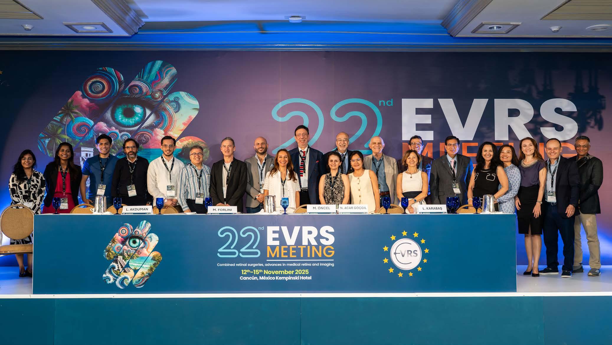
Retinal detachment and proliferative vitreoretinopathy (PVR) took over the afternoon. Talks covered long-term tamponade considerations, pneumatic vitrectomy and the growing role of swept-source OCT biomarkers. Dr. Rajeev Muni (Canada) provided a useful framework for pneumatic retinopexy and suprachoroidal viscopexy (SCVEXY).
Nearby, Dr. André Messias (Brazil), Dr. Gonzalez-Cortes and Dr. Hussein Almuhtaseb (UK) demonstrated how portable electroretinography is expanding diagnostic flexibility.

Dr. Kourous Rezaei (USA) delivered the day’s EVRS Award Lecture on the “aggregation of marginal gains,” highlighting how surgical mastery often grows through small, layered refinements. The RETINAWS session that followed echoed this idea as panelists dissected “heart stopping” and “hair greying” cases in a way that turned pressure points into practical teaching.
Sessions on diabetic retinopathy (DR) and imaging pushed the conversation into more technical territory. Tractional retinal detachment cases prompted discussion of blood-air exchange and tranexamic acid,
The Retina World Congress session continued in that vein. Prof. Fernando Arevalo (USA) delivered the Zivojnovic Award Lecture on intraocular foreign bodies, outlining decision paths shaped by trajectory, visibility and timing.
The Vit-Buckle Society session chaired by Dr. Danilo Iannetta (Italy) introduced one of the day’s practical case reviews. After a welcome from President Prof. Aleksandra Rachitskaya (USA), short videos from Dr. Matteo Forlini (Italy), Dr. Andrea Cardenas (Guatemala), Dr. Nur Acar (Turkey) and Dr. Daniel Moreno-Paramo

(Spain) demonstrated how frame-by-frame review turns difficult cases into shared problem solving.
The SPVR Symposium, chaired by Prof. Arevalo and Prof. Mauricio Maia (Brazil), covered modern subretinal hemorrhage management, giant retinal tear strategy, vitreoretinal interface updates and new thinking around endophthalmitis. A joint BRAVS–SARyV video symposium followed with case-driven exchanges that were grounded in everyday decision-making.
The South American Societies session shifted the lens toward resource-aware VR surgery. Moderators Dr. Samuel Gomez (Mexico) and Dr. Andrea Cárdenas (Guatemala) presented cases shaped as much by local constraints as by pathology.
Dr. Gomez walked through platelet-rich plasma for macular holes, focusing on how the technique fits into day-to-day practice rather than theory. Dr. Cárdenas followed with operating room strategies from Guatemala, showing how small adjustments can make a big difference when equipment options run thin.
One of the meeting’s most affecting moments came from Dr. Alejandro Olguin (Bolivia), who described “the cruel reality of

working in a Bolivian mine” and its connection to delayed highimpact trauma. Dr. Aldo Muñoz Quiñones (Peru) closed with clear guidance to adapt quickly, decide with confidence and make sure that every tool earns its keep.
By the time EVRS President Dr. Giampaolo Gini announced Athens as the 2026 venue, it already felt like the meeting had pointed the way forward. EVRS 2025 showed how much the field grows when surgeons treat complexity as a shared curriculum and speak honestly about what works, what fails and what still needs refining.
Vitreoretinal surgery may move in millimeters, but the conversations in Cancún made clear that those small shifts add up. The meeting brought together individuals who were willing to learn from each other in real time, and that mindset is what will carry the field into its next chapter.
Editor’s Note
The 22nd EVRS Meeting was held in Cancun, Mexico from 12th-15th November 2025. Media MICE is a media partner of EVRS. A version of this article was first published on MediaMICE.com.
Not all breakthroughs in ophthalmology come from devices, data or surgical techniques. At the ASCRS 2026, one project stepped outside the clinic–and still managed to leave a lasting clinical impression.
The Blind Canvas Project uses generative AI to transform lived experiences of patients with vision loss into large-scale artwork. Built from personal narratives of different individuals with conditions like retinitis pigmentosa (RP) and Stargardt disease, each piece is less an illustration and more an interpretation–one that invites clinicians to see beyond metrics and into meaning.

By centering patient voices instead of clinical outputs, the project reframes what “understanding” in eye care can really look like. It also highlights a gap many clinicians recognize which is the balance between technical precision and human connection.

As Ora Chief Medical Officer Dr. Gustavo De Moraes noted, progress is not just about what can be measured–but what we choose to understand more deeply.
In a field driven by outcomes, The Blind Canvas Project offers something different: a reminder that every diagnosis is a story–and sometimes, the most important insights aren’t found in charts, but in how long we’re willing to look.
Editor’s Note
The 2026 ASCRS Annual Meeting was held April 10–13 in Washington, D.C., United States. Reporting for this story took place during the event.








Ten thousand ophthalmologists, 250 international speakers and vibrant Rajasthani culture converge for “Green Vision: Ophthalmic Excellence with Sustainability.”
By Michaela Perez
The 84th Annual Conference of the All India Ophthalmological Society (AIOC 2026), held March 12-15 at the Jaipur Exhibition & Convention Centre, brought together 10,000+ ophthalmologists and 750 speakers under the theme Green Vision: Ophthalmic Excellence with Sustainability. Locally organized by the Rajasthan Ophthalmological Society, the conference demonstrated how science and culture can create an unforgettable experience.
Chris McBride, Media MICE’s Creative Content Director and MICE TV Producer, didn’t mince words about his most memorable moment: “The gala dinner. I’ve never seen such a big party at a conference. There was so much color and life and the performances were awesome.”
That festival atmosphere permeated the entire event. Ruchi Ranga, Media MICE’s Societies Relationship & Conference Manager and correspondent for MICE TV, witnessed it from day one: “Seeing thousands of ophthalmologists from across India and around the world gathered under one roof was truly inspiring. There was a palpable energy—from the inaugural ceremony to the packed scientific halls.”
The Rajasthan Ophthalmological Society, led by Dr. Virendra Agrawal and Dr. Mukesh Sharma, didn’t just host a conference— they staged a cultural showcase. AIOS Utsav, the Dhola mascot, traditional Rajasthani gifts and a Salim-Suleman cultural
night, created moments where delegates relaxed, connected and experienced Jaipur’s heritage.
“There was just stuff going on everywhere all day—dances, performances, camels on parade, photo opportunities. They really showcased Rajasthani culture,” Mr. McBride noted. “I would love to see more conferences showcasing local culture and food.”
International attendees particularly appreciated the warmth. “For many, this was their first exposure to Jaipur’s vibrant culture,” Ms. Ranga reported. “It made the conference feel not just educational, but memorable—an event where science and culture blended beautifully.”
The organizing committee’s invitation set a bold tone: “As healthcare professionals, we understand our responsibility extends beyond patient care to the health of our planet.”
The Green Vision theme encouraged delegates to explore innovative ways to make ophthalmology greener, more efficient and futureready—integrating eco-friendly practices with cutting-edge clinical care.
“Beyond scientific updates, the conference highlighted how ophthalmology is evolving—not just technologically, but ethically and environmentally,” Ms. Ranga observed. “The Green Vision theme encouraged participants to think about how medical


conferences, hospitals and practices can become more sustainable while delivering cutting-edge care.”
Beyond sustainability, AIOC 2026 reinforced a message Mr. McBride has observed across multiple India visits: “How much care everyone has for the underserved

population. India has a huge population with big unmet needs, but everyone here really focuses on trying to get eye care to the people that need it most—the poorest people in communities.”
The commitment showed everywhere—from screening programs to affordable technologies to rural clinic strategies. “Whether that’s through telemedicine, eye camps or anything else, they’re all figuring out ways to do that,” Mr. McBride said. “The world can learn a lot from that.”
MICE TV captured this ethos in action. In the opening of AIOC 2026 Day 1 Conference Report, India’s ophthalmology leader— Prof. Dr. S. Natarajan—shared his vision: “The mission is to make India blind-free from Kashmir to Kanyakumari. I want to start screening for eye problems using AI technology without a doctor, without a technician. I have a term called ‘anybody can screen for any eye problem.’”
His philosophy resonated: “We serve whether it’s a paying patient or a free patient. Every patient should be seen as your own father and mother.”
INNOVATION FROM HOMEGROWN TO GLOBAL
The exhibition floor buzzed with domestic innovation. Appasamy Associates CEO Mr. Senthil Kumar told MICE TV their mission is “to democratize ophthalmology and ophthalmic technology.”
“We’ve brought together a basket of new technologies ensuring better treatment for patients and easy service delivery for
ophthalmologists,” Mr. Kumar explained, showcasing the REVEL phaco machine with advanced fluidics, Rhexa automated capsulorrhexis system, MICROLASE with pulse matrix technology, CALYPSA swept-source optical biometer and its FOVi retinal imaging series.
HelpMeSee brought their manual small-incision cataract surgery (MSICS) training mission to AIOC, operating at five India centers plus facilities in China, Madagascar and the United States. “Our mission is to prevent cataract blindness, which is preventable,” Dr. Kimaya V. Chavan, their representative explained.
Furthermore, Mr. McBride observed: “There’s so much new tech and developments, especially domestic Indian developments. Any attendee who saw the show floor or attended sessions will come home with up-to-date knowledge about what’s going on in the world and in India, and how they can make it accessible for their patients.”

The International Agency for the Prevention of Blindness (IAPB) hosted their regional meeting at AIOC 2026. Dr. Rohit C. Khanna, IAPB’s South-East Asia regional chair, told MICE TV that partnerships with national societies amplify impact.
“India has 25,000-plus ophthalmologists. AIOS represents these ophthalmologists and they want to support IAPB at the Global Summit for Eye Health,” Dr. Khanna stated. Four societies—India (AIOS), Nepal (NOS), Indonesia (PERDAMI) and Sri Lanka (COSL)—signed a declaration of commitment.
“We can leverage the platform of AIOS to go to government and policymakers and influence policy much faster and more effectively than going individually,” Dr. Khanna explained.

With International Women’s Day celebrated earlier this month, MICE TV captured perspectives from Indian women ophthalmologists. Their motivations, in ophthalmology and in attending conferences such as the AIOC, ranged from work-life balance to serving rural communities to witnessing transformative surgical outcomes.
“The visual outcome after successful cataract surgery— first day 20/20 vision—that inspires me a lot,” one explained. Another emphasized her roots: “I’m a rural student, so I want to help my rural area people.”
Their excitement about AIOC 2026 focused on new techniques, machines, skills transfer sessions and networking. One word kept recurring: “Amazing.”
Dr. Taraprasad Das closed MICE TV’s conference report with a perspective spanning decades: “Conferences mean a lot because you see new people, young people, new equipment, new devices. It’s both social fun and intellectual stimulation.”
His advice to younger ophthalmologists? “When I plan for something, I don’t look at the peak of the mountain—I look at the next step only. Don’t worry too much for the last step, worry for the next step only.”
With 250 international and 500 national speakers across multiple scientific halls, the logistics were massive at AIOC 2026 in Jaipur. Yet the organizing committee delivered on their promise: registration fees remained unchanged, and additional halls accommodated emerging ophthalmologists.
Ms. Ranga, managing Media MICE’s relationship with AIOS, marveled at the execution: “Despite the magnitude, the team maintained remarkable attention to detail. The leadership ensured the event ran smoothly while maintaining a welcoming atmosphere.”
Her takeaway for conference organizers: “Build strong committees, empower volunteers and combine scientific excellence with thoughtful delegate experience.”
Conversations throughout AIOC 2026 centered on technological innovation, training and mentorship, and sustainability.
“Many attendees mentioned feeling energized by the exchange of ideas and the realization they’re part of a truly global community working toward better vision care,” Ms. Ranga observed.
On the other hand, Mr. McBride’s wish? “I really wish I had more time to listen to some of the great speakers because there seems to be amazing knowledge you could come away with—not just about techniques and the latest science, but also about how to care.”
Scouring the conference halls for MICE TV filming in the last three days, what struck Mr. McBride most during the day were amazing interviews with remarkable people. “But what was fun at the gala dinner was seeing them at night enjoying each other. Everyone who’s seen me at another conference makes sure to come say hello. India is chaotic, vibrant and colorful, but people always remember the small things and go out of their way to make you feel welcome.”
Ms. Ranga echoed the sentiments: “What made AIOC 2026 special was how it balanced scientific rigor, large-scale organization and cultural warmth. Events like this remind us that ophthalmology is about building a global network committed to improving lives.”
In conclusion, Mr. McBride offered this advice: “Try and come next year because they really put on a big show. They call it the ‘All India Ophthalmological Conference’, but more people from around the world should come and check out what’s going on in India.”
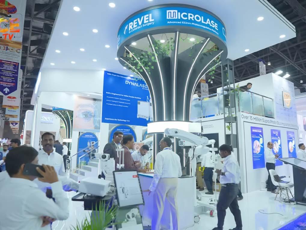
The 84th Annual Conference of the All India Ophthalmological Society (AIOC 2026) was held March 12-15, 2026 at Jaipur Exhibition & Convention Centre, organized by Rajasthan Ophthalmological Society. Reporting for this story took place during the event. A version of this article was first published on MediaMICE.com.