Professor Department of Ophthalmology and Visual Science The University of Chicago Chicago
KALEB ABBOTT, OD, MS, FAAO, FOWNS
Assistant Professor
Department of Ophthalmology
University of Colorado School of Medicine
Sue Anschutz-Rogers Eye Center Aurora, Colorado
MILE BRUJIC, OD, FAAO
AARON S. GOLD, OD, FAAO
Clinical Director
Murray Ocular Oncology & Retina Miami
Premier Vision Group Bowling Green, Ohio Professor of Ophthalmology
JOANN KANG, MD
Director, Cornea and Refractive Surgery Department of Ophthalmology and Visual Sciences Montefiore Medical Center Bronx, New York
NEEL D. PASRICHA, MD
Assistant Professor, Department of Ophthalmology
Associate Member, Department of Breast Oncology School of Medicine
University of California – San Francisco San Francisco
ACTIVITY DESCRIPTION
Antibody drug conjugates (ADCs) represent a newer class of oncology drugs associated with corneal toxicity. This activity summarizes a series of video discussions with optometrists and ophthalmologists about recognition and grading of pseudomicrocysts and other ocular adverse events (AEs), options for managing AEs, and communication strategies with patients and the oncology care team.
TARGET AUDIENCE
This accredited educational activity is designed for optometrists.
LEARNING OBJECTIVES
• Identify the clinical signs and symptoms of ocular side effects associated with a new class of oncology drugs, antibody drug conjugates (ADCs), including corneal issues, blurred vision, dry eye and microcysts
• Explain the importance of timely intervention for ocular side effects, including the use of preservative-free artificial tears and steroid drops, and implement evidence-based strategies to improve patient comfort and treatment adherence
• Develop strategies for effective comanagement between ophthalmologists, optometrists and oncologists, ensuring timely recognition and management of ocular toxicities that may require adjustments to cancer treatment plans
• Describe the standardized grading criteria for ocular side effects, differentiating between mild, moderate and severe presentations to guide appropriate management strategies
GRANT SUPPORT STATEMENT
This activity is supported by an unrestricted educational grant from AbbVie.
ACCREDITATION STATEMENT
Sponsored by:
Axiom Ophthalmic, LLC., is a COPE-Approved Administrator.
This activity, COPE Activity Number 133297, is accredited by COPE for continuing education for optometrists. This course is approved for 1.0 hour of CE.
Course #: 105694-SD
Activity #: 133297
COPE advises optometrists to contact the State or Provincial Board where they are licensed for verification of what is acceptable for license renewal.
TO OBTAIN CREDIT
To obtain credit for this activity, log in to the Axiom Learning Center and go to https:// www.axiomlearningcenter.com/courses-page/ course/26-01_sup or scan the QR code with your smartphone’s camera.
Once you’re logged into the course, you must complete the pre-test, view the activity, successfully pass the post-test, and complete the evaluation.
Only candidates with a passing score of at least 70% earn CE credit. Candidates have the opportunity to retake the post-test until a passing score is achieved. The course credit certificate will appear in the Certificates section of your account automatically upon successful completion of the course.
For any additional questions about obtaining credit, please email info@axiomophthalmic.com.
DISCLOSURE POLICY
It is the policy of Axiom Ophthalmic that faculty and other individuals in a position to control content of this activity disclose all financial relationships with COPE-defined ineligible companies within the past 24 months. Axiom Ophthalmic has policies in place that have identified relevant relationships and mitigated any real or apparent conflicts of interest prior to this educational activity.
Asim Farooq, MD, Chair, has the following financial relationships:
Meeting Support: ABB Optical, Art Optical, Contamac, Copper Vision, Johnson & Johnson Vision Care, Tangible Science and Walman Optical.
Research Support: Lentechs
Speaker: Sun Pharma
Aaron Gold, OD, Faculty, has the following financial relationships:
Advisory Board: Regeneron
Joann Kang , MD, Faculty, has the following financial relationships:
Consultant: OcuCell and TearDx.
Neel Pasricha, MD, Faculty, has the following financial relationships:
Consultant: AbbVie, Amgen, AstraZeneca, Glaukos, GlaxoSmithKline, Hexagon Bio and Iksuda Therapeutics
Research: Alcon
Stock in Private Company: Diatiro and Globe Biomedical
All other Axiom staff, planners, reviewers and writers have no financial relationships with ineligible companies.
All identified relevant financial relationships have been mitigated and the educational content thoroughly evaluated for fair, balanced, and safe, effective patient care.
OFF-LABEL STATEMENT
This educational activity may contain discussion of published and/or investigational uses of agents that are not indicated by the FDA. The opinions expressed in the educational activity are those of the faculty. Please refer to the official prescribing information for each product for discussion of approved indications, contraindications a nd warnings.
DISCLAIMER
The objective of Axiom Ophthalmic in developing continuing medical education activities is to promote the free expression and interchange of ideas and information for educational purposes. Axiom Ophthalmic does not accept responsibility for any opinions, positions, or statements contained or expressed in such material, and such opinions, positions and statements are not necessarily those of Axiom. Courses are intended to provide instruction leading to new knowledge and/or skills. Axiom Ophthalmic does not certify competence upon completion of the courses. The information presented is not meant to serve as a guideline for patient management. Any procedures, medications, or other courses of diagnosis or treatment discussed in this activity should not be used by clinicians without evaluation of patient conditions and possible contraindications on dangers in use, review of any applicable manufacturer’s product information, and comparison with recommendations of other authorities.
INTRODUCTION
Antibody drug conjugates (ADCs) represent a relatively new class of highly effective oncology drugs. However, these treatments are often associated with corneal toxicity. The corneal adverse events (AEs) present primarily as dry eye and pseudomicrocysts, among other findings.1
Experts expect these therapies to eventually move from their current status as salvage therapies to earlier lines of treatment. Therefore, eye specialists will likely see more patients presenting with ocular toxicities associated with these life-saving treatments
REFERENCE
and should be aware that additional patient visits for monitoring and increased communication with the oncology care team will be necessary.
Although there are additional products on the market, and even more in the pipeline, the following articles focus on three of the more common ADCs. The goal is to increase awareness of the overall class of medications and related ocular AEs and to highlight the importance of collaboration across providers in eye care and hematology/oncology.
Canestraro J, Hultcrantz M, Modi S, et al. Refractive Shifts and Changes in Corneal Curvature Associated With Antibody-Drug Conjugates. Cornea. 2022;41(6):792-801.
ANTIBODY DRUG CONJUGATES AND THE CORNEA
Signs and symptoms of corneal toxicity associated with ADCs
By Mile Brujic, OD, FAAO and Asim V. Farooq, MD
WHAT ARE ANTIBODY DRUG CONJUGATES?
Antibody drug conjugates (ADCs) represent a paradigm shift in the field of oncology. These novel, targeted therapies for cancer are more effective and are prolonging life and survival for many patients as they currently are utilized mostly as salvage therapies.1
ADCs are comprised of three different components. A monoclonal antibody is tethered to a cytotoxic drug—also known as a payload or a warhead—and a chemical linker connects the two.1,2 ADCs are being used to treat ovarian, cervical, B-cell lymphoma, multiple myeloma, breast cancer and others. There are currently about 15 ADCs approved by the FDA and more than 400 ADC candidates globally are currently undergoing clinical evaluation.3
A significant number of ADCs have been associated with ocular adverse events (AEs). This article will focus on ocular side effects of mirvetuximab soravtansine, tisotumab vedotin and belantamab mafodotin.4-6
Mirvetuximab is indicated for the treatment of ovarian cancer, tisotumab is primarily for cervical cancer, and belantamab— which was approved in 2020, withdrawn from the market by the manufacturer in 2022, and then reapproved in October 2025— is used to treat multiple myeloma.7
ADCs are hyper-specific for their target, and ocular toxicities did appear in the clinical trials, which resulted in a black box warning issued by the FDA for ocular side effects associated with ADCs. Eye care specialists need to be aware of this when caring for patients undergoing treatment for cancer, according to Mile Brujic, OD, FAAO.
“It’s incumbent upon us, as eye specialists, to be very present when it comes to these medications. Patients need to be seen by us before treatment with some of these infusions and then on a regular basis,” Dr. Brujic said.
It’s incumbent upon us, as eye specialists, to be very present when it comes to these medications. Patients need to be seen by us before treatment with some of these infusions and then on a regular basis.
OCULAR ADVERSE EVENTS ASSOCIATED WITH ADC s
Most individuals who are treated with ADCs experience at least some ocular adverse events. For example, with belantamab, ocular toxicity occurred in more than 90% of treated patients. About three-quarters of them had Grade 3 or Grade 4 toxicity. 8 Similarly, at least 50% of patients receiving tisotumab and mirvetuximab experienced ocular adverse events.4-6
“The fact of the matter is that these are agents that are targeting a specific marker on the tumor, but they are finding their way to the cornea,” said Asim V. Farooq, MD.
The fact of the matter is that these are agents that are targeting a specific marker on the tumor, but they are finding their way to the cornea.
PSEUDOMICROCYSTS
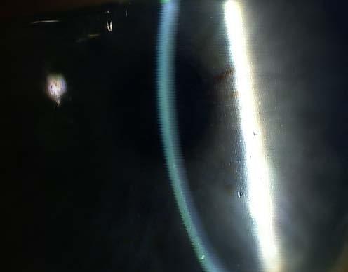
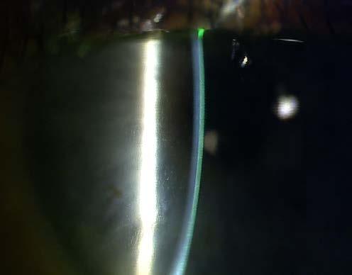
Lesions known as pseudomicrocysts are visible through slit-lamp examination as small semi-translucent wounds within the corneal epithelium (Figure). They develop initially in the periphery of the cornea and move toward the center. Experts believe the drug is being internalized either by limbal stem cells or epithelial cells early in their life cycle.9
There is some evidence the drug is being delivered to the cornea and is being internalized by these cells through a process called macropinocytosis, which is a form of cellular uptake that allows cells to sample their environment, as well as pick up nutrients.9
A change in visual acuity is observed with these pseudomicrocysts, which can be in part due to refractive shifts. When pseudomicrocysts are in the periphery of the cornea, they can often cause a hyperopic shift; whereas when they appear in the center, they can be associated with a myopic shift.10
When pseudomicrocysts are in the periphery of the cornea, they can often cause a hyperopic shift; whereas when they appear in the center, they can be associated with a myopic shift. 10
The lesions migrating toward the center tend to correlate with a more significant impact on vision. Notably, in some patients, the
lesions don’t make it to the center, but instead stay in the periphery and mid-periphery. We hypothesize that these epithelial cells die before making it to the center, but there doesn’t seem to be a uniform pattern. Whorl epitheliopathy is also sometimes visible on fluorescent staining, according to Dr. Farooq.11
Managing these temporary refractive shifts that occur while patients are being treated with ADCs can be challenging because it’s possible to have a hyperopic shift and a myopic shift, and therefore a new eyeglasses prescription may not help because it may change at any time.10
“We’ve actually seen both of these occur in clinical practice. Years ago…we created generic glasses, -1.00 D all the way to -8.00 D, and the same thing on the plus side, up to +6.00 D in 1.00 D steps. This was utilized for people who were contact lens wearers who maybe had contact lens emergencies, and they couldn’t put contact lens back in, but they needed some type of refractive correction,” Dr. Brujic said. “They’ve been a true blessing for some of these patients on ADCs who just need temporary prescription changes or differences. We literally loan them the glasses. The patient signs out the glasses, and then they bring them back at the follow-up visit.”
Macropinocytosis is one pathway of nonspecific off-target uptake.10 It is considered off-target because in many cases the target for these cancer drugs—the target antigen—is not expressed in the eye. And yet these lesions, known as pseudomicrocysts, still appear, along with other forms of ocular toxicity.12
Researchers have found from studies of a few different ADCs— both in vitro and in animal models—that macropinocytosis, a form of cell drinking, is the major pathway for uptake of the ADC into the corneal epithelium.12 Studies have shown that if a structural modification is made to the drug to decrease its ability to undergo macropinocytosis, there is less uptake into the corneal epithelium. This is an example of how, despite the cornea not expressing the target antigen, ADCs can affect vision.12
According to Dr. Brujic, the best way to visualize the pseudomicrocysts is to look beside where the slit beam shines through the cornea. High magnification is also useful.
“I really increase the magnification. We have a few different slit lamps in our office that go to different magnification settings, but we have one that goes to 25X,” Dr. Brujic said. “I love getting that 25X magnification on these individuals because I really find that you can hone in on the presence or absence of these microcysts.”
COMMUNICATING WITH PATIENTS
There are a few important points that should be discussed with patients. The first is that the lesions tend to be reversible with time, even for a patient with a grade of toxicity that requires dose reduction or dose delay based on exam findings or the impact on visual acuity.13
There are a few important points that should be discussed with patients. The first is that the lesions tend to be reversible with time, even for a patient with a grade of toxicity that requires dose reduction or dose delay based on exam findings or the impact on visual acuity.13
The second point is that these patients have most likely been prescribed ADCs because they have failed previous cancer treatments. Because of this, patients may theoretically be motivated to de-emphasize ocular symptoms to their eye care specialist, according to Dr. Brujic.
“Patients don’t want to be pulled off the ADC that may be keeping them alive,” Dr. Brujic said. “We must communicate to patients
REFERENCES
1. Hurwitz J, Haggstrom LR, Lim E. Antibody-Drug Conjugates: Ushering in a New Era of Cancer Therapy. Pharmaceutics. 2023;15(8):2017.
3. Parveen I, Aqil A, Sharma S, Qadir A, Rehman SA, et al. From concept to clinic: Cutting-Edge advances steering the next wave of antibody–drug conjugates innovation. J Drug Deliv Sci Technol. 2026;120:108271.
4. Coleman RL, Lorusso D, Gennigens C, et al; innovaTV 204/ GOG-3023/ENGOT-cx6 Collaborators.; innovaTV 204/GOG3023/ENGOT-cx6 Collaborators. Efficacy and safety of tisotumab vedotin in previously treated recurrent or metastatic cervical cancer (innovaTV 204/GOG-3023/ENGOT-cx6): a multicentre, openlabel, single-arm, phase 2 study. Lancet Oncol. 2021;22(5):609-619.
5. Wahab A, Rafae A, Mushtaq K, et al. Ocular Toxicity of Belantamab Mafodotin, an Oncological Perspective of Management in Relapsed and Refractory Multiple Myeloma. Front Oncol. 2021;11;11:678634.
6. Holtzman S, Letchinger R, McCarthy L, et al. Unnecessary exclusions: eligibility criteria in gynecologic oncology interventional clinical trials impairs access. Int J Gynecol Cancer. 2026;36(4):101927.
7. Riccardi F, Dal Bo M, Macor P, Toffoli G. A comprehensive overview on antibody-drug conjugates: from the conceptualization to cancer therapy. Front Pharmacol. 2023;14:1274088.
that if they experience any changes in vision prior to their followup, it’s important they call you to make sure you’re aware of those changes. The sooner we assess those changes, the sooner we can address them.”
CONCLUSION
The number of ADCs in the pipeline and on the market is steadily growing. Eye specialists should stay up to date on these therapeutics, so they know what to expect clinically, how to best manage these toxicities and be prepared to answer patient questions.
Scan the QR code with your smartphone’s camera to watch Drs. Brujic and Farooq discuss their experience with ocular toxicities associated with ADCs.
8. National Cancer Institute. Common Terminology Criteria for Adverse Events (CTCAE) v6.0 (MedDRA 28.0). Updated July 22, 2025. chrome-extension://efaidnbmnnnibpcajpcglclefindmkaj/ https://dctd.cancer.gov/research/ctep-trials/for-sites/adverse-events/ ctcae-v6.pdf. Accessed June 10, 2025.
9. Schwartz GS, LoVerde L, Gomes J, Holland EJ. Cornea, 4th edition. 2017; Chapter 157: Classification and Staging of Ocular Surface Disease, p. 1668-1680.
10. Canestraro J, Hultcrantz M, Modi S, Hamlin PA, Shoushtari AN, et al. Refractive Shifts and Changes in Corneal Curvature Associated With Antibody-Drug Conjugates. Cornea. 2022;41(6):792-801.
11. Deng SX, Borderie V, Chan CC, Dana R, Figueiredo FC, Gomes JAP, Pellegrini G, Shimmura S, Kruse FE; and The International Limbal Stem Cell Deficiency Working Group. Global Consensus on Definition, Classification, Diagnosis, and Staging of Limbal Stem Cell Deficiency. Cornea. 2019;38(3):364-375.
12. Zhao Q, Eichten A, Parveen A, Adler C, Huang Y, et al. Single-Cell Transcriptome Analyses Reveal Endothelial Cell Heterogeneity in Tumors and Changes following Antiangiogenic Treatment. Cancer Res. 2018;78(9):2370-2382.
13. Dy GK, Farooq AV, Kan JJ, Ocular adverse events associated with antibody-drug conjugates for cancer: evidence and management strategies. The Oncologist. 2024;29(11):e1435-e1451.
MANAGING OCULAR SIDE EFFECTS ASSOCIATED WITH ADCs
Prevention, monitoring and managing ocular AEs in patients receiving ADC therapies
By Aaron S. Gold, OD, FAAO and Joann J. Kang, MD
Antibody drug conjugates (ADCs), such as mirvetuximab soravtansine, tisotumab vedotin and belantamab mafodotin, are an effective, newer class of oncologic therapies associated with toxicities that are typically absent from traditional chemotherapies.
Ocular adverse events (AEs) occurred in more than 50% of treated patients in the mirvetuximab studies1,2 ; approximately 60% of treated patients in the tisotumab studies3 ; and more than 90% of treated patients in the belantamab trials, 77% of which were Grade 3 or 4 level AEs, based on the Common Terminology Criteria for Adverse Events (CTCAE), a standardized system managed by the National Cancer Institute that defines the severity of the cancer treatment-related AEs.4
Considering this rate of ocular risks, eye care specialists must be vigilant and monitor these patients carefully, according to Joann J. Kang, MD. The median time of onset is about 1.3 months, usually cycle one and two; however, toxicity may appear up to 10 months after treatment initiation, so continued monitoring of patients is required.5
A collaborative approach with the patient’s oncologist is also required to achieve the best outcomes. Eye care specialists are not experts in oncology and vice versa.
“Communicating with the oncologist about what’s going on with the eye puts it back into the oncologist’s hands regarding what they may do next to mitigate ocular toxicity,” Dr. Kang said. “Oncologists are not familiar with the eye as they don’t perform slit lamp biomicroscopy. They may not check formal visual acuity and may not even have a vision chart in their office.”
Communicating with the oncologist about what’s going on with the eye puts it back into the oncologist’s hands regarding what they may do next to mitigate ocular toxicity.
CLINICAL GUIDANCE
To help establish a standard of care for patients receiving ADCs, the American Academy of Ophthalmology (AAO) released a clinical statement in July 2025, intended to serve as guidelines for the ophthalmic management of patients on ADCs. 6 Treatment and screening guidelines vary by agent and eye care providers should refer to the package insert that accompanies the ADC for the most current guidelines. Slit lamp biomicroscopic evaluation and a fundus exam are required at baseline.
According to the AAO statement, eye care providers evaluate patients to:
1. Provide a baseline examination prior to the start of the ADCs to allow for assessment for change while on the therapy.
2. Provide counseling on the use of preservative-free artificial tears and sometimes topical corticosteroids during treatment cycles. Additionally, ocular surface vasoconstrictors may be recommended during the infusions (such as with topical brimonidine 0.2% and cold compress eye masks).
3. Provide education for patients on the symptoms of AEs and contact information to schedule a repeat examination if symptoms occur.
4. Provide follow-up visits to identify AEs early and tailor treatment and follow-up based on the grade or severity of the findings.
5. Provide an objective assessment for the oncologist so that they may decide if the next dose of the ADC can be given as scheduled, delayed, reduced or discontinued.
Baseline examination and prophylaxis
The baseline exam allows the eye care provider to establish baseline visual acuity prior to treatment, as well as the patient’s corneal health prior to beginning treatment with ADCs.
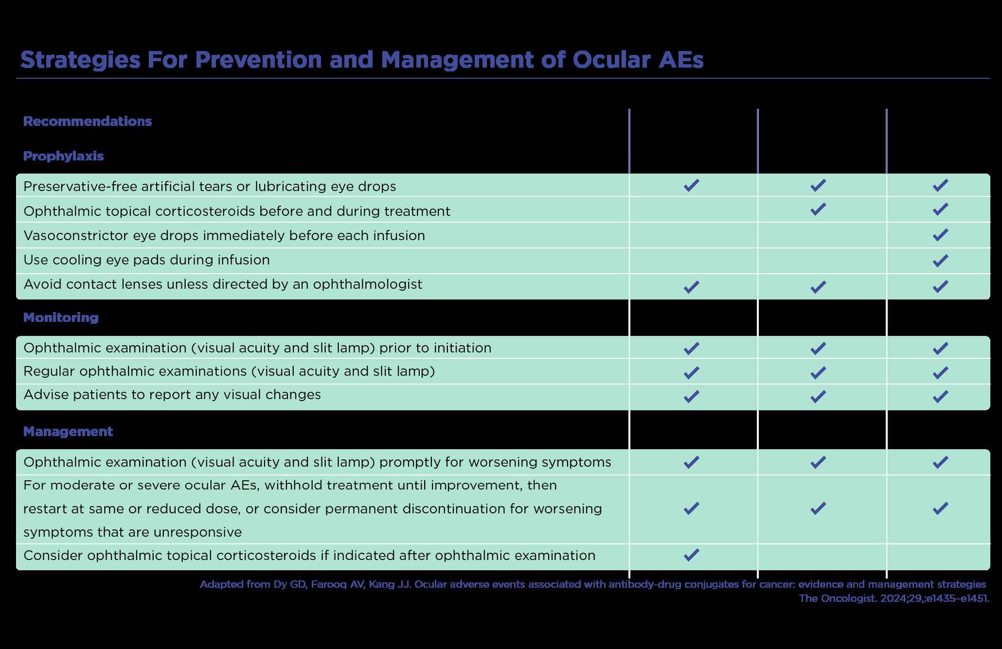
In addition to the AAO clinical statement, published studies have outlined specific strategies to help with the prevention and management of ocular AEs associated with ADCs (Figure 1). Dr. Kang said she prescribes preservative-free tears four times daily for all patients receiving ADCs and advises patients not to wear contact lenses.7
Aaron S. Gold, OD, FAAO, stressed the importance of preservative-free artificial tears for these patients, and that patients must be reminded to follow the dosing recommendations.
“One thing I always question is our patient’s regimen with their artificial tears, because I’m asking them to instill the drops every two hours, and oftentimes patients will say they are doing it twice a day,” Dr. Gold said. “They don’t understand that twice daily practically means they’re not using it at all.”
One thing I always question is our patient’s regimen with their artificial tears, because I’m asking them to instill drops every two hours, and oftentimes patients will say they are doing it twice a day.
1. These prevention and management strategies provide guidance for eye providers in caring for patients with ADC-related ocular toxicities.7
Figure
Dr. Kang shared her similar experience: “I am always reminding my patients that every two hours is at least eight times a day.”
Certain ADCs also call for use of corticosteroids or vasoconstrictors, so eye care specialists should familiarize themselves with these varying requirements based on the patient’s specific ADC. Ocular surface vasoconstrictors can be instilled immediately before the ADC infusion as a way to decrease the amount of the drug that enters the limbal vasculature, followed by cooling eye masks.7,8
“This is not done for every ADC, and it may differ per infusion center, depending on each center’s protocol. Some centers use a cooling pad for about 60 minutes also to decrease the load of ADC that gets to the eye,” Dr. Kang said.
In terms of prophylactic corticosteroids, Dr. Gold prescribes prednisolone acetate 1% four times daily (QID) for his patients receiving ADCs, although not all ADC clinical studies support this approach. There is support for the use of steroids immediately before and after infusions in patients receiving mirvetuximab and tisotumab, because some of those patients developed uveitis and conjunctivitis.9,10 However, in patients receiving belantamab, researchers found that steroids were not effective, therefore, prophylactic steroids are not universally recommended.11
Monitoring patients on ADCs
In addition to the baseline exam, Dr. Kang advises that an ocular exam be performed prior to every infusion of belantamab or tisotumab, and every other infusion for mirvetuximab—unless there is evidence of toxicity. In that case, the patient should return for an exam more frequently and this information must be shared with the patient and the oncologist.7,8
“After the baseline exam, depending on the patient’s schedule, you should have the patient come in, perform a slit-lamp exam, monitor them and regularly look at the corneas,” agreed Dr. Gold.
After the baseline exam, depending on the patient’s schedule, you should have the patient come in, perform a slitlamp exam, monitor them and regularly look at the corneas.
Dr. Kang said she sees patients on ADCs about every three weeks for continued monitoring and she notifies the oncologist personally if she observes any ocular AE that is Grade 1 or above: “That way they are aware, and we are monitoring that together.”4
Care team communication and tailoring treatment
Once an ocular toxicity associated with an ADC is observed— other than use of artificial tears and topical corticosteroids, when appropriate—the next option to prevent worsening toxicity is to discuss with the oncologist the possibility of ADC dose modification.7 Dr. Kang said by following the guidelines for monitoring and the prophylaxis discussed here, that she typically does not see patients progress to Grade 2 (See more about grading in the following article titled Recognition, Grading and Management of Ocular Toxicities).
“If someone has a corneal ulcer or has an impending perforation, you would, of course, provide the supportive eye care needed,” Dr. Kang explained. “But in terms of decreasing the ocular toxicity, for Grade 1, typically you can simply monitor the patient. If the toxicity is Grade 2 and above, it usually does require either decrease of the dose or delay of the dose. The decision to withhold or decrease the dose is at the discretion of the oncologist.”
If the toxicity is Grade 2 and above, it usually does require either decrease of the dose or delay of the dose. The decision to withhold or decrease the dose is at the discretion of the oncologist.
CASE STUDY
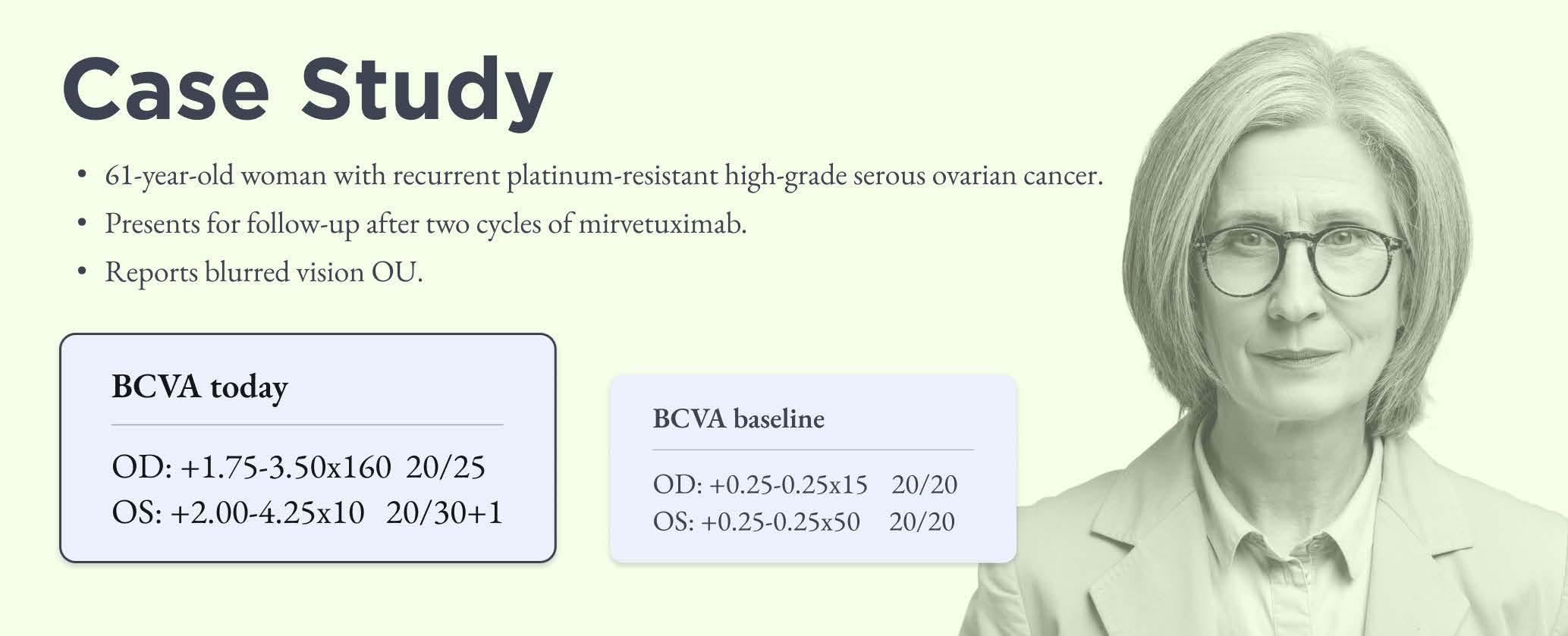
This case study highlights a 61-year-old woman being treated with mirvetuximab for ovarian cancer. She had baseline VA of 20/20 (Figure 2). When the patient returned after two cycles of mirvetuximab, she reported blurred vision and her VA was 20/25 OD and 20/30+1 OS. Without the baseline exam, Dr. Gold said he likely wouldn’t have thought much about the change in vision. However, the patient presented with high with-the-rule astigmatism that was not present before the baseline exam.12
Figure 2. Without the baseline exam, an abnormal finding related to the ADC would not have been discovered.
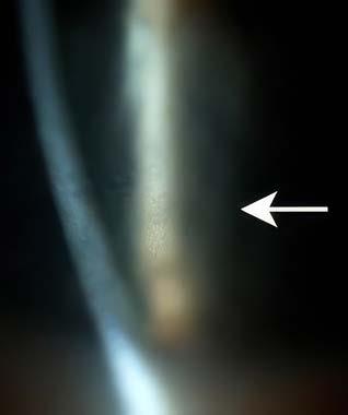
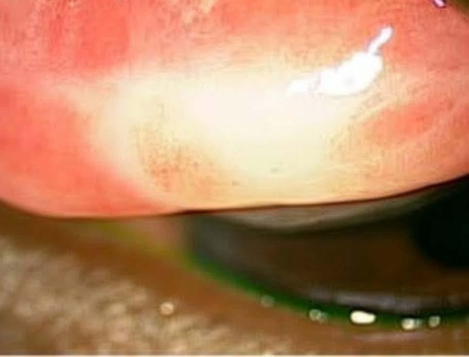
The slit lamp exam revealed mild microcyst-like epithelial changes (Figure 3) that appeared after two cycles of mirvetuximab. The patient went on to receive 10 total cycles before treatment was discontinued after progression of disease. These pseudomicrocysts
can be subtle and emerge bilaterally around the limbus in a ringlike pattern and migrate toward the central cornea with subsequent drug infusions.13
Figure 3. The slit lamp exam revealed the pseudomicrocysts associated with ADCs. Image courtesy of Aaron S. Gold, OD, FAAO.
“We have found that there are these topographic changes that are happening in the cornea induced by epithelial microcysts, but they are transient,” added Dr. Kang. “Because the limbal epithelial cells will keep regenerating, if a patient is off the medication, their cornea will go back to baseline.”
Because the limbal epithelial cells will keep regenerating, if a patient is off the medication, their cornea will go back to baseline.
Dr. Gold’s patient was lost to follow-up but when she returned to the clinic seven months later, “her corneas were clear and her visual acuity was good,” he said.
CONCLUSION
ADCs are expected to become more common, and therefore, eye care specialists will need to be familiar with ocular toxicities associated with ADCs in order to provide the best possible care for their patients with cancer.
REFERENCES
1. Hendershot A, Slabaugh M, Riaz KM, et a l. Strategies for prevention and management of ocular events occurring with mirvetuximab soravtansine. Gynecol Oncol Rep. 2023;47:101155.
2. Matulonis UA, Lorusso D, Oaknin A, et al. Efficacy and Safety of Mirvetuximab Soravtansine in Patients With Platinum-Resistant Ovarian Cancer With High Folate Receptor Alpha Expression: Results From the SORAYA Study. J Clin Oncol. 2023;41: 2436-2445.
3. Arn CR, Halla KJ, Gill S. Tisotumab Vedotin Safety and Tolerability in Clinical Practice: Managing Adverse Events. J Adv Pract Oncol. 2023;14(2):139-152.
4. National Cancer Institute. Common Terminology Criteria for Adverse Events (CTCAE) v6.0 (MedDRA 28.0). Updated July 22, 2025. Available at: https://dctd.cancer.gov/research/ctep-trials/ for-sites/adverse-events/ctcae-v6.pdf Accessed June 10, 2025.
5. Bouguerra Zina B, Rousseau F, Fauquier S, Sabatier R, Kfoury M. Practical clinical management of ocular adverse events related to Antibody-Drug Conjugates in gynaecological malignancies. Cancer Treat Rev. 2025;134:102867.
6. AAO. Antibody-Drug Conjugates and Ocular Toxicities: A Call for Standardized Guidelines. Updated July 2025. Available at: https://www.aao.org/education/clinical-statement/antibodydrug-conjugates-ocular-toxicities-call-st Accessed June 11, 2026.
7. Dy GK, Farooq AV, Kang JJ. Ocular adverse events associated with antibody-drug conjugates for cancer: evidence and management strategies. Oncologist. 2024;29(11):e1435-e1451.
8. Farooq AV, Degli Esposti S, Popat R, et al. Corneal Epithelial Findings in Patients with Multiple Myeloma Treated with Antibody-Drug Conjugate Belantamab Mafodotin in the Pivotal, Randomized, DREAMM-2 Study. Ophthalmol Ther. 2020;9(4):889-911.
9. Matulonis UA, Birrer MJ, O’Malley DM, et al. Evaluation of Prophylactic Corticosteroid Eye Drop Use in the Management of Corneal Abnormalities Induced by the Antibody–Drug Conjugate Mirvetuximab Soravtansine. Clin Cancer Res. 2019;25(6): 1727-1736.
10. Brahmer JR, Lacchetti C, Schneider BJ, et al; National Comprehensive Cancer Network. Management of ImmuneRelated Adverse Events in Patients Treated With Immune Checkpoint Inhibitor Therapy: American Society of Clinical Oncology Clinical Practice Guideline. J Clin Oncol. 2018;10;36(17):1714-1768.
11. Wahab A, Rafae A, Mushtaq K, et al. Ocular Toxicity of Belantamab Mafodotin, an Oncological Perspective of Management in Relapsed and Refractory Multiple Myeloma. Front Oncol. 2021;11:678634.
Scan the QR code with your smartphone’s camera to watch Drs. Gold and Kang discuss the best ways to manage ocular side effects associated with ADCs.
12. Canestraro J, Hultcrantz M, Modi S, et al. Refractive Shifts and Changes in Corneal Curvature Associated With Antibody-Drug Conjugates. Cornea. 2022;41(6):792-801.
13. Lindgren ES, Yan R, Cil O, et al. Incidence and Mitigation of Corneal Pseudomicrocysts Induced by Antibody-Drug Conjugates (ADCs). Curr Ophthalmol Rep. 2024;12(2):13-22.
RECOGNITION, GRADING AND MANAGEMENT OF OCULAR TOXICITIES
A review of prevention strategies and tips for care team collaboration
By Kaleb Abbott, OD, MS, FAAO, FOWNS and Neel D. Pasricha, MD
Antibody drug conjugates (ADCs) present unique challenges for the eyes, according to Kaleb Abbott, OD, MS, FAAO, FOWNS.
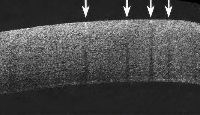
The primary ocular adverse effect associated with ADCs is corneal pseudomicrocysts, sometimes called microcyst-like epithelial changes. These pseudomicrocysts represent apoptotic epithelial cells and accumulation of the ADC itself within those epithelial cells. The drug is entering the cornea from the limbal vasculature. It begins in the periphery and may migrate to the center of the cornea (Figure 1).1,2
As this occurs, the vision for the patient is continuously changing. The patient may develop blurred vision as the corneal changes alter the refraction of light within the cornea. Vision is thought to fluctuate due to pseudocyst centripetal migration within the cornea.3
These patients can also develop corneal staining and in severe cases, epithelial defects or ulcers. Patients may report dry eye symptoms and eye pain, and ocular surface changes are often seen on exam. However, these AEs typically resolve when the treatment is modified or ceased.1
“One thing that’s important for patients to know, and for providers to know, is that these pseudomicrocysts are reversible,” Dr. Abbott said. “Once a patient stops taking the ADC, we do see them go away. They can go away in as little as two weeks, but sometimes it can take as long as a year. On average, it’s about two to four months. The vision does return to normal for the vast majority of these patients.”
When these ocular AEs associated with ADCs are observed by an eye care provider, the next step is to assign a grade, often based on a standardized system managed by the National Cancer Institute, that defines the severity of the cancer treatment-related AEs. 4 Once the grade is assigned, the eye care provider should share this information with the patient’s oncology team.
When ocular
AEs associated with ADCs are observed by an eye care provider, the next step is to assign a grade
that
defines the severity of the cancer treatmentrelated AEs. 4 Once the grade is assigned, the eye care provider should share this information with the patient’s oncology team.
ADC s AND OCULAR AE s
Belantamab mafodotin, mirvetuximab soravtansine, tisotumab vedotin and other ADCs are expected to move from salvage therapies to more first-line options, which will result in more eye care providers seeing these patients in their optometry and ophthalmology clinics, according to Neel D. Pasricha, MD.
In the clinical studies of belantamab mafodotin, more than 90% of patients developed some degree of ocular toxicity, with more than 75% having Grade 3 or Grade 4.5,6 This level of severity resulted in many patients requiring a dose modification by their oncologist.
“There are specific recommendations on how to modify the dose based on the ocular toxicity grade the patient has,” said Dr. Pasricha. “So, the role of optometrists and ophthalmologists is really important in helping decide each patient’s treatment.”
The mirvetuximab soravtansine trials reported high rates of corneal toxicity (>50%; any grade), with 11% having an ocular AE severe enough to result a dose reduction.7,8
In tisotumab vedotin trials, 60%% of treated patients reported ocular AEs9; the most common included conjunctivitis (26%) and dry eye (23%). Grade 3 ulcerative keratitis was reported in (2%) of patients.10 Dr. Abbott has treated several patients undergoing treatment with tisotumab vedotin and said these are some of the most challenging cases of ADC-associated ocular toxicity because they can develop cicatrizing conjunctivitis and scarring of the conjunctiva.11
“I’ve had patients who have developed symblephara from this and I’ve had patients who have gone on to develop entropions,” Dr. Abbott said. “There are a lot more severe epithelial changes with tisotumab vedotin that require more aggressive treatment compared with belantamab mafodotin and mirvetuxumab soravtansine.”
Dr. Pasricha echoed Dr. Abbott’s comments: “I agree and I think because of the tissue factor expression in the conjunctiva, you uniquely get those on-target severe cicatrizing changes that can happen. And unlike the off-target corneal toxicity, which you correctly point out is reversible, that conjunctival toxicity sometimes is not. If you develop subepithelial fibrosis or symblepharon, those don’t go away just by stopping the drug.”
So, the role of optometrists and ophthalmologists is really important in helping decide each patient’s treatment.
Because of the tissue factor expression in the conjunctiva, you uniquely get those ontarget severe cicatrizing changes that can happen. And unlike the offtarget corneal toxicity, which you correctly point out is reversible, that conjunctival toxicity sometimes is not. If you develop subepithelial fibrosis
or
symblepharon, those don’t go away just by stopping the drug.
GUIDELINES FOR PROPHYLAXIS, MONITORING AND MANAGEMENT
There are basic guidelines for eye care providers for prevention and management of these ocular AEs.12 Despite prophylactic approaches, ocular AEs occur in more than 75% of patients on select ADCs, and the number will likely increase with newer ADCs.1
“For some drugs, like belantamab mafodotin, we’re going to see that go even higher because any Grade 2 ocular toxicity is going to lead to dose reduction first, and dose delay second. I think we’re going to see over 90% of patients going down that pathway,” Dr. Pasricha said.
PREVENTION STRATEGIES
Corticosteroids
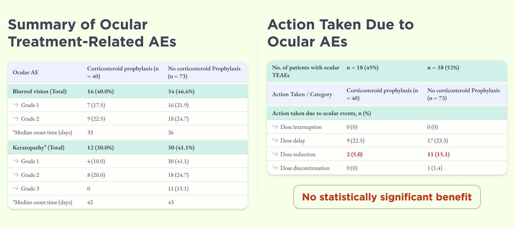
Studies have shown that corticosteroids have some efficacy in preventing ocular AEs with certain ADCs. In one particular study with mirvetuximab soravtansine, researchers looked at a group that were treated with topical steroids versus a group who were not. The results indicated that there was no statistically significant difference, except when related to the dose reduction (Figure 2). The results revealed the steroid-treated group had a lower rate of dose reductions compared to the nontreated group.13
“In my anecdotal experience, patients who are symptomatic with eye pain from the corneal toxicity will sometimes have some symptom relief with topical steroids,” said Dr. Pasricha.
Figure 2. Results of this study revealed no statistical difference in prevention of AEs with use of corticosteroids, but there was a lower rate of dose reductions in patients receiving topical steroids.13
The study looked at blurred vision, keratopathy and dose interruptions, but not necessarily how the eyes felt to those study patients. With some ADCs—including tisotumab vedotin—patients can develop a significant amount of eye pain and discomfort, which corticosteroids can provide some relief, according to Dr. Abbott.
Vasoconstriction
Vasoconstriction has shown some efficacy in reducing ocular AEs. Results of a preclinical study showed that use of brimonidine delayed the onset of pseudomicrocysts in rabbits treated with mirvetuximab soravtansine. However, corneal toxicity still occurred in 100% of the treated population by Day 14. For tisotumab vedotin, a human ocular substudy testing an ocular prophylaxis regimen that included vasoconstriction, among other therapies, did find a reduction in overall ocular AEs: from 80% to 60%. Specifically, a reduction of 80% to 30% was observed in conjunctival adverse events. These results are the reason vasoconstriction is recommended for patients undergoing treatment with tisotumab vedotin (Figure 3).14,15
ADCs are becoming increasingly common and will continue to play a larger role in patient care.
There are additional ongoing studies investigating options for reducing ocular AEs associated with ADCs, including a study of mirvetuximab soravtansine in which study participants are randomized (1:1) to primary prophylactic steroid eye drops versus primary prophylactic vasoconstricting eye drops.16
Serum tears
Although the data is limited on the use of serum tears to prevent ocular toxicities associated with ADCs,17,18 Dr. Abbott said he has used them for patients receiving tisotumab vedotin and mirvetuximab soravtansine because they improve the level of discomfort.
“One of my patients on mirvetuximab soravtansine said she felt that her vision improved after she started using serum tears, but it was more subjective rather than objective,” said Dr. Abbott. “I think the jury is still out in terms of whether serum tears are helpful in these cases.”
An issue for debate is the timing of the blood draw, according to Dr. Abbott, because if the ADC is entering the patient’s bloodstream, when the patient has their blood drawn can make a difference.
One of my patients on mirvetuximab soravtansine said she felt that her vision improved after she started using serum tears, but it was more subjective rather than objective. I think the jury is still out in terms of whether serum tears are helpful in these cases.
“One thought is that if the patient has already received the ADC systemically, and you’re drawing serum for the serum tears, are you going to be introducing more toxicity to the patient?” asked Dr. Pasricha.
However, there are some data from other systemic disorders that indicates blood draws can occur after the patient has started therapy.19 The main driver of toxicity in these patients is thought to come from the blood rather than the tears. 20 Therefore, even though the ADC enters the tears and the free toxic payload and intact ADC is detectable in the tears, it appears to be the blood effect that drives the toxicity, according to Dr. Pasricha.
Systemic or topical antibody saturation
Antibody saturation is another approach thought to reduce ADCrelated ocular toxicities that unfortunately failed. Researchers tried without success to administer an ADC’s monoclonal antibody and antigen both systemically and topically as a way to essentially saturate the ADC target. 21
“ADCs are becoming increasingly common and will continue to play a larger role in patient care. That makes it especially important that we keep exploring new strategies to help manage their ocular side effects,” Dr. Abbott said.
GRADING SCALES
Established AE grading scales, such as the National Cancer Institute’s Common Terminology Criteria for Adverse Events (CTCAE) that originated in 1998, were devised to help standardize classification when recording AEs of drugs and treatments.4 However, for eye care providers, it’s often difficult to “speak the same language” when communicating with the oncology team.
“Oncologists are very comfortable in managing systemic side effects with normal chemotherapies. But ADCs represent a new class of chemotherapies that are associated with ocular toxicity,” Dr. Abbott said. “Oncologists are not as comfortable with the ocular AEs, especially as their patients are saying their vision is now blurry. So, they really rely heavily on what we are seeing in our eye examinations, and we must communicate what we are seeing to make sure that these patients aren’t having unnecessary dose interruptions.”
One challenge with interspecialty communication relates to the grading scales. The grading scales in place for years have several limitations, including mixing visual acuity with clinical findings, exclusion of photos and use of ambiguous terms that can result in dose interruptions for patients.
The grading scales in place for years have several limitations, including mixing visual acuity with clinical findings, exclusion of photos and use of ambiguous terms that can result in dose interruptions for patients.
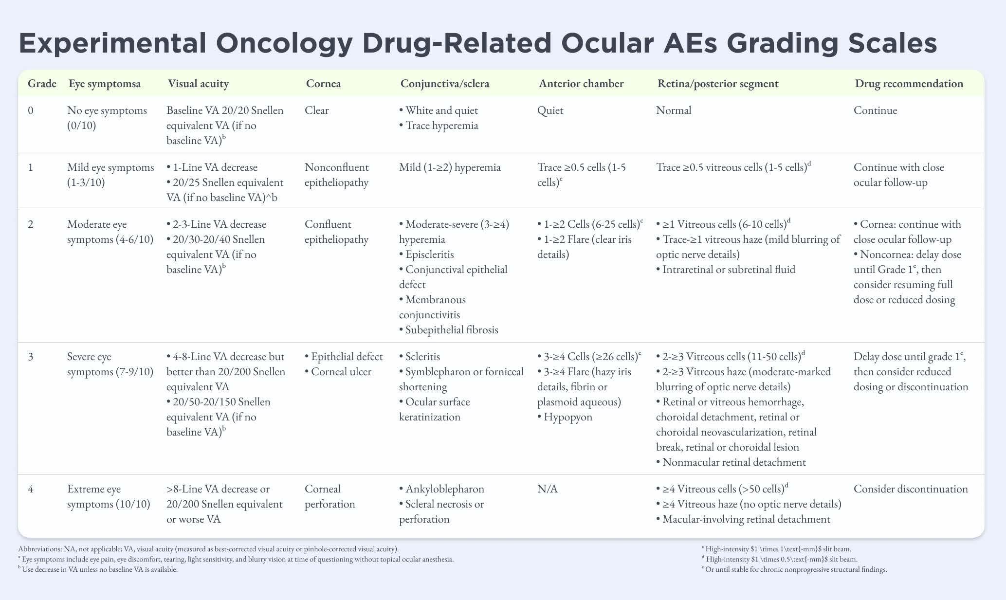
As a way to facilitate consistent reporting, standardize appropriate experimental oncology drug dose modification recommendations and promote patient safety, Dr. Pascricha and colleagues formed a collaborative, multicenter, interspecialty working group consisting of oncologists and academic ophthalmologists from 11 U.S. academic centers and ophthalmologists from the FDA to form a consensus on new experimental oncology drug-related ocular AE grading scales. 22
The goal was to improve the grading system so it was more uniform and would result in less confusion and fewer opportunities for varying interpretations of clinical findings. The new scale (Figure 4) separates eye symptoms from visual acuity changes, and includes six different scales.
Figure 4. Experimental oncology drug-related ocular AEs grading scales created by collaborative multicenter interspecialty working group. 22
“One important thing to note is that for ADCs, this corneal pseudomicrocyst pathology seems very important in one of the unifying factors, but on the corneal scale, that was purposely left out because those are difficult to see and, in isolation, don’t always cause problems,” Dr. Pasricha explained. “Instead, epitheliopathy—which is easier to see and will cause vision changes or eye pain—was what was used for the Grade 1 and Grade 2 corneal changes.”
The impact of this consensus will become more significant as the number of ADCs on the market increases because, currently, each approved ADC drug has its own scale. The differing scales can create confusion for the care team and make it difficult to keep track of the separate scales and associated protocols, and what to grade on that scale. These issues were outlined in the call for standardized guidelines for addressing ADC-related ocular AEs issued in July 2025 by the American Academy of Ophthalmology, 23 which Dr. Pasricha was a contributing author (See more on this in the previous article titled Managing Ocular Side Effects Associated with ADCs).
“The key is that we need better interdisciplinary care. We need optometrists, ophthalmologists, oncologists and hematologists,
all working together with the industry and the FDA, to figure out how we can do this better,” Dr. Pasricha said.
The key is that we need better interdisciplinary care. We need optometrists, ophthalmologists, oncologists and hematologists, all working together with the industry and the FDA, to figure out how we can do this better.
CONCLUSION
Eye care providers should document accurately what is happening with these patients in their eye exams and document the findings in a way that makes sense to the oncology team. Providing an objective assessment, having good notes and sharing with the care team are important steps so the oncology team can decide if a dose interruption is needed.
Developing partnerships and bridging the gap between oncology and eye care allows everyone to determine how to help the patient remain on these potentially life-saving cancer therapies, according to Dr. Pasricha.
“We need to work together. A lot of times, we as eye care providers see the eye as separate from the body. But in the future, we will see that the eye is intricately connected to the body,” Dr. Abbott said. “We will need multidisciplinary coordination of care, and we need to be communicating what we see within the eye to help out our oncology colleagues.”
8. Matulonis UA, Lorusso D, Oaknin A, Pignata S, Dean A, et al. Efficacy and Safety of Mirvetuximab Soravtansine in Patients With Platinum-Resistant Ovarian Cancer With High Folate Receptor Alpha Expression: Results From the SORAYA Study. J Clin Oncol. 2023;41:2436-2445.
9. Arn CR, Halla KJ, Gill S. Tisotumab Vedotin Safety and Tolerability in Clinical Practice: Managing Adverse Events. J Adv Pract Oncol. 2023;14(2):139-152.
10. Coleman RL, Lorusso D, Gennigens C, González-Martín A, Randall L, et al.; innovaTV 204/GOG-3023/ENGOT-cx6 Collaborators. Efficacy and safety of tisotumab vedotin in previously treated recurrent or metastatic cervical cancer (innovaTV 204/GOG-3023/ENGOTcx6): a multicentre, open-label, single-arm, phase 2 study. Lancet Oncol. 2021;22(5):609-619.
11. Lent-Schochet D, Tauber S, Seagrave Z, Paley GL, Farooq AV. Ocular surface disease related to tisotumab vedotin-tftv. Gynecol Oncol Rep. 2025;57:101676.
12. Dy GD, Farooq AV, Kang JJ. Ocular adverse events associated with antibody-drug conjugates for cancer: evidence and management strategies. The Oncologist. 2024;29,:e1435–e1451.
13. Matulonis UA, Birrer MJ, O’Malley DM, Moore KN, Konner J, et al. Evaluation of Prophylactic Corticosteroid Eye Drop Use in the Management of Corneal Abnormalities Induced by the Antibody–Drug Conjugate Mirvetuximab Soravtansine. Clin Cancer Res. 2019;25(6): 1727-1736.
14. Warbington A, Ab O, Laksmikanthan S, Dalton S, et al. Nonclinical ocular toxicity of a maytansinoid payload-antibody drug conjugate: Ocular tissue distribution, lesion pathogenesis, and mitigation. Abstract 2591. Presented at: AACR Annual Meeting; Apr 5-10 2024; San Diego.
Scan the QR code with your smartphone’s camera to watch Drs. Abbott and Pasricha discuss ocular AE prevention strategies, grading scales and care team collaboration.
REFERENCES
1. Lindgren ES, Yan R, Cil O, et al. Incidence and Mitigation of Corneal Pseudomicrocysts Induced by Antibody-Drug Conjugates (ADCs). Curr Ophthalmol Rep. 2024;12(2):13-22.
2. Hurwitz J, Haggstrom LR, Lim E. Antibody-Drug Conjugates: Ushering in a New Era of Cancer Therapy. Pharmaceutics. 2023;15(8):2017.
3. Canestraro J, Hultcrantz M, Modi S, Hamlin PA, Shoushtari AN, et al. Refractive Shifts and Changes in Corneal Curvature Associated With Antibody-Drug Conjugates. Cornea. 2022;41(6):792-801.
4. National Cancer Institute. Common Terminology Criteria for Adverse Events (CTCAE) v6.0 (MedDRA 28.0). Updated July 22, 2025. Available at: https://dctd.cancer.gov/research/ctep-trials/for-sites/ adverse-events/ctcae-v6.pdf. Accessed June 10, 2025.
5. Wahab A, Rafae A, Mushtaq K, Masood A, et al. Ocular Toxicity of Belantamab Mafodotin, an Oncological Perspective of Management in Relapsed and Refractory Multiple Myeloma. Front Oncol. 2021;11:678634.
6. Hungria V, Robak P, Hus M, Zherebtsova M, Ward C, et al. Belantamab Mafodotin, Bortezomib, and Dexamethasone for Multiple Myeloma. N Engl J Med 2024;391:393-407.
7. Hendershot A, Slabaugh M, Riaz KM, Moore KN, O’Malley DM, et al. Strategies for prevention and management of ocular events occurring with mirvetuximab soravtansine. Gynecol Oncol Rep. 2023;47:101155.
15. Kim SK, Ursell P, Coleman RL, Monk BJ, Vergote I. Mitigation and management strategies for ocular events associated with tisotumab vedotin. Gynecol Oncol. 2022;165(2):385-392.
16. NCI. A Study of Ocular Toxicity Evaluation and Mitigation During Treatment With Mirvetuximab Soravtansine in Participants With Recurrent Ovarian Cancer With High Folate Receptor-Alpha Expression. Available at: https://www.cancer.gov/research/participate/ clinical-trials-search/v?id=NCI-2024-05878 Accessed June 18, 2026.
17. Sharma A, Riaz K, Gill M, et al. Reversible HER2 antibodydrug conjugate-induced ocular toxicity. Canadian Journal of Ophthalmology. 2021;57:118-126.
18. Kreps EO, Derveaux T, Denys H. Corneal Changes in Trastuzumab Emtansine Treatment. Clin Breast Cancer. 2018;18(4):e427-e429.
19. Harrington MT, Su L, Adamski J, Patel DR, Shusko AR Jr, Shen JF. Autologous Serum Tear Production in Anticoagulated Patients. Cornea. 2025;44(3):373-376.
20. Rao M, Wu L, Chen H, et al. Hematological toxicity of anti-tumor antibody-drug conjugates: A retrospective pharmacovigilance study using the FDA adverse event reporting system. PLoS One. 2025;20(10):e0334513.
21. Loberg LI, Henriques TA, Johnson JK, Miller PE, Ralston SL. Characterization and Potential Mitigation of Corneal Effects in Nonclinical Toxicology Studies in Animals Administered Depatuxizumab Mafodotin. J Ocul Pharmacol Ther. 2022;38(7): 471-480.
22. Pasricha ND, et al. Multicenter Interspecialty Consensus on Experimental Oncology Drug-Related Ocular Adverse Event Reporting. JAMA Ophthalmol. 2026;144(1):62-68.
23. AAO. Antibody-Drug Conjugates and Ocular Toxicities: A Call for Standardized Guidelines. Updated July 2025. Available at: https:// www.aao.org/education/clinical-statement/antibody-drug-conjugatesocular-toxicities-call-st Accessed February 10, 2026.