



—and in healthcare, what works under pressure in developing countries is starting to look uncomfortably relevant in the developed world.

In developing countries, the problem is obvious: not enough doctors, not enough nurses, not enough infrastructure, not enough resources. So anything that reduces cost or expands access doesn’t just help— it changes who gets treated at all.
Meanwhile, in developed countries, the demographic wave has arrived. The baby boomers are older now, and their diseases have followed them. And suddenly, the same problem appears again—not enough doctors, not enough nurses, not enough infrastructure—at least not enough to treat everyone in time. So again, anything that reduces cost or expands access begins to matter.
What’s good for the geese in Gabon is good for the ganders in Germany.
Cataract surgery makes the point cleanly. It’s life-changing, and in economic terms, it can turn someone from dependency back into participation. At the highest standard, it is not cheap. But it can be made cheaper—manual small-incision surgery, a $4 PMMA lens, no phacoemulsification—bringing the cost down to under $20 per eye.
That’s deliberately provocative. No one is abandoning phaco in developed systems, nor should they. But the existence of a lower-cost pathway matters. It shows that access can be expanded if the model is rethought.
We saw something similar in corneal cross-linking. The shift from mercury vapor lamps to 365 nm LEDs removed cost, fragility and scale limitations. The technology became smaller, more portable, easier to deploy. The medical device requirements still define the floor—calibration, safety, control—but the overall trajectory is clear: lower cost, greater mobility, more patients treated.
Form factor matters. A device that can move will work harder than one tied to a single room.
And before treatment, there is screening. In keratoconus, disease progresses until detection, and vision lost in that interval is often permanent. High-end tomography remains the standard, but it is expensive. A smartphone with a Placido disc adaptor is not. It does not replace Scheimpflug or OCT, but as a screening funnel, it is materially better than nothing, particularly for children, where early detection changes the trajectory.
Cheaper is not better medicine. The goal is not to lower standards. But budgets are tightening everywhere, and the constraint is real. The question is how to extend care without degrading it.
For that, the solutions developed under necessity are not compromises. They are, increasingly, templates.

Best,
Mark Hillen, PhD Director of Communications ELZA Institute, Zurich, Switzerland Editor-At-Large | CAKE
Operation Miracle The price of sight and the politics of global eye




How Japan became the world’s ICL powerhouse Inside the CATALYS™ cOS7.0 Regional Expert Meeting at APAO 2026
Lane, Long Game Raising the Bar in Cataract Surgery With FLACS The Curable Crisis
Closing the gap between possibility and reality A surgical masterclass
Vision and Cocktails
Five
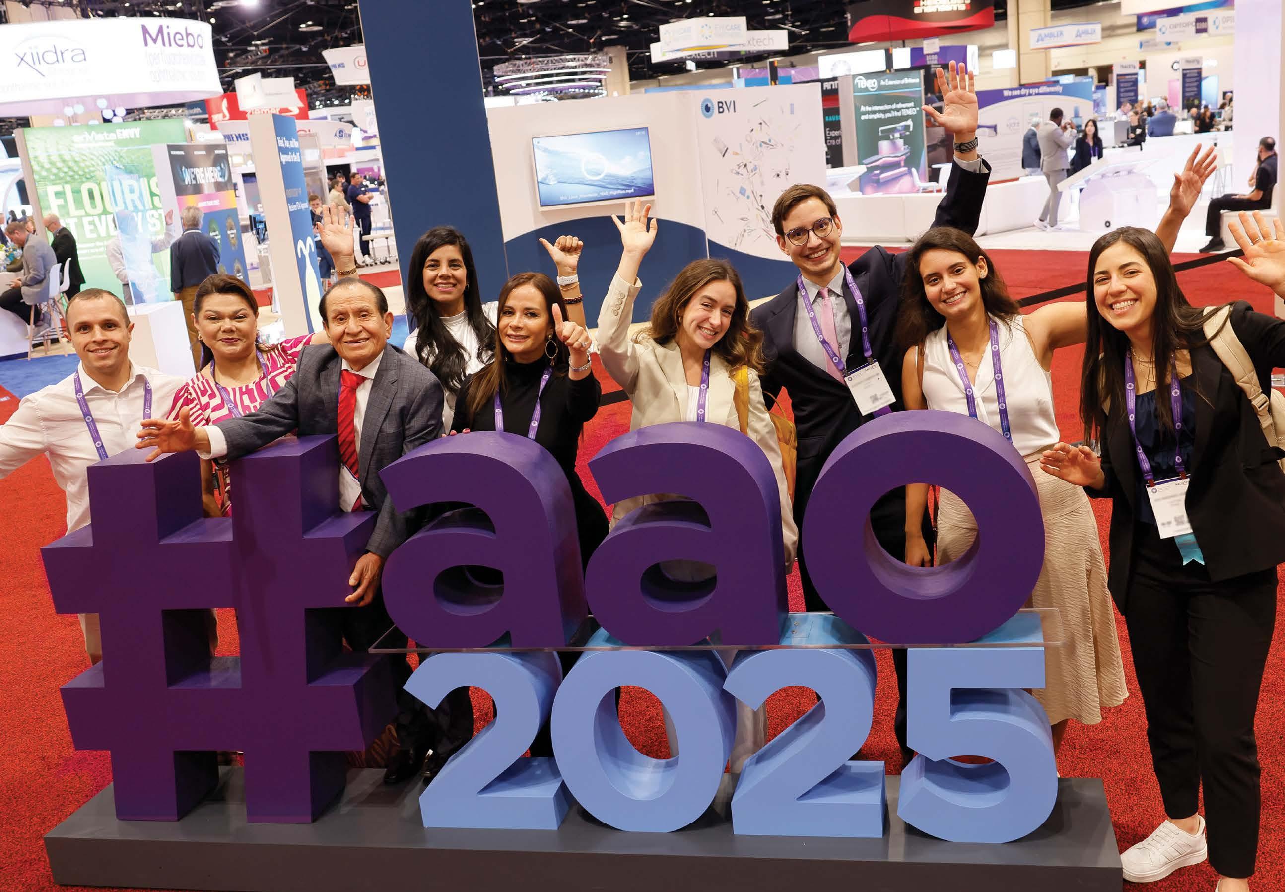

Registration Opens
• Members: June 24
• Nonmembers: July 8
Where All of Ophthalmology Meets® aao.org/2026
Join colleagues from around the world at AAO 2026, Oct. 9–12, in New Orleans, including Subspecialty Day* (Oct. 9 & 10) and the AAO 2026 Expo (Oct. 10–12).
Expand connections, strengthen skills, and deepen insights that elevate patient care through:
• Subspecialty Day*
• Hands-on Skills Transfer labs*
• The largest ophthalmic Expo
• Unparalleled global networking
• Illuminate, the Saturday night celebration supporting ophthalmology*
*Select programs and events require tickets or separate registration.


DR. HARVEY S. UY
University of the Philippines; Peregrine Eye and Laser Institute, Manila, Philippines harveyuy@gmail.com
DR. FRANCIS MAH
Scripps Clinic Medical Group La Jolla, California, USA Mah.Francis@scrippshealth.org

DR. WILLIAM B. TRATTLER
Center For Excellence In Eye Care Miami, Florida, USA wtrattler@gmail.com
DR. CATHLEEN MCCABE
The Eye Associates Sarasota, Florida, USA cmccabe13@hotmail.com


PROF. BURKHARD DICK
University Eye Hospital Bochum Bochum, Germany burkhard.Dick@kk-bochum.de
PROF. DR. SORCHA NÍ DHUBHGHAILL
Brussels University Hospital (UZ Brussel) Brussels, Belgium nidhubhs@gmail.com
Society Friends


We are looking for eye care professionals who can contribute articles to CAKE. Interested? Let's talk! Send us an email at editor@mediamice.com.
To place an advertisement, advertorial, symposium highlight, video, email blast or other promotional packages, contact sales@mediamice.com.

Matt Young CEO & Publisher
Gloria D. Gamat Chief Editor
Diana Truong Associate Editor
Andrés Pacheco
Art Director
Chris McBride
Editorial Creative Lead
Nhat Vo Raquel Jiménez Graphic Designers
Writers Chow Ee-Tan
Hazlin Hassan
Kendra Bruning
Marhiel Garrote
Michaela Perez
Tan Sher Lynn
Artwork credits to: Andrés Pacheco, Chris McBride, Lieu Truong, Yura Yu
Dr. Sashwanthi Mohan Contributor
Ruchi Ranga
Societies Relationships & Conference Manager
International Business Development
Brandon Winkeler Robert Anderson Sven Mehlitz



HOW JAPAN BECAME THE WORLD’S ICL POWERHOUSE
While the rest of the world was busy chasing the next big thing in refractive surgery, Japan quietly built a decades-long evidence base, invented safer procedures and started rewriting the global rulebook. Here’s how patience became a superpower.
By Hazlin Hassan
At a time when new vision correction technologies tend to spread fast and ask questions later, Japan has taken a different road: slower, more deliberate and, in many ways, more durable. The work of the Japan ICL Study Group around implantable collamer lenses (ICLs) illustrates how that philosophy plays out in clinical practice and, increasingly, in global standards.
Japan’s standing in ICL research was not engineered by design. It grew from a sustained willingness to study outcomes over the long term, aided by an early and deliberate comparative framework.
When the country adopted both ICLs and LASIK simultaneously in 1997, clinicians had an immediate opportunity to observe two very different refractive approaches side by side. Rather than committing to a single technique, clinicians tracked outcomes in parallel. Over time, that comparative lens—combined with rigorous follow-up—began to shape preferences.
Conventional ICL surgery, however, had a significant drawback: it interfered with the circulation of aqueous humor, carrying a 1% to 2% risk of postoperative cataract development. Dr. Kimiya Shimizu (Japan), the first surgeon to introduce the ICL procedure in Japan, developed a technical solution, the ‘Hole ICL’, in which a microscopic central perforation in the lens restores normal fluid dynamics.
A third factor, less visible but arguably most consequential, was structural. “Clinical research was conducted in collaboration with university institutions,” Dr. Shimizu noted. That academic involvement standardized data collection and ensured findings were subject to rigorous scrutiny beyond the individual clinic.
Japan’s broader posture toward refractive surgery helps explain why ICL gained traction there in the first place. The country has long maintained a degree of skepticism toward corneal refractive procedures, a wariness with deep historical roots.
“Radial keratotomy was already performed in Japan in the 1940s, and since then, corneal refractive procedures have long been viewed with a certain degree of skepticism,” Dr. Shimizu explained.
That caution was consequential. “When procedures such as LASIK experienced rapid commercial expansion, academic societies in Japan objectively highlighted potential issues, which led to a relatively rapid decline in their widespread adoption,” he recalled.
This was not a rejection of refractive surgery as a discipline. Procedures perceived as less invasive to corneal architecture and more predictable over time began to attract serious attention.


Much of the Study Group’s work has centered on a specific technical challenge: postoperative vaulting, defined as the distance between the implanted ICL and the natural crystalline lens. Vaulting that falls outside an acceptable range can precipitate complications including cataract formation and angle closure.
Rather than treating vaulting as a variable to manage after surgery, the group worked to predict and control it preoperatively. “We developed an original calculation formula and a standardized orientation strategy for ICL sizing by using anterior segment OCT [optical coherence tomography], based on the relationship between postoperative vaulting and associated complications,” Dr. Shimizu shared.
The practical consequences of this shift—from estimation to measurement—have been meaningful. By using anterior segment OCT to assess internal ocular structures with greater precision, surgeons can select lens sizes more accurately, resulting in fewer extreme vaulting outcomes and less complications attributable to sizing errors.

The Study Group’s operating model is straightforward: participating surgeons contribute data, review outcomes and refine techniques collectively.
Through collaborative data sharing within the group, the accuracy of ICL sizing improved, enabling more appropriate postoperative ICL vaulting.
The same framework governs how the group approaches ongoing challenges. “We actively share information on complications as well as emerging technologies, allowing our approaches to continue evolving daily,” said Dr. Shimizu.
Complications are not treated as outliers. Instead, they are utilized as data points that feed back into surgical planning and patient selection.
Clinical philosophy alone does not drive adoption; patient expectations matter too.
In Japan, where myopia prevalence is high and patients tend to prioritize long-term safety over novelty, ICL has found a receptive audience. “With the introduction of the Hole ICL, the adoption of
ICL has increased rapidly and it has become the first-line option for refractive correction surgery in Japan,” said Dr. Shimizu.
The procedure’s reversibility is central to its appeal. Unlike corneal reshaping, an ICL can be removed or exchanged if a patient’s refractive needs change over time—a meaningful advantage for younger patients. “One important lesson is the recognition that most refractive surgery patients are relatively young, which makes long-term safety a critical consideration,” he observed.
That demographic reality pushes the calculus away from shortterm outcomes and toward durability. “Therefore, when selecting refractive surgical procedures, it is essential to base decisions on academic and scientific evidence rather than on commercial considerations,” Dr. Shimizu added.
Some of the Study Group’s technical contributions are now filtering into international practice. “One notable influence has been the shift in ICL sizing from traditional white-to-white measurements to angle-to-angle measurements using anterior segment OCT, enabling more accurate postoperative vaulting,” Dr. Shimizu said.
This reflects a broader movement in ophthalmic surgery toward individualized, imaging-guided treatment planning.
According to Dr. Shimizu, the Study Group’s current research agenda points to a gradual expansion beyond myopia to include the treatment of hyperopia.
There is also interest in less conventional territory. “In addition, given the unique advantage of ICLs being exchangeable, we are interested in exploring their potential application in pediatric amblyopia treatment,” Dr. Shimizu shared. That work is still in development, but it signals how the technology may extend beyond its current scope.
In a specialty where new ideas arrive quickly and consensus is hardwon, Japan’s approach is a good reminder that the kind of progress that lasts tends to take its time.
CONTRIBUTOR

Director general of the Department of Eye Center at Sanno Hospital, Japan, specializing in cataract and refractive surgery. He is also a consultant to STAAR Surgical.
kimiyas@med.kitasato-u.ac.jp
At a packed side meeting during the 41st Asia-Pacific Academy of Ophthalmology Congress (APAO 2026) in Hong Kong, ten of the region’s most experienced cataract surgeons who have experience with the CATALYS™ cOS7.0 premium software gathered around a single question: what does genuinely better cataract surgery look like in 2026?
The occasion was Johnson & Johnson (Florida, United States) CATALYS TM cOS7.0 Regional Expert User Meeting moderated by Dr. Fam Han Bor (Singapore)—an intimate, candid session that blended scientific discussion with real-world case sharing. The takeaway? The latest iteration of the CATALYS TM femtosecond laser platform is doing more than incremental upgrades.
Dr. Brian Schwam (United States), senior director of Global Medical Affairs at Johnson & Johnson Vision, opened the meeting by positioning the CATALYS™ journey as one defined by sustained innovation. He highlighted the cOS7.0 premium software version as a key milestone in the ongoing evolution, while underscoring that further advancements are already in development.
While femtosecond lasers are often discussed as a single category, the discussions made clear that not all femtosecond laser-assisted cataract surgery (FLACS) platforms are experienced equally in practice. Differences in patient interface design, laser speed, and workflow integration meaningfully shape how—and how often—surgeons are willing to use the technology.
The clinical rationale for FLACS has occasionally been contested in the literature, but the surgeons in the room were largely unmoved by studies suggesting parity with manual techniques.
Dr. Gaurav Luthra (India), who has used CATALYS™ for over eight years, addressed this tension head-on. “There have been so many studies which tried to prove that femtosecond-assisted cataract surgery is no better than conventional surgery, but I would like to strongly disagree,” he said. “It adds so much value to surgeries.”
His point? Comparing the two modalities purely on cost misses what the technology actually delivers. Clinicians like himself—high-volume
From workflow hacks to microns-and-degrees precision, a closed-door gathering during APAO 2026 revealed how seasoned cataract surgeons are using CATALYS™ cOS7.0 to move past “good enough” and redefine what premium, repeatable cataract surgery really looks like—case by case, eye by eye.
Sponsored by Johnson & Johnson Vision
cataract surgeons—continue to reach for the laser because it delivers a level of consistent and reproducible outcomes, supporting confidence and precision in cataract surgery. The distinction is not whether excellent surgeons can achieve excellent results manually—but whether those results can be delivered with the same level of precision, every time, across every eye. In that context, femtosecond laser assistance is less about replacing surgical skill and more about standardizing its best outcomes.
Dr. Luthra noted that his transition to CATALYS™ cOS7.0 was transformative in terms of workflow. Where he once dreaded lining up more than three or four laser patients due to speed and miosis concerns, he can now comfortably run seven or eight patients through CATALYS™ cOS7.0 in roughly thirty minutes.
Dr. Guangbin Zhang (China) offered a particularly compelling account of platform evolution. Over the past decade, his center has worked with five different FLACS systems, and CATALYS™ cOS7.0 is now their goto choice. He attributed this decision to three core factors: precision, safety, and efficiency—each of which is directly enabled by the platform’s underlying technology.
The CATALYS™ Liquid Optics Patient interface avoids direct corneal applanation—a design choice that surgeons noted helps maintain more stable intraocular pressure, preserve imaging quality, and support confidence across a wider range of eyes. In parallel, its advanced OCT imaging delivers a detailed three-dimensional view of the anterior segment, offering particular value in the planning and execution of more complex cases.
Dr. Zhang shared two cases that illustrated the point: a young patient with posterior polar cataract where arcuate incision was used to manage regular astigmatism while avoiding the risks of a toric intraocular lens (IOL), and an older patient with moderate astigmatism where a toric IOL was implanted with CATALYS™’s toric mark incisions, achieving high accuracy and excellent postoperative outcomes.
“I think it’s very convenient for astigmatism prediction, whether arcuate incision or the toric lens element,” he concluded.
A significant thread running through many of the presentations was the enhanced astigmatism management capability in CATALYS™ cOS7.0 premium, arguably the most clinically impactful upgrade in the new release.
Dr. Luthra walked through his center’s workflow in detail, highlighting how the system now allows seamless import of biometry data from devices such as the IOLMaster 700 (ZEISS; Oberkochen, Germany), with patient names and keratometry values populating automatically, alongside enabling automatic iris registration. The surgeon can then select between arcuate incisions and toric incisions, or applying surgeon-specific nomograms preferences— to optimize the post-operative outcome.
For a patient with very high corneal astigmatism (7.6 D), where the highest available toric IOL would still leave around two diopters uncorrected, Dr. Luthra combined a high-cylinder toric lens with an arcuate incision to manage the residual astigmatism.
His data from 24 eyes showed an average toric axis deviation of just 2°, with 96% of cases within 3° of target. “That gives us pretty good peace of mind to use this,” Dr. Luthra concluded.
Prof. Tae Im Kim (Korea) echoed the value of the toric axis marking system, demonstrating how slit-lamp-based reference marking, combined with CATALYS™ ’s digital iris registration, enables highly accurate IOL alignment, even in eyes where the visual axis does not coincide with the pupil or capsule center.
Perhaps the most impassioned defense of routine FLACS came from Prof. Soon-Phaik Chee (Singapore), who presented a remarkable video of a severely subluxated cataract managed with the femtosecond laser— a case that, by conventional wisdom, would typically be referred to a vitreoretinal surgeon.
Using CATALYS™ cOS7.0 to create a perfect capsulotomy in a lens that was barely in position, her fellow successfully completed the surgery, preserving the capsular bag and ultimately enabling placement of a multifocal IOL. That patient did not receive a MF IOL but potentially could have.
The message was simple: consistent, well-centered capsulotomies have advantages that extend decades into a patient’s life. A beautifully circular rhexis is permanent. It protects against IOL decentration, supports premium lens performance and reflects the standard of care a surgeon sets for every patient who walks through the door.

One of the meeting’s most instructive sections involved a series of complex case presentations that pushed CATALYS™ cOS7.0 to its limits—and demonstrated that it holds up well.
Prof. Kim presented an elderly patient with a very dense anterior subcapsular cataract with fibrosis lying directly in the path of the planned capsulotomy. Her approach? Enlarge the capsulotomy from 5.3 mm to 6.5 mm, increase the pulse energy, and reduce horizontal spacing to ensure a complete cut through the dense fibrotic tissue. The result was a successful continuous curvilinear capsulorhexis (CCC), a well-centered lens and no need for further surgical maneuvers.
Dr. Li-Yen Pan (Taiwan) shared a series of edge cases where CATALYS™’s speed and OCT clarity opened up options that other platforms would not allow. For small-pupil cases—a common challenge in his center— CATALYS™ ’s capsulotomy speed of 1.3 seconds allows surgeons to position the cut very close to the iris margin without significant risk of intersection, something that is not safely achievable with slower systems. Additionally, he shared his experience adjusting the capsulotomy settings to reduce capsulotomy creation time to under 1 second.
“Rhexis is forever,” Prof. Chee noted. “Even 20 years later, your residents will look at it. It’s not easy to be consistent. You can be very good, but you don’t get a perfect rhexis every time. Femto makes you a consistent surgeon with consistent outcomes.”
Across sessions, CATALYS™ cOS7.0 was less often discussed as a laser and more as a surgical operating system— one that integrates diagnostics, planning, execution, and verification into a coherent digital process.
Throughout the discussion, Dr. Schwam clarified an important point for existing CATALYS™ users: core cOS7.0 version is available at no additional cost as an upgrade for current platform users worldwide. The 7.0 premium version—which includes the enhanced astigmatism management workflow with integrated biometry import from the IOLMaster 700, toric planning and arcuate incision tools—is available as a separate upgrade globally.
For the surgeons in the room in Hong Kong, the verdict was clear: CATALYS™ cOs7.0 premium represents a meaningful step forward— not just in hardware, but in the sophistication and integration of the surgical workflow surrounding it. In a specialty where the difference between a good outcome and a great one is measured in microns and degrees, that matters.
Editor’s Note
The 41st Asia-Pacific Academy of Ophthalmology Congress (APAO 2026) was held from February 5-8, 2026 in Hong Kong. Reporting for this story took place during the event.


Emma Jolley (United Kingdom) doesn’t mince words when asked what drives her work in global eye health. “It’s a very strong sense of injustice for me,” she said. “Cataract surgery is there. It’s quick. It’s available, and yet people don’t have it.”
After nearly 14 years in the field, Jolley, global technical lead for Health & Disability Research at Sightsavers, UK, has seen the numbers. New research from the International Centre for Eye Health—to which Jolley contributed—predicts effective cataract surgical coverage will increase by just 8.4 percentage points globally between 2020 and 2030. In Africa, the predicted increase is only 4 percentage points.1
The World Health Assembly’s target, endorsed in 2021, was 30 percentage points.
Burundi, a low-income country in East Africa, recorded effective cataract coverage of 2.1%—the lowest of any country surveyed. Qatar achieved 77.7%. The surgery is the same. The gap is everything else.
“It’s not a failure of science,” Ms. Jolley said. “There’s very good surgery for treating cataract and it’s very cost effective. It’s really a failure of systems.”
Surgery itself might be quick, but getting people to that operating theater isn’t. In many African countries, there aren’t enough surgeons. Where they exist, they’re concentrated in capital cities. Theaters and supply chains aren’t reliable. Eye care isn’t well integrated into primary health care.
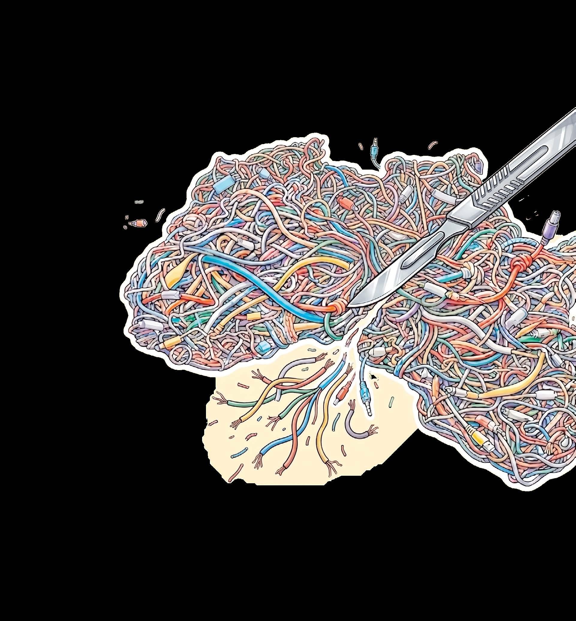
CLOSING THE GAP BETWEEN POSSIBILITY AND REALITY
Even where services exist, community-level challenges persist. Long distances. Prohibitive costs for rural populations. For women and people who don’t control household finances, barriers multiply.
And let’s not forget about cost. “In a lot of countries in Africa, eye care services are not provided for free,” Ms. Jolley said. “If it’s a balance between feeding the family and having cataract surgery, it’s a difficult decision.”
Women in Africa have significantly lower effective cataract coverage than men, roughly 20% compared to 27%. The gap is expected to persist through 2030.1
It’s an eternal question. Unfortunately there’s no clear magic bullet.
She continued, “In a lot of situations, it’s not just being a woman that’s the problem. It’s about being a woman who doesn’t have control of household resources or maybe being an older woman who is a widow and doesn’t have the social support to access services.”

It’s financial resources. Social isolation. Cultural norms. Ms. Jolley sees potential in partnering with women’s groups. “They understand what challenges are facing women in their settings. And they have networks of support, particularly for
It’s not just about how many people get surgery. It’s about whether they can see afterward. Some systems focusing on numbers can inadvertently let quality suffer. “The problem is there might not be enough time to adequately do preoperative counseling,”
For clinic directors, Ms. Jolley offers specific guidance on integrating refractive error services. Getting people to the surgical center is difficult. Asking them to return for refraction and spectacles compounds barriers. “What we need to do is think about how we can move services closer
Surgery needs an operating theater, but postoperative follow-up can happen closer to people’s homes.
“The closer that can be provided, the more you’re going to overcome those distance barriers and cost barriers that affect particularly women and other
This requires developing the broader health workforce: primary care staff and ophthalmic clinical officers who can support ophthalmologists and deliver refractive services. Biometry still isn’t available everywhere. National procurement systems need appropriate supplies, including suitable ranges
The African region recorded the highest rate of intraoperative complications at 27.5%.1 Ms. Jolley points to the World Health Organization’s (WHO’s) recommendations for improving quality. But she emphasizes routine monitoring data. “Integrating one of these outcome monitoring tools—for example, the one that WHO launched—I think that’s really important for surgeons to continually improve services.”
Mali and Senegal, both in West Africa, showed real progress. Burundi did not. What’s the difference?
“The countries making progress are those that have taken that long-term view to planning and investment,” Ms. Jolley said.
Sightsavers worked with both countries for years. Surveys formed the basis of robust plans. Eye health system assessments identified strengths and weaknesses. Countries built strategies and rolled out workforce development. “It’s become a very well-oiled machine because they know exactly what they want to achieve and what they need to do to get there,” Ms. Jolley said.
Districts with dedicated operating theaters, strong leadership and good evidence are empowered to deliver services. Regular services lead to higher demand over time. People see neighbors return with good outcomes. Trust spreads.
Ms. Jolley is clear about where ultimate responsibility lies. “Ultimately governments are responsible and must take the lead for the health of their populations. But they need to be supported by donors, development partners, NGOs.”
Cataract services need financing as part of essential health care. NGOs like Sightsavers can generate evidence, build capacity and demonstrate what works. “But ultimately, responsibility for implementing at scale and ensuring long-term sustainability has to be with national and public ownership.”
If there’s one thing Jolley wishes more people understood, it brings her full circle. “Cataract blindness persists not because it’s hard to treat or we don’t know how to do it,” she emphasized. “It’s because the systems are failing to meet the people who need the care the most. That is a policy choice, and it’s one that we can fix if we want to.”
1. McCormick I, Ouchtar Y, Macleod D, et al. Effective cataract surgical coverage in adults aged 50 years and older: Empirical estimates from population-based surveys in 68 countries and modelled estimates for 2000–30. Lancet Glob Health. 2026;14:e367-377.
2. Global eye care targets endorsed by Member States at the 74th World Health Assembly. World Health Organization. May 27, 2021. Accessed April 16, 2026.
3. Blindness and vision impairment. World Health Organization. February 10, 2026. Accessed April 16, 2026.

ejolley@sightsavers.org


VISION COCKTAILS, CAPSULE RUPTURES TREATMENT OPTIONS AND CUTTING-EDGE REFRACTIVE OPTICS. TELEON’S SHOWING AT APAO 2026 MADE ONE THING CLEAR. PREMIUM CATARACT SURGERY ISN’T ABOUT THE FANCIEST LENS, BUT ABOUT SMART CHOICES, SOLID TECHNIQUE AND KNOWING HOW TO ADAPT WHEN CASES GET COMPLICATED.
At the 41st Asia-Pacific Academy of Ophthalmology Congress (APAO 2026), Teleon Surgical (Spankeren, Netherlands) took the stage not once, but twice. First came a lively evening symposium on intraocular lenses (IOLs), followed by a deep-dive Femtis® instruction course.
One moment, speakers were comparing restoring vision to carefully mixed cocktails; the next, they were dissecting surgical videos frame by frame. The takeaway? Premium surgery requires more than premium lenses. It demands a breadth of options, technical refinement and, above all, a responsibility to match the right solution to the right patient— every single time.
A PORTFOLIO WITH A PURPOSE (AND A PLAN)
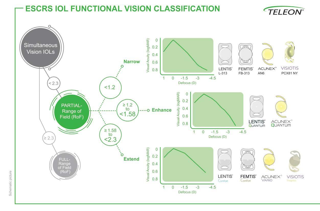
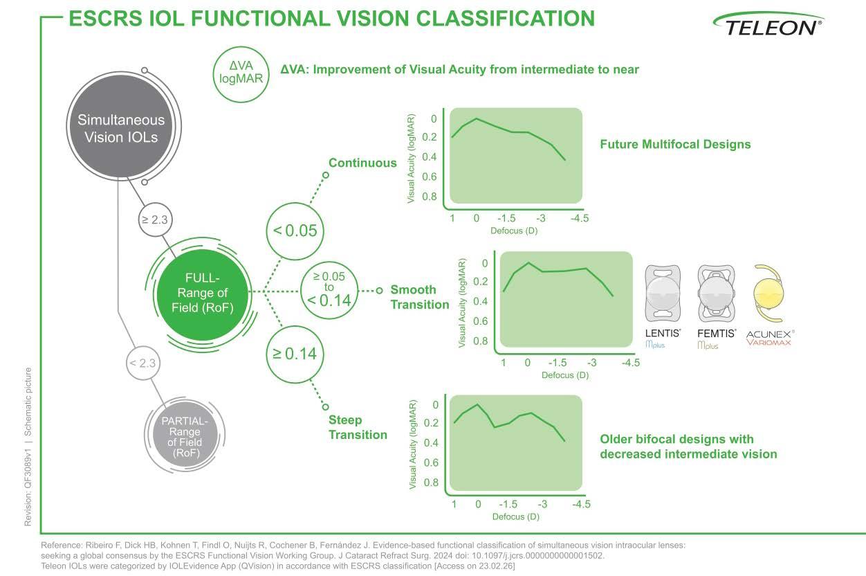
Teleon’s approach begins with patientcentered lens selection and extends all the way to fixation biomechanics and effective lens position control. A new functional range-of-vision classification introduced by the

European Society of Cataract and Refractive Surgeons (ESCRS)
Functional Vision Working Group offers a clearer framework for navigating today’s expanding IOL landscape.1
Lenses are categorized according to range of focus (RoF) and change in visual acuity (ΔVA), grouping them into enhanced, extended and full-range options—each aligned with different visual demands and lifestyle priorities.
The challenge, of course, lies in matching optical design to patient expectations, anatomy and surgical realities. Teleon’s portfolio is built to meet that challenge head-on.
• Enhanced range: LENTIS® and ACUNEX® Quantum
• Extended range: LENTIS® Comfort MF15/20, Visiotis Progress, ACUNEX® Vario
• Full range: LENTIS® Mplus MF30, MplusX, ACUNEX® VarioMax
Between traditional monofocals and extended depth of focus (EDOF) lenses sit the LENTIS and ACUNEX Quantum platforms, designed to expand intermediate vision without compromising distance acuity. Their patented Q-zone aspheric design maintains stable performance even with decentration or tilt. Clinical studies demonstrate excellent distance acuity, crisp intermediate function and minimal halos. 2
For patients exploring premium optics without dramatic trade-offs, Quantum offers a smooth and accessible entry point.
Teleon’s LENTIS Comfort MF15/20 lenses extend the RoF to approximately 1.58 D to 2.3 D, making them suitable for patients who work on computers or drive at dusk. By avoiding diffractive optics, the Comfort platform aims to reduce common dysphotopsia. Toric standard versions correct up to 5.25 D of astigmatism, expanding eligibility for patients often excluded from multifocal solutions.
At the full-range end of the spectrum, the LENTIS MF30, MplusX and ACUNEX VarioMax provide distance-to-near freedom through asymmetric, segmental optics. Compared with classic ringed multifocals, this design is associated with fewer halos and smoother focus transitions.
The mood outside the lecture theater set the tone with light bites, wine and easy conversation among colleagues. Inside, chaired by Prof. Jodhbir Mehta (Singapore), the session unfolded as a series of case-driven “recipes”, each illustrating how nuanced lens selection becomes when guided by personality, profession and visual demand.
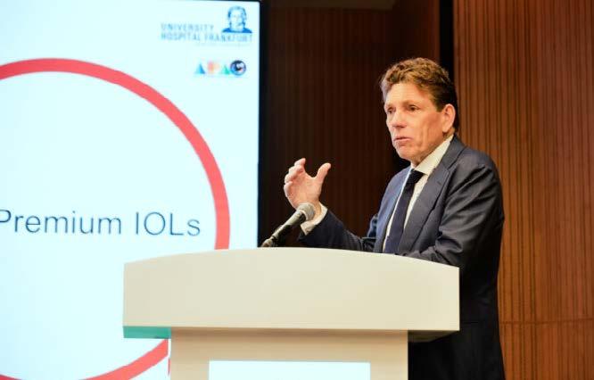
The Daily Driver Spritz. Prof. Thomas Kohnen (Germany) opened with a pragmatic exploration of premium IOL selection. His “Daily Driver Spritz” metaphor captured the idea of balance: refreshing, reliable and built for everyday use.
Drawing on a monocentric study evaluating bilateral implantation of the ACUNEX VarioMax IOL, he presented three-month outcomes assessing distance, intermediate and near acuity, spectacle independence and optical phenomena.3
The findings were notable. Visual acuity was strong across all distances, patient satisfaction was high and 86% achieved spectacle independence. Only two of seventeen patients required glasses for specific tasks, while contrast sensitivity remained robust.
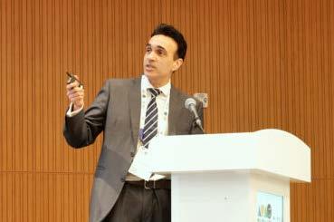
The Singapore Sling. Prof. Mehta presented a case emblematic of modern premium expectations. A 68-year-old IT consultant sought spectacle independence, particularly for computer use.
The case progressed routinely until posterior capsule rupture (PCR) occurred—an uncommon but sobering reminder that even premium surgery is not immune to complication.
In such moments, expectations become as critical as anatomy. Prof. Mehta emphasized adaptability by reassessing fixation, adjusting lens strategy and preserving visual goals without compromising safety. The FEMTIS lens platform allowing rhexis fixation was the perfect choice. The
“Singapore Sling” was his metaphor for controlled improvisation.
“PCR in a premium patient is something we all fear. Even though rare, it can happen,” he explained. “Change is the key.” Premium surgery, he suggested, is defined less by flawless cases than by how surgeons respond when conditions shift.
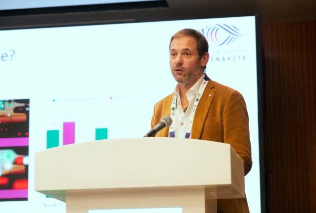
The Hugo. Dr. Florian Kretz (Germany) turned the spotlight to extended range optics, sharing his experience with the LENTIS Comfort Toric MF15.
Addressing concerns around dysphotopsia, comparative analyses showed no significant difference in unwanted photic phenomena when compared with monofocal IOLs. Ease of implantation, reliable toric stability and high contrast sensitivity translated into consistently high patient satisfaction.4,5
For presbyopic patients seeking enhanced intermediate function without the adaptation demands of fullrange multifocality, blended strategies incorporating extended lenses offer a measured and effective approach.


The Saffron Mirage. Dr. Neha Dholakia (India) brought narrative depth to lens selection through a character-driven case. Her patient, a 57-year-old businessman, wanted clear distance and intermediate vision, drove at night, and expressed strong aversion to halos and contrast loss.
An EDOF option risked insufficient near acuity, so she selected a segmental optic full-range design, the LENTIS Mplus MF30.
The outcome delivered on expectations. Distance vision was clear, contrast preserved and halos or glare were absent. The patient declined additional near enhancement, satisfied with functional independence.
The Farsighted French 75. Dr. Lena Beckers (Germany) closed the evening with a memorable portrait of “Jacques,” a patient for whom compromise was not an option. Perfect distance vision was non-negotiable, and halos would prompt relocation to another country rather than adaptation (the patient professed). Therefore, Dr. Beckers chose LENTIS Comfort MF15.
The humor underscored a serious truth. Patient psychology matters as much as optical physics. Premium surgery demands careful listening, expectation alignment and frank discussion
• Premium IOL selection must be lifestyle-driven
• Blended strategies remain powerful tools
• Complication management is integral to premium care
• Communication defines satisfaction as much as refractive accuracy
The “vision cocktail” metaphor proved apt: success lies in proportion, pairing and knowing when to adjust the mix.
Introduced by Dr. David Lubeck (United States), the Femtis instruction course the next day focused on the technical foundations of refractive cataract surgery. Premium outcomes depend on reducing variability, particularly in effective lens position and vault.
From calculation to prediction. Dr. Fam Han Bor (Singapore) reframed IOL power determination as a shift from calculation to prediction.

Traditional vergence formulas remain efficient and reliable but may falter in eyes with atypical anatomy. AI-driven models such as Hill-RBF, Kane and PEARLDGS leverage large real-world datasets to improve performance, particularly in complex cases.
No single formula, however, performs best in all situations. AI offers adaptability but brings limitations, including dependence on data quality and limited transparency in algorithmic reasoning.
“AI reframes biometry as prediction, not calculation,” Dr. Bor concluded. “The largest gains are in difficult eyes. Clinical judgement remains essential. Understanding the technology and its limitations are keys to using it wisely.”
The bottom line? AI enhances accuracy but does not replace clinical judgment. Surgeons must understand both the power and the boundaries of predictive technology.
Phakic IOL placement. Dr. Robert Ang (Philippines) delivered a meticulous review of phakic IOL implantation, emphasizing patience and attention to detail, particularly during lens loading and haptic management. Many complications, he noted, arise from haste rather than complexity.
Scleral and iris fixation. Dr. David Lubeck (United States) addressed stability in compromised capsules, including transscleral haptic fixation. While predictability may not match in-the-bag placement, outcomes can remain stable and visually satisfying when techniques are mastered.
He encouraged surgeons to develop fixation skills early, reinforcing that premium surgery requires preparedness for complexity.
Rhexis fixation. Returning to the podium, Prof. Mehta tackled effective lens position—the persistent source of refractive “noise.”
Even with advanced formulas, variability in lens position limits accuracy. By fixating the lens to a predictable anterior capsuleplane, surgeons can reduce uncertainty and enhance refractive predictability.
He demonstrated how optimized clipping sequences shorten the learning curve. Although early cases increased surgical time, experience rapidly reduced procedures to near-routine duration.

Teleon at APAO 2026: Tailoring IOLs to Patient Vision with Top Experts
The rapid evolution of IOL technology opens exciting possibilities—but it also raises expectations.
Today’s surgeons need more than familiarity with a brochure. They need a working understanding of optical design, supporting laboratory data and real-world lens behavior.
For Teleon, combining a broad IOL portfolio with refined technique and tight surgical control turns personalized vision from an aspiration into everyday practice.
Visit Teleon’s booth at APACRS 2026 from June 4-6, in Pattaya, Thailand, at the Pattaya Exhibition and Convention Hall (PEACH).
Editor’s Note
The Asia-Pacific Academy of Ophthalmology Congress 2026 (APAO 2026) was held from February 5-8, 2026 in Hong Kong. Reporting for this story took place during the event. A version of this article was first published on MediaMICE.com.
1. Ribeiro F, Dick HB, Kohnen T, et al. Evidence-based functional classification of simultaneous vision intraocular lenses: seeking a global consensus by the ESCRS Functional Vision Working Group. J Cataract Refract Surg. 2024;50(8):794-798.
2. Borkenstein, AF, Borkenstein, EM. Clinical Performance of New Enhanced Monofocal Intraocular Lenses: Comparison of Hydrophobic C-loop and Hydrophilic Plate-Haptic Platform. Adv Ther. 2003;40: 4561-4573.
3. Data presented by Prof. Thomas Kohnen at ESCRS 2024 Barcelona.
4. Pedrotti E, Mastropasqua R, Bonetto J, et al. Quality of vision, patient satisfaction and long-term visual function after bilateral implantation of a low addition multifocal intraocular lens. Int Ophthalmol. 2018;38(4): 1709-1716.
5. Sugawara K, Ito R, Horiguchi H, et al. Surgical outcome comparisons of multifocal IOLs of LENTIS Comfort LS-313 MF15 and Tecnis Eyhance DIB00V. Int J Ophthalmol. 2023;16(12):2004

A recently published new joint position statement brings ophthalmology and optometry closer together on one of pediatric eye care’s trickier balancing acts: how to fit, monitor and eventually stop contact lens-based myopia control with greater consistency across the Asia-Pacific region.
Sponsored by CooperVision
For years, the treatment a myopic child received across the Asia-Pacific region depended heavily on which professional happened to be guiding their care. An optometry practice and an ophthalmology clinic in the same city could approach the same patient in entirely different ways.
This fragmentation exists despite a mountain of evidence-based options. The struggle has never been a lack of tools; it has been the absence of a shared clinical map.
At CooperVision’s Luminary event in Kuala Lumpur, the room kept circling an uncomfortable fact. “The majority of the myopic children everywhere are still not on myopia control intervention today,” the company’s APAC Head of Professional Affairs in Myopia Tacy Song said, noting that the regional gap no longer sits between evidence and awareness but between evidence and habit.1
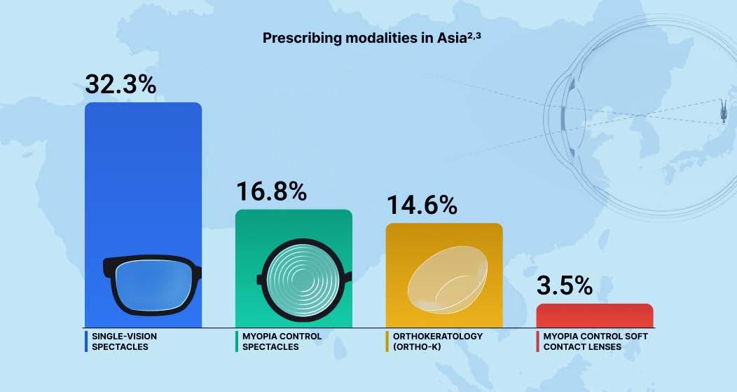
That gap is visible in the prescribing mix. Across Asia, singlevision spectacles remain the primary modality for progressing young myopes at 32.3%. Myopia control spectacles follow at 16.8%, with Ortho-K at 14.6%. Myopia control soft contact lenses sit at just 3.5%. 2,3
For a region carrying the world’s heaviest myopia burden, these figures point to a category established in literature but inconsistent in daily use. Adoption is stalled by mismatched product approvals and children filtered through different professional cultures.
In a Japanese survey highlighted at the meeting, 40% of respondents said that guideline availability would increase the intent to recommend.1 A child’s refraction may be identical in Seoul or Taipei, yet clinical confidence around contact lens treatment is not.
The new joint position statement is a clinical handshake between disciplines that have historically operated in parallel.
Thirty-four eye care professionals from Chinese Taipei, Hong Kong SAR, Japan, Malaysia, Singapore and South Korea completed an online survey before an expert subgroup met in Kuala Lumpur in July 2024 to debate divergence and vote through the final recommendations. 2
Dr. Kate Gifford (Australia) described the process as an effort to stop leaving this work to local improvization.1 A common framework gives ophthalmology and optometry a better chance of speaking the same clinical language despite local regulatory differences.
By drawing on expertise across six major regional hubs, the document replaces individual intuition with a collaborative standard. This shift is essential because the window for intervention is narrow and the cost of delay is measured in permanent ocular changes.
Labeling the rise of myopia an “epidemic” almost feels like an understatement given the current trajectory. Dr. Sandra Block (United States), immediate past president of the World Council of Optometry (WCO), argued the situation has reached “pandemic” status, affecting nearly every corner of the globe.1
In parts of East and Southeast Asia, 90% of young adults are now myopic,
a figure that shifts the condition from a refractive nuisance to a public health crisis.
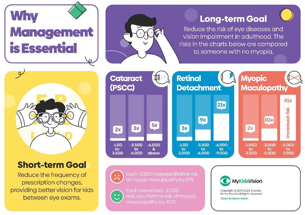
“Currently…we’re seeing one in three children with myopia worldwide. Most of those are in Asia,” stated Elizabeth Lumb, director of Global Professional Affairs at CooperVision.1 The goal is the mitigation of a 67% increased risk of ocular disease for every additional diopter of progression.1 When a child’s eye elongates, they’re losing future health.
Source: mykidsvision.org

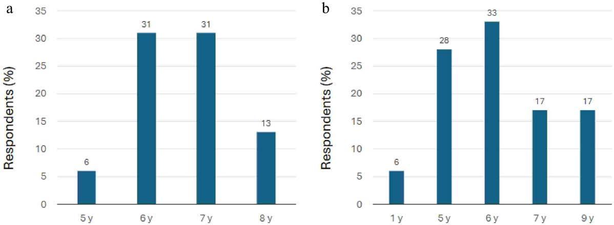
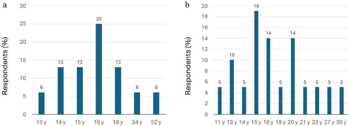
The reliance on a child’s age as the primary clinical gatekeeper is a blunt instrument for a delicate problem. The expert consensus now allows for contact lens fitting as young as five years old, extending the horizon of care deep into a patient’s 20s. 2
This recognizes that biological readiness and caregiver support are more accurate predictors of success than a date on a birth certificate.
“Children aren’t treated for one or two years,” Ms. Lumb pointed out, noting the clinical relationship must span the entire duration of childhood.1 The treatment journey is a growth management marathon where the clinician keeps the eye as close to physiological “normal” as possible.
A child progressing fast at six does not become easier to manage simply because contact lenses feel more culturally comfortable at ten.
Standardization requires a rigorous devotion to the slit lamp and the topography map. For soft lenses, the preferred route is a trial lens fit supported by over-refraction. 2
Ortho-K, by its nature, demands a more expansive technical workflow. Beyond the manual calculations, digital software driven by topography and refractive error has become an
Category
Patient Selection
Fitting Protocol
Key Fit Parameters
Follow-up (Year 1)
Success Metrics
Visual Goal
Discontinuation
appropriate standard, provided it’s validated by fluorescein pattern evaluation. 2
These benchmarks prevent “practice drift,” the slow erosion of standards that occurs when a clinician becomes too comfortable with a familiar modality. The underlying logic of the assessment needs to be identical to ensure consistent care regardless of the clinic’s letterhead.
Myopia Control Soft Contact Lenses (MCSCLs)
Age 5 to early 20s
Trial lens fitting; Slit-lamp exam & over-refraction
Comfort, vision, centration primary gaze position
3, 6 and 12 months (1 and 9 months optional)
≤ 0.50 D progression / < 0.200.30 mm axial growth
6/6 (6/9 monocular is acceptable)
Generally age 18+; Stability < 0.25 D over 1 year
Orthokeratology (Ortho-K)
Age 5 to early 20s
Trial lens, empirical calculation or digital software
Slit-lamp, corneal topography, fluorescein pattern
1 day, 1 month, 3 and 6 months (9 months optional)
≤ 0.50 D progression / < 0.200.30 mm axial growth
6/6 (6/9 monocular is acceptable)
Generally age 18+; Stability < 0.25 D over 1 year
The statement’s clinical weight comes from the role of axial length as a metric of success. While refraction is the traditional metric, the physical elongation of the eye is the true indicator of disease progression. 2
The IMI Clinical Management Guidelines pushed the field toward measuring it at least every six months, but the APAC framework pulls it further into the actual rhythm of care.4
Treatment success is pegged to progression of 0.50 D or less over one year. Acceptable axial length change is set at less than 0.20 to 0.30 mm for children aged 9 and under, and 0.20 mm or less for those 10 and older. 2 Corrected monocular acuity of 6/9 or better is acceptable, while 6/6 is still the goal.5
These numbers are the result of a massive evidence base, including the seven-year MiSight 1-day study. This data showed that dual-focus lenses can maintain 6/6 vision while significantly slowing the elongation that leads to pathology. 2,5,6 This level of granularity turns the follow-up visit into a precise tactical adjustment.

What is myopia? | 3D animation of myopia
One of the most neglected aspects of myopia management is knowing when to walk away. The panel suggested that age 18 is a reasonable starting point for discontinuation, but only if the eye has demonstrated stability, defined as progression between 0.00 D and 0.25 D over a full year. 2 If the axial length increase remains at 0.10 mm or less, the clinician can finally consider the treatment complete. 2
Even as clinicians explore combination therapies involving lowdose atropine (0.01% to 0.05%), the goal remains a disciplined exit. 2 The objective is to transition the patient into adulthood with an eye that is stable, healthy and no longer under the constant threat of progressive elongation.
The success of these guidelines depends on breaking down the silos that have traditionally separated eye care providers. Dr. Block insists that vision health must be integrated into the wider healthcare model rather than being treated as an isolated specialty.1 This requires a commitment to communication, not just between doctors, but between the clinician and the family.
Implementation is the final frontier. The APAC position statement is the necessary scaffolding for a region that has been building its defenses piece by piece. Eye care professionals are essentially negotiating for time, trying to slow a biological clock that wants to run too fast.
Every tenth of a millimeter we save is a victory against a future of preventable blindness, but those victories are only won when every clinician is finally reading from the same map.
1. Song T, Lumb E, Block S. APMMS 2025 Luminary Event. CooperVision Asia Pacific. 2025. Conference symposium.
2. Foo LL, Gifford K, Baek SH, et al. Joint position statement on standardizing the clinical use of contact lenses for myopia control from ophthalmology and optometry practices in the Asia-Pacific region. Vis Neurosci. 2026;43:e013.
3. Wolffsohn JS, Whayeb Y, Logan NS, et al. IMI—global trends in myopia management attitudes and strategies in clinical practice—2022 update. Invest Ophthalmol Vis Sci. 2023;64(6):6.
4. Gifford KL, Richdale K, Kang P, et al. IMI—clinical management guidelines report. Invest Ophthalmol Vis Sci. 2019;60:M184-203.
5. Chamberlain P, Hammond DS, Arumugam B, et al. Six-year cumulative treatment effect and treatment efficacy of a dual focus myopia control contact lens. Ophthalmic Physiol Opt. 2023;43:13240.
6. CooperVision. MiSight® 1 day (omafilcon A) soft (hydrophilic) contact lenses for daily wear package insert. June 2021.

APAC Joint Position Statement for Standardizing the Clinical Use of Contact Lenses for Myopia


The evolution of minimally invasive glaucoma surgery (MIGS) in Canada mirrors a broader shift in glaucoma care: less wait-and-see, more act-while-it-matters.
At the heart of this transition is a reframing of the disease itself.
As Dr. Cindy Hutnik explained, “A number of factors have contributed to phaco-MIGS not only being integrated into the glaucoma management paradigm but also moving surgery from a late option to much earlier consideration.”
Central to this shift is the recognition that structural damage precedes functional loss by a wide margin, where “up to 75% of optic nerve structural damage can happen before functional damage is detected.”
The rise of interventional glaucoma challenges the long-standing, stepwise paradigm that begins with medications and delays surgery until progression is evident.
“In the last few years, the concept of interventional glaucoma has emerged with evidence to support being proactive and preventative—rather than being reactive—to preserve as much vision as possible while acknowledging the importance of healthrelated quality of life for patients,” she said.
“A sufficient body of evidence has shown that MIGS, either solo or combined with cataract surgery, can provide safe and effective control of intraocular pressure [IOP], thus positioning this as an effective risk reduction strategy especially if offered earlier in the treatment paradigm,” she added.
For years, MIGS hovered at the edges of glaucoma care. In Canada, it moved to the center, nudged by data, discipline and a long-awaited funding green light. The payoff is a more proactive approach that’s hard to ignore.
By Tan Sher Lynn
CATARACT SURGERY, RECONSIDERED
Canada’s success also stems from recognizing a simple overlap hiding in plain sight: cataract and glaucoma populations often share the same patient.
“Age and elevated IOP are two of the most significant risk factors for glaucoma progression,” Dr. Hutnik said. “Cataracts are one of the most common ocular changes that happen as we age. The location of the lens in the eye causes it to anatomically have an indirect, and sometimes direct, effect on the drainage anatomy of the eye.”
This anatomical and demographic alignment makes combined surgery a strategic move rather than a convenient add-on. “If a patient who is already on anti-glaucoma medications has a cataract, doing a combined phaco-MIGS procedure at the time of cataract extraction has been shown to be a very effective way to reach a safe target IOP while reducing the dependency on medication,” she said.
The integration of MIGS has also reshaped consultation dynamics and surgical planning. “In every patient with a surgically indicated cataract and risk of glaucoma progression, I discuss MIGS, moving from an option to a recommendation when there is medication intolerance or early disease progression,” she added.
Canada’s adoption story is not one of momentum alone. It’s one of method. High-volume surgical centers played a decisive role in building the evidence base that would ultimately support adoption.

“The framework used by the Canadian Agency for Drugs and Technologies in Health [CADTH] included a clinical review of safety and effectiveness, cost effectiveness, legal, social and ethical issues. A significant amount of the early experience and evidence that was considered by the CADTH to determine the optimal use of MIGS devices and procedures was generated in Ambulatory Surgical Centers,” Dr. Hutnik noted.
“High volume centers that were able to evaluate and assess quality parameters such as safety, effectiveness, efficiency, outcomes and complications were instrumental in providing sufficient data for CADTH to make a positive recommendation for MIGS.”
While Canada’s Health Technology Assessment process is often criticized for its pace, its role in enabling adoption is equally significant. “The process was rigorous, objective and evidencebased but (because of this) was lengthy, lasting over two years,” Dr. Hutnik acknowledged. “Whereas the CADTH report was completed in 2019, public funding in Ontario was only made available in 2025.”
The delay constrained early access, but it ultimately paved the way for broad, equitable adoption once approval was secured.
MIGS training in Canada is closely linked to high-volume, publicly funded centers, allowing for rapid skill acquisition and smoother integration into routine practice.
“I strongly support a broad-based postgraduate training curriculum that provides residents with the knowledge, skills and exposure to state-of-the-art management options. Ophthalmologists are surgeons and, as such, should graduate from residency programs with fundamental competencies in all the subspecialties,” she noted.
She also emphasized the importance of anatomical mastery early in training. “I…believe that the success of angle-based surgeries depends upon a detailed understanding of the anterior chamber angle anatomy which should be a core competency requirement in residency training,” she said. “This may be best achieved during glaucoma subspecialty training with exposure as early as possible in the program. Waiting until fellowship may limit the experience needed to have the confidence and skills.”
Earlier exposure, in turn, translates into broader access and more confident adoption in practice.
If one lever determined the pace of adoption, it was financial, not clinical.
Although glaucoma surgery is classified as medically necessary under the Canada Health Act and therefore intended to be publicly funded, access to MIGS was initially constrained by the absence of provincial reimbursement for devices and associated procedural costs.
“The lack of provincial reimbursement was the most significant barrier in equitable access to care,” Dr. Hutnik stated plainly.
Once reimbursement was secured, uptake accelerated rapidly. “Funding models are a key factor in determining access to care,” she remarked. “Whereas there likely is no perfect health care system globally, the Canadian system—albeit cautious as evidenced by the extent of evaluation needed for public funding—does have a relatively straightforward public funding model once approved.”
For countries looking to replicate Canada’s trajectory, the lesson is less about any single intervention and more about coordination.
“The three most important factors are awareness, engagement and collaboration among the stakeholders with all sharing the common goal of cost-effective delivery of quality health care,” Dr. Hutnik stated.
“The successful adoption of the MIGS model was possible by the active engagement of provincial physician groups with their medical associations and government, as well as strong national leadership by the Canadian Glaucoma Society of Canada with advocacy by the Canadian Ophthalmological Society and awareness campaigns by the Glaucoma Research Society of Canada,” she reflected.
“Collectively, these physician groups…shared a commitment to work together with the government to ensure Canadians have equitable access to the care needed to prevent blindness from glaucoma.”
In the end, Canada’s MIGS journey extends beyond devices and procedures. It reflects the power of timing, alignment and a system structured to translate evidence into access.

Dr. Cindy Hutnik
Department head at the Ivey Eye Institute in London, Ontario, and a board member of the Glaucoma Research Society of Canada, among her many roles.
cindyh@sjhc.london.on.ca

Sponsored by Bausch & Lomb
From smoothing corneas by the micron to ditching drops without a single stent, this APAO 2026 session showed that incremental innovation can still deliver headline-worthy results. Four surgeons, four subspecialties, and one clear message: precision—when paired with pragmatism—is quietly rewriting what “good outcomes” look like in everyday practice.
Bausch & Lomb’s (Laval, Canada) surgical portfolio took center stage at the 41st Asia-Pacific Academy of Ophthalmology Congress (APAO 2026) with a symposium that moved briskly from cornea to canal, and did so with confidence.
Four speakers walked the audience through how customized ablation profiles, enhanced monofocal platforms, trifocal technology and laser glaucoma treatment are sharpening outcomes—with clinical data quietly doing the heavy lifting.
WHEN
Dr. Julian Tagal (Malaysia) opened with a question many refractive surgeons ask behind closed doors: can we really do better than standard ablation profiles for normal eyes—and what about the decidedly non-normal ones?
OCTAVIUS with TECHNOLAS TENEO™ 317 Model 2, approved in Europe in August 2024, pairs topography with the market’s fastest eye tracker (1.7 kHz) to selectively target elevation abnormalities averaging 0.33 microns in normal corneas, as Dr. Tagal explained.
His bilateral study—ProScan in one eye, Octavius in the other— showed similar visual acuity at three months, but Octavius edged ahead on higher-order aberration control. Not statistically significant, but intriguing.



“There was a hint that Octavius could minimize or reverse wavefront abnormalities by correcting elevation abnormalities,” Dr. Tagal noted.
The real payoff came in twelve aberrant cornea cases using Octavius with the Athens Protocol—topography-guided photorefractive keratectomy (TG-PRK) plus corneal collagen cross-linking (CXL) with ablation under 50 microns.
A 26-year-old keratoconus patient with corneal maximum curvature (Kmax) 55 D saw Kmax drop by 3 D and asymmetry fall from 5.8 to 2.6 D at three months, with a 10-micron reduction in the elevated vertex area. A post-LASIK ectasia patient achieved a similar Kmax reduction along with a 0.5-micron decrease in higher-order aberrations. A corneal scar patient gained two lines of best-corrected vision.

The clinical pearl: “Octavius selectively smooths irregular elevation while steepening and flattening as designed,” Dr. Tagal stated. The software sees what standard profiles miss and corrects it with micron-level precision.
Dr. Yau Kin (Hong Kong) addressed a familiar cataract dilemma: patients want distance and intermediate vision, without paying for—or living with—the side effects of multifocal IOLs.
He noted that 84% use smartphones, 70% use computers and 57% use tablets. “Intermediate vision is no longer optional. It’s essential for daily living,” Dr. Yau emphasized.
In Hong Kong’s public sector, only conventional monofocals are provided. In private practice, insurance does not cover premium IOLs, leaving patients to pay out of pocket.
Dr. Yau highlighted that enVista Aspire™, launched in Asia at the end of 2025, uses posterior-surface higher-order aspheric coefficients to create 1.25 D of continuous depth of focus. Optical bench testing showed this without compromising distance vision, he reported.
He has converted a high percentage of his practice, implanting over 100 lenses. His unpublished 13-patient retrospective series showed over 90% achieving uncorrected distance vision better than 0.2 logMAR, with 76% reaching the same intermediate target.
“All feedback has been very positive. No major side effects or complaints,” Dr. Yau reported.
A 74-year-old housewife achieved bilateral J2 near vision unaided—unexpected for a 1.25-D add. An 86-year-old on aspirin reached 0.8 distance vision, maintained an active lifestyle, and avoided the dysphotopsia risks associated with multifocals.
Even Dr. Yau’s father, a retired businessman, received bilateral enVista Aspire IOLs. At one month, he had 1.0 distance vision and 0.8 intermediate unaided, swam daily and needed reading glasses only occasionally.
The clinical pearl: “Best for patients with active lifestyles and strong dependence on distance and intermediate vision—computer users, housewives, smartphone users. If patients can afford a few hundred Hong Kong dollars more, they usually choose this platform.”
Prof. Chameen Samarawickrama (Australia) focuses on younger professionals, with a mean age of 61, who work on computers, drive at night, play sports and expect glasses independence.
“They have high visual needs and high expectations. They won’t forgive compromised distance or bothersome halos,” he explained.
His “hierarchy of needs” for premium IOL success starts with consistent refraction, followed by excellent distance, strong intermediate and near vision, and minimal dysphotopsias. Miss one layer, and dissatisfaction follows.
enVista® Envy, launched to Asia-Pacific at APAO 2026, is designed to close the gaps seen with older trifocals. Built on the glistening-free enVista platform, enVista Envy uses 22 apodized diffractive rings to deliver 4 D of continuous vision without abrupt sweet-spot transitions. Apodization allows maximum light transmission, supporting good contrast and low scatter across lighting conditions.
Prof. Samarawickrama reported that the dysphotopsia profile is fantastic, with severe dysphotopsias at only 6% for Envy compared to 16% with older trifocal technologies.* He noted that toric correction down to 0.64 D on the cornea (0.9 at the IOL plane) addresses residual astigmatism—the most common cause of dissatisfaction.
Patient selection, he stressed, is critical. “You can’t just throw it in an eye. You have to be selective.”
His screening protocol includes a higher-order aberration cutoff of about 0.4. “If you’re sub-0.4, that’s a good eye for trifocal.”

He also routinely screens for keratoconus, which at a prevalence of 1 in 84 is more common than many realize.
Every patient undergoes toric calculator assessment. “If the calculator says a toric would be beneficial, we implant it,” Prof. Samarawickrama shared. “Nailing refractive outcomes is what helps this lens work.”
In his first 22 eyes, 86% of which received toric lenses, six-week outcomes showed 95% achieving 20/20 distance and 91% reaching J4 near.
“Mean absolute error is very low. Constants are optimized from the start,” Prof. Samarawickrama reported. “Envy has become my trifocal of choice. Patients saw rings and halos but weren’t bothered.”
Prof. Dan Lindfield (United Kingdom) closed the session by turning to glaucoma, introducing Elios™, an excimer laser that creates ten trabecular meshwork ostia of 210 microns each, for a total treatment area of 2,100 microns.
The appeal is its point-and-shoot simplicity. No adjustment is required for angle pigmentation, pseudoexfoliation or trabecular color. Cold, non-thermal ablation produces clean openings without damaging the posterior wall of Schlemm’s canal.
“It’s not doing damage to the posterior border. It leaves that intact and allows Schlemm’s to drain naturally,” Prof. Lindfield explained.
His surgical approach uses temporal and superior ports to treat more than 180° of trabecular meshwork. “You’re not sure where collector channels are diseased,” he said. “By spreading 10 shots wide, you’re more confident you’ll hit the areas that need help.”
Setting realistic expectations matters. “[With] MIGS surgery, you’re not going to get a pressure of 10 or 12,” Prof. Lindfield stated. “This is mainly lifestyle surgery to get patients off medication.”
Prof. Lindfield cited eight-year study data showing the effect lasts eight years and longer, with three-year imaging from colleagues showing patent channels and blood reflux from Schlemm’s canal when pressing on the sclera.
His UK real-world data from 138 eyes across four centers showed a reduction of >20% on pre-laser pressures, with nearly half medication-free at one year.
The clinical pearl: “What Elios is really good for is reducing medication burden—getting patients off drops—saving their ocular surface from the fat atrophy and disease that drops cause.”
As for selection, Prof. Lindfield’s threshold is refreshingly low. “Anyone with glaucoma or ocular hypertension on drops gets offered it.”
From smarter topography to fewer drops, the session takeaway was simple: precision, when thoughtfully applied, has a way of making everyday practice a little more elegant and outcomes a lot more predictable.
Editor’s Note
The Asia-Pacific Academy of Ophthalmology Congress 2026 (APAO 2026) was held from February 5-8, 2026 in Hong Kong. A version of this article was first published on MediaMICE.com.
*Shultz M, Wiley W, Liang E, et al. Visual and PatientReported Outcomes of a Novel Full Visual Range Intraocular Lens Versus a Monofocal Intraocular Lens: A Randomized Multicenter US Trial. Am J Ophthalmol. 2025;280:493-507.

For a century, corneal transplantation meant removing the entire cornea and suturing in a replacement. “It’s philosophically a bad idea,” said corneal specialist Dr. Jack Parker (United States), offering a blunt assessment of the procedure that dominated the field for so long.
He isn’t being provocative. Penetrating keratoplasty (PK)— full-thickness corneal replacement—worked in a narrow technical sense. Some patients regained vision, but the trade-offs were significant.
The cornea’s avascularity slows healing. Sutures remain in place for years, with risks of breakage or infection. Surface irregularity from stitching often limits visual quality. Long-term steroid use to prevent rejection introduces its own complications, including glaucoma and immunosuppression.
For decades, corneal surgeons replaced the whole to fix a part. In Rotterdam, that logic was flipped, ushering in precision-layer surgery with better outcomes, steeper learning curves and a global rollout still catching up.
By Michaela Perez
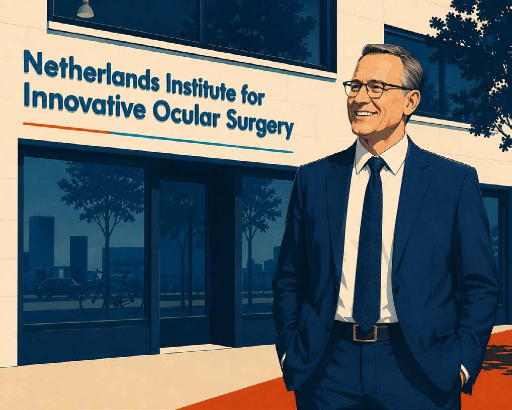
Based in Alabama, Dr. Parker is the only American trained at the Netherlands Institute for Innovative Ocular Surgery (NIIOS) in Rotterdam, where Dr. Gerrit Melles pioneered a fundamentally different approach.
“Most of what a cornea specialist did before Dr. Melles was to convince patients that they didn’t want to have this surgery,” Dr. Parker said.
Rather than accepting those limitations, Dr. Melles set out to rethink the procedure. In most cases of corneal failure, only the posterior layer—the endothelium—is affected. Replacing the entire cornea, he reasoned, was biologically excessive.
Dr. Melles’ first iteration, DLEK (deep lamellar endothelial keratoplasty), demonstrated the concept but was technically
demanding. He refined the approach into DSAEK (Descemet stripping automated endothelial keratoplasty), which gained rapid adoption due to clear advantages over PK.
Yet Dr. Melles viewed DSAEK as transitional. “He immediately recognized that it was just a stepping stone on the way to DMEK,” said Dr. Parker.
DMEK—Descemet membrane endothelial keratoplasty—isolates and transplants only the Descemet membrane, roughly 10 to 15 microns thick.1 The results were transformative, with faster recovery, superior acuity and rejection rates below 1% through two years. 2
This shift reshaped practice patterns in the United States. Endothelial keratoplasty (EK) displaced PK as the standard of care, with DSAEK initially leading adoption.3 By 2015, DSAEK accounted for more than half of all EK procedures while DMEK comprised a smaller share. By 2021, utilization had nearly equalized, reflecting gradual uptake of the more technically demanding technique.4
Dutch registry data illustrated real-world outcomes. Between 2011 and 2018, 752 DMEK procedures were recorded, with mean visual acuity improving from 0.45 logMAR preoperatively to 0.12 logMAR at one year. Sixty-seven percent of eyes achieved 20/25 or better.5
Outcomes improved over time, with procedures performed after 2015 demonstrating higher survival rates, underscoring a systemlevel learning curve.5
THE SECRET WASN’T JUST THE SURGEON NIIOS wasn’t structured like other clinics. It integrated an eye bank—Amnitrans EyeBank Rotterdam—alongside an R&D lab, surgical practice and the NIIOS Academy training center under one roof. 6
“Dr. Melles told me that having an in-house eye bank, along with a corneal clinic, was very important for developing new surgical techniques,” Dr. Parker explained. “You need access to the tissue. It’s almost impossible to be working with the tissue in some fundamental transformative way if you’re just giving somebody else suggestions for what to do.”
Most places separate these functions. Eye banks prepare and distribute tissue, while surgeons operate without direct insight into preparation protocols. The absence of a feedback loop limits iterative innovation.
“I don’t know of any such centers anywhere else in the world where you have an eye bank and you have the surgeon in the clinic, in one location,” Dr. Parker said.
This integration—linking tissue preparation, surgical execution and patient outcomes—enabled the refinements that led to DMEK.
Despite superior outcomes, DMEK hasn’t completely replaced DSAEK. Why? According to Dr. Parker, this is the reason: DSAEK is unambiguously superior to PK, and while DMEK is better than DSAEK, “it’s less undeniably better. It’s [only] more subtly better.”
Economic and technical factors contribute. Reimbursement is equivalent for both procedures, and most patients don’t actively request DMEK. The technique itself is more demanding, requiring precise donor preparation, complex graft handling and managing higher detachment rates.4
The learning curve is well described. One study reported rebubbling rates of 20% in the first 45 cases, declining to 4.4% in the final 45. Visual outcomes tend to stabilize after about 25 cases.4
Dr. Parker sees this in practice. “Detachment is DMEK’s greatest complication, and it has seemingly nothing to do with surgical experience,” he noted. “A brand new trainee and Dr. Gerrit Melles have the same detachment rate.”
If experience doesn’t reduce detachments, the limitation may lie more in tissue behavior than surgical technique. Reassuringly, graft detachment is “generally not a vision or patient threatening complication. It’s just an annoyance.”
An estimated 4.9 million people worldwide live with corneal blindness, concentrated overwhelmingly in low- and middleincome countries where eye banking infrastructure, tissue preparation technology and surgical training remain limited.7

Dr. Parker identifies policy as a primary constraint rather than tissue availability. “There are many countries where they don’t really have an eye banking infrastructure, and maybe that’s for legal or political reasons. People just don’t donate their eyes upon their death.” Regulatory barriers often extend to tissue importation.
He points to Taiwan as an example. Despite its advanced healthcare system, DMEK wait times can exceed two years. “In the United States, you can have a DMEK today if you want one,” Dr. Parker said.
DMEK’s technical complexity further limits scalability. In resource-constrained environments, DSAEK remains the more practical option despite comparatively inferior outcomes. 8
In 2016, Dr. Melles attempted to replicate the Rotterdam model in the United States with the launch of NIIOS-USA. The goal was to establish the same integrated ecosystem in a different regulatory and research environment.
The experiment failed. Without a U.S. medical license, Dr. Melles could not operate. Without a functioning eye bank, the infrastructure underpinning innovation was absent. “What he had was his ideas,” Dr. Parker said. “So he would sit in a room and think and strategize and plan, but it was a little bit more difficult to bring those ideas to fruition.”
The COVID-19 pandemic compounded these challenges, and Dr. Melles eventually closed NIIOS-USA entirely.
But a new American experiment is now underway. Amnitrans USA, modeled after Rotterdam’s eye bank, is set to open in Birmingham, Alabama. Focused on advancing corneal transplantation techniques, it represents another effort to translate the integrated model.
“The entity already exists,” Dr. Parker said. “We hope to be processing tissue this year.”
The Rotterdam model—integrating eye banking, surgical practice and training—worked spectacularly in a well-resourced European setting. Whether it can be adapted to underserved systems remains an open question.
For the millions living with corneal blindness in regions without access to tissue banking, where donations rates are low and import restrictions persist, the issue is not theoretical. Access, not optimization, remains the unmet need.
1. Melles GRJ, Ong TS, Ververs B, van der Wees J. Descemet membrane endothelial keratoplasty (DMEK). Cornea. 2006;25(8):987-990.
2. Price MO, Price FW Jr. Descemet’s membrane endothelial keratoplasty surgery: Update on the evidence and hurdles to acceptance. Curr Opin Ophthalmol. 2013;24(4):329-335.
3. Helen Keller Eye Research Foundation. Gerrit Melles. Biography. Helen Keller Prize for Vision Research. 2017. Available at www.helenkellerfoundation.org/gerrit-melles/. Accessed on March 30, 2026.
4. Hurley DJ, Murtagh P, Guerin M. Ultrathin Descemet Stripping Automated Endothelial Keratoplasty (UT-DSAEK) versus Descemet Membrane Endothelial Keratoplasty (DMEK)-a systematic review and meta-analysis. Eye (Lond). 2023;37(14):3026-3032.
5. Dunker SL, Veldman MHJ, Winkens B, et al. Real-world outcomes of DMEK: A prospective Dutch registry study. Am J Ophthalmol. 2021;222:218-225.
6. Netherlands Institute for Innovative Ocular Surgery. NIIOS. Available at www.niios.com/. Accessed on March 30, 2026.
7. Oliva MS, Schottman T, Gulati M. Turning the tide of corneal blindness. Indian J Ophthalmol. 2012;60(5):423-427.
8. Huang M, Yin G, Hoang T, et al. Comparative study of effectiveness and safety between non-Descemet stripping endothelial keratoplasty and Descemet stripping endothelial keratoplasty for endothelial decompensation. Front Med (Lausanne). 2025;12:1499422.
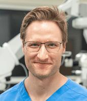
Dr. Jack Parker
Secretary and Director of the Melles Research Foundation, United States. He is the only American to have trained at the renowned NIIOS in Rotterdam, the Netherlands. jack.parker@gmail.com CONTRIBUTOR

In ophthalmic surgery, visualization is everything. The catch is that the best view has traditionally come with a tradeoff: hours at the oculars, shoulders forward, neck angled, and a surgical field only one person can truly see at a time. Heads-up 3D visualization is changing that dynamic by moving the surgeon’s perspective to a shared screen, bringing the team into the same visual plane while supporting a more natural posture.
segment surgeons. Incorporating advanced 3D 4K camera technology, METIS HU combines a 55-inch 3D 4K monitor with a compact floor stand to maximize your working space. By replacing traditional eyepieces with a heads-up view, you benefit from better visualization, improved workflow, lower illumination, and enhanced teaching opportunities.
Whether you already own a METIS 900 ophthalmic microscope or are considering
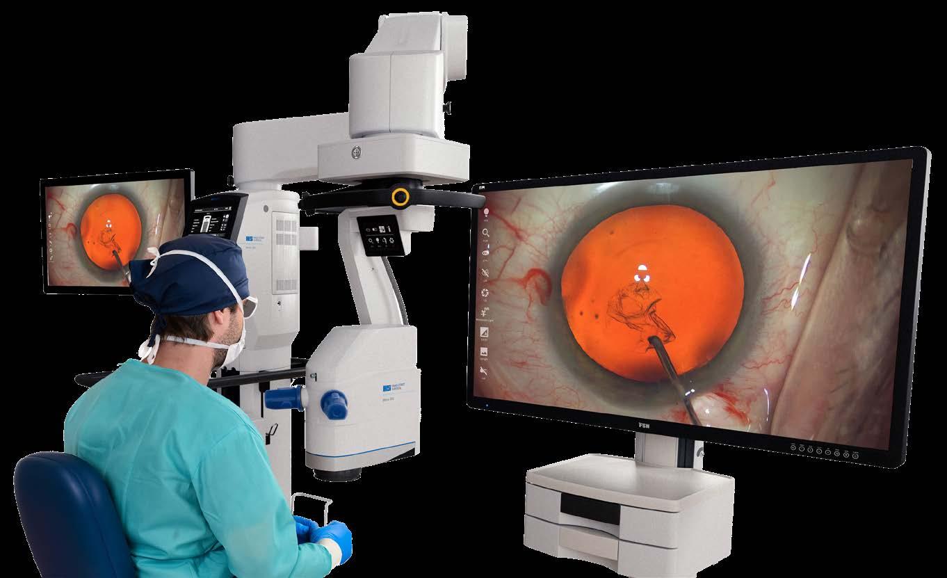
Haag-Streit’s (Köniz, Switzerland) answer is the METIS 3D Digital HeadsUp Module (METIS HU). METIS HU expands the trusted METIS 900 ophthalmic microscope with an integrated digital 3D visualization platform, designed to deliver a state-of-the-art solution for an enhanced surgical experience.
METIS HU is designed to meet the needs of progressive anterior and posterior
digital visualization for the first time, METIS HU offers a flexible configuration and convenient ocular conversion process, giving you the freedom to work how you want.
It’s your METIS HU, and your pathway to enhanced surgical performance.
METIS HU supports ophthalmic surgery with stunning detail and clarity. The high
Sponsored by Haag-Streit
image resolution, combined with a large 3D display, delivers a greater depth-of-field, together with a 3D stereo 4K camera that can constantly adapt to light conditions. The result is enhanced vision that facilitates a more accurate manipulation of delicate intraocular structures.
Haag-Streit has been an innovator in surgical ergonomics for decades, and METIS HU is the latest iteration in this legacy. Benefit from an upright sitting position and a compact design that provides a clear view of the screen without excessive sideways head tilting.
Together, these features can reduce the neck and back strain that can result when using traditional oculars for lengthy procedures.
“The METIS HU has wonderful image clarity that is as good as a regular light path microscope. There is no significant image lag,” said Dr. Edward Meier (United States) of Apex Eye in Cincinnati, Ohio.
“One of the best things is that you can use digital illumination to brighten the image while using a much dimmer microscope light. This is much more comfortable for the patient while still delivering an excellent view and red reflex. As a purposebuilt, heads-up scope, the ergonomics are fantastic. At the end of a long day, my neck & back are much less tired than when using a traditional microscope.”
You don’t need to dispense with the ocular setup altogether because METIS HU gives you the freedom to choose.
Take full advantage of digital visualization when desired, or work with the ocular setup when it’s needed. With a convenient conversion process, you’re in full control with METIS HU.
When using METIS HU, surgeons can draw on the efficient workflows embedded into the METIS 900 microscope to support productivity during procedures. Motorized features, activated by a touch of the hand switch, replace cumbersome knobs, while the foot switch offers wireless control of up to 14 functions.
For even greater efficiency, any digital function can be mapped to the foot or hand switch for customization and ease of use.
METIS HU incorporates digital features designed to support workflow efficiency. Whether adjusting the settings mid-case or pulling up on-screen guidance, these tools are designed to stay within reach with a single, sterile touch.
The METIS HU also includes a digital quick zoom that can reveal fine detail with a tap of the foot, along with image inversion without the need for separate system add-ons.
METIS HU technology is designed to support work at lower light levels compared with an analog microscope setup. As a result, patients may benefit from lower light toxicity and greater comfort during procedures.
At the same time, the system is intended to maintain accurate color representation and true-to-life visuals to support effective surgery.
METIS HU is designed to support an immersive learning environment, where assistants, trainees, and other OR personnel can observe surgical procedures exactly as the surgeon does on a large 3D monitor. High-quality 3D videos and images can
be captured during surgery with a touch of the hand switch or a tap of the foot, then shared for analysis and review.
Multiple 3D monitors can also be linked in a teaching environment, allowing remote observation in real time. In practice, that shared view can turn live cases into teachable moments rather than post-op explanations.
“Screen-based surgery with METIS HU is simply a delight, whether in the anterior segment, posterior segment, or with combined surgery,” said Dr. Christopher Riemann (United States) of the Cincinnati Eye Institute. “The resolution is superb and the unique feature of being able to actively adjust the camera settings intraoperatively
METIS HU is designed to deliver highquality 3D visualization. With advanced digital processing, color, contrast, and brightness are continually optimized to produce a clear, natural image. Surgeons can select from various filters, such as “redfree,” to enhance contrast and improve visibility of retinal structures.
Combined, these features are intended to support better visualization that can aid efficiency and safety across a range of operating scenarios.
As heads-up viewing becomes more common across ophthalmic subspecialties, the focus shifts from whether digital
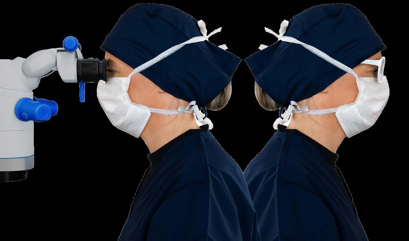
provides a level of control that surgeons will love. We can’t fix what we can’t see and with METIS HU, we can see beautifully.”
METIS HU can be controlled and personalized from the microscope’s 27-inch touchscreen display, including setups for multiple users. Designed with surgeons and their teams in mind, the system interfaces with the Microscope Imaging & Operation System (MIOS), which is a single user interface with interactive menus designed to guide surgical workflow.
From customizing monitor settings and surgeon profiles to recording and sharing patient data, these functions are brought together within one central point of control.

visualization belongs in the OR to how it fits into a surgeon’s existing workflow.
For surgeons who are heads-up curious, METIS HU is designed to bring a digital 3D workflow into the METIS 900 ecosystem while preserving the option to revert to oculars, which may make adoption feel like a controlled transition rather than a point of no return.
For more information, visit the METIS HU page.
Availability of the METIS 3D Digital Heads-Up Module may vary by region. Please contact your local Haag-Streit distributor for details.
Editor’s Note
A version of this article was first published on MediaMICE.com.


THE PRICE OF SIGHT AND THE POLITICS OF GLOBAL EYE CARE
High-volume cataract, low-cost access and a not-so-neutral supply chain. When surgery scales across borders, so does the agenda.
By Kendra Bruning
Outside a clinic near Havana, Dr. Robert Huish (Canada) struck up a conversation with a small group of taxi drivers. They had come from St. Lucia, an island that, at the time, had a single ophthalmologist serving its entire population. Dr. Huish asked one of them why he had made the trip.
“I can’t see,” the man said. “If I’m blind, I’m not a very good taxi driver, am I?”
5,500 5,000 4,500 4,000 3,500 3,000 2,500 2,000 1,500 1,000 500

settings in wealthier urban centers, serving patients who can afford to pay.3
Cataract surgical rates in developed countries range between 4,000 and 6,000 procedures per million people per year. Across much of Latin America, that figure falls between 500 and 2,000, dropping lower still in the poorest areas. Sight deprivation is nearly four times more common in the developing world than in wealthy nations.3

He was there for a cataract operation. The surgery was free. His flight, accommodation and post-operative care were all covered. Within weeks, he would be back behind the wheel.
That exchange captures the operating logic of Operación Milagro (Operation Miracle), launched in 2004 by Cuba and Venezuela on the premise that if reversible blindness was stripping people of livelihood and independence, organized medicine could reach them anyway, regardless of what they could pay.
Over the next two decades, the program scaled up to become one of the most ambitious and politically charged eye care initiatives in the Global South. It moved from flying patients to Cuba for surgery to building ophthalmological centers closer to home, eventually reporting between 4 and 6 million eye procedures performed across 34 countries.1,2
Supporters saw proof that high-volume surgery could be organized around need rather than purchasing power. Critics saw medicine entwined with diplomacy. Both were part of the story.
In Latin America and the Caribbean, an estimated 2.8 million people are blind, and another 11.2 million are visually impaired, most from low-income households. Two-thirds of those conditions are reversible.3
The region has roughly 19,000 ophthalmologists, a figure that should, in theory, be sufficient. In practice, most work in private
Cuban teachers deployed to Venezuela as part of a national literacy campaign noticed that many prospective students could not see and, therefore, could not learn. The program launched in July 2004 as a direct response, beginning with patients identified through active screenings in Venezuela’s most remote communities.4
“Too often, international medical assistance is too narrow and too downstream,” said Dr. Huish, whose research has examined Cuban medical internationalism across multiple countries.
“Brigades may be deployed for a specific disease or outbreak. Postdisaster relief also gets a lot of international buy-in, but it is often too late for too many,” he continued. “The important piece of Operación Milagro is that despite focusing on ophthalmology, it is inherently an upstream intervention that has impacted millions.”
The important piece of Operación Milagro is that despite focusing on ophthalmology, it is inherently an upstream intervention that has impacted millions.
Dr. C. William Keck (United States), a founding board member of Medical Education Cooperation with Cuba (MEDICC), witnessed surgeries on Venezuelan patients in Cuba firsthand.
“Cuba’s pragmatic health decision-making is based first on science, and next on available resources,” he said. “As Cuba’s capacity in the medical and surgical subspecialties grew, the relative simplicity and tremendous impact of sight-restoring surgery became obvious.”
The original target was 5,000 surgeries per year for the region’s most underserved populations. Four years later, the program was averaging that number every single month.4
Patients, identified through community screenings in Venezuela, were flown to Havana with a family member. Surgery was performed at the Ramón Pando Ferrer Ophthalmology Institute, with transportation, meals, lodging and medicine all covered in full.4
Dr. Keck, who observed procedures in Cienfuegos during this period, recalls speaking with Venezuelan patients afterward. “Restoring sight had a great emotional and economic impact on the individuals served and their communities,” he said. “The Venezuelan patients I spoke with were awestruck by the results.”
By the end of 2004, 14,000 Venezuelans had undergone surgery in Cuba. From 2005, Jamaica, Dominica and St. Lucia were among the Caribbean countries that joined, and the range of treatable conditions grew to include pterygium, diabetic retinopathy, retinopathy of prematurity, glaucoma, ptosis, strabismus and retinitis pigmentosa.4
A third phase shifted the model toward local sustainability, with Cuba donating ophthalmological centers and surgical stations to recipient countries. By 2008, those centers had performed more than one million eye procedures across 32 countries, with 47 facilities and 85 surgical stations operating across 11 nations.4
By 2017, the network had grown to 69 centers across 15 countries. 2 Cuban and Venezuelan authorities reported cumulative figures of between 4 and 6 million eye procedures across 34 countries by 2019, though those totals are self-reported and the higher figure incorporates Venezuelan domestic operations under the broader Misión Milagro infrastructure rather than the international program alone.1,2
Venezuela supplied oil; Cuba supplied personnel. The arrangement operated within the framework of ALBA, the Bolivarian Alliance for the Peoples of Our America.1
Dr. Sarah Blue (United States) found in her research that by 2005, Cuba’s non-tourism services exports—the majority of which were
medical services—had doubled from $1.2 to $2.4 billion, surpassing tourism as the country’s primary source of hard currency. Medical export income equaled 28% of Cuba’s total export receipts in 2006.5
“The big difference between Cuban solidarity and Western aid is that Cuba committed to programs that put service before budget lines,” said Dr. Huish. “With solidarity, the programs are established based on cooperation, before worrying about how much will be spent. There is less worry about the limits of a project with the Cuban model, more of a concern about ‘how much can we get done.’”
WHAT SURGERY COULD, AND COULDN’T, FIX Official program sources claimed complication rates below 1%, with post-operative capsule rupture cited as the most common issue. 4 Those claims are unsupported by any published peerreviewed evidence.
A 2012 paper in MEDICC Review examining 210 patients treated in San Cristóbal de Táchira, Venezuela, is the only published outcomes study with patient-level data from the program. 6
Among the 78 cataract patients, 55.2% achieved optimal visual acuity (20/40 or better) within three months of surgery, with a complication rate of 15.4%. Among the 132 pterygium patients, 90.9% recovered optimal vision, though the complication rate reached 41.7%. 6 Both visual acuity outcomes fall short of benchmarks in well-resourced settings, where success rates above 90% are standard for cataract procedures.
Of the 210 patients, 94.2% returned to work prematurely because they couldn’t afford not to. 6 The poverty that had made surgery necessary was also constraining the recovery it was meant to enable.
A 2010 editorial in Arquivos Brasileiros de Oftalmologia noted that many procedures performed—including early-stage pterygium correction and strabismus surgery—had limited direct impact on blindness prevention, and that independent verification of reported procedure totals was not possible given the near-total absence of peer-reviewed documentation.7
“As a ‘one and done’ experience for an individual, it is a ‘miraculous’ transition,” said Dr. Keck, who also questioned what the program left unaddressed. “Another public health question to be asked is what can be done about the prevention of blindness so that the need for surgical intervention is diminished over time.”



“By restoring vision to people who work physical jobs, livelihoods return to communities,” said Dr. Huish. “Because cataract operations are quick and highly successful, it becomes an important pathway to make these upstream impacts not just to the patients, but for their families and communities.”
Dr. Blue’s research found that 73% of Cuba’s specialized ophthalmologists in 2007 were women. Her research argues more broadly that international missions opened a rare path to hardcurrency earnings for health workers across the Cuban system, a workforce that included many Afro-descendant professionals for whom conventional routes to dollar income had been largely closed off since dual economy took hold in the 1990s.5
Some local ophthalmology societies in recipient countries objected. The Uruguayan Society of Ophthalmologists argued that the Cuban presence was unnecessary, since the country already had the equipment to perform the same procedures. The devices existed in private clinics, serving patients who could afford private care. In El Salvador, Operación Milagro’s arrival spurred local societies to advocate for a national cataract program where none had previously existed.4
Leaked diplomatic cables confirmed that the U.S. government worked to discourage President Felipe Calderón from allowing the program to operate in Mexico.1 In August 2006, the United States enacted the Cuban Medical Professional Parole program, offering fast-track asylum processing to Cuban medical personnel working in third countries, a policy Dr. Blue discussed in her research. The aim was to accelerate defections from the medical workforce sustaining programs like Operación Milagro.5

“Cuba’s medical internationalism has been charged as political profiteering, mostly by the U.S. State Department, which committed a $10 million annual budget during the time of George W. Bush to undermine Cuban cooperation by actively recruiting Cuban and Cuban-trained doctors to migrate to the United States,” said Dr. Huish.
“The State Department has also put enormous pressure on countries in the global south to reject Cuban offers of medical assistance, but offers no form of medical assistance to replace it.”
For Dr. Blue—who interviewed Cuban doctors participating in international missions—humanitarian intent and political utility were never in conflict to begin with.
“Cuban medical missions can be characterized as a win-win,” she said. “Cuban policymakers could exploit its human capital and promote positive international diplomacy while also providing sustainable economic options for Cuban professionals at home.”
The doctors she spoke with valued the difference they were making and largely saw missions as an opportunity to apply their training
and humanitarian values.5 The J-1 visa program, she notes, recruits foreign doctors to serve in underserved American communities for below-market wages, which is an arrangement that Washington has never characterized as exploitation when applied to its own healthcare gaps.
Sending tens of thousands of medical personnel abroad restructured the home health system. Family doctor offices closed. Waiting times lengthened. Frustration with longer waits and shorter consultation hours became a tangible consequence felt on Cuban streets.5
In August 2006, a retired Bolivian soldier checked in for cataract surgery at a Cuban-run center in Santa Cruz de la Sierra under a false name. The Cuban doctors treated him without knowing who he was.
When his son wrote to the Santa Cruz newspaper El Deber to thank them, the letter arrived days before the 40th anniversary of Che Guevara’s death, and the patient’s identity became international news. 8
Who was the patient? Mario Terán Salazar, the Bolivian army sergeant who had executed Guevara on October 9, 1967. Cuba’s official newspaper Granma published an editorial declaring that forty years after Terán had tried to destroy a dream and an idea, Guevara had returned to win another battle.1
In a 2014 interview with the Spanish newspaper El Mundo, Terán disputed the official version of events. “It is not as they say that they restored my sight,” he said. “I was not blind. I simply had a cataract.” 8

A program performing 4 to 6 million eye procedures across 34 countries did not check identities at the door. For some, that universality was the point. For others, it was the problem.


Venezuela’s economic and political collapse severely strained the program’s capacity. Cuba’s own domestic pressures complicated the sustained deployment of personnel abroad. What often outlasted the bilateral agreements was the infrastructure left behind, though 2026 has shown how vulnerable even that residual value can be.
On March 4, 2026, Jamaica formally notified Cuba of its decision to end the technical cooperation agreement governing Cuban medical deployments on the island. Honduras lost more than 150 Cuban medical staff the same week, and Guyana announced the departure of more than 200 Cuban doctors days later.9
Jamaica acknowledged U.S. pressure directly, stating that concerns raised by Washington over workers’ freedom of movement and salary payments had prompted a review of existing arrangements. Cuba accused Jamaica of yielding to those pressures. By late March 2026, Cuba had announced the full withdrawal of all 277 Cuban medical personnel from the island.9
Sending tens of thousands of doctors abroad reshaped Cuban healthcare from the inside. The neighborhood-based primary care network was hollowed out, waiting times grew and the intimate,

block-level system Cubans had relied on for decades gave way to a centralized polyclinic model.5
“Ideally, after demonstrating its value, the program should be taken over by facilities and professionals in the recipient country,” said Dr. Keck. “This kind of intervention should not be taken on without pursuing it with the involvement of the host country’s health system and a clear assessment of what can be done relative to sustainability.”
In Latin America and the Caribbean, two-thirds of blindness cases stem from entirely reversible conditions. 3 The taxi driver from St. Lucia was able to drive himself home. The question that the program spent twenty years failing to fully answer is why treating him required a geopolitical arrangement in the first place.

1. Kirk JM. Operación Milagro: Bringing vision to millions. In: Kirk JM, ed. Healthcare Without Borders: Understanding Cuban Medical Internationalism. University Press of Florida. October 6, 2015.
2. Yaffe H. We Are Cuba! How a Revolutionary People Have Survived in a Post-Soviet World. Yale University Press. April 6, 2020.
3. Silva JC, Bateman JB, Contreras F. Eye disease and care in Latin America and the Caribbean. Surv Ophthalmol. 2002;47(3):267-74.
4. Gorry C. Sight for sore eyes: Cuba’s vision restoration program. MEDICC Rev. 2008;10(2):49-51.
5. Blue SA. Cuban medical internationalism: Domestic and international impacts. J Lat Am Geogr. 2010;9(1):31-49.
6. Triana-Casado I, Socarrás Lovio OA, Rondón Paz N. Cataract and pterygium surgery results in Venezuelan patients treated in the Misión Milagro program. MEDICC Rev. 2012;14(3):37-40.
7. Furtado JM, Cade F, Lansingh VC, et al. Is Misión Milagro an effective program to prevent blindness in Latin America? Arq Bras Oftalmol. 2010;73(5):397-8.
8. Olmedo I, Toro JJ. El hombre que mató al Che. El Mundo. November 14, 2023.
9. Charles J. Cuba pulls medical brigades from Guyana and Jamaica after more than 50 years. Miami Herald. March 10, 2026.
Dr. Robert Huish Associate professor and interim associate dean of research in the Department of International Development Studies at Dalhousie University (Canada).
Dr. C. William Keck Professor emeritus and chair emeritus of the Department of Community Health Sciences at Northeastern Ohio Medical University (United States).

Dr. Sarah Blue Jones professor of Southwestern Studies and an associate professor of Geography and Environmental Studies at Texas State University (United States).
huish@dal.ca keck@lek.net sb74@txstate.edu

Watercolor on paper
Softly blooming hues and gentle architectural lines transform a simple house into a vessel of memory, one of a childhood refuge.
Returning to painting after years devoted to medicine, the artist approaches watercolor as a form of meditation, embracing its immediacy and unpredictability to mirror the way memories shift and settle over time.
CONTRIBUTOR

Comprehensive ophthalmologist and oculoplastic surgeon at Eye Physicians of North Houston, Texas. She is also a watercolorist whose subjects range from architecture to portraits.




Two decades into a textbook academic career, one glaucoma surgeon steps off the well-lit stage—and into the surgical unknown—where dense cataracts, scarce resources and zero safety nets redefine what it means to operate. The result? Sharper skills, stripped-back dogma and a reminder that the real gold standard isn’t fancy tech—it’s impact.
By Chow Ee-Tan
For nearly two decades, Prof. Dr. Sameh Mosaed (United States) of the University of California, Irvine, built a distinguished career in academic ophthalmology, refining her expertise in glaucoma, training the next generation of surgeons and contributing extensively to research.
But somewhere along that journey, a realization began to take hold: excellence within one community, however impactful, was only part of a much larger picture.
“I believe the turning point came not from dissatisfaction, but from awareness,” said Dr. Mosaed. “I became increasingly aware of the staggering disparity in how eyecare is distributed globally.”
“Take for instance cataract surgery —one of the most effective and transformative interventions in medicine—which can restore sight within minutes. Yet, for many around the world, it remains out of reach.”
That awareness led her beyond the wellequipped surgical theaters of Southern California to resource-limited settings across the globe, where the absence of care often means a lifetime in darkness. As cofounder of the ophthalmic branch of Women for World Health, she stepped into a role that demanded not only surgical skill, but a calling rooted in equity, adaptability and purpose.

“By stepping into global mission work, I am able to turn that conviction into action, ensuring that the life-altering miracle of clear vision is accessible to everyone, regardless of the circumstances they were born into,” she said, adding that there are countless major and minor details that ensure missions are highly efficient and successful.
“It seems like an overwhelming responsibility, but on each mission—after my first cataract patient sits up from the surgical bed and realizes that they can see again—it is all 1000% worth it,” she enthused.
In her academic practice in the United States, she operates with access to advanced technology, comprehensive support systems and a wide array of surgical tools. In contrast, her mission work demands an entirely different mindset.
“The cataracts are entirely opaque—black, white or even sometimes ruby-red,” she shared. “These are complex cases, often compounded by comorbidities such as diabetic eye disease or end-stage glaucoma, and managed in environments with limited resources.”
It takes a certain degree of confidence to tackle the red cataract behind a poorly dilating pupil with no access to automated vitrectomy or additional supplies in case of complications. These constraints have sharpened her skills. Operating without the usual safety nets has made her more precise, more resourceful and ultimately, a stronger surgeon.

“I am still never relaxed during cataract surgery, no matter how seemingly routine,” she noted.
Within the context of a global surgery playbook, Dr. Mosaed advocates for a critical reassessment of what is truly essential in surgical practice. In resourcerich environments, many processes have become standard, not necessarily because they are indispensable but because they’re convenient.
Her experience in global missions challenges that assumption.
“It is important to identify which routine practices are based on sound evidence and which are unnecessary and wasteful,” she explained. From reusing phaco tips and surgical packs to minimizing draping and extending the use of consumables, efficiency becomes both a logistical necessity and an ethical one.
This approach allows surgical teams to maximize their reach without compromising safety. It also fosters a mindset shift, encouraging surgeons to rely less on specific tools and more on fundamental principles.
“Any blade will cut, any forceps will hold. I am just as happy to take whatever my surgical tech hands me,” she said. “Flexibility is the mindset. Surgeons must be prepared to pivot when the situation calls for it and adapt to whatever is available.”
But beyond technique and efficiency lies something more profound: the human impact of restoring sight.
“In underserved communities, where access to care is often non-existent, cataract surgery is not merely a medical intervention, it is a life-altering event,” said Dr. Mosaed. “These efforts make the difference, literally, between light and dark for hundreds of people.”
Moments such as a young mother seeing her baby’s face for the first time are not rare. They occur on every mission. These experiences “charge our emotional batteries and reinforce the purpose behind the work,” she said.
It is such endeavors that transformed her career from a profession into a worthy calling.
Leading global surgical missions requires more than clinical expertise. It demands the ability to build cohesive teams across cultures, systems and varying levels of experience.
For Dr. Mosaed, the foundation is a shared commitment to patients. This mindset of ‘patients first’ allows team members to be more flexible in their roles, where rigid hierarchies give way to collaboration, with team members often taking on multiple responsibilities to ensure the success of the mission.
Thus, paring down the surgical wish list to the basic essentials, sourcing these essentials and finding ways of stretching things to maximum utilization while maintaining safety.
“It really is threading the needle in some cases, but we have created the formula for successful future missions,” she said. And despite the physical and logistical challenges, the atmosphere remains buoyant.
“Everyone’s spirits are high, and we feel supported by one another,” she said. “The bonds formed through our work are enduring as they are rooted in shared purpose and collective achievement.”
In an increasingly interconnected world, Dr. Mosaed believes that the future of ophthalmology lies in collaboration and exchange. Her recent research comparing surgical outcomes between global outreach settings and resource-rich environments
revealed comparable results.* It is an encouraging sign that high-quality care is achievable across contexts.
Equally important is the mutual learning that takes place during these missions, she stressed.
“The local doctors are often happy to teach extracap techniques to our surgeons, while we teach them glaucoma surgery and phaco skills,” she said. “This exchange not only enhances surgical capability, but fosters a deeper understanding between practitioners from different backgrounds.”
To her, a well-rounded anterior segment surgeon is one who is willing to adopt new techniques and has the courage to put herself in a challenging or vulnerable situation for the benefit of others.
Looking ahead, Dr. Mosaed hopes to see global collaboration continue to expand— not merely as an act of service, but as an integral part of surgical education and practice. Her own journey reflects this evolution. From an accomplished academic surgeon to a global advocate for equitable eyecare, she continues to restore sight with dignity and hope.
*Chen LE, Vahidi R, Kesler-Diaz A , Mosaed S. Comparison of cost-utility, visual acuity, and humanistic outcomes of cataract surgery performed in the United States versus international outreach campaigns. J Clin Med. 2025;14(9):3037.

Prof. Dr. Sameh Mosaed Professor of Ophthalmology and chief of Glaucoma at the Gavin Herbert Eye Institute, UC Irvine. She also serves as vice-chair of Faculty Development, as well as medical director and co-founder of the ophthalmic arm of Women for World Health. smosaed@hs.uci.edu



Few patients casually fly for surgery, except when the system makes it feel easy. In Turkey, ophthalmology has become an exportable system, where accreditation, data and logistics do the heavy lifting, and trust is engineered from a distance.
By Marhiel Garrote
Why would a patient travel thousands of miles for something as critical as sight?
It’s a question that sits uneasily at the intersection of medicine, mobility and modern healthcare economics.
Vision is not elective in the way cosmetic procedures might be framed. It’s functional, immediate and deeply tied to quality of life. And yet, for the past two decades, a growing number of patients have boarded flights for treatment.
The easy explanation has always been cost. But in Turkey’s case, that answer feels incomplete. What has emerged is not just a price advantage, but a system that makes care believable from a distance—and crucially, repeatable at scale.
Long before Turkey became shorthand for medical travel, its earliest international draw was ophthalmology. LASIK, cataract surgery and corneal transplantation quietly formed the foundation of what would later expand into a multi-specialty industry.
In an exclusive interview, Emin Çakmak, chairman of the Turkish Healthcare Travel Council, said the roots of this model go back roughly 25 years, when a small group of highly trained Turkish physicians returned from the United States and began building specialized eye care centers at home.
“We started with ophthalmology patients. Laser, cataract and many other [eye] surgeries… These patients are coming from Germany, from England, from Switzerland,” he said, describing the earliest wave of international patients.
Mr. Cakmak, also recalled an early moment that, in hindsight, neatly captured the scale
of ambition behind the sector’s growth. At the time, the Turkish president had taken a direct interest in the development of specialized eye care and engaged in discussions with both a professor and a clinic owner planning to expand a small practice into a dedicated eye hospital.
“They want to go to the next level, to build a specialty eye hospital,” Mr. Cakmak said. “But they were still looking for a name.”
According to Çakmak, the conversation turned unexpectedly strategic. “The president said, ‘In the world, we have seven billion people…[that’s] 14 billion eyes,” he recalled. “So the potential is very big.”
That framing led to the name: Dünyagöz, or ‘World Eye.’ The hospital would go on to become one of Turkey’s first dedicated ophthalmology centers, bringing together subspecialities under one roof.
What distinguishes Turkey today is not simply that it offers eye surgery at lower cost. It’s the surrounding ecosystem— clinical, logistical and regulatory—that has been deliberately structured to reinforce credibility.
“Why was Turkey chosen as the destination for eye procedures?” Mr. Cakmak said. “Affordability, quality and accessibility.”
The pricing gap is real. Procedures like LASIK or premium intraocular lens implantation often cost significantly less than in Western Europe or the United States. But cost alone does not explain patient flow.
“Turkey is not a cheap destination at all,” he said. “But it is more [attractive] than Europe because the cost structure is different.”
That distinction matters. Patients weigh quality, safety and reputation alongside cost.¹ Price may open the door, but trust closes the decision. In Turkey’s case, what closes it is the perception of a coherent system.
International accreditation plays a central role. Many hospitals in Turkey have pursued recognition from bodies such as Joint Commission International (JCI), alongside European frameworks like Temos International Healthcare Accreditation.
“When we started this journey, Turkey was the number one in the world with JCI accreditation,” Mr. Cakmak noted.
If there is a defining feature of Turkey’s ophthalmology model, it may be how aggressively it leans into data.
“All patients—foreign or Turkish—must be registered in the Ministry of Health system. We can see what operation was done, which doctor performed it, and what the result was,” he said, describing a national system that tracks clinical outcomes.
The implication is significant. In ophthalmology, where outcomes are often reduced to visual acuity, complication rates and patient-reported satisfaction, transparency becomes a powerful currency.

Globally, research has shown that public reporting of outcomes influences both patient choice and provider behavior, reinforcing quality through visibility.² In Turkey, that principle appears to be embedded structurally rather than layered on top.
Clinical credibility alone does not move patients across borders. Logistics do.
A scan of Turkish eye clinics websites reveals a striking consistency: bundled packages that include flights, accommodation, transfers, translation services, postoperative follow-up and even a city tour.
“You pay once, and everything is arranged,” Mr. Cakmak said. “What is less visible, but equally important, is the policy layer behind this.”
He explained, “When the clinic buys the ticket of their patient, the Turkish government pays 50% of the cost,” referring to travel incentives tied to medical
tourism. The result is a patient journey that minimizes friction at every step.
For ophthalmology, this is particularly effective. “Usually the operation is done in one day,” Mr. Cakmak said. “Second day, third day [the patient] is free.”
He also noted broader efforts to promote medical tourism, including international healthcare travel exhibitions bringing together hundreds of organizations abroad to showcase Turkey’s capabilities, ophthalmology included.
For all its structure, Turkey’s ophthalmology sector operates within a wider ecosystem where quality can vary, particularly outside top-tier centers. Research on medical tourism consistently highlights this asymmetry: patients often rely on indirect signals—accreditation, branding, testimonials—rather than firsthand clinical evaluation.³
Social media has amplified this tension. A handful of adverse outcomes can travel fast, shaping perception far beyond their statistical frequency. The industry’s response has been to reinforce safeguards.
1. Lunt N, Smith RD, Mannion R, et al. Implications for the NHS of inward and outward medical tourism: A policy and economic analysis using literature review and mixed-methods approaches. Southampton (UK): NIHR Journals Library. January 2014.
2. Alhawajreh MJ, Paterson AS, Jackson WJ. Impact of hospital accreditation on quality improvement in healthcare: A systematic review. PLos One. 2023;18(12):e0294180.
3. Hanefield J, Lunt N, Smith R, Horsfall D. Why do medical tourists travel to where they do? The role of networks in determining medical travel. Soc Sci Med. 2015;124:356-63.
“We have malpractice insurance for hospitals, doctors and also complication insurance,” Mr. Cakmak said.
The system can standardize trust signals, but it cannot eliminate uncertainty entirely.
In a landscape shaped by distance, uncertainty and information gaps, the winners may not be those who offer the lowest price, but may be those who make quality most legible.
Turkey’s ophthalmology sector offers a case study in how that legibility can be constructed, through accreditation, training pipelines, outcomes tracking and carefully designed patient experience systems.
Mr. Cakmak, however, frames the model less as a system and more as a principle.
“We do not look at international patients as an economic object,” he said. “We first serve under the understanding that every human in the world has the equal right of getting good quality healthcare.”
Economics, as it turns out, comes after.

Emin Çakmak Chairman of

A multinational cohort study published in Neurology reported lower rates of incident glaucoma among migraine patients treated with calcitonin gene-related peptide (CGRP)-targeting therapies compared with those on conventional preventative medications.
Researchers evaluated adults with migraine across multiple international real-world databases, comparing patients initiating CGRP inhibitors against matched cohorts on older preventive therapies. The analysis included more than 73,000 adults treated between 2018 and 2024, with follow-up of up to three years for glaucoma incidence.¹
CGRP inhibitors were associated with approximately 25% lower glaucoma risk over that period. The association remained consistent across database cohorts and sensitivity analyses, and the study was rated as providing Class II evidence.¹
The relationship between migraine and glaucoma has surfaced repeatedly in population-based ophthalmic research, with earlier studies linking migraine to elevated primary open-angle glaucoma risk, particularly among younger patients without hypertension.² Much of that discussion has centered on vascular dysregulation and ocular perfusion instability, pathways that overlap uncomfortably with longstanding questions in glaucoma biology.
A MIGRAINE THERAPY WITH OPTIC NERVE IMPLICATIONS? GLAUCOMA’S VASCULAR STORY THICKENS.
By Marhiel Garrote


The analysis did not account for ophthalmic screening frequency, treatment adherence, or structural progression markers such as OCT retinal nerve fiber layer measurements and visual field change. Follow-up duration and medication exposure windows also varied across databases. No glaucoma progression-related safety signal emerged.

Several mechanistic questions remain open: whether outcomes differ between monoclonal antibody therapies and oral migraine medications, and whether migraine severity itself
CGRP is involved in vascular and pain signaling connected to migraine, making it a plausible point of intersection. The current study examined whether therapies targeting this pathway altered glaucoma incidence, though it did not report separate outcomes by glaucoma subtype, such as normal-tension versus primary openangle glaucoma.
The findings add a neurovascular dimension to the evolving understanding of glaucoma, a disease increasingly characterized as a multifactorial neurodegenerative process involving vascular, inflammatory and metabolic pathways, not simply
The consistent signal across multiple real-world cohorts may fuel further investigation into vascular regulation and optic nerve resilience. The pressure to look beyond IOP, it seems, is building.
Editor’s Note
A version of this article was first published on MediaMICE.com.
1. Chou CC, Wu JW, Lin HJ, et al. Glaucoma risk associated with calcitonin gene-related peptide inhibitor use in migraine: A multinational cohort study. Neurology. 2026;106(11):e218035.
2. Xu C, Li J, Li Z, Mao X. Migraine as a risk factor for primary open angle glaucoma. Medicine (Baltimore). 2018;97(28):e11377.
3. Gupta N, Yucel YH. Glaucoma as a neurodegenerative disease. Curr Opin Ophthalmol. 2007;18(2):110-114.


Global leaders are being called to turn vision commitments into action and perhaps finally bring eye health the political attention it has long needed. The International Agency for the Prevention of Blindness (IAPB) and the Government of Antigua and Barbuda have extended invitations to heads of government, ministers and business leaders for the first-ever Global Summit for Eye Health, scheduled for November 2, 2026, in St. John’s.
The invitation comes five years after the United Nations (UN) Resolution on Vision established eye health as a global priority. Organizers say the Caribbean gathering is intended as a key accountability moment—one that moves global commitments beyond declarations and toward concrete country-level action addressing the estimated one billion people living with avoidable sight loss.
Her Royal Highness The Duchess of Edinburgh, Global Ambassador for IAPB, hosted the invitation launch at St James’s Palace in London. The event brought together international dignitaries and eye health sector representatives to mark the Summit’s official announcement.
Prime Minister Gaston Browne of Antigua and Barbuda addressed the gathering via video link, issuing a direct call to global leaders. “This is not only a profound human challenge—it is an economic one because the evidence is clear. Expanding access to vision care would create millions of jobs and unlock substantial gains in education, productivity and wellbeing,” he stated. “Investing in eye health is not charity. It is an investment in inclusive growth, resilience and shared prosperity, particularly for small states like Antigua and Barbuda.”

Five years after vision hit the UN agenda, leaders are being summoned to prove it wasn’t just talk.
By Michaela Perez

The invitation targets decision-makers who can commit national resources, integrate eye health into development plans, and establish accountability mechanisms for measurable progress.
The Global Summit for Eye Health will be hosted by the Government of Antigua and Barbuda in technical collaboration with the World Health Organization and supported by IAPB.
The initiative is structured around three pillars—Act, Allocate, Accelerate—designed to catalyze commitments to integrate eye health into national development plans, increase sustainable financing and scale proven solutions. Twelve high-impact partners spanning philanthropy, finance, civil society and the private sector are supporting the effort.

“Five years on from the UN Resolution on Vision, the Summit will focus on practical commitments that expand access to affordable, quality eye care services and strengthen health systems to meet rising demand,” said IAPB Chief Executive Peter Holland.
“By bringing leaders together in Antigua and Barbuda we hope that countries, businesses and civil society make formal public commitments to take specific actions, allocate resources or achieve measurable targets in pursuit of our shared goal of ending avoidable sight loss,” he added. “We hope the Summit will serve as a clear expression of political and organizational will, providing a benchmark for accountability long into the future.”





The invitation explicitly asks attending leaders to arrive prepared to make formal public commitments, whether policy changes, budget allocations or measurable targets with defined timelines.
Organizers emphasized that the case for eye health now rests as much on economics as on humanitarian need.
The report The Value of Vision: The Case for Investing In Eye Health*, launched in September 2025 at the United Nations General Assembly, provides the economic framework behind the Summit’s call to action.
The analysis estimates that investing $7 billion in eye health in lowand middle-income countries (LMICs) between 2026 and 2030 could generate $199 billion in economic benefits, representing a 1:28 return on investment and placing eye care among the highest-yield global health interventions.*
0 million fewer people

Walton Webson, Permanent Representative of Antigua and Barbuda to the United Nations and founder and co-chair of the UN Friends of Vision Group, summarized the Summit’s goal, stating, “The Global Summit for Eye Health is about turning words on paper to action for people—from paper to people.”
The invitation extends to government leaders, private sector executives, NGO leadership and funding institutions capable of mobilizing resources and implementing policy changes at scale. If the organizers have their way, November’s gathering in Antigua and Barbuda could mark the moment when eye health moves from admirable rhetoric to measurable action.
More information about the Global Summit for Eye Health is available on the official website, with updates shared via Instagram @GlobalSummitforEyeHealth.
18 million fewer people with unoperated cataract
39 million fewer people with distance uncorrected refractive error 100
198 million fewer people with near vision impairment
A $7 billion investment in eye health could prevent 255 million cases of sight loss globally by 2030—a 24% reduction in avoidable blindness. Courtesy of visionatlas.iapb.org.
If the one billion people currently living with avoidable sight loss received necessary care, the global economy would gain $447 billion annually. Additional projected benefits include 3.3 million years of schooling gained, 5,800 transport deaths averted, 211,000 transport injuries prevented, 1.7 million cases of depression averted, 12,000 deaths averted and 80,000 years of life gained.*
Editor’s Note
A version of this article was first published on MediaMICE.com.

These projections underpin the Summit’s economic argument, positioning vision care investment as a driver of workforce productivity, educational attainment and broader economic development—particularly relevant for small island developing states and emerging economies.


*The Value of Vision: The case for investing in eye health. IAPB Vision Atlas. September 2025.