
ON THE ROAD AGAIN
From Global Stages to Imaging Suites PAGE 14



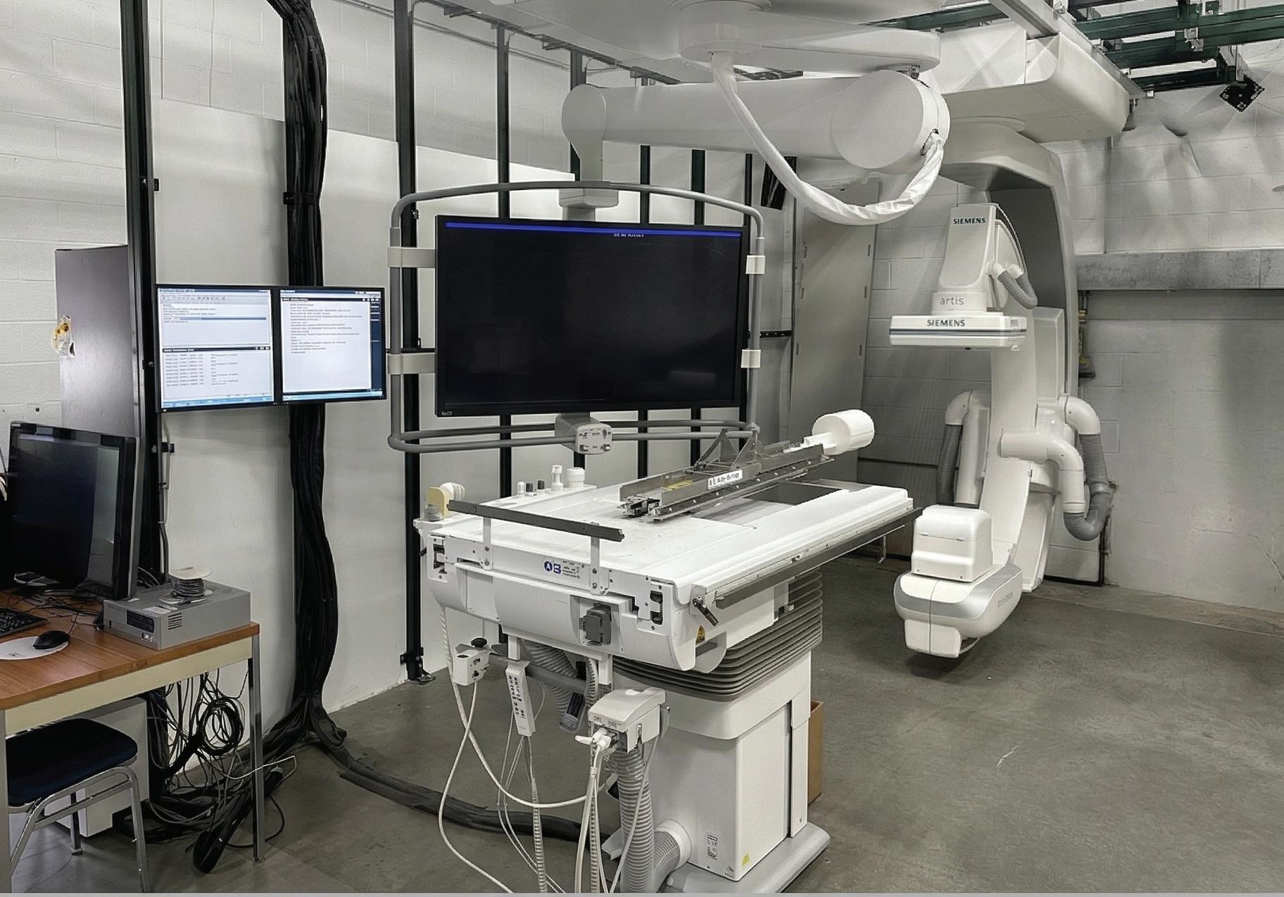
CATH LAB TRAINING
NOW AVAILABLE!
Artis Q & Artis Zee August 10-21 Contact us today for details!
RSTI provides proven, cost-effective imaging service training built on real-world systems, experienced instructors, and outcomes that strengthen HTM teams long after class ends. Engineered for learning. The Industry Experts for OVER 4O years!




FEATURES
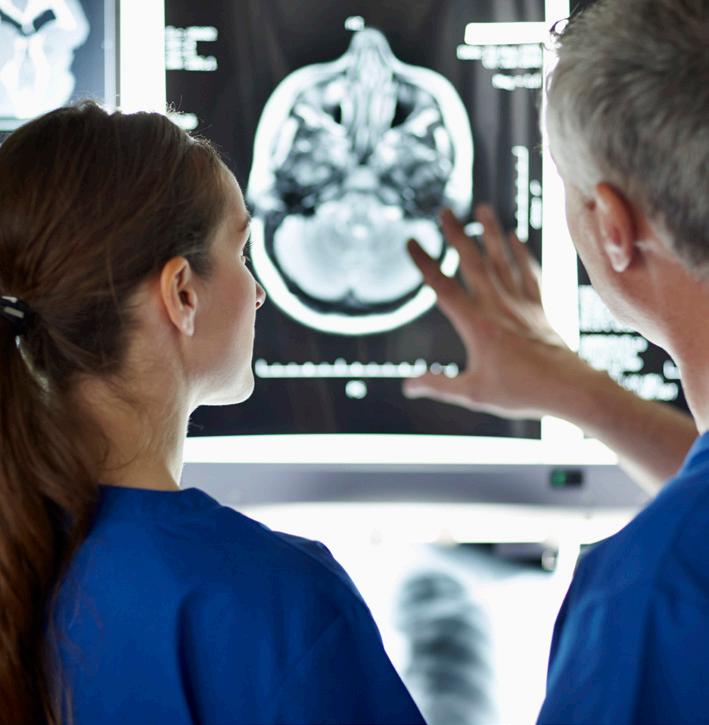
EMOTIONAL INTELLIGENCE
Researchers at the American Psychological Association found that “multitasking” can reduce a person’s productivity by up to 40%.
COVER STORY
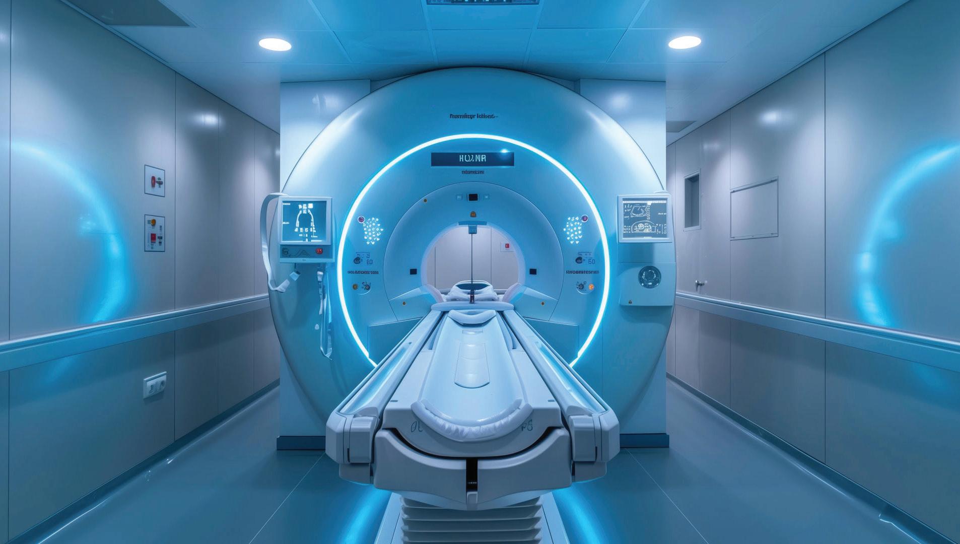
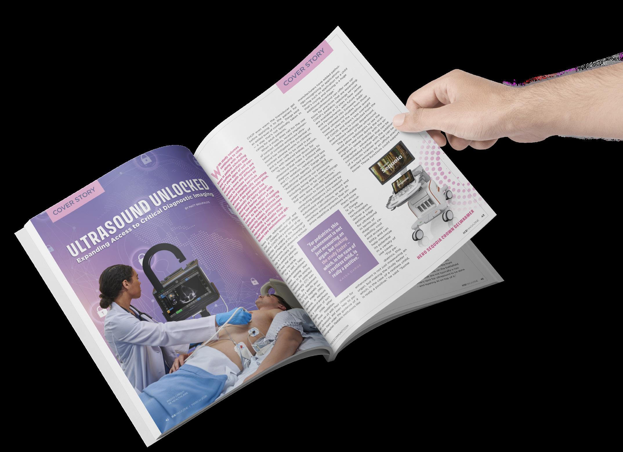
Experts share tips for planning any capital projects involving medical imaging equipment, including new construction.
BLUEPRINTS FOR TOMORROW: Imaging Construction Planning for the

RISING STAR
Amber Washington was nominated as a Rising Star in the diagnostic imaging field by Oklahoma City Indian Clinic Radiology Director Lisa Walling, CRA, R.T (R).

BLUEPRINTS TOMORROW: Construction & the Future
29
PRODUCT FOCUS
Manufacturers share some of the latest MRI devices available in the U.S.
IMAGING NEWS
Catch up on the latest news from around the diagnostic imaging world. 18

MEDICINE FOR THE SOUL Study using advanced imaging found that a “beer belly” is associated with more harmful changes in heart structure than overall body weight alone.
President
Vice
Vice
Senior
Megan
Beth
Kimberly
Megan
Karlee
Kristin
Webinars
Cindy
Kennedy
Accounting
Diane
RISING STAR
AMBER WASHINGTON, RT, CT, M

Am ber Washington, RT, CT, M, holds an associate degree in applied science and serves as a radiologic technologist with the Oklahoma City Indian Clinic. She was nominated as a Rising Star in the diagnostic imaging field by Oklahoma City Indian Clinic Radiology Director Lisa Walling, CRA, R.T (R).
Oklahoma City Indian Clinic (OKCIC) is a contractor of the Indian Health Service to provide culturally sensitive health and wellness services from talented and devoted providers. From pediatric and prenatal care to family medicine, OKCIC services not only included basic medical care but also dental, optometry, behavioral health services, fitness, nutrition and other family programs.
ICE Magazine found out more about Amber in a recent Q&A where she revealed that she loves crafting when she isn’t providing outstanding patient care.
Q: WHERE DID YOU GROW UP?
A: Born and raised in Oklahoma.
Q: WHERE DID YOU RECEIVE YOUR IMAGING TRAINING/EDUCATION?
A: Metro Technology Center, Oklahoma City, associate degree in applied science from Oklahoma State University, Oklahoma City, OK.
Amber Washington, RT, CT, M, thrives in her role supporting the local community in the state she grew up in.
Q: HOW DID YOU FIRST DECIDE TO START WORKING IN IMAGING?
A: While working as a certified nurse’s aid in the hospital I would see radiologic technologists taking X-rays and it piqued my interest in going into the imaging field.
Q: WHAT IS THE MOST REWARDING ASPECT OF YOUR JOB?
A: Helping patients that are searching for answers to why something unusual is happening with them.
Q: WHAT DO YOU LIKE MOST ABOUT YOUR POSITION?
A: The ability to help my co-workers navigate issues that they are having. Also being able to cross train my co-workers in CT.
Q: WHAT INTERESTS YOU THE MOST ABOUT THE IMAGING FIELD?
A: The advancements in technology.
Q: WHAT HAS BEEN YOUR GREATEST ACCOMPLISHMENT IN YOUR FIELD THUS FAR?
A: Becoming registered in three different modalities.
Q: WHAT GOALS DO YOU HAVE FOR YOURSELF IN THE NEXT 5 YEARS?
A: Advance to a leadership role in the radiology department. •
FUN FACTS
FAVORITE HOBBY: Crafts (embroidery, beading, crocheting, woodworking)
FAVORITE SHOW: Anything on HGTV
FAVORITE FOOD: Mexican
1 THING ON YOUR BUCKET LIST: See the Northern Lights
SOMETHING YOUR CO-WORKERS DON’T KNOW ABOUT YOU: I played tennis in high school.

FOCUS IN
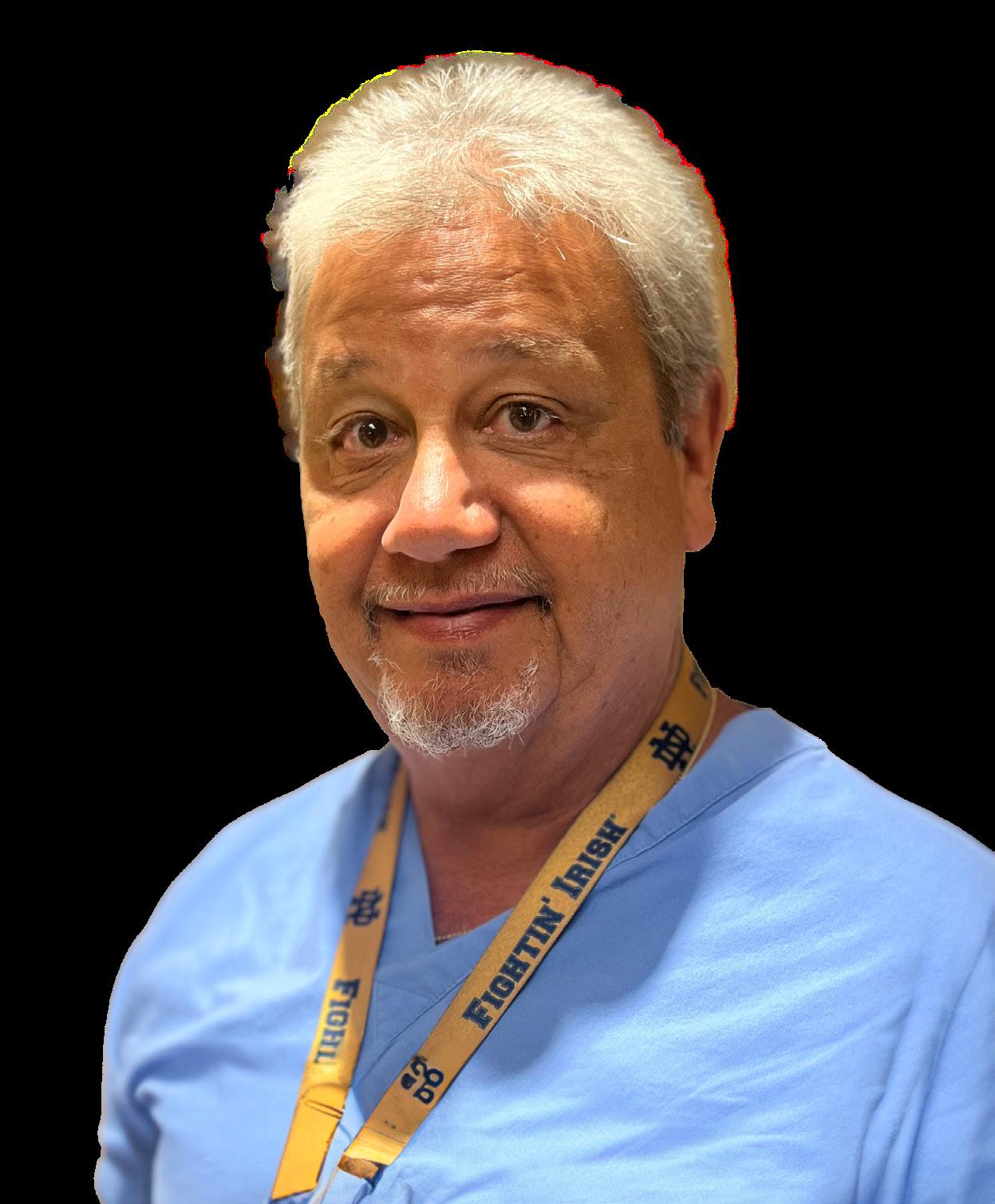
RICH EGAN, RT (R), CRA, MS, MBA
Or lando Health Melbourne Hospital is a comprehensive medical and surgical acute care facility serving the Brevard County Space Coast in Florida as a trusted healthcare provider since 2002. The 119-bed hospital has all private rooms and is uniquely designed for patient comfort and a positive patient experience. A dedicated team of physicians, nurses, clinicians and medical professionals is committed to delivering the highest level of quality and compassionate care, according to the facility’s website.
The hospital provides a complete scope of care, with advanced technology and expertise in a number of specialties, including cardiovascular care, digestive health, emergency care, orthopedics and surgical services. Recognized for high standards, it has earned full hospital and laboratory accreditation from The Joint Commission. It is the first hospital in Brevard County to receive spine surgery certification for its specialized spine program.
Orlando Health Melbourne Hospital Radiology Director Rich Egan, RT (R), CRA, MS, MBA, plays an important role in delivering quality and compassionate care to patients. His road to a radiology career was a bumpy one – literally.
“I knew I wanted to be a radiology technologist ever since I was 16 years old. I grew up in Chicago and broke my femur in a motorcycle accident,” he shares. “While I was in traction, every couple of days, two female radiology techs
came to my room to take follow-up portable X-rays to check on my fractured femur. I found it fascinating and asked them questions each time they came. They always looked like they were having fun even when they crashed the AMX portable into the wall, laughing the entire time. From that point on, I knew I wanted to be a rad tech.”
Now, Rich is the one loving every minute of his imaging job.
“The other day, I realized that I have been a radiology technologist for 40 years and have enjoyed every minute of it,” he says. “I love taking care of patients and my staff. And it’s the only thing I know. Healthcare is always changing and throwing obstacles in your way and it’s fun trying to figure things out.”
Rich has seen lots of changes during his four decades in diagnostic imaging. He sees those changes coming faster and more frequently as technology advances at a rapid pace.
“I would have to say Ai is becoming more and more involved in healthcare, especially radiology. It’s interesting to see the tools they have compared to 5 years ago,” Rich says.
Looking back, Rich shares the impact others have had on his career and professional development.
only then can you build trust.”
“I have had two mentors in my career. The first was when I was a manager and worked for a very knowledgeable imaging director. He taught me how to have fun while being very productive at work,” Rich says. “We had such a great time and worked very well together. I became a director shortly after working with him and use a lot of his wisdom to this day.”
“The next one is who I currently work with and is the president of the hospital. He teaches me every day to take your time when making decisions,” he adds. “I’ve always been a shoot from the hip problem solver, but he teaches me to this day, about thinking of the consequences of your decision.”
“I love taking care of patients and my staff. And it’s the only thing I know. Healthcare is always changing and throwing obstacles in your way and it’s fun trying to figure things out.”
“When I first started, I was in my student clinical rotation at a hospital in Chicago and attended a staff meeting. The director told the staff that if they or their family ever needed surgery, to use this hospital. The very next week, the director had elective surgery at another hospital,” he says. “From then on in my career, I have always done what I say I’m going to do. You need to be honest with your staff and
RICH EGAN
RT (R), CRA, MS, MBA, Orlando Health Melbourne Hospital Radiology Director
1. What are you reading currently? I read radiology periodicals, including ICE Magazine to stay in touch with the state-of-the-art equipment that is being offered.
2. Favorite movie? Tie: “Blues Brothers” and “Animal House”
3. What is something most of your coworkers don’t know about you? Nothing, I’m an open book.
4. What is one thing you do every morning to start your day? Work out
Another aspect of Rich’s professional development and career success can be attributed to his involvement with AHRA: The Association for Medical Imaging Management.
“I have been a member of AHRA since 2004 when I received my CRA credential. I also have been on their Membership Committee for the past year and welcome anyone who would like to join AHRA as this is the most beneficial organization that I know,” he adds. “I have made so many lifelong friends that I see at the annual and local meetings.”
Orlando Health Melbourne Hospital patients and co-workers benefit from the fun-loving and dedicated Radiology Director Rich Egan. His dedication to continuing education and career growth benefits his colleagues and the patients who depend on the department for quality care.
In other words, the patients at Orlando Health Melbourne Hospital are in good hands. •
5. Best advice you ever received? Have fun with everything you do.
6. Who has had the biggest influence on your life? My grandmother who was an English teacher in the 1940s for the blind. She corrected my English on the thank you cards I sent her. She sent them back just like a teacher did
7. What would your superpower be? Invisible Man.
8. What are your hobbies? Golf, fishing, and playing the horses
9. What is your perfect meal? Ribeye and salad.
Clock Off THE
RICHARD ADRIAN RODRIQUEZ, BMET, CBET, TEAM LEAD, INTELAS HEALTHCARE OF SAN ANTONIO, TEXAS
BY MATT SKOUFALOS
As a biomed specializing in medical imaging equipment for Intelas Healthcare of San Antonio, Texas, Richard Adrian Rodriquez travels across the country, performing on-site preventive maintenance, corrective maintenance and administrative work as part of a national field service team.
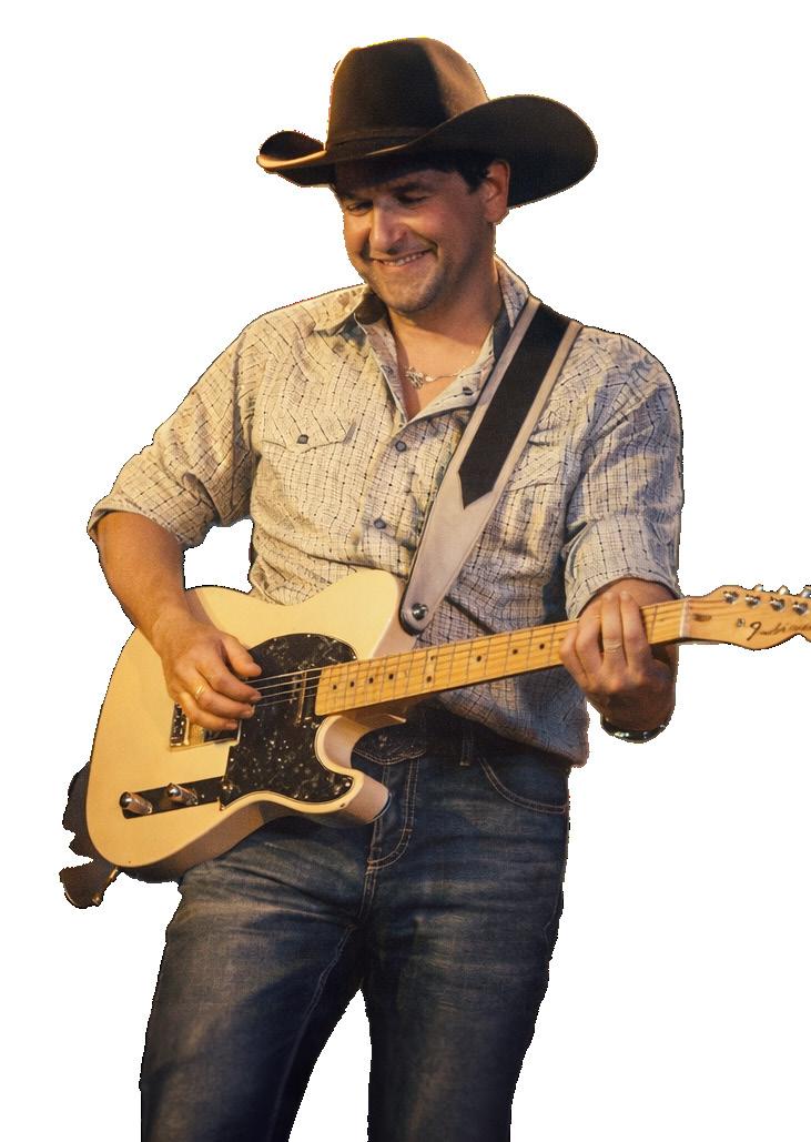
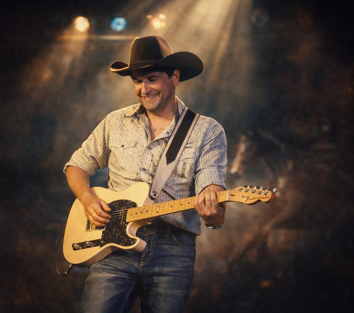
But for the better part of three decades, Adrian traveled across the world, performing Tejano and country music for fans in far-flung regions of the globe, recording Grammy-nominated albums, and rubbing elbows with some of the best players in the industry.
His interest in biomedical work developed partly from a love of tearing down electronic devices and repairing them, a skill he developed early in high school. He also remembers visiting his mother at the hospital where she worked, and her pointing out the professional who came in to repair the equipment. “‘Isn’t that interesting?’ she said, and it was,” Adrian recalled. “I always wanted to be a musician, and they supported me in that, but they told me to have a backup skill.”
A childhood of tearing apart and reassembling electronics led Adrian to earn an associate’s degree in electronics after high school. But a childhood of absorbing and replaying guitar-driven rock from bands like AC/DC, Van Halen, Rush and Journey led to a career as a hired-gun guitarist.
Despite having an arena-rock soul, “there weren’t any cover bands making money in the clubs” with that repertoire by the time Adrian started playing gigs. However, in the late 1980s and early 1990s, the mainstream explosion of country music brought artists like Garth Brooks, Alan Jackson, Travis Tritt, and Brooks and Dunn into the popular consciousness. As radio stations and
clubs adapted their formats to accommodate that interest, he learned new tunes to pick up work.
“More country bands started calling me,” Adrian said. “They were paying, and they were busy, so I started playing more, and traveling regionally.”
By 1995, he was playing five nights a week in the house band at a nightclub in Lubbock, Texas, where Tejano singer Shelly Lares regularly toured with her band. After a few times through, they became friendly enough with Rodriquez that when Lares began to lean more into country music, “they needed a picker and thought of me,” he said.
“I came to San Antonio for an audition and I was in,” Adrian said.
A radio rocker turned country, Adrian slid into the Tejano sound with some slight adjustments. He’d grown up listening to his father playing guitar in conjunto bands, “doing the things that were hot in his generation,” and so was prepared to leverage his Fender telecaster for some arpeggiated “chicken pickin’” just as easily as for rock ’n’ roll distortion. If the songs needed a different voicing, he picked up the 12-string bajo sexto and acoustic guitar.
“Tejano music is mostly ranchero type songs that come from German accordion and polka and cumbia rhythm,” Adrian said. “Spanish people grabbed onto the accordion and the polkas; that evolved into Tejano (Tex-Mex), which incorporated more keyboards and brass and more orchestral instruments and sounds.”
The queen of Tejano music was Selena Quintanilla-Perez, known better by her stage name, Selena. Lares was a top performer in the genre, having grown up with Quintanilla-Perez. Tragically, Quintanilla-Perez was murdered by the president of her fan club at the age of 23, cutting short a career that was poised for mainstream crossover. Her albums have continued


to sell long after her death in 1995, but a broader embrace of Tejano music never followed.
“Selena’s death was hard on everybody,” Adrian said. “She was on the verge of crossing over into pop; Shelly was crossing over into country.”
Adrian began working with Lares in October 1996, a professional touring and recording relationship that has extended across three decades. They still get together occasionally for projects, but today, Lares runs her own record label as an artist-in-resident at the University of Texas-San Antonio. There, she established the first Tejano music course to mentor young Latin artists to preserve the genre and culture.
While working with Lares, however, Adrian played in Japan, Korea, Guam, China, and most of the United States. He played on two Grammy Award-nominated recordings. He’s met musicians from some of his favorite bands, including Los Lobos, members of George Strait’s Ace in the Hole Band, and singer-songwriters like Dean Dillon, Randy Rogers and many others.
“I’ve done so much more in this business than I thought I would ever do,” he said. “I was just lucky and blessed to be out there.”
In the midst of his music career, Adrian met his wife Kara, in 2009. They were married in 2013, but Kara fell ill in 2016, and sadly passed away in 2019. Her diagnosis compelled Rodriquez to pursue a mid-life career change, and so he returned to school to earn a degree in biomedical engineering.
“Being a musician is feast or famine,” Adrian said. “My wife had always picked up the slack and floated us; when she got sick, I knew I had to get a job. I graduated around the time she passed away, and that threw me into the workforce.”
“Usually a musician might have a day job to support their creative pursuits; in my case, playing music supports the day job,” he said.
If the Tejano music scene lost its mainstream momentum with Selena’s passing, the emergence of the digital music business upended the recording industry altogether. Today, most of Adrian’s gigging is to feed his passion and pick up lucrative side work
when it’s available. Music has become “a nice little side hustle” that he enjoys on weekends and the occasional evening, playing out with several San-Antonio-area acts like Jerry DeLeon and Jake Botello, and producing an album for singer Mario Flores.
“It’s very hard to make any money at it other than publishing, touring and playing live,” he said. “There’s no such thing as CD sales anymore. You just put your stuff up there, drive people to it, and hope people come out and support live music.”
Despite those factors, the human connection in live performance still resonates. A kind word from an audience member, or a moment of recognition in passing reminds Adrian about the fundamental power of reaching people through music.
“I thank God that what talent I had was able to reach somebody,” he said. “It’s really rewarding when somebody comes up to you and says, ‘Thanks for being here.’ If it makes people happy, great, if it helps someone, great.”
“It’s about helping people, and that’s why I got into healthcare as well,” he said. “That’s my way of helping mankind: keeping the equipment up and running. It takes a special kind of heart to be a nurse or a teacher; I grew up with my mom setting that example for me.”
Adrian views his career in healthcare as keeping his mother’s legacy alive; “contributing what you can to humanity.”
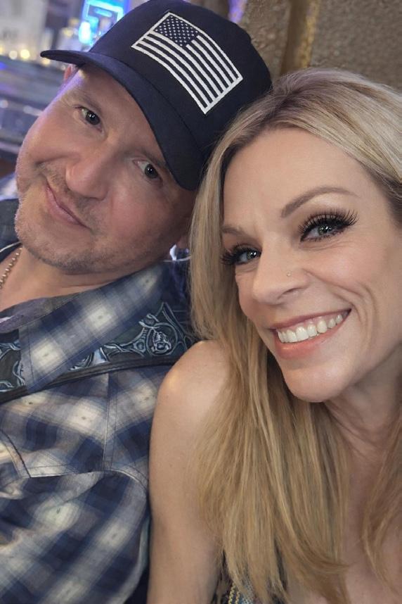
He’s also found salvation from the tragedy of losing Kara. In the wake of her passing, Adrian began to evaluate his own life’s path, and made what he said are changes for the better. Along the way, he also met Shari, a speech therapist to whom he’s now engaged to be married. The family includes their adult children, his daughter Brooklyn and her son Austen; a Chihuahua, a Yorki and three Chinese Crested dogs.
“Shari’s changed my outlook on life,” Adrian said. “She’s a beautiful, amazing woman, inside and out, and I’ve learned so much from her.”
“Every day I thank God for her” he said. “She’s my purpose. With God on our side, and her by mine, there’s nothing we can’t do together.” •
Richard Adrian Rodriquez spent about 30 years traveling across the world, performing Tejano and country music for fans. He plays more local gigs these days and is engaged to Shari who he says, “changed my outlook on life.”
ICE Debut
Everyone has an origin story. The SalesMaker Carts story started with, of all things, a copier. If you’ve ever tried moving a copier, you know it’s a process unlike any other. Honestly, it’s terrible. But it did lead to an idea that turned into the company you’re reading about right now.
What began with the moving of a single copier, now boasts two stocking warehouses nationwide. It just goes to show that you never know where a great idea will come from.
All of the carts are handmade. It might not be the most efficient way to craft carts, but at SalesMaker Carts it’s the only way. When something is made by hand, there are no shortcuts.
“We also only use the best components. At SalesMaker Carts, we define the best as the finest grade aluminum. We chose this type of metal for a few reasons. It’s known for exceptional strength. It’s lightweight. And it’s forgiving. These are all incredible traits to have when you’re in the business of crafting carts,” the company website explains.
“We got into this business simply because we saw a need for making transporting equipment easier,” it adds. “But the more we dove into the mechanics of this industry we discovered an amazing monetary benefit, as well. SalesMaker Carts can help protect your most important asset – your people.”
Operations Manager Nathan McKay recently shared more information about the company and its plans for 2026 and beyond.
Q: CAN YOU TELL US A LITTLE ABOUT YOUR COMPANY?
MCKAY: At SalesMaker Carts, we specialize in supplying durable, mobile equipment solutions that improve efficiency in site-to-site and oftentimes off-campus work requirements. Our high-quality carts help teams move equipment, technology and materials safely and easily. We offer gurney-style folding carts that serve as mobile workstations for technicians and sales reps, compact computer carts and even heavy-duty stair-climbing dollies for material handling.
Q: HOW DOES YOUR COMPANY STAND OUT IN THE IMAGING SPACE?
MCKAY: Many imaging departments don’t need highly customized, expensive transport systems – they need reliable, mobile workstations that simply work every day. That’s where we come in. Since 1992, our carts have been built with a strong emphasis on durability, mobility, and space efficiency. One of our biggest differentiators is our folding cart design. Unlike traditional fixed workstations, our folding carts can be easily stored when not in use, or alongside your equipment in the back of vehicles when off-campus work is necessary.
Q: WHAT IS ON THE HORIZON FOR YOUR COMPANY?
MCKAY: In the near term, one of our biggest goals is to continue growing our presence in medical and imaging environments. Our carts are already used to transport sensitive diagnostic equipment, including ultrasound and lab systems, and we see a clear opportunity to further tailor our solutions to grow in that space. A lot of imaging departments are still using workarounds – stationary desks, makeshift carts or multiple pieces of equipment – when a purpose-built mobile solution could simplify their workflow. We want to make sure more healthcare providers understand that there are better, safer and more efficient options available.
Q: IS THERE ANYTHING ELSE YOU WOULD LIKE ICE MAGAZINE READERS TO KNOW?
MCKAY: Flexibility matters more than ever. Imaging departments are evolving, with more portable diagnostics and multi-use spaces becoming the norm. Having equipment that can adapt – rather than being fixed in one place – gives facilities a lot more control over how they operate. And finally, we want people to know that we’re approachable. Whether it’s a large hospital system or a small outpatient imaging center, we take pride in working directly with customers to find practical solutions that fit their specific needs – not just selling a product off the shelf. •
For more information, visit SalesMakerCarts.com

Imaging News
A LOOK AT WHAT’S CHANGING IN THE IMAGING INDUSTRY
SCANLABMR UNVEILS MRI SIMULATION UPGRADE
ScanLabMR, a global leader in MRI and CT simulation and education software, has announced a “groundbreaking upgrade” to its MRI simulator that introduces the industry’s first scientifically accurate visualization of key image artifacts within a simulated clinical environment. The new capability allows users to observe and interact with artifacts as they occur during scanning – most notably phase aliasing, commonly known as wrap artifact – providing an unprecedented level of realism in MRI training and education.
Phase aliasing/wrap artifact is one of the most frequently encountered artifacts in clinical MRI practice. Until now, MRI simulation platforms have been limited in their ability to realistically demonstrate how parameter choices directly impact image outcomes. With this latest upgrade, ScanLabMR closes the gap between theory and clinical reality.
The enhanced simulator now dynamically displays varying degrees of phase aliasing directly within generated MRI images. The amount of wrap experienced by the user is determined by a true cause-and-effect relationship between key imaging parameters, including:
• Field of View (FOV) selection
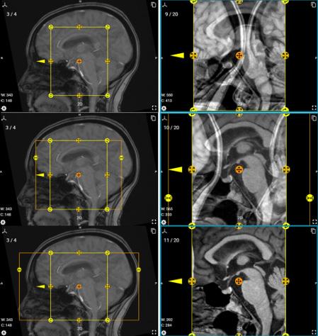
• Oversampling/Wrap Prevent/No Phase Wrap settings
• Phase encoding direction choices
This advancement allows learners to immediately visualize the consequences of their decisions, reinforcing practical understanding through experiential learning rather than abstract grading alone.
QT IMAGING RECEIVES
FDA CLEARANCE FOR BREAST ACOUSTIC CT SCANNER ENHANCED CONFIGURATION
QT Imaging Holdings Inc. has announced receipt of U.S. Food and Drug Administration (FDA) 510(k) clearance for an updated configuration of its Breast Acoustic CT scanner, the company’s 3D ultrasound tomographic breast imaging system. The newly cleared enhanced configuration improves visualization and expands coverage of posterior breast tissue.
“The updated configuration cleared by the FDA incorporates a tilted transmitter geometry that improves tomographic imaging coverage of breast tissue located near the chest wall, which is an area that can be extremely challenging to capture,” said Bilal Malik, Ph.D., chief science officer of QT Imaging. “This design delivers more complete coverage and improved overall
diagnostic utility by optimizing the scanner’s ability to include posterior breast tissue that is often difficult to image with standard approaches. We remain dedicated to developing and refining innovative technologies that provide greater precision and confidence to deliver the best outcomes for women regardless of breast density.”
The QTI Breast Acoustic CT scanner generates both reflection-mode and transmission-mode ultrasound data to reconstruct 3D tomographic images. Its proprietary software quantifies fibroglandular tissue volume (FGV) and the ratio of fibroglandular tissue to total breast volume (TBV), providing valuable insight for breast health assessment and monitoring.
HARRISON.AI RECEIVES NEW FDA CLEARANCE
Harrison.ai has announced FDA 510(k) clearance for acute infarct triage on non-contrast CT brain. Acute infarct is one of the most difficult findings to identify on non-contrast CT. Existing stroke AI focuses on large vessel occlusion in one or two vascular territories. The rest go untriaged. Harrison. ai’s triage covers six vascular territories and mechanisms of infarct, prioritizing ischemic strokes across the full geography of the brain.
Most stroke AI works on CT angiography (CTA), an advanced scan typically obtained later in the diagnostic workflow. Harrison.ai triages on the non-contrast head CT, the very first scan, helping ensure suspected ischemic stroke patients are prioritized for follow up including advanced imaging sooner. It triages actual brain tissue injury in ACA, MCA, PCA, cerebellar, basilar, and watershed infarcts, not just vessel occlusions.
No other AI has received FDA clearance for acute infarct triage on non-contrast CT brain. This is not a marginal improvement, but rather a fundamental shift.
“Acute infarct on non-contrast CT is one of the hardest findings in radiology. Most radiologists will welcome the help in bringing these cases to the front of the queue,” said Dr. Jarrel Seah, chief medical officer and practicing radiologist at Harrison.ai. “That’s why we felt it was important to build it.”
Harrison.ai’s acute infarct triage achieved up to 89.2% sensitivity on thin slices and 85.7% on thick slices, with greater than 80% sensitivity and specificity on multiple operating points for both thin and thick slice series. Ground truth was established using advanced imaging as the reference standard, meaning the AI was validated against confirmed infarcts that were not guaranteed to be visible on non-contrast CT.
The FDA-reviewed 510(k) submission for this clearance included direct performance comparisons against existing stroke AI approaches. The closest FDA-cleared comparator on non-contrast CT, a large vessel occlusion (LVO) triaging device, demonstrated 63.5% sensitivity and 95.1% specificity for identifying vessel occlusion only. Harrison.ai achieved greater than 80% sensitivity and greater than 80% specificity over multiple operating points for identifying actual tissue injury across all six vascular territories and mechanisms of infarct. This is a more comprehensive approach and triages stroke in more areas that might not be picked up with an LVO device.
The submission also included comparison to a widely deployed, FDA-cleared CT angiography-based large vessel occlusion triage system as a reference device. That system reported 91.3% sensitivity and 85.6% specificity in its most recent clearance. However, CTA requires contrast injection and is typically obtained after non-contrast CT in the acute stroke workflow. The system identifies vessel occlusion; it is not indicated for triage of acute infarction.
Harrison.ai triages actual ischemic tissue damage on the first scan a patient receives, across all major vascular territories of infarct, which can lead to more suspected strokes being prioritized sooner.
Combined with seven other FDA-cleared CT brain findings, Harrison.ai now delivers the most comprehensive critical exam coverage on non-contrast CT brain of any AI triage platform. Eight findings on a single first-line imaging modality. Both hemorrhagic and ischemic stroke covered in one integration. Harrison.ai is also currently one of only two radiology AI companies with a device eligible for Medicare New Technology Add-on Payment (NTAP).
BAYER EXPANDS MEDRAD
MRXPERION MR INJECTION SYSTEM SUPPORT
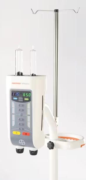
Bayer has announced that the U.S. Food and Drug Administration (FDA) granted 510(k) clearance for expanded capabilities of its MEDRAD MRXperion Magnetic Resonance (MR) Injection System, a power injector designed to support efficient workflow and consistent contrast delivery across a broad range of MR environments.
This updated clearance expands MRXperion’s labeled field strength compatibility to support magnetic resonance imaging (MRI) scanners with magnetic fields up to and including 7T (Tesla) range, offering radiology departments greater flexibility within diverse MR suite configurations. It also introduces Imaging Scanner Interface 2 (ISI2) capability, which enables communication between the injector and scanner system, helping streamline exam coordination and support operational efficiency.
“MRXperion already has a strong track record of delivering dependable imaging performance across clinical settings, and this clearance reinforces and expands on its established reputation as a flexible solution that supports coordinated, efficient workflows across MR systems,” said Bayer’s Jill Carbone. “The clearance also exemplifies our continued innovation in MR solutions to consistently meet the needs of today’s imaging providers.”
MR systems are deployed across healthcare settings with differing technical and workflow requirements. MRXperion is engineered to support this full spectrum of use, from routine clinical imaging to advanced research applications. The clearance further enhances Bayer’s integrated MR portfolio spanning contrast media, injection systems, software and workflow solutions.

FUJIFILM SHOWCASES AI-DRIVEN SOLUTIONS
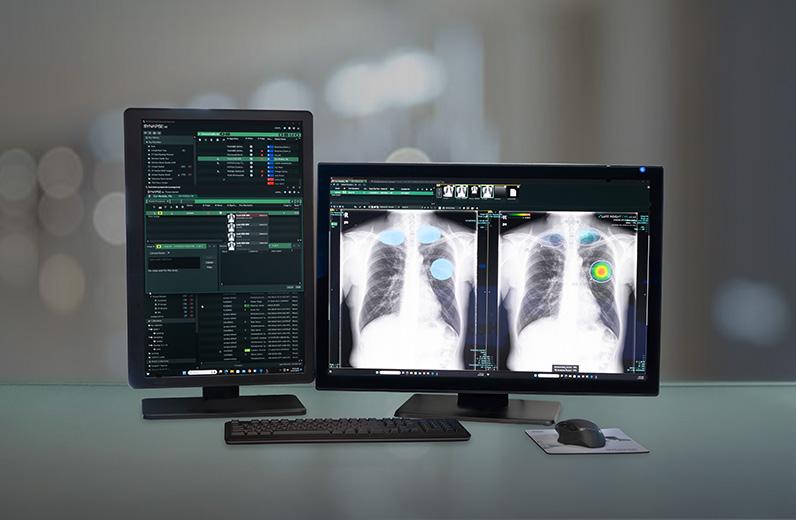
FUJIFILM Healthcare Americas Corporation showcased how its latest AI-powered enterprise imaging solutions are enabling healthcare organizations to enhance workflows for faster patient throughput, reduce administrative burden, and support more personalized care at the 2026 Healthcare Information and Management Systems Society (HIMSS) conference.
“AI delivers exceptional impact in medical informatics when it’s seamlessly embedded into the clinical workflows,” said Bill Lacy, senior vice president, medical informatics global business, FUJIFILM Healthcare Americas Corporation. “At HIMSS, we’re spotlighting our secure, AI-powered Synapse enterprise imaging portfolio, and sharing real-world examples on how these solutions are accelerating radiologist interpretation, improving operational efficiency and creating a more sustainable workflow for radiologists – all while keeping patient care at the center of innovation. We are also highlighting our ever-expanding AI activity in cardiology, pathology, and non-imaging AI models.”
Fujifilm’s AI-driven medical informatics solutions highlighted include:
• Synapse AI Orchestrator: An open imaging workflow orchestrator that uses an advanced rules
engine to seamlessly bring preferred imaging algorithm results directly to Fujifilm’s Synapse Enterprise PACS workflows. Designed for true interoperability, Synapse AI Orchestrator allows healthcare IT staff to deploy and maintain a multitude of AI applications for clinical workflows across the enterprise.
• Synapse Worklist Orchestrator: Fujifilm’s flexible, workload balancing and workflow/worklist orchestration solution transforms how studies are assigned, and how images are interpreted. The solution automatically assigns diverse studies from a variety of sites based on unique conditions set by the organization with the option to ensure assignment equity across subspecialists, if desired. Synapse Worklist Orchestrator uses AI powered learning modules and supports complex enterprise workflows with patented assignment algorithms that are tailored to Synapse Enterprise PACS (radiology, cardiology, pathology) readers. Additionally at HIMSS for the first time, Fujifilm highlighted Synapse One, the company’s comprehensive, tailor-made workflow solution designed for unique outpatient imaging needs, in North America.

NEXT-GENERATION DIAGNOSTIC VIEWER NOTCHES
U.S. FDA 510(K)
CLEARANCE
GE HealthCare has announced that View, the powerful viewer within the Genesis Radiology Workspace, has received 510(k) clearance from the U.S. Food and Drug Administration (FDA). Designed as a fast, diagnostic, zero-footprint viewer, View serves as the core anchor of the Genesis Radiology Workspace. This next-generation solution aims to transform radiology workflows, unify the user experience, and empower radiologists with greater efficiency and precision.
Studies show that radiologists spend up to 44% of their time on non-interpretive tasks, such as navigating complex interfaces or waiting for images to load – directly impacting productivity and turnaround times. Radiologists are looking for solutions that can help them reduce turnaround time and improve their efficiency.
“View is designed to meet radiologists wherever they are – providing secure, high-performance access to diagnostic
imaging through a truly cloud-native experience,” said Scott Miller, CEO of solutions for enterprise imaging, GE HealthCare. “By removing traditional infrastructure barriers, we’re enabling faster collaboration, greater flexibility, and more streamlined workflows across care teams. View within the Genesis Workspace reflects our commitment to modernizing radiology in ways that support clinicians and, ultimately, improve patient care.”
View is engineered to optimize the radiology workspace by empowering professionals to work from virtually anywhere with full diagnostic confidence. It delivers high-performance 2D and 3D visualization alongside AI-enabled tools to support fast, precise clinical decision-making. In addition, View provides direct access to the full suite of Advanced Visualization applications, enabling seamless, comprehensive analysis within a single workflow.
$2 MILLION UPGRADED MRI SUITE UNVEILED IN BRONX
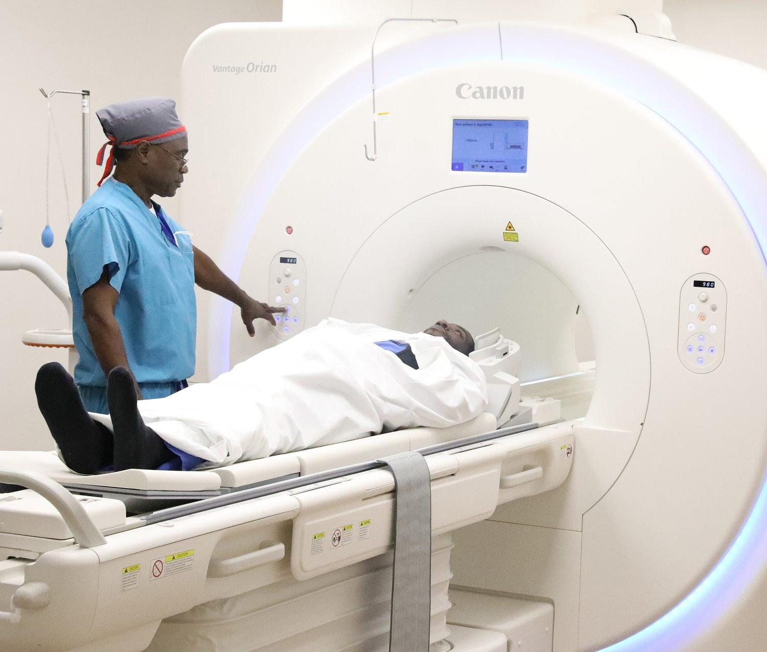
NYC Health + Hospitals/Jacobi | North Central Bronx recently unveiled a new $2 million MRI suite at the North Central Bronx campus with renovated rooms and upgraded MRI machines. The new Canon Vantage Orian Encore MRI machines offer faster scan times while delivering clearer, more distinct images, which will boost diagnostic functions and reduce wait time for patients. To improve the patient’s comfort, the system uses a video projection to help relax patients with a calming, virtual experience as well as technology that reduces scanner noise to improve the patient experience.
“We believe in providing high-quality care to our communities,” said Alfredo Jones, chief operating officer at NYC Health + Hospitals/North Central Bronx. “This new MRI system is part of our commitment to enhancing imaging services at North Central Bronx
Hospital. Upgrading our existing MRI unit with new technology allows us to enhance productivity and imaging quality while offering our patients greater comfort and safety to deliver outstanding clinical performance.”
“The opening of our new MRI suite marks a transformative moment for NCB,” said Dr. Chinyere Anyaogu, deputy chief medical officer at NYC Health + Hospitals/North Central Bronx. “This investment is more than just advanced technology; it’s a promise to our community that we will deliver the highest quality of care, right here at home. By expanding access to stateof-the-art diagnostics, we’re not only improving patient outcomes today, but also building a healthier future for generations to come.” •
DIRECTOR’S CIRCLE
Magnetic resonance imaging (MRI) continues to play an increasingly vital role in modern healthcare, with advancements in technology, expanding clinical applications and growing patient demand shaping how imaging departments operate. From improving patient comfort and safety to navigating staffing challenges and optimizing workflow, today’s MRI leaders are balancing innovation with efficiency in a rapidly evolving landscape. In this Director’s Circle roundtable, industry experts share insights, strategies and best practices for managing MRI services. Participants in this Director’s Circle article are:
• AdventHealth Central Florida Director of Operations and Imaging Safety Cory Whitehouse;
• Scripps Memorial Hospital La Jolla MRI Supervisor Megan Comia; and
• University Hospitals of Cleveland MRI Research Technologist Martin Allen Ayres.
Q: MRI UTILIZATION CONTINUES TO GROW ACROSS MANY SPECIALTIES. WHAT CLINICAL APPLICATIONS OR SERVICE LINES ARE DRIVING THE MOST GROWTH IN MRI AT YOUR ORGANIZATION RIGHT NOW?
WHITEHOUSE: In our community, orthopedics and sports medicine (knee, shoulder, hip), neuro-spine and brain, and oncology (prostate, breast, liver) and a high number of abdomen and pelvis scans are where we see the most demand and growth. In our ambulatory imaging centers, we are seeing growth in MRI year over year. In 2025, our 13 ambulatory imaging centers cared for 13.5% more community members requiring an MRI than in 2024.
COMIA: One of the biggest areas of growth for us right now is cardiac MRI. As technology and protocols continue to improve, we’re able to support more advanced cardiac imaging that provides detailed functional and structural information for cardiology teams. Another major driver of growth has been imaging patients with implanted devices. As more patients present with pacemakers, defibrillators, and other implants, we’ve spent a lot of time refining our workflows and standard processes to safely accommodate these exams. Through close collaboration with cardiology, radiologists, and device representatives, we’ve been able to expand access to MRI for many patients who previously may not have been considered candidates. That has significantly broadened the types of patients we’re able to serve.
AYERS: Growth is strongest in neuro, musculoskeletal (MSK) and oncology imaging. Neurology demand continues to rise for stroke evaluation, MS monitoring and cognitive assessment. Neurosurgical volume also remains steady, particularly for preoperative planning, postoperative follow-up and complex anatomical imaging. MSK imaging continues to be robust as orthopedic practices rely on MRI for soft tissue injuries and surgical planning. We’re also seeing growth in imaging for device patients, including those with pacemakers and other MR conditional implants. As more devices receive MR conditional labeling and protocols become standardized, providers are increasingly comfortable referring these patients for MRI.
Q: PATIENT EXPERIENCE HAS BECOME A MAJOR FOCUS IN IMAGING. WHAT STEPS HAS YOUR DEPARTMENT TAKEN TO IMPROVE COMFORT, REDUCE ANXIETY OR MAKE MRI EXAMS MORE PATIENT-FRIENDLY?
WHITEHOUSE: Patient comfort and anxiety are priorities in our MRI departments because we know that a relaxed patient leads to better image quality and better overall experience. We’ve taken a multi-step approach that starts before the patient even enters the scanner. First, we focus on communication and education. We explain the exam clearly and set expectations about noise and scan times and encourage patients to ask questions. For anxious PT or first timers, we walk them through the process step by step so there are no surprises. Inside the MRI suite, we use comfort focused tools such as earplugs, headphones with music, positioning cushions, covers and careful coil placement to minimize discomfort. We also give every patient a call button and reinforce that they can stop the exam at any time if they feel uncomfortable. To help reduce claustrophobia, we use shorter scan protocols, when possible, offer mirrors as visual aids when available, and maintain regular communication during the scan to reassure the patient and keep them informed of progress. We also adjust our approach based on individual needs such as allowing extra time or adjusting times for pediatric or elderly patients. We sometimes recommend a mild sedation before proceeding as well. Overall, our goal is to treat each patient as an individual, combine technical excellence with empathy, and create an MRI experience that feels safe, respectful and patient centered.
COMIA: MRI can be an intimidating exam for many patients, so patient experience really starts with the technologist. Our team has many experienced technologists who are very skilled at coaching patients through the exam, explaining what to expect and recognizing when someone may need extra reassurance. That level of experience and communication makes a big difference in helping patients feel comfortable and confident during the scan. We’re also fortunate to have care team assistants and a dedicated MRI nurse who help support patient preparation and coordination. This allows our technologists to stay focused on the exam while ensuring patients feel supported throughout the entire process. From an operational standpoint, we also focus on keeping workflows efficient, so patients aren’t waiting unnecessarily or spending longer in the scanner than needed. For patients who struggle with anxiety or claustrophobia, we’ve also explored simple supportive tools such as aromatherapy tabs that can help create a calmer environment during the exam. Often the biggest difference comes down to preparation, communication and having a team that is confident guiding patients through the process.
AYERS: We’ve made patient comfort a central part of the MRI process. Clear communication, thorough instructions,
and opportunities to ask questions help set expectations before the exam begins. During the scan, we provide music, blankets and additional padding for comfort. Patients may use mirrors to reduce the feeling of being enclosed or choose to keep their eyes covered if they prefer. For those with claustrophobia or anxiety, technologists take a slower, more supportive approach and offer reassurance throughout the exam. A screened support person may accompany the patient when appropriate. We offer designated slots for conscious sedation and can coordinate anesthesia for patients who require it due to anxiety, claustrophobia or pain. In pediatric cases, our team works closely with sedation services and child life specialists to provide age-appropriate support and make the imaging process less intimidating.
Q: STAFFING REMAINS A CHALLENGE FOR MANY IMAGING DEPARTMENTS. WHAT STRATEGIES HAVE YOU FOUND EFFECTIVE FOR RECRUITING, TRAINING OR RETAINING SKILLED MRI TECHNOLOGISTS?
WHITEHOUSE: As the demand for our MRI techs grow because of extraordinary growth and demand in the Central Florida community, we sought ways to allow our techs to focus more time on scanning while limiting the additional duties such as IV insertion, room turnover and walking patients back to the rooms. We began hiring paramedics for these tasks in late 2025. We continue to hire paramedics for our 13 locations. We have implemented an extensive cross training program where we offer tech training in our centers while they continue to earn a paycheck. Once they complete competencies through ARRT, we pay their fees to sit for their registry. We also offer non-clinical team members the ability to get their ARMIT through Tesla Institute which we fully pay for, and they use our sites in a clinical location. We are also encouraging our paramedics to also grow to become MRI techs utilizing the Tesla Institute as well.
COMIA: One of the most important things for retention is creating an environment where technologists feel respected, supported and genuinely enjoy coming to work. MRI can be a demanding modality, so building a strong team culture makes a big difference. I make it a priority to stay connected with the team, whether that’s checking in during the day, celebrating wins, or creating opportunities for staff to share ideas and contribute to improving workflows. When technologists feel like their experience and input matter, they tend to take pride and ownership in the department. We also focus heavily on mentorship and cross-training so technologists can continue expanding their skills, particularly with more complex exams and devices. Investing in technologists and supporting their growth helps build confidence and keeps the team engaged long-term. At the end of the day, people want to work in a place where they feel valued and where they can continue to grow, and that’s something we actively try to cultivate in our department.
CORY WHITEHOUSE
ADVENTHEALTH

MEGAN COMIA
SCRIPPS MEMORIAL HOSPITAL LA JOLLA
AYERS: Promoting a positive and supportive workplace has been essential for attracting and retaining skilled MRI technologists. Flexible scheduling options help reduce burnout and support a healthier work-life balance. We encourage professional development through continuing education, involvement in quality initiatives and participation in protocol-related projects. A culture centered on respect, open communication and shared goals continues to play a major role in long-term retention. To further strengthen day-to-day workflow, we’ve added MRI tech extenders on all shifts. They assist with routine tasks and help maintain steady operations. Each extender is currently enrolled in an imaging program with plans to become an MRI technologist, creating a clear internal pathway for developing future staff while supporting current operational needs.
Q: MRI SAFETY IS ALWAYS EVOLVING AS MORE IMPLANTS AND DEVICES BECOME MR-CONDITIONAL. HOW IS YOUR TEAM MANAGING SCREENING, SAFETY PROTOCOLS AND TECHNOLOGIST EDUCATION TO KEEP PATIENTS SAFE?
WHITEHOUSE: MRI safety continues to advance quickly – especially with the rise of MRI “conditional implants” such as pacemakers, neurostimulators, ortho hardware, ear implants and more. Keeping patients safe requires a combination of rigorous screening, updated safety SOPs and ongoing tech training supported by governing bodies like the ACR and recent safety literature. Tech education also plays a central role, with ongoing training required to keep pace with new implant designs and updated standards. The latest guidance highlights the importance of understanding MR-safe, MR-conditional and MR-unsafe classifications, as well as the potential hazards posed by thermal injuries, projectile risks and device interactions. Continuous learning – supported by ACR resources and real-world safety

MARTIN ALLEN AYRES
UNIVERSITY HOSPITALS OF CLEVELAND
articles – ensures techs are prepared to make informed decisions and respond appropriately to emerging challenges in the MRI environment. As implants become increasingly MRI-compatible, a combination of thorough screening, robust protocols and ongoing education remains essential to protecting patients and maintaining the highest standard of MRI safety. We also encourage our MRI techs to get their MRSO certification which we fully fund.
COMIA: MRI safety is something we take very seriously, especially as more patients present with implanted devices and complex medical histories. Our approach is built around strong screening processes and collaboration across multiple teams. We work closely with cardiology, radiologists, device representatives and referring physicians to ensure each case is reviewed carefully before scanning. Education is also a big part of our safety culture. We have MR safety officers on our team and encourage technologists to attend safety seminars and stay engaged with current literature. Sharing new information and articles with the team helps ensure everyone stays informed as guidelines and device recommendations continue to evolve. MRI safety is always changing, so maintaining open communication and ongoing education helps us keep patients and staff safe.
AYERS: Our team follows a comprehensive screening process for every implant or device before an exam is scheduled. All required conditions – such as magnet strength, coil restrictions, SAR or B1+rms limits, spatial gradient considerations, and any time or positioning requirements – are outlined on a dedicated MRI implant clearance document for technologists. Schedulers receive any special instructions needed for a safe visit, such as bringing a device controller or using a specific magnet. For more complex cases, tech-
nologists collaborate with radiologists, our MRSE and other clinical partners to ensure all safety criteria are met before imaging begins. Ongoing safety education is prioritized through refreshers, in-service training and vendor-supported learning. A centralized Microsoft Teams channel gives staff quick access to device manuals, manufacturer guidance, gradient maps, SOPs and other safety tools, ensuring up-to-date and consistent practice.
Q: HOW ARE WORKFLOW TOOLS SUCH AS REMOTE SCANNING, PROTOCOL STANDARDIZATION OR ADVANCED SCHEDULING SYSTEMS HELPING YOUR TEAM HANDLE HIGHER MRI VOLUMES?
WHITEHOUSE: Our most utilized protocols were obtained and reviewed by subject matter expert MRI techs. Recommendations were made for revisions by decreasing the number of sequences and obtaining the section chief input and approval with the goal of shaving off unnecessary sequences. On top of that, we are preparing to install software that will allow us greater efficiency and clearer images. We are also preparing for a pilot with two AI software organizations and finally, the implementation of remote scanning to solve for sick calls and other staffing strains. We believe that when these initiatives are combined, we will see greater efficiencies, clearer images and less time on the table for the patients, allowing us to increase our community access. Our plan is to measure each initiative’s impact on the scan time as they are implemented.
COMIA: Protocol standardization has been one of the biggest tools for improving workflow. When protocols are consistent across scanners, it helps technologists move efficiently between exams while maintaining diagnostic quality and consistency. With seven MRI scanners across our campus, we’re also constantly refining scheduling strategies at each location to balance inpatient, outpatient and specialty exams. The goal is to maintain strong volume while still creating a workflow that allows technologists to move through exams efficiently and seamlessly. MRI operations can be complex, so thoughtful scheduling, clear communication and standardized protocols help ensure we maintain both quality and efficiency across the department. We are currently exploring remote scanning however we have not implemented it yet.
AYERS: Standardizing protocols across scanners has
helped streamline exams, minimize variation and maintain consistent image quality. Improved scheduling tools ensure exam length, location and resources are appropriately matched, reducing delays and creating a smoother patient flow. Remote support capabilities – recently implemented at one of our sites – allow experienced technologists to provide real-time assistance or adjust protocols as needed. This improves responsiveness, consistency and overall efficiency as imaging volumes continue to grow.
Q: WHAT ELSE WOULD YOU LIKE TO SHARE WITH READERS REGARDING MRI?
WHITEHOUSE: Some vendors are offering AI to improve efficiency and accuracy. Some talk about up to 75% decrease in scan time with AI reconstruction. There is more than one player in the radiology tech remote scanning arena and should be compared for their lead time to staff, cost and minimum number of hours per week. If considering remote scanning, one may want to consider training one’s own team internally first and allow them to reap the benefits of additional hours versus a more costly approach of outsourcing. Look for smaller footprint MRIs. With the high cost of construction and helium-free or reduced helium magnet design to save costs if you must suddenly squelch the system.
COMIA: MRI is a very special modality in that it’s constantly evolving, both in technology and in the clinical questions it can answer. As capabilities continue to expand, so does the importance of strong collaboration between technologists, radiologists, nurses, physicians and operational leaders. When teams communicate well and work together, MRI can be incredibly powerful, not only in the quality of diagnostic information it provides, but in the experience we create for patients along the way. It’s a modality that requires both technical expertise and teamwork, and when those come together, the impact on patient care is significant.
AYERS: MRI technology continues to advance quickly, offering increasingly precise diagnostic information. As capabilities expand, maintaining high safety standards, strong imaging quality, and a positive patient experience becomes even more important. The expertise of MRI technologists – supported by teamwork, collaboration and ongoing education – remains central to providing safe, high-quality care. •
PRODUCTS

Market Report
STAFF REPORT
Th e magnetic resonance imaging (MRI) market refers to the production, distribution and use of MRI, which is a non-invasive imaging technology that produces three-dimensional, detailed anatomical images. Magnetic resonance imaging is a medical imaging technique used in radiology to generate pictures of the anatomy and the physiological processes inside the body. MRI scanners use strong magnetic fields, magnetic field gradients, and radio waves to form images of the organs in the body. It provides better soft tissue contrast than CT and can differentiate better between muscle, water, fat and other soft tissue.
According to Statifacts, the global magnetic resonance imaging (MRI) market size was valued at $1.086 billion in 2024 and is predicted to be worth around $2.619 billion by 2034, growing at a CAGR of 9.2% between 2024 and 2034. Expanding applications of MRI in clinical and research settings, ongoing technological innovation in MRI hardware and software, and demand for non-invasive and high-resolution diagnostic imaging are driving the growth of the market.
Statifacts adds that North America led the global market in 2024 and is expected to maintain its dominance because of strong healthcare infrastructure and tech investments. The Asia-Pacific region is projected to see the fastest growth from 2025 to 2034, driven by rising healthcare demands and medical advancements.
The closed system segment dominated in 2024 and is expected to retain its lead because of its safety and efficiency. The open system segment is set to gain significant market share from 2025 to 2034, thanks to its flexibility and cost-effectiveness.
The high-field-strength segment led in 2024 and is expected to remain dominant because of superior imaging precision. The midfield-strength segment is expected to grow significantly, offering a balance of performance and cost.
The neurology segment led in 2024 and is set to continue growing with rising neurological conditions and imaging advancements. The musculoskeletal segment is expected to
grow at the highest CAGR, driven by demand for orthopedic and sports medicine imaging.
The hospital segment dominated the market in 2024 and is expected to maintain its position as the key end-user. The ambulatory surgical center segment is anticipated to grow the fastest due to increasing outpatient procedures.
The news release also reports the following MRI trends:
• Integration of Artificial Intelligence (AI): AI is increasingly being used in MRI systems to accelerate scan times, enhance image quality, and assist in automated diagnosis and reporting.
• Rising Demand for High-Field and Ultra-High-Field MRI: There is growing adoption of 3T and 7T MRI systems, especially in neurology and research, due to their superior resolution and diagnostic capabilities.
• Development of Portable and Point-of-Care MRI Devices: Advances in low-field MRI technology are enabling the creation of compact, mobile systems suitable for emergency, rural, and bedside applications.
• Patient-Centric System Design: Manufacturers are focusing on improving patient experience through open-bore systems, noise reduction technologies, and faster scan protocols to reduce discomfort.
• Market Growth in Emerging Economies: Increasing healthcare investment, growing awareness, and expanding diagnostic infrastructure are fueling MRI adoption in regions like Asia-Pacific, Latin America and parts of Africa. According to Precedence Research, the global AI in medical imaging market size is calculated at $2.57 trillion in 2026 and is expected to hit approximately $22.97 trillion by 2035, expanding at a healthy CAGR of 27.57% from 2026 to 2035. The growing imaging patient volumes and strong focus on earlier detection of diseases will drive market growth.
“Increasing imaging volumes, growing chronic disease prevalence, and radiologist shortages are accelerating the adoption of AI across CT, MRI, X-ray, and ultrasound imaging. AI enables faster, more accurate diagnoses, improves workflow efficiency, and reduces clinical workload, positioning it as a critical technology shaping the future of diagnostic healthcare worldwide,” the firm states.
At the same time, healthcare providers are leveraging AI to manage rising scan volumes and mitigate a 30 to 40% global radiologist workforce gap, it adds. •
Product Focus
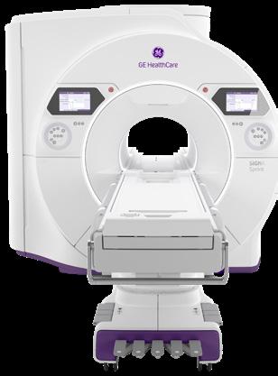
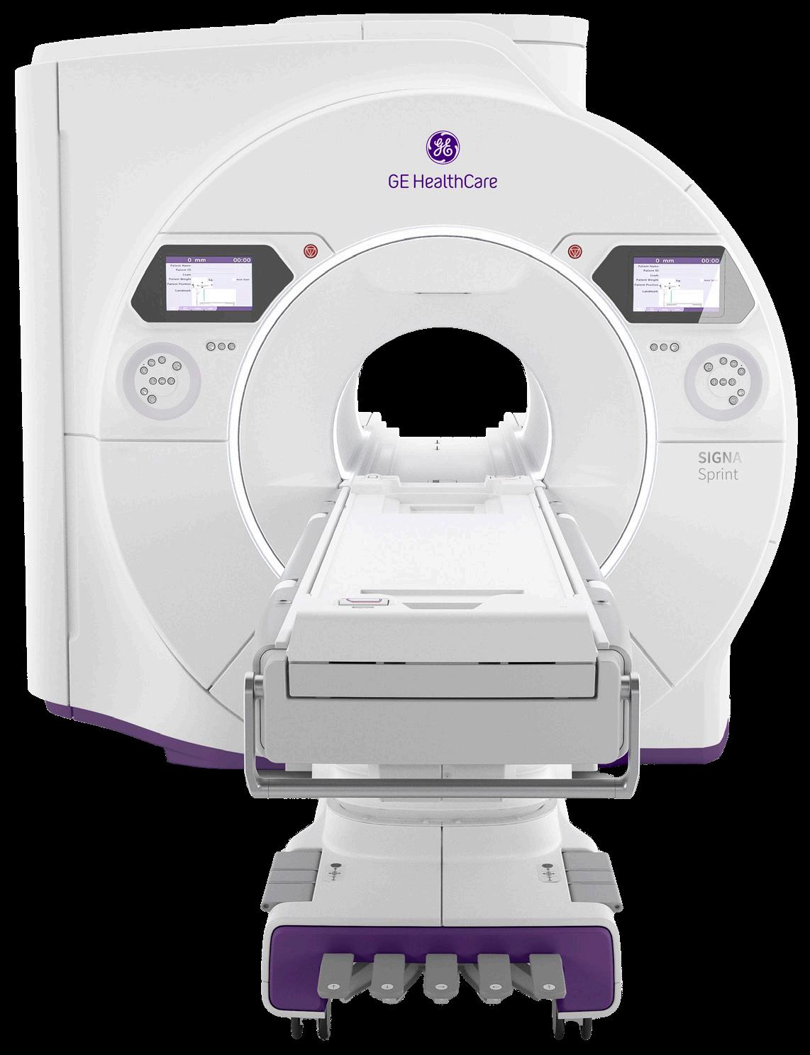
GE HEALTHCARE SIGNA Sprint Elite
SIGNA Sprint Elite is an ultra-premium wide bore 1.5T high-performance gradient MRI system designed to unlock advanced imaging possibilities in cardiology, oncology and other clinical and research areas. With the goal of delivering higher gradient benefits previously attainable on only 3.0T systems, SIGNA Sprint Elite is designed to offer high-performance scanning made simple at 1.5T, with exceptional diffusion imaging, a critical tool in oncology diagnosis and treatment planning. SIGNA Sprint Elite is also designed to equip clinicians and researchers with the ability to expand the current 1.5T boundaries, aiming to enable fast clinical translation and pioneering research.
With a high-gradient performance of 65/200 per axis for fast imaging and exceptional image quality, SIGNA Sprint Elite is designed to deliver crystal-clear visualization of sub-millimetric structures and deep learning solutions to support diagnostics and treatment response monitoring in oncology patients. This system supports clinicians’ shift to quantitative MRI and a deeper understanding of tissue characteristics, going beyond basic assessment of anatomy. The adoption of deep-learning reconstruction techniques for accelerated cardiac MRI aims to reduce the time and expertise needed to interpret scans and drive consistency and reliability.
*Disclaimer: Products are listed in no particular order.
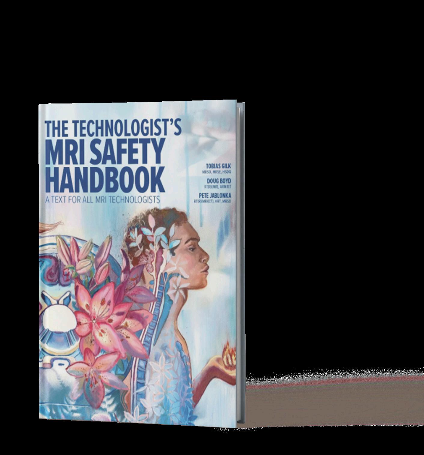
MRI SAFETY HANDBOOK
A resource for MRI technologists is “The Technologist’s MRI Safety Handbook,” authored by industry leaders Tobias Gilk, Doug Boyd and Pete Jablonka. This indispensable guide offers a much-needed resource for those just beginning in MRI safety – and for seasoned technologists seeking to deepen their understanding. This foundational text is designed to equip MRI technologist trainees and early-career professionals with a comprehensive understanding of MRI safety principles and practices. The handbook addresses a critical gap in MRI safety education by offering practical, accessible training that begins with the basics and builds toward advanced knowledge. It emphasizes the “whys” behind protocols, empowering technologists to practice safely and respond confidently to evolving safety challenges. The hardcopy edition is available now. An eBook version is coming soon.
To learn more, visit thependrakegroup.com.
EYAS MEDICAL IMAGING INC.
Ascent3T
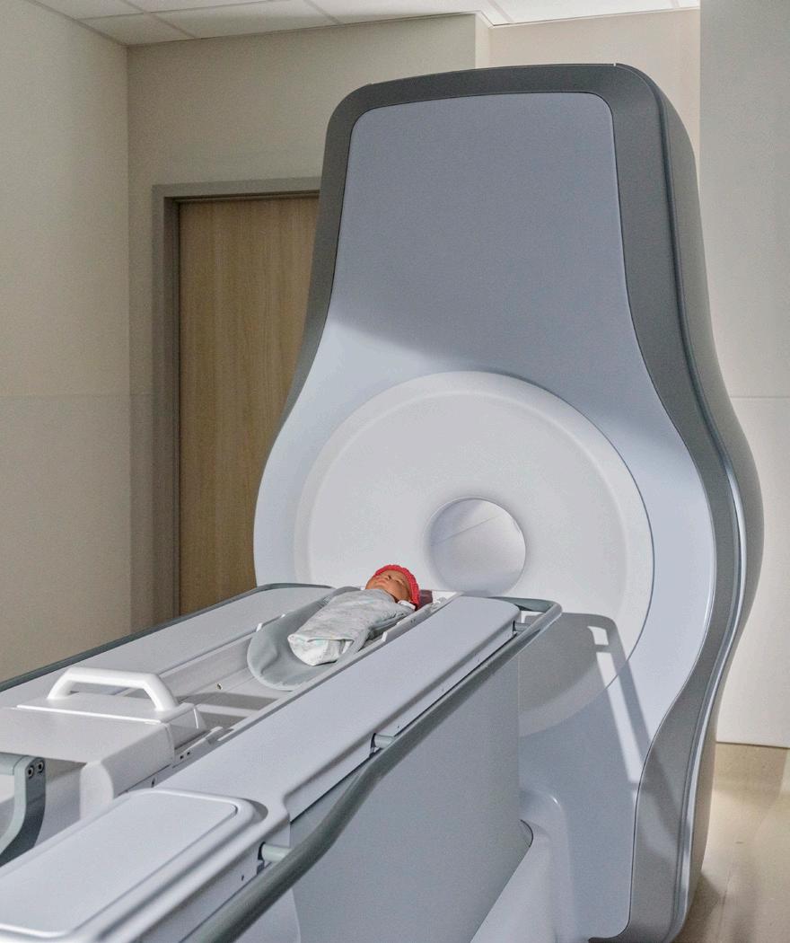
Eyas Medical Imaging Inc. has received U.S. Food and Drug Administration (FDA) 510(k) clearance for its Ascent3T Neonatal Magnetic Resonance Imaging (MRI) System, a whole-body magnetic resonance scanner designed and optimized specifically for neonate and infant anatomy, including head, body and extremities. The Ascent3T is the first high-field, 3 Tesla (3T) dedicated neonatal MRI system in the world, according to a press release. It utilizes a 3T magnet, enabling a more comprehensive and precise diagnostic tool that provides healthcare professionals with detailed imaging of vital anatomy, including the brain, lungs, heart and abdomen. The Ascent3T addresses the technical limitations of using an adult-size MRI system to image babies and provides clinicians with improved ability to visualize and diagnose disease in the neonatal patient population.

INKSPACE IMAGING Snuggle Pediatric Body Array Coil 4
The InkSpace Imaging Snuggle pediatric body array coil is designed specifically for pediatric patients, according to a press release. Young patients often find MRI exams challenging, and the Snuggle coil was created to make the experience more comfortable and less intimidating. Its light, blanket-like design and soft, flexible structure gently wrap around the patient. The Snuggle coil’s high-density array and flexible design allow sharp, high-resolution images and more efficient exams across a range of pediatric anatomies. These advantages can reduce the need for additional imaging or follow-up procedures while helping technologists work more efficiently through easier handling, faster setup and shorter scan times, helping to improve throughput and reduce repeat exams. Recently cleared through the U.S. Food and Drug Administration (FDA) 510(k) process, InkSpace Imaging developed the Snuggle coil to provide a more child-friendly alternative to traditional rigid devices.
SIEMENS HEALTHINEERS
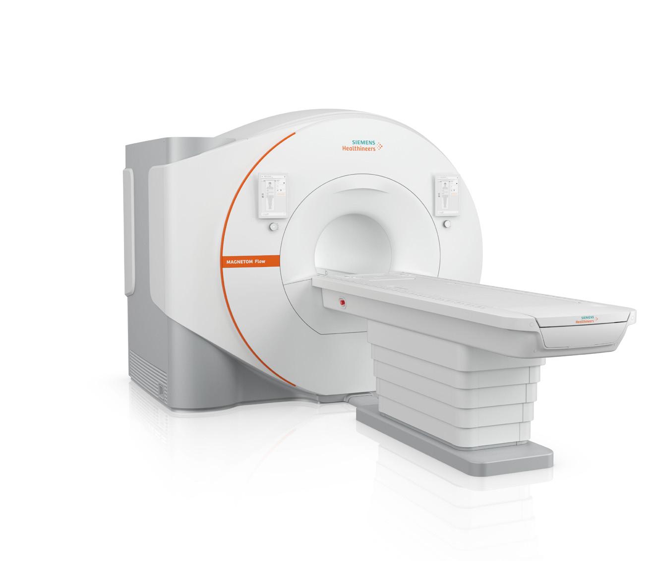
Magnetom Flow. 70 cm Platform
The new 70 cm bore Magnetom Flow. 1.5 Tesla (T) magnetic resonance (MR) imaging platform from Siemens Healthineers has a closed helium circuit and no quench pipe to reduce helium dependency. The upgradable platform covers the entire range of MR applications and offers image reconstruction based on artificial intelligence (AI) for shorter scan times with improved image quality. It requires just 0.7 liters of liquid helium for cooling, compared with conventional MRI scanners that typically require over 1,000 liters. Its lack of a quench pipe and the scanner’s compact size can reduce installation requirements and costs compared to other 1.5T MR scanners. The platform reduces annual energy consumption up to 56% compared with the previous generation of Siemens Healthineers MR scanners.¹ AI supports automated workflows, including new cardiac workflows that simplify and standardize the setup of scan parameters. High-density contour coils are available in four sizes.
¹ Data on file.
PRODUCTS
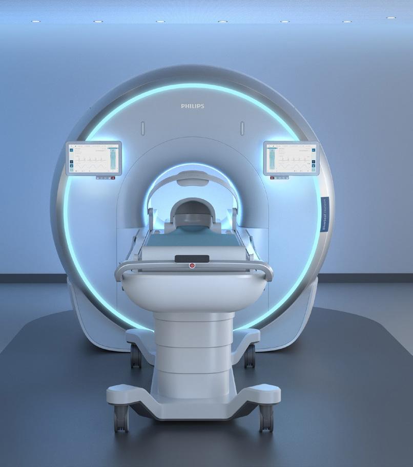
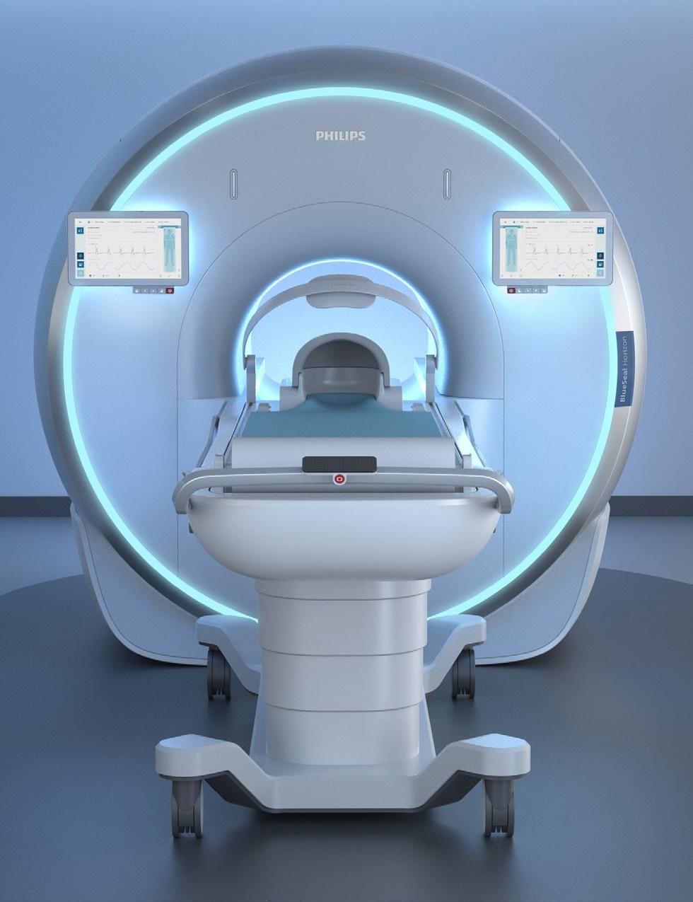
PHILIPS BlueSeal Horizon
Royal Philips unveiled BlueSeal Horizon, an entirely new 3.0T MRI innovation platform that includes the world’s first helium-free 3.0T magnet, at RSNA 2025. A pivotal advance in MRI innovation, helium-free 3.0T is a major scientific achievement set to have significant impact for health providers and patients, according to a press release announcing the MRI.
Key AI-powered innovations include:
• SmartPlanning: Expanding to include cardiac imaging, this AI-driven feature will automate time-consuming planning steps. What once required multiple manual actions can now be completed in a single click, achieving automated planning in as little as 30 seconds.
• Real-time Scan Preview: Powered by NVIDIA’s accelerated computing platform and Open Models (Segment and Generate), this innovation aims to enable faster 3D image reconstruction, denoising, and artifact reduction, so radiologists can preview scans, adjust image quality and speed parameters in real time, and optimize workflow efficiency for more timely diagnosis.
• SmartSpeed Precise: Dual AI technology will enable scans up to three times faster and images up to 80% sharper, helping clinicians capture more detail in less time.
• SmartReading: This tool will integrate cloud-based AI reading and reporting tools directly on the MR system, specifically for neurology and oncology applications. •
Simplifying The Imaging Equipment Ownership Experience
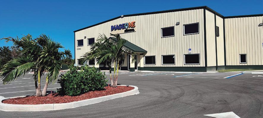
Image One Medical is the only Engineer-Owned medical equipment service group that is fully dedicated to Florida’s amazing base of Imaging Centers, Hospitals, and Cancer Treatment Centers. We have a mission: Self perform on every aspect of our business. Specialize on specific modalities: Pet CT, CT & MRI, and Focus in a key geographic region. Fort Myers I Fort Lauderdale I Tampa I Orlando
FLORIDA BASED
Dealer and servicer of PET CT, CT and nuclear medicine
• Equipment service: full coverage plans
• Equipment sales: installation, relocation and project management
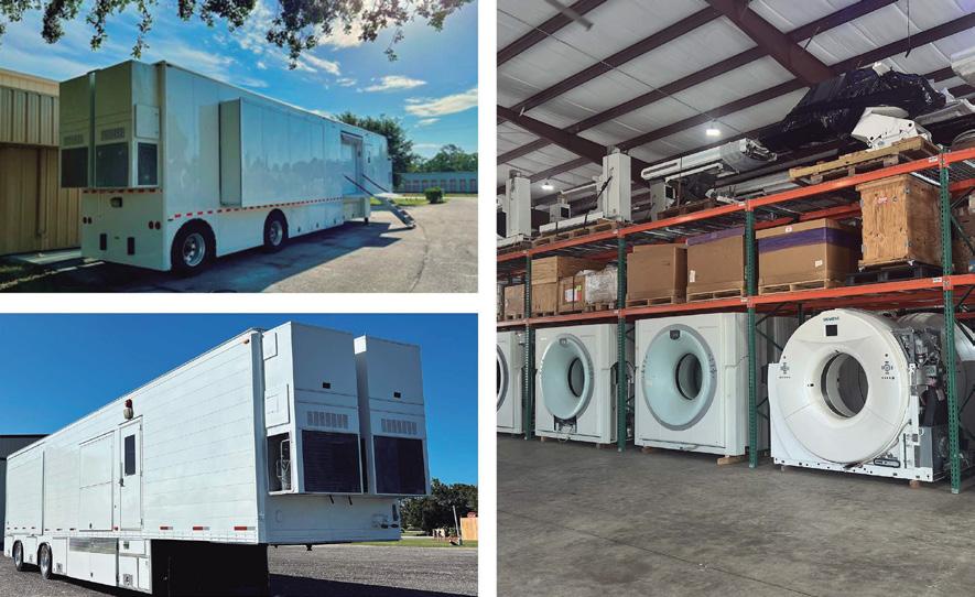
• Mobile coach construction, refurbishment, maintenance and management
OUR EXPERTISE
• Self performing team of in-house engineers
• High level problem solving for Siemens and GE PET CT, CT and nuclear medicine
• In-house inventory of 40+ systems and parts for repair
• 24 hour service with quick uptime guarantee
BLUEPRINTS FOR
Imaging Construction &
By Matt Skoufalos
For as much as medical imaging technology remains a cornerstone of future healthcare lines of service and treatment planning, decision-makers don’t always contemplate the longevity of the facilities constructed to house the devices that will provide those services.
When it comes to planning any capital project, especially one involving medical imaging equipment, it’s a truism that mistakes made early in the process can have unintended, negative consequences that stifle future capacity and growth opportunities, said Tobias Gilk, senior vice president of RAD-Planning and founder of Gilk Radiology Consultants. RAD-Planning is a consulting firm made up of architects dedicated to radiology, nuclear medicine, and radiation therapy projects.
As imaging systems are focused on faster acquisition of higher-quality images, facilities must be designed to maximize patient throughput. For MRI services, that means allowing for the inclusion of preparatory infrastructure – patient interview and screening spaces, sub-acute waiting spaces, changing areas – to support patient turnover at rates that achieve manufacturer-advertised thresholds. It also means contemplating the needs of current and future patient populations, including those with implanted medical devices, and
those who require higher acuity care delivered by additional medical staff and supportive technologies.
“Most facilities are still focused on, ‘How small can I make the scanner room?’” Gilk said. “‘How can I shoehorn this new scanner into a poorly suited existing space, minimize my cost, [and] accelerate my design and construction project?’”
“Those short-term gains often come at the expense of the longer-term, more profitable gains to come,” he said. “Lowest first cost compromises the gains that exist and will continue to grow in image acquisition.”
Many facilities will try to save on design costs by working with architectural partners recommended by vendors providing the equipment they’ve selected, or by re-using old templates from prior installations. However, vendor-provided architectural designs often are built around minimum service clearances required for device maintenance, and aren’t necessarily inclusive of zoning and construction codes, staffing thresholds, or the evolving needs of patients and the medical equipment that will deliver their care in the future, Gilk said.
“If we’re going to copy and paste the next MRI like we did the previous one, the default mode completely ignores all the changes that are undergoing in radiology and MRI in 2026,” he said. Vendors delivering an “off-the-shelf” plan have “every motivation to make these
spaces as small as possible so they can appear to have a competitive advantage over the other guys,” Gilk said.
Yet when space savings is presented as delivering cost savings, or when spaces are designed and built to bare minimums, that approach can limit future growth while also impeding clinical and design flexibility. Instead, he said, decision-makers should include in their calculations people who understand the workflow that imaging space will facilitate and its constituent parts. That conversation should touch on the patient population it will serve and how those people will circulate within the building, the technological needs of support staff that will attend to them and manage the medical technology involved in their care, and how to plan for emergent circumstances in that process.
“Instead of taking the copy-andpaste imaging suite and looking at it as, ‘Here’s a design; what do we want to change?’ work for the solution from the point of view of ‘How do we want to deliver patient care?’” Gilk said.
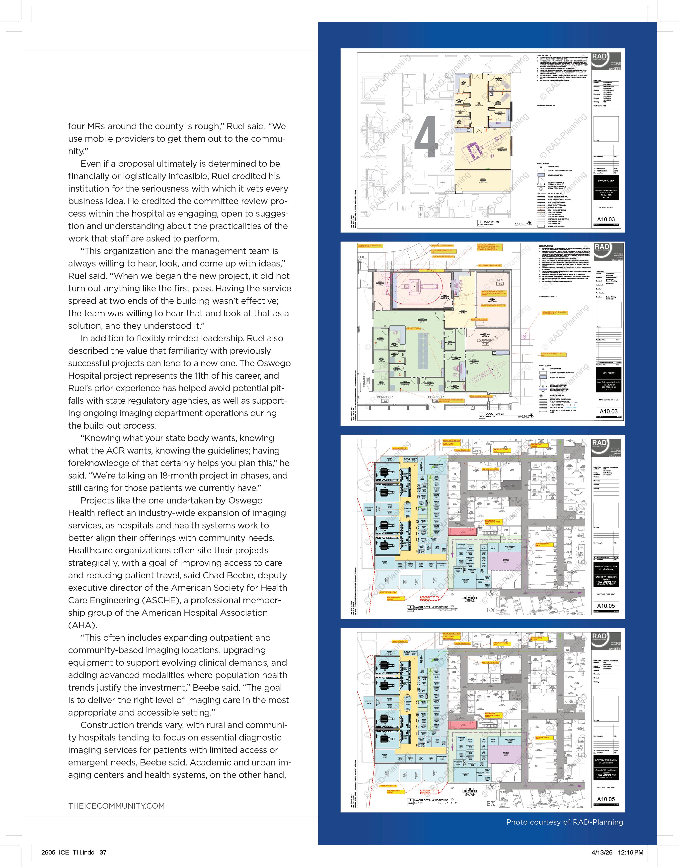
Through RAD-Planning, Gilk was a consultant on the Imaging Services Design Guide produced for the U.S. Department of Veterans Affairs Office of Construction and Facilities Management. It’s a freely available toolkit for healthcare facilities operators to consult when contemplating the design of spaces that will serve medical imaging patients. The design guide also includes
Planning for the Future FOR TOMORROW
three-dimensional planning files that they can overlay on their own spaces with the equipment they plan to include there, offering a starting point for facilities conversations.
Gilk also identified as another key facet of the design process interpreting the language of architectural professionals into concepts that imaging professionals can grasp. Speaking about architectural drawings as cross-sectional views of a building, for example, can help cross-sectional radiologists regard a floor plan as “a coronal view of a building with a 1-meter slice thickness,” he said.
“Sometimes just translating what they’ve seen a billion times before can help them immediately build their competence and confidence,” Gilk said.
Another aspect of facilities and project planning that often can’t be standardized is regulatory approval, which varies across states (and even among municipalities within the same state). In some parts of the country, clearing regulatory hurdles “can take many months or a year,” Gilk said. Even excluding those demands, however, the work of needs identification and outlining the scope of a project can be condensed to a much more compact timeline when all stakeholders are involved from the beginning and have a shared vision of the project outcome. In architectural terms, this is described as “programming,” but
it boils down to a clear understanding of the types of work that will be done in the space, the people who will perform it, and the equipment they’ll need to do it.
“We always look at every project with the intent to future-proof, but square-footage is the biggest thing for us. We have a landlocked building. We used a shoehorn and got every inch of space out of this.”
- David Ruel
“Once you have a needs assessment [that includes] equipment, spaces, [and] operational models to support, you can come up with different ways to arrange the boxes and architectural expressions of what that might look like,” Gilk said. “The needs assessment – what architects call programming – [is expressed as]
how we translate that activity of what the building needs in order to support the performance needed.”
Comprehensive needs assessments and programming work must contemplate future throughput volume increases, technological applications and potential re-uses of the space that will outlive the people tasked with its design by many years. Defining the underlying mission statement of a project involves modeling its immediate, idealized workflow as well as “crystal-ball-gazing” anticipating subsequent changes that can affect technologies with lifespans of a decade-and-a-half or longer, Gilk said.
“Odds are the replacement piece of equipment is going to require different power, or air conditioning, ceiling height, width and length of the room,” he said. “Squeeze this down to the bare minimums, and you’re also committing yourself to unnecessary costs of demolishing and rebuilding that room to serve the same purpose for the next piece of equipment.”
In anticipating operational changes and organizational flexibilities in the clinical demands for a piece of medical imaging equipment, Gilk notes the “massive disintegration” that has de-centralized imaging services into specialty and subspecialty deployments: freestanding imaging centers dedicated to women’s health, cardiac imaging in support of cardiology units,
COVER STORY
and sports medicine imaging associated with orthopedic groups, to name just a few. Those applications have needs as varied as the patient populations they serve, and planning their successful implementation requires more than a cookie-cutter effort, he said.
“Today, radiology and imaging services are being distributed throughout the enterprise because we’re finding that these can be so useful in patient- or condition-specific areas,” Gilk said. “The same CT scanner is going to have wildly different needs in a cardiology or electrophysiology practice versus being adjacent to your emergency department. They may require wildly different spaces and equipment to be able to support the care, and must be carefully tailored to a specific clinical mission.”
When considering the lifespan of an imaging facility build-out, Gilk begins by asking clients whether they own or lease their properties. An outpatient imaging center with an eight-year strip mall lease may be forced to confront more limitations on its future flexibility than a community hospital making an investment that might have to last for decades.
Thinking about how much and in what ways medical imaging practices have changed in the past decade-plus in terms of patient populations, referrers, and studies performed can lend some insight into the future demands of the space, he said. An additional foot or two of width can facilitate the ease of equipment replacement or the inclusion of supportive devices, like anesthesia machines, respiratory equipment, and infusion pumps, buying future flexibility for clinical practices and support personnel as well as technology.
Finally, as important as future-proofing designs can be, so, too must consideration be given to receive input from the professionals who will be working in the space.
“Failing to have those kinds of conversations with clinical stakeholders and front-line staff like technologists often
leads to projects that may look beautiful on ribbon-cutting day, but people start working in it and wonder, ‘Why did nobody ask me how we deliver patient care?’” Gilk said.
“Make sure the voices of the folks who are involved in the clinical care process are involved with this,” he said. “There’s no shortage of gorgeous-looking facilities that do not meet the needs of contemporary patient care. It’s really important to get somebody who’s had a lot of experience with radiology facilities, and not just in a retrospective sense. It’s most expensive and most mission-disruptive to go back in and fix problems that you should have thought of when you built the facility in the first place.”
“Today, radiology and imaging services are being distributed throughout the enterprise because we’re finding that these can be so useful in patient- or condition-specific areas.”
- Tobias Gilk
“It’s important to get it right the first time,” Gilk said, “and it’s important to recognize where the limits of your own knowledge are. Oftentimes, folks who run design and construction projects don’t have that immediate point-of-care understanding. They’ll do things with the best of intention, but it requires that folks really understand the workflow, the process, the patient needs.”
For smaller community hospitals, some of the biggest challenges related to rolling out an imaging facilities project involve financial and physical constraints.
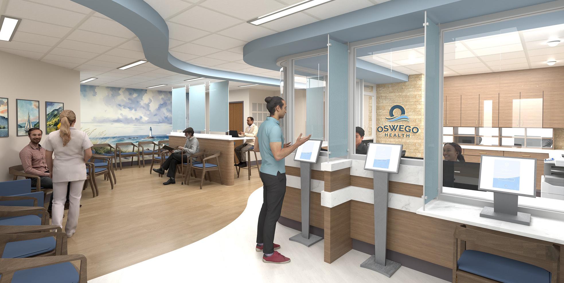
In October 2025, Oswego Health in Oswego, New York, announced a $14-million renovation and expansion project at Oswego Hospital that will streamline patient flow, expand the emergency department, consolidate imaging services into a centralized location and align that department to streamline patient access to it.
David Ruel, vice president of ancillary services at Oswego Health, said that the biggest consideration the project team faced was how to wrangle all of the functions of that work into less than 18,000 square-feet of space.
“We always look at every project with the intent to future-proof, but square-footage is the biggest thing for us,” Ruel said. “We have a landlocked building. We used a shoehorn and got every inch of space out of this.”
The upgrades also include procuring two, state-of-the-art CT scanners to improve patient access to services. By situating the CT devices in close proximity to the emergency department, and consolidating two imaging centers within the building into a shared space, the physical redesign also facilitated workflow improvements that allow staff to be more responsive to patient needs.
“My techs are producing 40 to 50 scans a day,” Ruel said. “The need is out there; the resources are few and far between. One of the reasons I brought the department back together was so I can use one tech to run two rooms as opposed to four techs.”
Overall, Ruel said, a small community health system like Oswego doesn’t have the resources to de-centralize its imaging services. The hospital upgrade project was offset by a state Healthcare Facility Transformation grant and nearly $600,000 in community contributions. Despite ever-increasing study volumes, “when scanners are $2 million apiece and finances are limited, trying to put
may host advanced imaging services that offer greater complexity of care, support research and professional education, or specialized clinical services.
In whichever environment a project is undertaken, “building infrastructure capacity for medical imaging requires careful coordination of power, cooling, space and compliance requirements,” Beebe said.
On the power side, electrical systems should be sufficient not only to support normal operations, but also continuity during extended outages, he said, making “redundancy, emergency power, and protected circuits for critical imaging equipment” necessary components of any imaging project.
“Cooling requirements are equally important,” Beebe said; “imaging rooms must be designed to manage both equipment and room heat loads through adequate chilled water capacity, ventilation and humidity control.”
Like power systems, cooling systems are expected to be able to maintain uptime during unforeseen complications, and that resilience doesn’t only extend to device uptime.
Healthcare organizations rank physical and cybersecurity risks among the top threats to their facilities, staff and
patients, and are taking greater care to contemplate them at the top of any planning discussion.
In the 2026 edition of the annual hospital construction survey conducted by Health Facilities Management (HFM) magazine and the American Society for Health Care Engineering, some 300 healthcare professionals were polled about their perspectives on budgeting, construction and equipment purchasing trends. Their topline takeaways for construction involved the integration of physical and cybersecurity countermeasures.
In a brief accompanying the results, writers Chris Dimick and Jamie Morgan described the work of security-related design features as necessary to prevent physical and verbal attacks on staff and patients, cybersecurity breaches, child abduction and pharmaceutical theft. Among those security features most recommended are surveillance cameras and lighting, parking lot fencing, vehicle bollards, weapons control systems and control plans that can contemplate the eventuality of access restriction or facilities lockdowns.
Just as important as physical security is cybersecurity: only 34 percent of respondents said their security officials and
information technology teams collaborate in developing “physical environment-related cybersecurity measures,” and only 38 percent said facilities management teams are involved in those conversations. Connecting new and legacy clinical systems, equipment, and software is especially important in managing vulnerabilities established by the Internet of Things (IoT), like web-enabled controls on heating, ventilating and air-conditioning (HVAC) and fire safety systems.
Acute care hospitals remain the top building project currently under construction among survey respondents, followed by ambulatory care, and, for the first time, central energy plants, as increased power demands are pressing facilities to reduce costs and carbon emissions.
Ultimately, the blueprint for tomorrow’s imaging environment is less about square footage and more about foresight. As technologies evolve, care models decentralize, and patient expectations rise, the most successful projects will be those that begin with clarity of purpose. By aligning clinical workflows, infrastructure and long-term strategy from the outset, healthcare leaders can create spaces that not only meet today’s demands, but adapt seamlessly to what comes next. •

CLOSING THE IMAGING WORKFORCE GAP STARTS WITH EDUCATION
The imaging workforce gap is no longer a future concern – it is a present operational reality.
Across healthcare systems, imaging departments are facing increasing pressure. Modalities are more advanced than ever. Artificial intelligence integration, hybrid systems, and evolving software platforms demand deeper technical expertise. At the same time, many experienced imaging engineers are approaching retirement, and fewer professionals are entering the field with modality-specific training.
The result isn’t just a staffing challenge. It’s a financial and operational one.
Imaging departments are significant revenue drivers for hospitals and outpatient centers. When equipment is down, patient throughput slows. Delays ripple across departments. Reliance on external service providers increases. Service contracts tighten budgets. Downtime becomes more than an inconvenience – it becomes a measurable cost.
Consider a common scenario. A CT system goes down midweek. The in-house team can perform basic diagnostics but lacks modality depth to isolate the root cause. An OEM visit is scheduled. The earliest availability is two days out. Patients are rescheduled. Staff overtime increases. Revenue is deferred. What could have been resolved internally in hours stretches into days – not because of effort, but because of training gaps.
The workforce gap carries a price.
But so does solving it – and the return on that investment is substantial.
Forward-thinking healthcare organizations are shifting from reactive staffing models to strategic workforce development. Instead of simply filling roles, they are building capability. Crosstraining biomeds into imaging. Developing in-house modality depth. Creating succession plans that anticipate, rather than respond to, retirements and turnover.
Education is no longer optional. It is infrastructure.
For over four decades, the imaging service industry has evolved dramatically. Since 1985, training has moved from analog systems to fully digital platforms, from single-modality
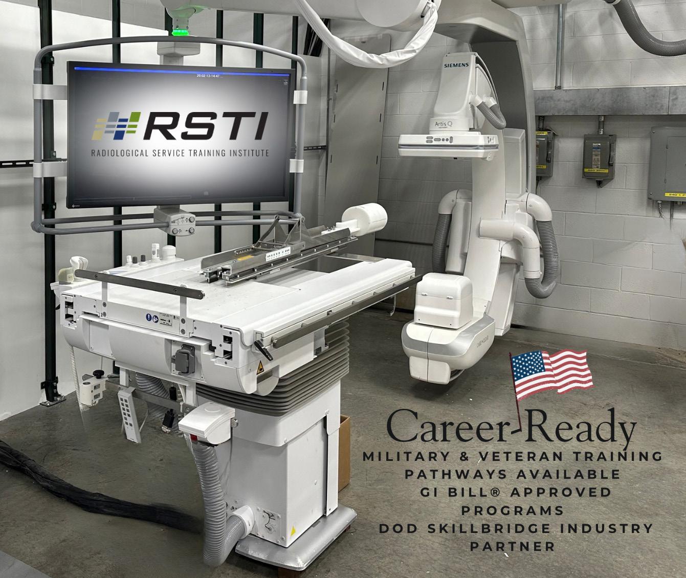
focus to multi-vendor, multi-modality ecosystems. Yet one principle remains constant – engineers who understand systems at a deeper level reduce downtime, increase confidence in the field, and strengthen operational stability.
Investing in education directly impacts:
• Faster troubleshooting
• Reduced dependence on OEM service contracts
• Increased in-house service capability
• Stronger employee retention and career progression
Organizations that prioritize structured, hands-on education are not simply improving skill sets – they are protecting revenue and improving patient care continuity.
Workforce development also means expanding the pipeline itself. Transitioning military professionals into imaging service roles through approved GI Bill and SkillBridge programs provides a disciplined, technically capable talent stream. Supporting biomeds who want to advance into imaging creates upward mobility within existing teams. Multi-vendor exposure ensures engineers can adapt as technology evolves.
The workforce challenge is real. But it is solvable.
This year, RSTI president, Todd Boyland has been recognized as Educator of the Year at the 2026 TechNation TechChoice Awards. The honor reflects more than individual achievement. It represents a long-standing commitment to raising the standard of imaging education and mentorship within the healthcare technology management community.
Excellence in education does not happen by accident. It is built over time – through experience, adaptability, and an unwavering focus on developing engineers who are prepared for real-world complexity.
As imaging systems continue to advance, the gap between minimal training and true technical mastery will widen. The organizations that invest intentionally in education today will be the ones best positioned for stability tomorrow.
Closing the imaging workforce gap does not begin with a job posting.
It begins with education. •
THE FEEDBACK TRAP:
WHY EVEN GOOD LEADERS JUMP TO DEFEND (AND HOW TO BREAK THE HABIT)

DIRECTOR’S CUT
BY NICOLE DHANRAJ
Soooo, I got feedback recently, and while I know the right thing to do is stay quiet and listen to understand … I didn’t. (Sighing heavily as I type this.)
As the conversation went on, I caught myself jumping in explaining my decisions, offering context and trying to paint the full picture.
My intent wasn’t to dismiss anything. I genuinely wanted the other person to know I cared and giving context to the situation.
But even if your intent is good, the impact can still shut people down.
When we respond too quickly, when we rush to justify or clarify, people start thinking:
• “Maybe I shouldn’t bring things up.”
• “It’s not worth the pushback.”
• “They don’t really want to hear it.”
And in a high-stakes environment like a radiology department – where communication directly affects patient flow, safety, morale and efficiency – that’s the last thing any leader wants.
We interrupt not because we’re dismissive, but because we’re operating in “Listening to Respond” mode, your mind is focused on your next statement instead of their actual message.
We’re going to discuss a tiny, two-second habit that breaks this defensive cycle and transforms your impact from dismissive to collaborative.
THE TWO-SECOND PAUSE THAT CHANGES EVERYTHING
The fundamental truth of handling feedback
is this: It’s not about agreeing. It’s about fully receiving what someone is trying to tell you. When someone gives you feedback, your brain immediately tries to protect your reputation and your decisions. That defensive spark is completely normal especially when decisions are high-stakes and fast-paced. But a normal reaction doesn’t mean it’s the helpful one.
Before responding, try building in a very small habit: Pause, For two seconds. This moment gives you space to:
• Calm the instant defensive reaction.
• Stay focused on the speaker and their experience.
• Listen to understand without preparing a rebuttal.
• Absorb the actual message, not just the keywords you need to defend against. It’s simple, but transformational (trust me on that).
WHEN YOU DON’T LISTEN FULLY, PEOPLE NOTICE
When we rush to clarify or jump in, even the most well-intentioned defense can come across as:
• Invalidating
• Dismissive
• Minimizing
• Corrective
And over time, people naturally step back. They think twice before speaking up. They stop raising concerns that actually matter. The intent may be connection, but the impact becomes distance.
WHY SILENCE IS SOMETIMES THE BEST RESPONSE
I have been on this journey for improving my listening and have seen the difference. You don’t owe an instant answer to every piece of feedback. That silence isn’t discomfort, it’s maturity.
Thoughtful leaders use the pause to transition into reflection:
“I appreciate you sharing that, let me sit with it before I respond.”
“You’ve given me a lot to think about.” This shows your team you value their voice more than your need to explain.
THAT TIME I GOT CALLED OUT (A SCENARIO YOU KNOW TOO WELL)
The urge to defend is so strong, even I, the one talking about this, totally failed.
My conversation that started with feedback, I immediately went on the offense. I was explaining, detailing and justifying everything.
My conversational partner had the courage to stop me dead in my tracks and ask the most humbling question a leader can ever hear:
“Are you going to listen?” And um, that was directed a few times as I was really in my trap.
Oof. That was the necessary punch to the gut. It proved my defensive intent was completely ruining my impact.
the opportunity to stop, regroup and hear you out completely. I value your candor and your perspective on this.”
This process, shutting down the defense immediately and then providing reflective acknowledgment later didn’t make me look weak.
It showed my team that I was committed to changing my behavior. It rebuilt the trust I had just broken. It shows that even when you stumble, you can recover and that’s the kind of maturity they respect.
REFLECTION FOR THE WEEK
The next time someone gives you feedback, ask yourself:
“Am I listening to understand … or listening to defend?”
And since impact is everything when you’re a leader, challenge yourself with this commitment:
“Am I willing to use the pause, acknowledge my stumble, and demonstrate the maturity that builds trust, even when it feels awkward?”
The fundamental truth of handling feedback is this: It’s not about agreeing. It’s about fully receiving what someone is trying to tell you.
In that moment, I immediately stopped talking. I used the pause, and though it felt awkward, I simply allowed the conversation to continue, focusing only on receiving their message.
I didn’t try to wrap up the emotional moment right then and there; I just focused on hearing them out. But later that day, I followed up with an email to properly close the loop on that tense moment, showing I had truly reflected:
“Thank you. I want to acknowledge something specific. When you had to ask, ‘Are you going to listen?’ that was a necessary correction. I genuinely appreciate you giving me
When people feel genuinely heard, they keep coming back, creating the culture every radiology manager wants: open communication, trust, and continuous improvement.
Your intent may be positive, but your team’s perception is what truly defines your leadership.
Of note, the hardest part isn’t listening; it’s learning to recover when you fail. If you want to discuss strategies for regrouping trust, crafting an authentic apology, or clearly communicating your commitment to change to your team, please reach out. Let’s talk about turning your defensive moments into defining leadership moments. It is a journey, mine about 15 years in the making. •
Nicole Dhanraj, is a radiology administrator and workforce strategist focused on military-to-healthcare transition pathways. Radiology leaders interested in exploring this approach are encouraged to connect with her directly at nicoledhanraj@gmail.com.
BUILDING IMAGING ENVIRONMENTS FOR TODAY’S HEALTHCARE DEMANDS
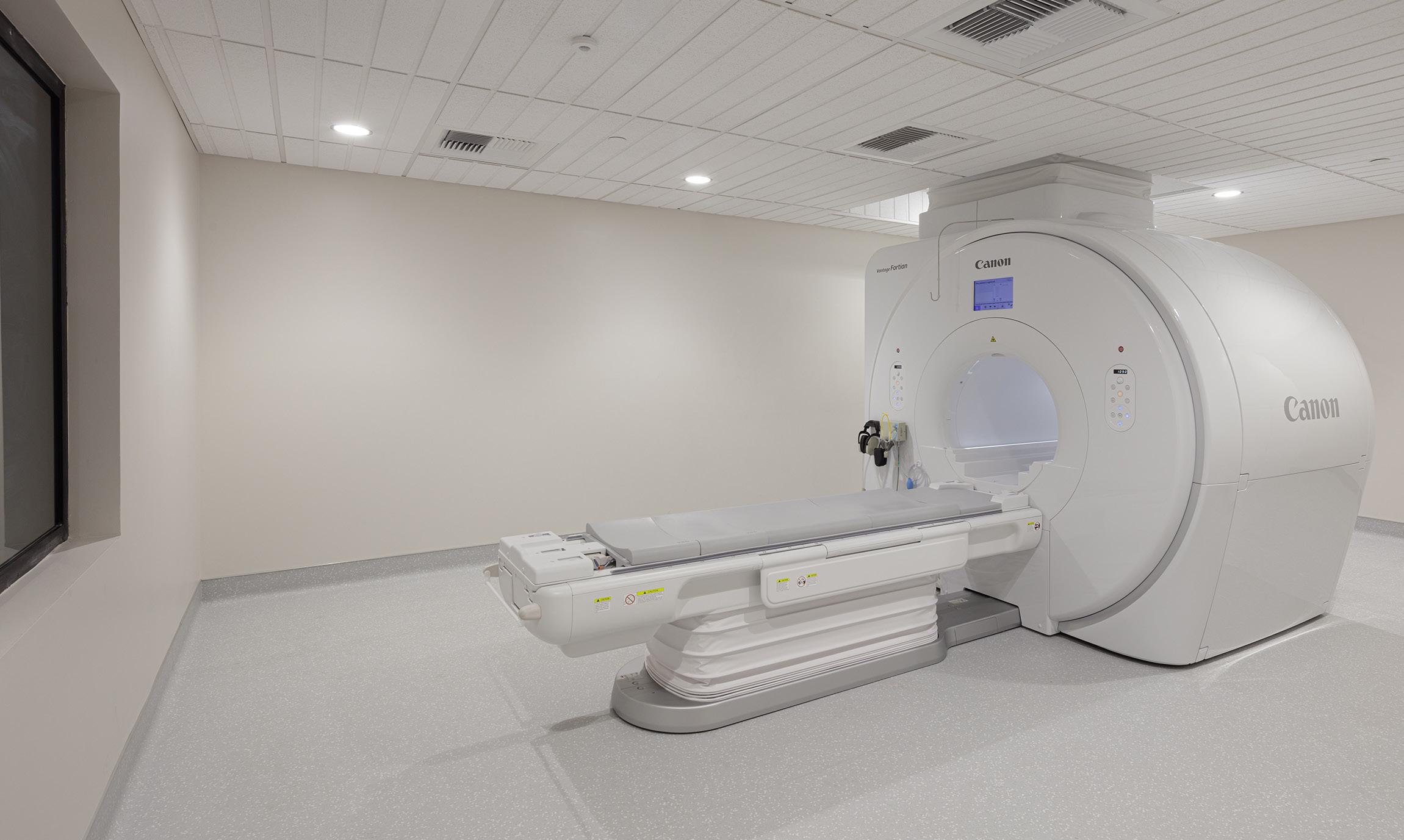
In today’s healthcare landscape, imaging plays a critical role across all areas of care, from acute clinical environments in active hospitals to the growing demand for outpatient and preventative diagnostic facilities. As technology continues to advance, so does the complexity of the spaces required to support it. Delivering these environments successfully, often within fully operational and highly sensitive facilities, requires a specialized approach grounded in experience, precision, and collaboration.
At Colin Construction Company, healthcare construction has been our sole focus since 1981. Over the past four decades, we have delivered hundreds of projects across the western United States and beyond, with a clear mission to lead projects that improve patient care. Imaging environments represent one of the most complex and rapidly evolving sectors within healthcare construction, and one where our team continues to lead through experience and innovation.
EXPERTISE THAT EXTENDS BEYOND THE BUILD
Imaging projects require far more than traditional construction knowledge. Each modality, whether MRI, CT, interventional radiology, radiation oncology, clean rooms, compounding pharmacies, or other advanced systems, introduces highly specific requirements that directly impact performance, safety, and long-term reliability.
Our team approaches these projects with a deep understanding of modality-specific requirements and constructability. As a vendor turnkey certified contractor with all major equipment manufacturers, we provide a level of coordination that connects design teams, equipment vendors, and facility stakeholders. This distinction, uncommon among general contractors, ensures that every detail aligns with the technical standards required for today’s advanced imaging systems.
Early engagement is just as critical. Decisions around equipment selection, existing infrastructure, and project delivery methods all influence cost, schedule, and feasibility. By partnering early, we help clients navigate these complexities with clarity and reduce risk from the start.
BUILDING WITHIN ACTIVE AND SENSITIVE HEALTHCARE ENVIRONMENTS
A large portion of imaging construction takes place inside fully operational healthcare facilities, where construction must coexist with ongoing patient care. These environments demand a higher level of planning, communication, and execution.
Infection control, life safety requirements, noise mitigation, and carefully phased construction are central to success. Our teams are experienced in working within these constraints, maintaining a constant focus on patient safety, staff coordination, and uninterrupted operations.
Through detailed planning and consistent communication, we ensure that stakeholders, from clinicians to facility managers, remain informed and supported throughout the

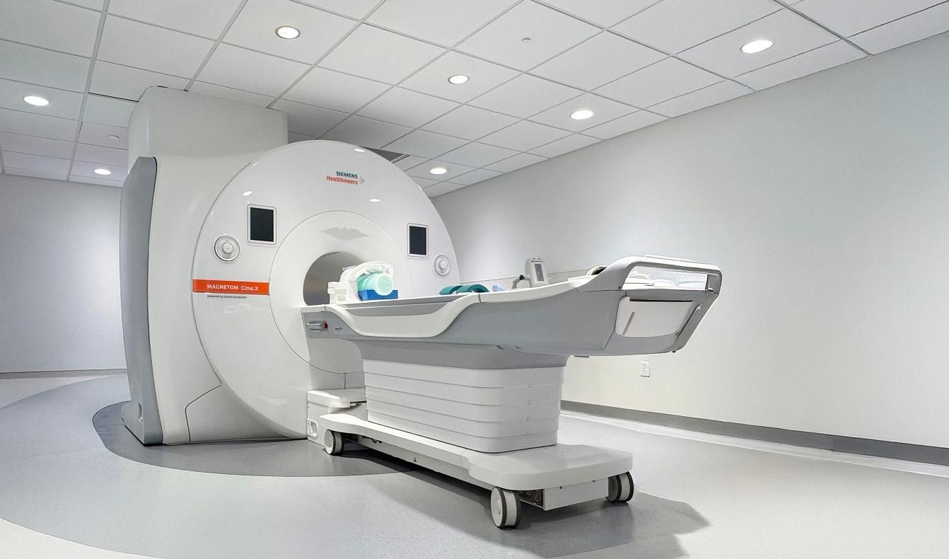
process. This approach has been a key factor in our longstanding client relationships and high rate of repeat work.
ADAPTING TO THE UNIQUE DEMANDS OF EVERY IMAGING PROJECT
No two imaging projects are the same. Some require efficient conversion of existing spaces, while others demand highly complex solutions in constrained environments. Our experience across this range allows us to tailor our approach to meet the specific needs of each facility.
In Newport Beach, California, our team converted 5,000 square feet of office space into a modern MRI suite designed to support advanced imaging technology. The project included structural enhancements for the MRI system, along with upgrades to mechanical, electrical, and plumbing systems, all while preserving the existing structure. The result is a space that delivers both technical performance and a patient-focused experience, with high-end finishes and a calming, thoughtfully designed environment.
On a project in San Francisco, our team took on a highly complex MRI build, delivering a design-build facility within a shelled basement located below the water table. The work involved extensive dewatering and re-waterproofing directly beneath a future MRI room, all while maintaining a long-term waterproofing warranty.
When unexpected water intrusion occurred, our team quickly implemented a manual dewatering operation, managing thousands of gallons of water daily over nearly two months. Working closely with engineers, inspectors, and specialty consultants, we maintained strict environmental
compliance while keeping construction moving forward. Through careful coordination and consistent communication, we delivered the project without schedule delays and transformed a highly constrained space into a fully operational imaging facility.
A PARTNER IN ADVANCING PATIENT CARE
As healthcare continues to evolve, imaging remains central to both diagnosis and prevention. Whether within active hospital campuses or outpatient environments, these projects require a partner who understands not just construction, but also the impact on patient care, clinical workflows, and facility operations.
At Colin Construction Company, our approach is rooted in collaboration, communication, and commitment. We understand that every imaging project comes with unique challenges, and we bring the experience and technical expertise needed to navigate them successfully.
In an industry where precision is critical and disruption is not an option, experience matters. For over 40 years, that experience has been the foundation of everything we build, helping healthcare providers deliver better outcomes for the communities they serve.
We’d welcome the opportunity to connect and explore how we can support your next project. Serving the Western United States, including Alaska, Arizona, California, Colorado, Montana, Nevada, Oregon, Utah, and Washington. •
Contact us at 530-272-3357 or visit www.colincc.com.
FRONTIER OPERATIONS: THE SKILLS THAT DON’T AGE AS THE AI SURFACE AREA EXPANDS

PACS/IT/AI
BY MARK WATTS
We are leading different teams and must recognize the needs of progressing at the rate of artificial intelligence. As AI capability accelerates, the mistake many teams make is treating “frontier work” as a moving set of tools or models to master. It is an approach that never stabilizes. The surface area keeps expanding, releases keep landing and whatever felt advanced six months ago becomes table stakes. What persists is not the tooling, but a small set of operational skills that let humans stay effective as the boundary between human and agent work keeps shifting. These skills don’t depend on knowing what the model can do today. They depend on staying calibrated as that answer keeps changing.
The first of these is boundary sensing. This is the ability to maintain a live, operational intuition for where the human/agent boundary currently sits in a given domain. Not where it sat last quarter, not where the marketing claims it sits, but where it actually holds under real workloads. Boundary sensing is not static knowledge you acquire once. It is a continuous calibration task. Every model release, every improvement in long context handling, every new tool use pattern nudges that boundary. Teams that fall behind usually don’t fail because they underestimate AI. They fail because their intuition is stale. They either cling to human work that has quietly become automatable, or worse, they offload judgment to agents that have crossed just past their reliability edge. Frontier operators develop the habit of constantly retesting assumptions, not because they love novelty, but because the ground is literally moving under them.
The second persistent capability is seam design. This is an architectural skill more than an individual one. Seam design is about structuring work so that transitions between human phases and agent phases are clean, verifiable and recoverable. In mature systems, you can point to exactly where an agent hands off to a human, what artifacts are produced at that seam, how those artifacts can be inspected, and how the process recovers if something goes wrong. This is closer to how a good engineering manager thinks about system boundaries than how a single contributor thinks about completing tasks. Poor seam design produces brittle workflows where humans are forced to reverse engineer agent behavior or blindly trust outputs they can’t audit. Good seam design lets small numbers of humans supervise large numbers of agents without losing situational awareness. As agent counts scale, seams become the real control surface.
Third is failure model maintenance. Early language models failed loudly. They produced nonsense, hallucinated obvious facts, and broke in ways that were easy to spot. Frontier models fail differently. Their failures are textured and subtle. They produce analysis that sounds correct but rests on a misunderstood premise. They generate code that works perfectly on the happy path and quietly breaks on edge cases. They summarize research with 98 percent accuracy and fabricate the remaining 2 percent with high confidence. These failures are dangerous precisely because they look competent. Frontier operators maintain an up to date mental model of how agents fail at the current capability level. Not that they fail, but how. That men-
tal model has to evolve alongside the models themselves. Without it, verification collapses into either blind trust or excessive manual review, neither of which scales.
The fourth capability is short-term capability forecasting. This is not about long-range prediction or speculative AGI timelines. It’s about making reasonable six to twelve month bets on where the boundary is likely to move next and investing accordingly. Frontier operators read trajectories rather than headlines. They watch which failure modes are shrinking, which tasks are becoming more reliable, and which forms of human scaffolding are disappearing. From that, they reposition workflows ahead of time. This is probabilistic positioning, not linear extrapolation. You don’t need to be perfectly right. You need to be directionally sensible often enough that your organization isn’t constantly retooling in panic mode.
The fifth and arguably most important skill is leverage calibration. In an agent-rich environment, human attention becomes the scarcest resource. The bottleneck is no longer execution. It’s judgment about where execution matters. Leverage calibration is the ability to decide, with high quality, where human attention creates the most value and where it doesn’t. This includes knowing when not to intervene. Consulting firms like McKinsey have already described operating models where two to five humans supervise 50 to 100 agents running end-to-end processes.
That only works if the humans are spending their time on the right decisions: setting objectives, inspecting seams, stress testing edge cases, and updating failure models. Humans who try to stay “hands on” everywhere become the constraint.
What ties these five skills together is that none of them are about raw intelligence or prompt cleverness. They are operational disciplines. Boundary sensing keeps intuition current. Seam design makes scale safe. Failure model maintenance keeps trust calibrated. Capability forecasting prevents wasted effort. Leverage calibration protects the most limited resource in the system. Together, they form a stable core that holds even as the surface area of knowledge explodes outward.
Frontier operations, in this sense, is not about chasing the edge for its own sake. It is about staying oriented while the edge moves. Teams that internalize these skills stop being surprised by capability jumps. They absorb them. The tools will keep changing. The models will keep improving. But the operators who master these five capabilities remain effective, not because they know what’s coming next, but because they’ve built systems and instincts that adapt when it does. •
Mark Watts is an experienced imaging professional who founded an AI company called Zenlike.ai.
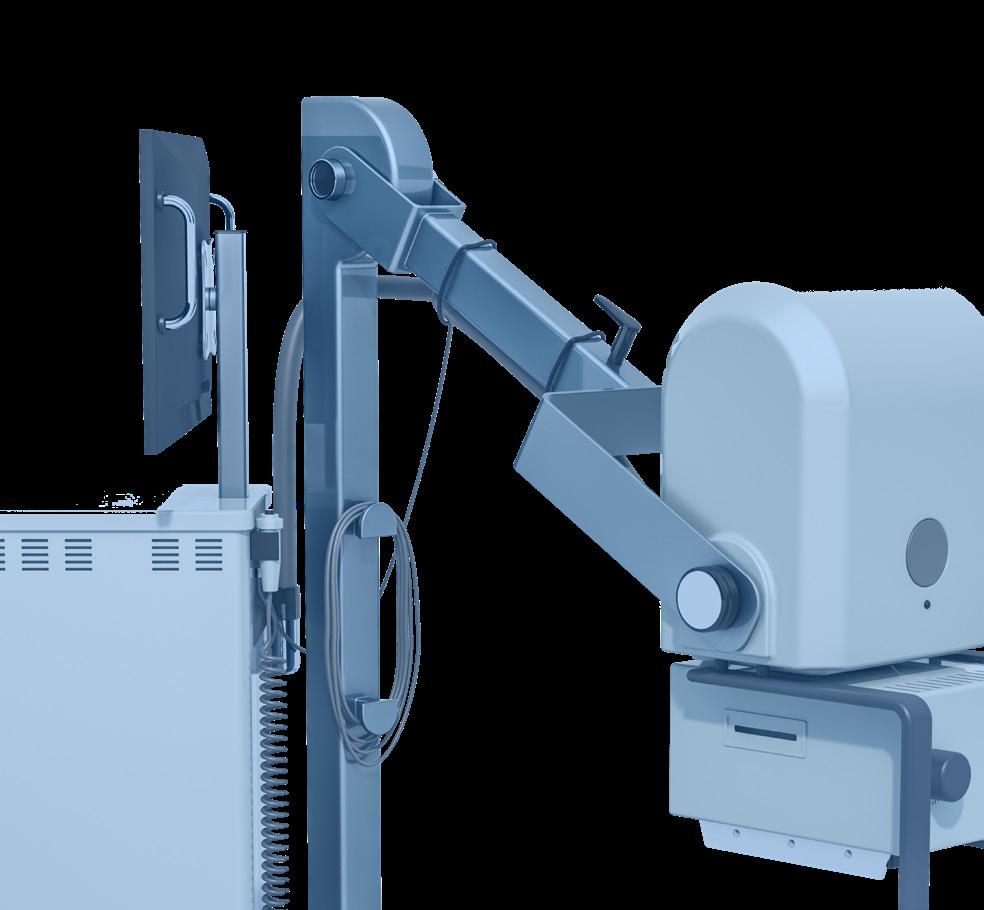


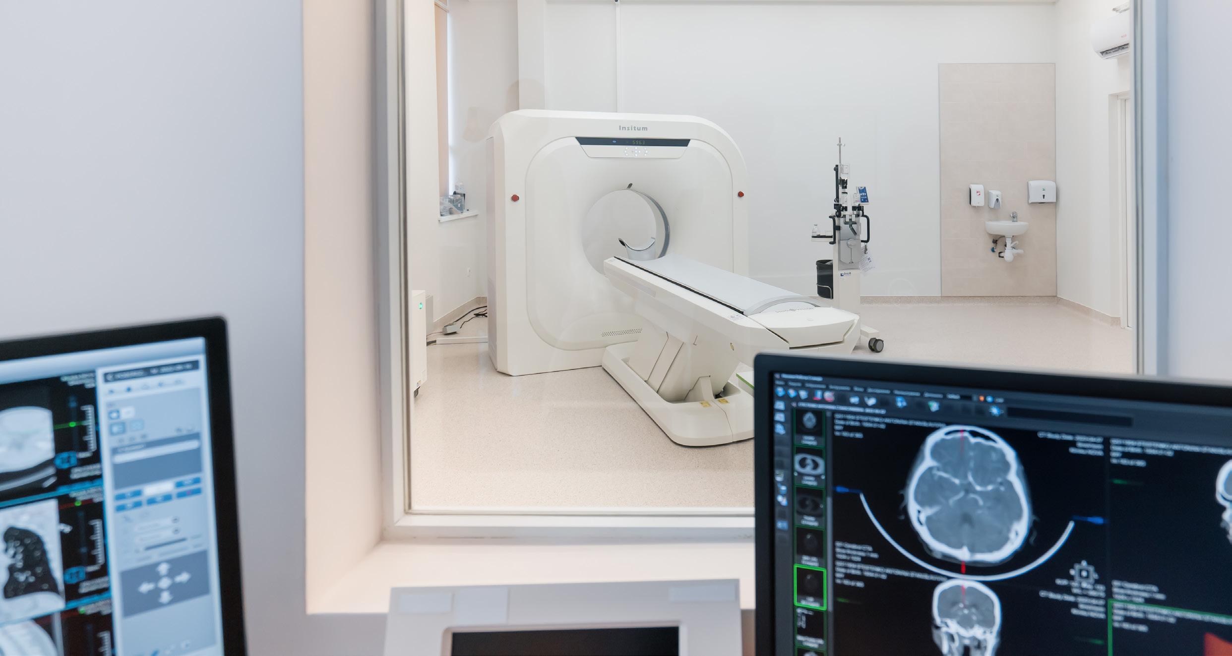
ENVIRONMENT IS PART OF EXAM:
WHEN ‘PERFECT’ AND ‘MODERN’ DESIGN FEEL
UNSAFE
BY NICOLE DHANRAJ DEI
In my previous article, I shared a moment that really humbled me. I was standing in a brandnew imaging department, one of those places that looks like a million bucks on a blueprint. It had the latest equipment, the sleekest finishes and a layout (even from the parking) that was theoretically flawless.
But the patient volume just wasn’t there. When I asked why, it was blunt. It wasn’t about the technology. The patients quietly said: “It feels too modern.” Or, more heartbreakingly, “It doesn’t feel like it was built for us.”
That hit me hard. It was a reminder that patients don’t experience a facility as “architecture.” They experience it as a feeling, especially when they’re anxious, in pain or walking in with a history of healthcare experiences they didn’t choose.
To lead a successful department, we have to understand a framework that is just as vital as clinical protocol: “environmental determinants of Care.” These are the physical, social and symbolic features of a space that shape whether patients feel safe enough to engage.
IT’S NOT ‘DECOR!’ IT’S BIOLOGY
We often think of design as an aesthetic choice. But for a patient, design is a message. When the brain is under stress, it stops processing logic and starts scanning for threats.
I think of Environmental Determinants in three layers that decide whether a patient trusts us, or quietly opts out: 1. Physical Layer: “Can I steady myself here?” This is what a patient sees, touches and hears.
• The Wayfinding Tax: Have you ever
been late for an appointment and couldn’t find the right door? That spike in cortisol makes a patient defensive before they even meet the technologist. Gosh, not to mention the anger and frustration adding more emotional intensity to the interaction. And God forbid, the patient gets to the front desk and they say, we “no-showed you.”
• The Sensory Load: A cold, echoing hallway doesn’t just feel “clean.” It can feel interrogative or even sterile, this is the place where I can die.
2. Social Layer: “Am I alone in this?” This is how the space shapes human interaction.
• The “Fortress” Desk: High, glass-shielded reception desks signal that staff need protection from patients. It creates an “us vs. them” dynamic immediately.
• Visibility of Help: Trust is built through line-of-sight. If a patient can see a staff member, their nervous system settles. If they hit a “dead zone,” anxiety rises.
3. Symbolic Layer: “Do I belong here?” This is the layer we most often miss. Ultra-minimalist, high-end design can signal emotional distance. To a manager, it looks like efficiency. To a patient, it can look like exclusion. Even with minimalism and sleek modern design, warmth and invitation are important.
GOWN JOURNEY: A CASE STUDY IN VULNERABILITY
Think about the moment a patient puts on a gown. In that second, their social status disappears. They are physically exposed and psychologically vulnerable. I’ve walked through departments where patients once changed have to walk past the main public waiting room to reach a sub-waiting area. We may have a policy that says “Patients first,” but the environment is telling them, “Your dignity is secondary to our floor plan.”
And if a patient feels exposed, they aren’t going to be “compliant” during a scan. They’ll be tense. A tense body leads to motion artifacts, repeat sequences, and a schedule that starts slipping before lunchtime. The interaction can also be forced and cold with their underlying embarrassment, vulnerability and frustration.
SIGNAGE
AUDIT: CHANGING THE TONE WITHOUT RENOVATING
You don’t need a renovation budget to improve the symbolic environment. Look at your signs.
• Do they say: “RESTRICTED AREA: DO NOT ENTER”? (A command that triggers defensiveness).
• Or do they say: “STAFF AREA: We will come get you shortly”? (A boundary that offers reassurance).
Institutional language tells patients they’re being processed. Partnership language tells them they’re being cared for. Read that again.
10-MINUTE “HUMAN AUDIT”
Try this tomorrow. Walk your floor, not as a manager, but as a person.
• Arrival: Can I find my way if I’m too anxious to think clearly?
• Change: Do I feel protected or on display in my gown?
• Wait: Does this space expect me to comply, or does it support me to succeed?
THE BOTTOM LINE
Imaging departments don’t lose volume because their technology is old. They lose volume because their environment is “loud.”
When you treat the environment as part of the exam, you stop managing facilities and start managing human trust. In imaging, trust isn’t just a “nice-tohave.” It’s a performance lever.
One metric to track for 30 days: Track Exam Interruptions (pauses, extended time or rescheduling due to anxiety/confusion). It’s one of the fastest ways to see where the environment is working against you.
Next, I’ll explain how environments don’t just influence comfort – they actively construct trust •
Nicole Dhanraj, is a radiology administrator and workforce strategist focused on military-to-healthcare transition pathways. Radiology leaders interested in exploring this approach are encouraged to connect with her directly at nicoledhanraj@gmail.com.
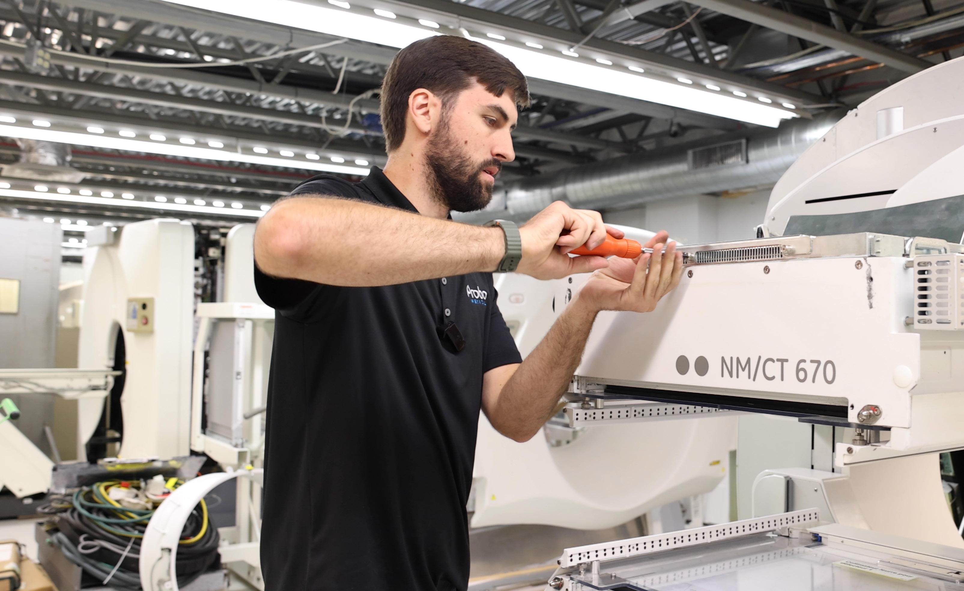
Solving Parts Procurement for Imaging Techs
SOLVING PARTS PROCUREMENT FOR IMAGING TECHS
Downtime in diagnostic imaging is more than an inconvenience, it's a critical issue that For repair technicians, getting equipment such as ultrasound, MRI, and CT scanners back priority. However, sourcing the right high-quality replacement parts often presents challenges entire repair process.
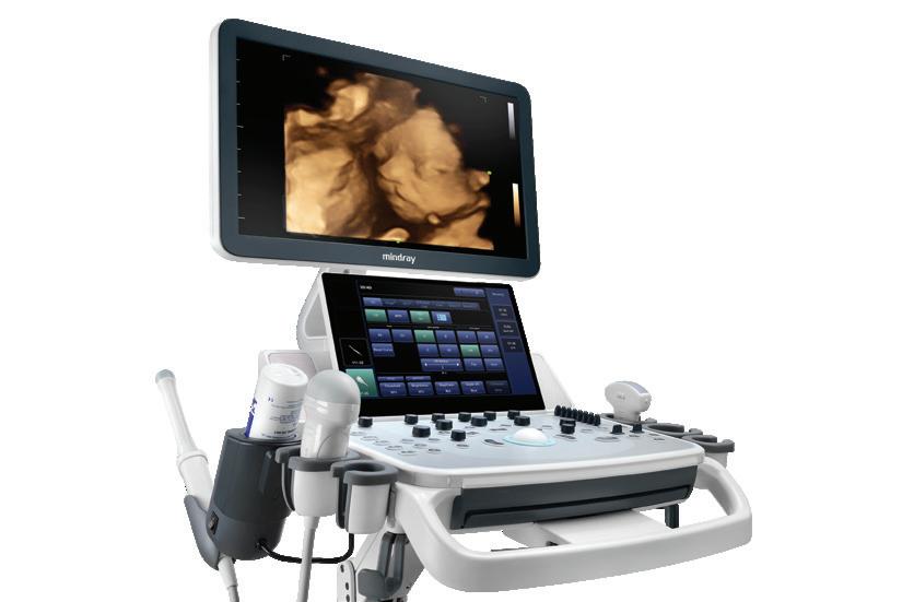
Downtime in diagnostic imaging is more than an inconvenience; it’s a critical issue that can delay patient care. For repair technicians, getting equipment such as ultrasound, MRI, and CT scanners back online quickly is the top priority. However, sourcing the right high-quality replacement parts often presents challenges that can slow down the entire repair process
The Challenge of Availability
THE CHALLENGE OF AVAILABILITY AND COMPATIBILITY
One of the most common concerns that repair technicians see is simply finding the required part. Whether it’s older machines with components no longer manufactured, or newer equipment with higher demand that backorders the part for weeks. Once a part is ordered, technicians need to ensure it’s compatible. A component might look right, but subtle differences in manufacturing runs or software ver-
One of the most common concerns that repair finding the required part. Whether it’s older longer manufactured, or newer equipment with backorders the part for weeks. Once a part is ensure it's compatible. A component might look differences in manufacturing runs or software Installing an incompatible part not only wastes damaging other components of the imaging
Scan QR code to view our complete list of products and services
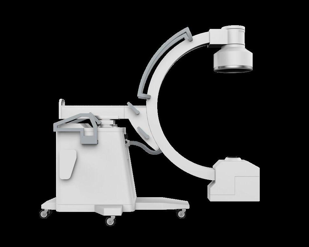

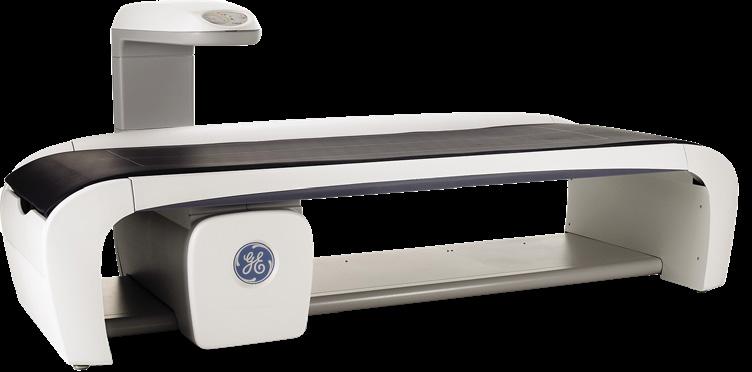
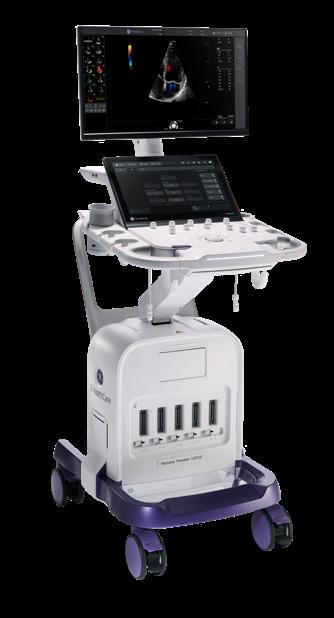
Navigating Cost & Quality Concerns
sions can render it useless. Installing an incompatible part not only wastes time but can also risk damaging other components of the imaging system.
The cost of OEM parts can be extremely high, putting a strain on operational budgets. This pressure often leads technicians to explore the third-party market, which introduces another major concern: quality. While a refurbished or third-party part typically is a fraction of the cost, if it fails prematurely, the initial savings are lost. Nothing hurts a technician's credibility more than getting a DOA part and having the system down an additional day. To remain proactive, technicians must consider the cost-benefit against the potential risks of poor quality if there is no product warranty when sourcing parts.
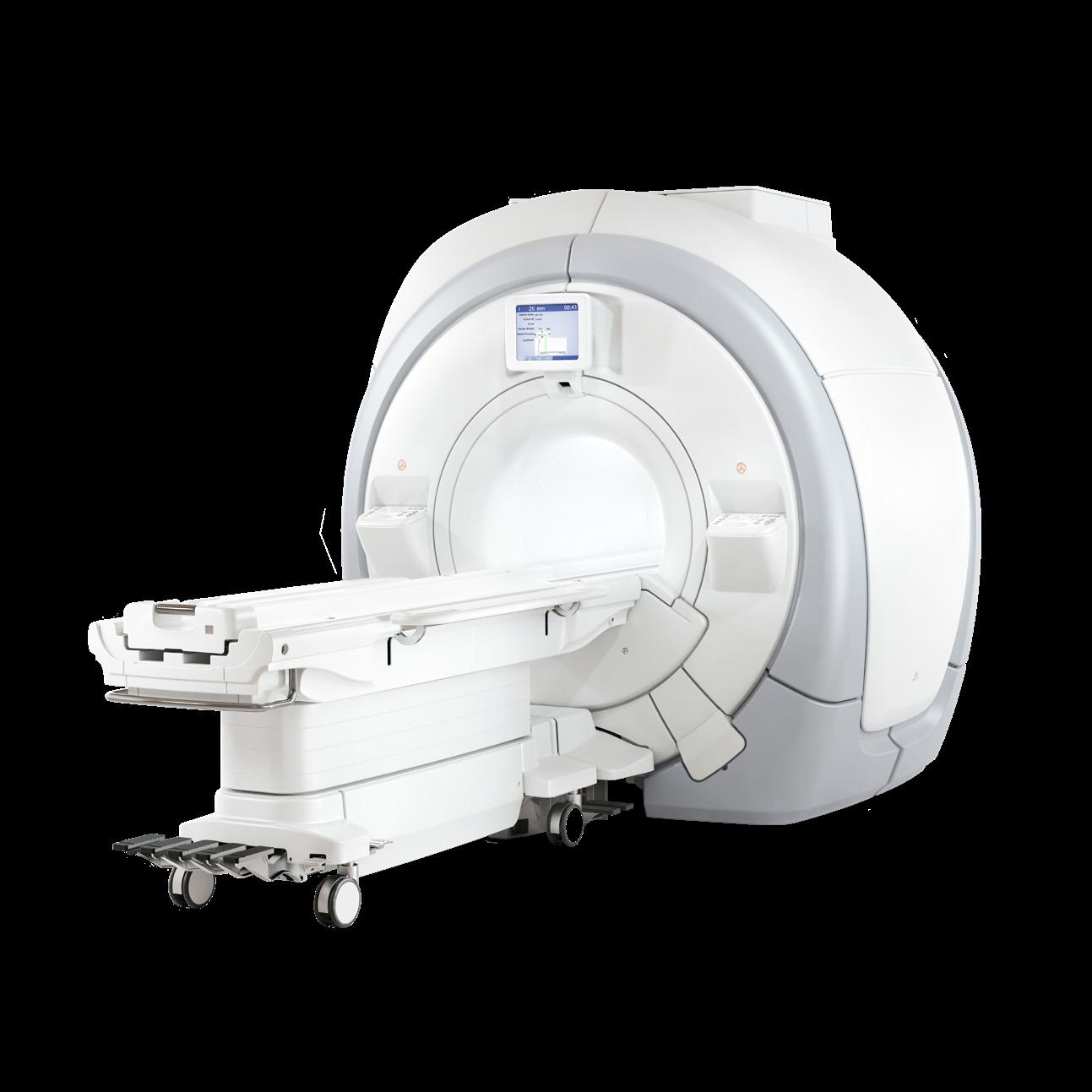
NAVIGATING COST AND QUALITY CONCERNS
The cost of OEM parts can be extremely high, putting a strain on operational budgets. This pressure often leads technicians to explore the third-party market, which introduces another major concern: quality. While a refurbished or third-party part typically is a fraction of the cost, if it fails prematurely, the initial savings are lost. Nothing hurts a technician’s credibility more than getting a DOA part and having the system down an additional day. To remain proactive, technicians must consider the cost-benefit against the potential risks of poor quality if there is no product warranty when sourcing parts.
Solutions for Smarter Sourcing
SOLUTIONS FOR SMARTER SOURCING
Overcoming these obstacles requires a strategic approach to parts procurement. The team at Probo Medical offers effective solutions to keep your equipment running smoothly with our high-quality, reliable parts. With locations around the globe, Probo Medical offers a huge inventory of guaranteed parts for nearly every modality of diagnostic imaging.
Overcoming these obstacles requires a strategic approach to parts procurement. The team at Probo Medical offers effective solutions to keep your equipment running smoothly with our high-quality, reliable parts. With
Our comprehensive quality assurance program includes inspection and testing of: CT, MRI, Ultrasound, Nuclear Medicine, Cath, C-arm, X-ray, BMD, PETand PET/CT, and Flexible & Rigid Endoscopes. Our expert harvesting departments test and certify all parts and accessories at our ISO 13485-managed facilities, as they follow stringent Quality Management System requirements. Plus, we offer same-day shipping, a six-month warranty, and free technical support with every part to ensure you get the most out of your repair.
inventory of guaranteed parts for nearly every modality of diagnostic imaging. Our comprehensive quality assurance program includes inspection and testing of: CT, MRI, Ultrasound, Nuclear Medicine, Cath, C-arm, X-ray, BMD, PET and PET/CT, and Flexible & Rigid Endoscopes. Our expert harvesting departments test and certify all parts and accessories at our ISO 13485-managed facilities, as they follow stringent Quality Management System requirements. Plus, we offer same-day shipping, a six-month warranty, and free technical support with every part to ensure you get the most out of your repair.
Our goal is to be the single, reliable source you turn to for any part or accessory. Our experienced team is here to help you find the right imaging parts for your specific equipment model. Whether you’re replacing a worn component or upgrading your system, we provide expert guidance and exceptional customer service every step of the way. •
For more information, visit probomedical.com.
Our goal is to be the single, reliable source you turn to for any part or accessory. Our experienced team is here to help you find the right imaging parts for your specific equipment model. Whether you're replacing a worn component or upgrading your system, we provide expert guidance and exceptional customer service every step of the way.
WHAT YOUR BRAIN WISHES YOU KNEW
One of the most persistent paradoxes of workplace life is that the busiest people aren’t always the most productive. Walk through any office and you’re likely to spot at least a few people who act perpetually slammed, yet have little to show for their frenzy. Think of a performer who is spinning plates, but the plates keep crashing.
During a recent training session, a group of middle managers described their days to me in almost identical terms. They were constantly bouncing from task to task, and struggling to meet deadlines was their norm.
What we’re talking about here is the need to find effective time management techniques. The problem is that effective time management methods are often different for each person. In other words, what works brilliantly for one person may be completely useless for someone else.
A SKILL THAT BACKFIRES
Every once in a while, a skill will get touted as a “must have” ability. One such skill, called “multitasking,” was born out of the computing industry. It referred to a computer processor’s ability to perform multiple tasks concurrently.
In human application, multitasking is the ability to bounce back and forth between differing tasks to get more done in less time. A few decades ago, this word was expected to be seen in resumes and job descriptions, and even in performance reviews. The problem? Despite the idea that multitasking was a productivity superpower, the concept has actually been debunked.
Researchers at the American Psychological Association found that switching back and forth between tasks can reduce a person’s productivity by up to 40%. Why? Because the human brain needs time to reorient itself to the requirements of whatever task one is doing.
Consider a study out of the University of California, Irvine,

EMOTIONAL INTELLIGENCE
BY DANIEL BOBINSKI
which found that it takes an average of 23 minutes and 15 seconds to fully regain focus after a single interruption. Apply that across a typical workday and it becomes clear why so many people feel exhausted and yet unproductive.
Microsoft’s 2025 Work Trend Index adds even more context: the typical employee is now interrupted every two minutes during core work hours, amounting to roughly 275 disruptions per workday.
I first became aware of multitasking’s downside during a conversation with Robert Croker, the former chair of the human resource training and development department at Idaho State University. Croker, who is certified in brain-based learning, says the brain simply isn’t wired for multitasking.
“It’s a common misconception that a brain is like a computer,” Croker says. “A computer is designed to multitask. The human brain is not designed to function optimally in a multitask environment.”
More recent neuroscience backs him up. Researchers at Wake Forest University, using fMRI and EEG monitoring, confirmed that what most people call multitasking is actually just rapid, sequential task-switching, and that every switch carries a measurable cognitive cost. Those costs add up when one considers that the average worker now switches tasks more than 300 times per day.
If you want personal evidence that the quality of completed tasks drops sharply when people attempt two things simultaneously, just notice how quickly your driving decisions deteriorate the moment you pick up your phone.
AN OLD-FASHIONED FIX
Do any time-management techniques actually work well for most people? The answer is yes, but no, it’s not using a time management app. It’s the old-fashioned habit of better planning.
The simple act of thinking through your tasks and prioritizing what needs to be done allows your brain to pre-map the work ahead. In reality, when you plan, your brain’s amygdala is doing what it does best – preparing your brain to
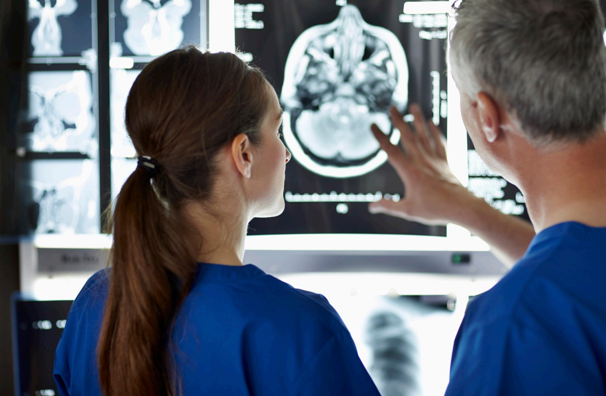
notice the things that matter most. Think of it as laying the groundwork for efficiency.
The second universally helpful tip may shock you. It comes from workplace-organizer Julie Morgenstern, and it’s simply this: don’t check your email first thing in the morning.
You heard that right. Once you get to work, you should not be checking email right away. Morgenstern says the best way to improve productivity is by building a task list first. She says it’s even better if you create the day’s task list the evening before. Then, when you arrive at work, you can immediately tackle your highest-priority, revenue-generating work when you arrive.
WHY YOU MIGHT FEEL OVERWHELMED
A 2025 survey found that 58% of professionals check their email before they even get out of bed. Additionally, workers now spend up to 28% of their entire workweek just managing their inboxes. For most people, that isn’t productivity; it’s a well-disguised time sink.
If your job genuinely requires an early inbox check, experts suggest setting the timer on your phone to three minutes. After beginning the time, scan your inbox for anything truly urgent, and then close your email when the timer goes off.
When I first raised this idea with some of my clients, every one of them insisted it was “not possible.” Then, coincidentally, the Monday following that training session, their company’s
email server went down and stayed down the entire day.
The next time I met with the group, their leader admitted, almost sheepishly, “We got a phenomenal amount of work accomplished that Monday. I honestly think we should make every Monday a no-email day.” Their accidental experiment proved Morgenstern’s point better than anything else could have.
One workplace I know actually established a policy that employees should check email only three times a day: at 11 a.m. to handle written correspondence before lunch; at 1 p.m. to address follow-up responses; and again at 4 p.m. to adjust the next day’s schedule. To stave off potential problems, the company informs clients and vendors of this policy so everyone knows what to expect.
The bottom line? Multitasking has been shown to make us less effective, not more. And although email remains one of the most powerful communication tools ever developed, it becomes a liability the moment it becomes the center of everything we do. •
Daniel Bobinski has a doctorate in theology and is a best-selling author and a popular speaker at conferences and retreats. For more than 30 years he’s been working with teams and individuals (1:1 coaching) to help them achieve excellence. He was also teaching Emotional Intelligence since before it was a thing. Reach him by email at DanielBobinski@ protonmail.com or 208-649-6400.

MEDICINE FOR THE SOUL
‘BEER BELLY’ LINKED TO HEART DAMAGE IN MEN
Al arge new study using advanced imaging found that abdominal obesity, sometimes referred to as a “beer belly,” is associated with more harmful changes in heart structure than overall body weight alone, especially in men. The findings were presented at the 2025 meeting of the Radiological Society of North America (RSNA). They point to actions patients and doctors can take to identify potential risks and intervene earlier to protect the heart.
“Abdominal obesity, a high waist-to-hip ratio, is associated with more concerning cardiac remodeling patterns than high body mass index (BMI) alone,” said study lead author Jennifer Erley, M.D., radiology resident at University Medical Center Hamburg-Eppendorf, Germany.
“It appears to lead to a potentially pathological form of cardiac remodeling, concentric hypertrophy, where the heart muscle thickens but the overall size of the heart doesn’t increase, leading to smaller cardiac volumes. In fact, the inner chambers become smaller, so the heart holds and pumps less blood. This pattern impairs the heart’s ability to relax properly, which eventually can lead to heart failure.”
Taking into account BMI, a measure of general obesity calculated from a person’s weight and height, and waist-to-hip ratio (WHR), a measure of abdominal
obesity, the researchers studied cardiovascular MRI images of 2,244 adults aged 46 to 78 (43% female) without known cardiovascular disease. Abdominal obesity reflects an accumulation of visceral fat, which is stored deep around internal organs and strongly linked to harmful cardiovascular effects. All of the study participants are part of the ongoing Hamburg City Health Study, a long-term population study in Germany.
According to BMI, 69% of males and 56% of females in the study were overweight or obese. Using WHR, 91% of the males and 64% of females met the World Health Organization (WHO) criteria for obesity.
General obesity based on BMI was more often linked to enlarged heart chambers across all participants. Abdominal obesity was associated with thickening of the heart muscle and smaller heart chamber volumes. These changes were more prominent in men, particularly in the right ventricle, which pumps blood to the lungs. This may reflect early cardiac stress on the heart related to how abdominal fat affects breathing and lung pressure.
Obesity also was linked to subtle heart tissue changes in men, detectable only with advanced cardiac MRI, potentially signaling early heart stress before symptoms or diagnosable disease. These associations persisted even after accounting for other cardiovascular risk factors, including arterial hypertension, smoking, diabetes and cholesterol.
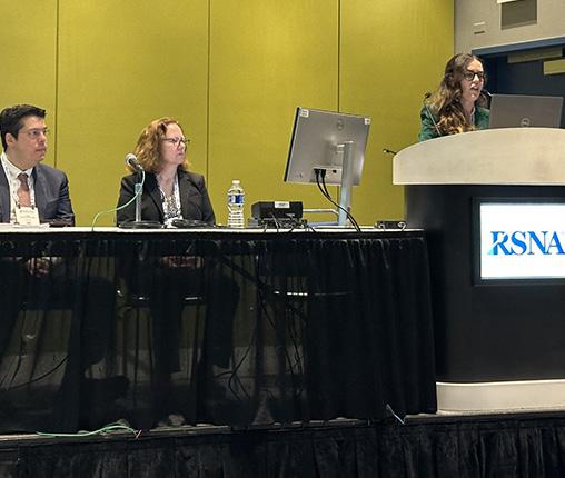
“The sex-specific differences suggest that male patients may be more vulnerable to the structural effects of obesity on the heart, a finding not widely reported in earlier studies,” Erley said. “Rather than focusing on reducing overall weight, middle-aged adults should focus on preventing abdominal fat accumulation through regular exercise, a balanced diet and timely medical intervention, if necessary.”
The more extensive heart damage seen in men could be due to an earlier onset of more severe abdominal obesity, or the cardioprotective effect of estrogen in women, Erley said, though more research is needed.
With a tape measure, anyone can calculate their WHR at home by dividing their waist circumference at its narrowest point by their hip circumference at its widest point. A ratio above 0.90 for men and 0.85 for women is an indicator of abdominal obesity and is associated with increased risk of cardiovascular disease, according to the WHO.
The authors also encourage clinicians to be proactive in checking and flagging abdominal obesity early on to improve health outcomes.
“From the perspective of a radiologist, when we see this cardiac remodeling pattern, we currently think of cardiomyopathy, hypertensive heart disease or some other form of disease, but we don’t clinically draw the line to obesity in our reports,” Erley said. “This study should alert radiologists and cardiologists to be more aware that this remodeling could be attributed independently to obesity.” •
Co-authors are Jonas H. Lund, M.D., Isabel Molwitz, M.D., Ersin Cavus, M.D., Gerhard B. Adam, M.D., Peter Bannas, M.D., Enver G. Tahir, M.D., and Mathias Meyer M.D.

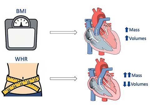
1. Pictogram showing the results of the research. An increase in waist-to-hip ratio (WHR) is associated with a higher left ventricular (LV) mass and lower ventricular volumes. Its association with right ventricular (RV) volumes is weaker in women than in men. An increase in body mass index (BMI) is associated with ventricular dilatation and a higher LV mass, although this relationship is also weaker in women.

Figure
Jennifer Erley, M.D., presents her research at RSNA 2025.
SCRAP BOOK
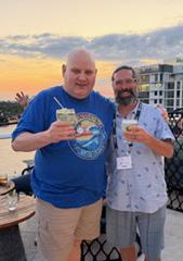
The Association of Medical Service Providers (AMSP) recently held its annual Winter Event in Ft. Lauderdale, Florida. AMSP is a national association of independent medical service providers whose common goal is a dedication to the support and improvement of the member organizations by providing process improvement, revenue growth initiatives, expertise sharing and regulatory compliance.
1. AMSP President Jason Olineo with Associated X-ray and AMSP Treasurer Bill Laird with Brandywine Imaging toast a successful winter event at the rooftop reception.
2. Steve Holmes presents a session at the meeting.
3. (from left to right) Paul Harris with Chesapeake Medical, Chelsea Harris, Jason Olineo, John Olineo and Mike Halsey were all smiles at the vendor reception.
4. Russ Perry with Televere is seen with Nanox’s Joe Spratt and Tim Budrewicz at the Wednesday rooftop reception.
5. Seko Logistics team members John Lynd and Said Huskic are seen at the Tuesday Member Dinner. 1 2 3 5 4
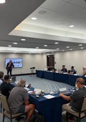



IT ALL STARTS ONLINE
ICEWEBINARS.LIVE
1.REGISTER
Register to view the webinars each month
2.WATCH
Watch recorded webinars on-demand
3.EARN
ARRT Category A CE credits pending approval
ICE Break


•