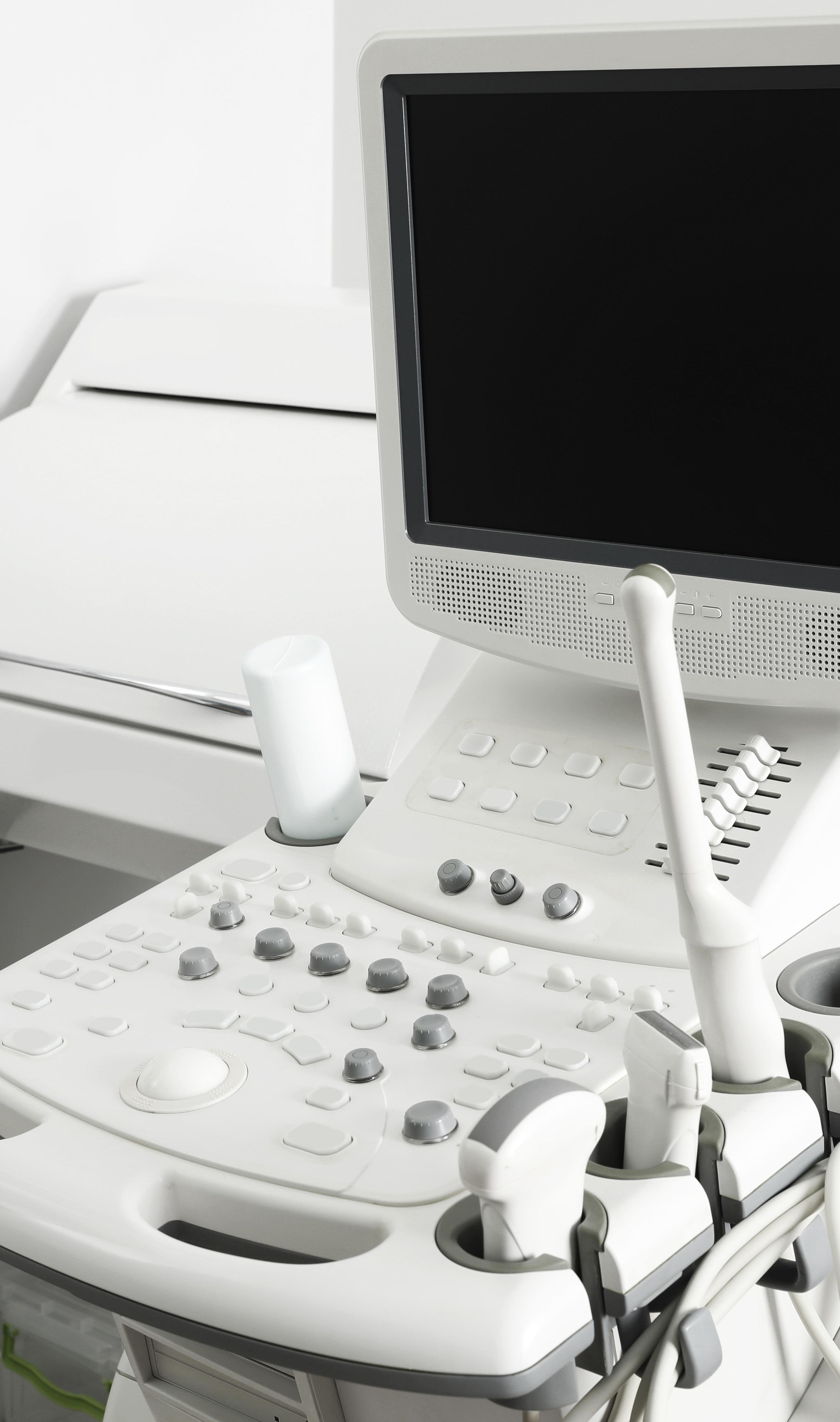
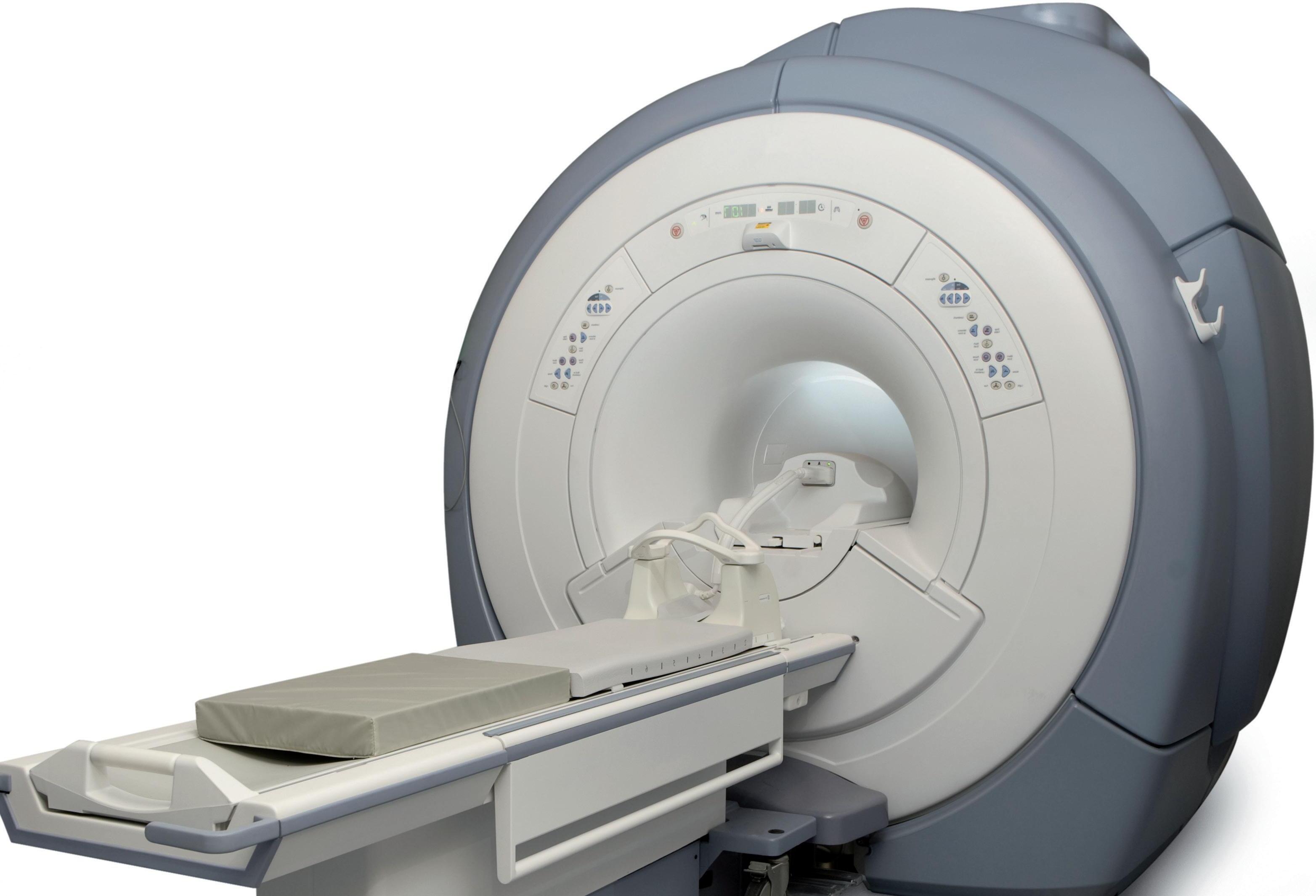
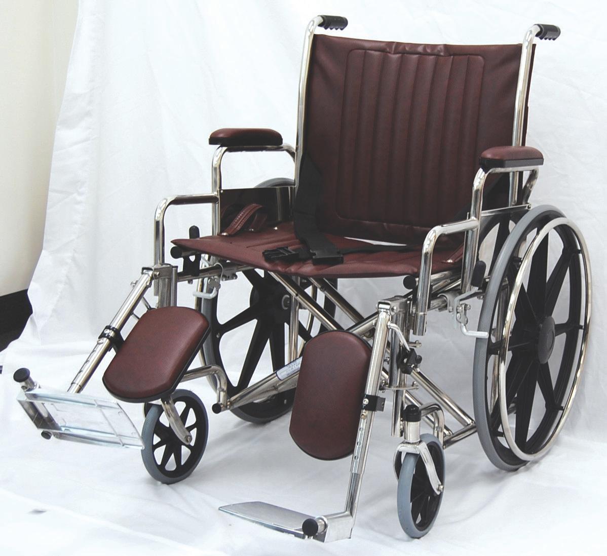
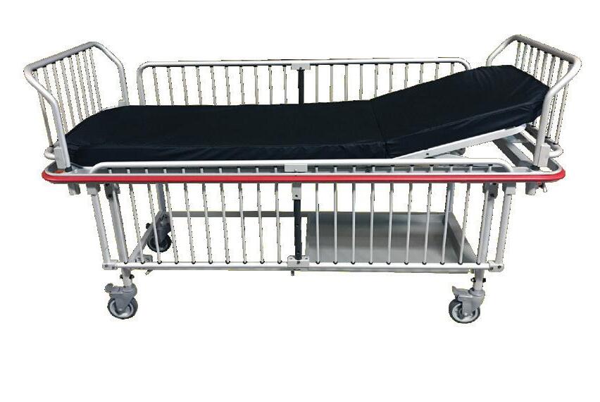
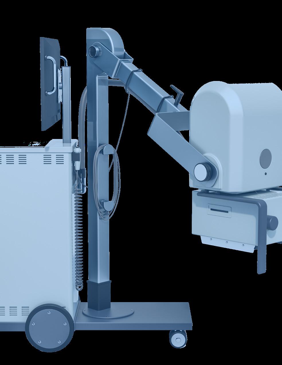
Welcome to Downtime Trace. Our platform gives healthcare facilities a faster, more costcontrolled way to find service providers for their medical equipment.
Access Vetted Service Technicians Nearby
Our network of qualified gig technicians is growing fast. More providers means more options, faster response times, and better pricing for your team.
Take Control of Your Costs
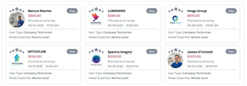
Compare multiple bids at once and select the best value instead of relying on a traditional vendor list.
Save Hours in Your Workflow
Connect with available providers in minutes, for emergency repairs, PMs, or gap coverage. Every service request, bid, approval, and payment runs through one platform, so your team spends less time on phone calls, emails, and paperwork.
DRINKS ICE ON
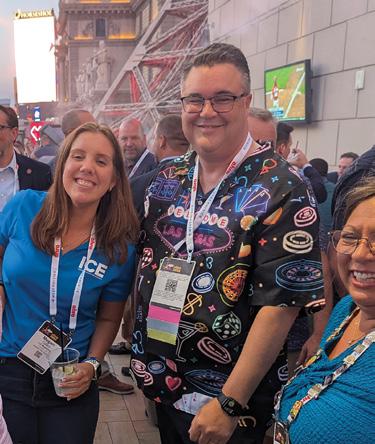
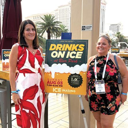
JOIN ICE MAGAZINE FOR OUR ANNUAL DRINKS ON ICE NETWORKING EVENT AT AHRA 2026.
PROUDLY SPONSORED BY:
JULY 13 6:30-7:30PM
FALLS POOL BAR AT THE ORLANDO WORLD CENTER MARRIOTT
Scan the QR code to RSVP for this year’s DRINKS ON ICE. Pick up your bracelet at ICE Magazine’s booth #736 or IAC’s booth #329 during Monday’s exhibit hall hours.
*Event is open to all AHRA attendees and exhibitors and current clients of MD Publishing.
FEATURES
DEI
The three environment layers — physical, social, and symbolic — combine into one critical operational outcome: therapeutic trust.
COVER STORY
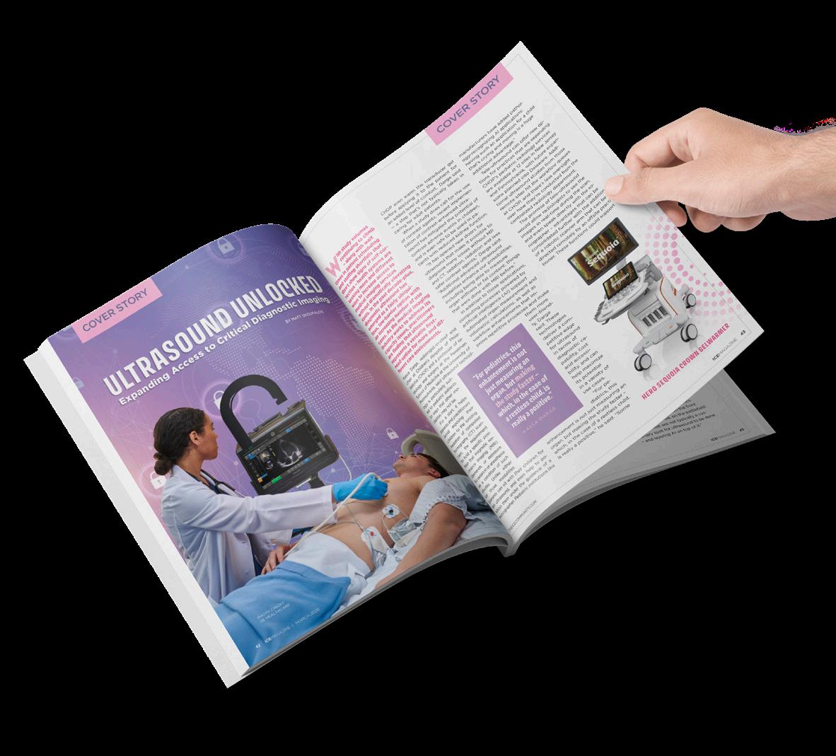
Advocacy is an essential aspect of quality health care and includes patients, colleagues and causes.
RISING STAR
Kaleida Health Imaging Specialist I Kyle Koscinski believes in continuing education.
JUNE 2026
PRODUCT FOCUS
The demand for contrast media is growing to meet the need for medical imaging and the rising burden of chronic disorders.
IMAGING NEWS
Catch up on the latest news from around the diagnostic imaging world.
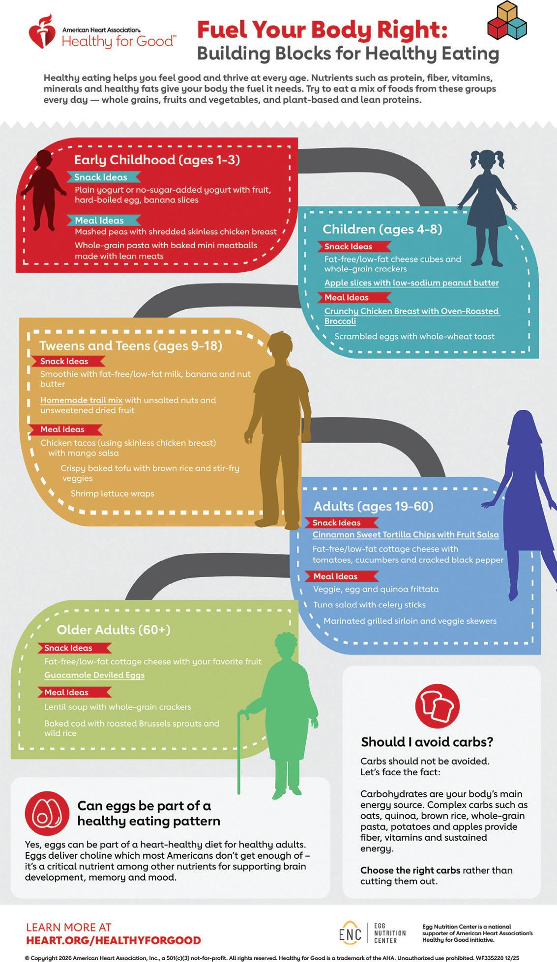
Making health-conscious decisions with your at-home menu doesn’t have to be complicated, but it does require commitment and a little more foresight at the grocery store. 52
MEDICINE FOR THE SOUL
President
Senior
RISING STAR
KYLE KOSCINSKI, KALEIDA HEALTH IMAGING SPECIALIST I
Kyle Koscinski’s plans to expand his knowledge in as many areas of imaging as possible.
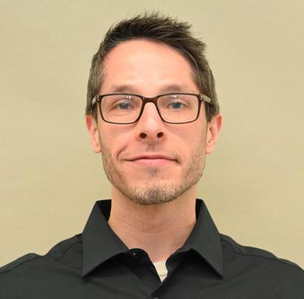
Kaleida Health Imaging Specialist I Kyle Koscinski believes in continuing education. He holds two engineering degrees as well as Siemens certifications. ICE Magazine recently found out more about this Rising Star.
Q: WHERE DID YOU GROW UP?
A: Depew, New York
Q: HOW DID YOU FIRST DISCOVER YOUR FIELD?
A: Recommended field from a relative.
Q: WHY (OR HOW) DID YOU CHOOSE TO GET INTO THIS FIELD?
A: It was the first job field that responded to my application to work. After gaining experience, I decided to stay and invest in a future in the field. The main appeal at the time: healthcare was considered one of the more reliable jobs to have for life.
Q: WHAT DO YOU LIKE MOST ABOUT YOUR POSITION?
A: The complexity of the equipment being worked on makes it engaging; more fulfilling.
Q: WHAT INTERESTS YOU THE MOST ABOUT YOUR CAREER FIELD?
A: The direction the field can go in 10, 20 and 50 years from now. How fast technology evolves; it’s exciting to think about what new equipment/techniques will be used and worked on in the future.
Q: WHAT HAS BEEN YOUR GREATEST ACCOMPLISHMENT IN YOUR FIELD THUS FAR?
A: Getting to the position I am in currently (imaging) after starting from the lowest entry position.
Q: WHAT GOALS DO YOU HAVE FOR YOURSELF IN THE NEXT 5 YEARS?
A: The main goal is expanding my knowledge in as many areas of imaging as I can through various means and OEM training. •
FUN FACTS
FAVORITE HOBBY: Cooking
SUPERPOWER OF CHOICE: Getting somewhere quickly/ teleporting
FAVORITE FOOD: Braised beef ribs with asparagus
1 THING ON YOUR BUCKET LIST: Seeing nanobots used in healthcare
SOMETHING YOUR CO-WORKERS DON’T KNOW ABOUT YOU: I have an autoimmune disease
FOCUS IN
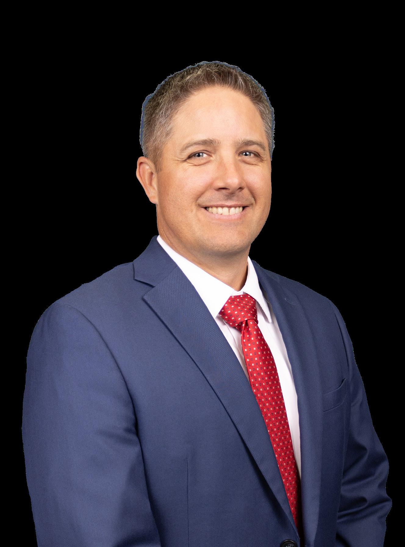
CASEY DYE, MHA, RT (R) ARRT
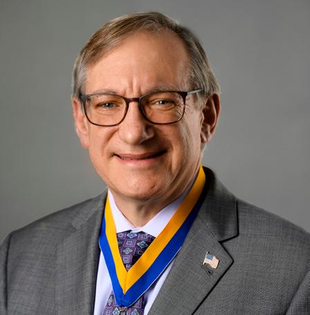
Ma disonhealth Executive Director of Diagnostic Services Casey Dye, MHA, RT (R) ARRT, started his imaging career with a goal to help people get back to living their best lives.
It is a never-ending goal because every time he and the healthcare team at the Rexburg, Idaho, hospital did just that there was soon another patient to help.
Casey developed another goal while tending to his first one. The new goal is to serve as a mentor and a leader who cares about his colleagues and their careers.
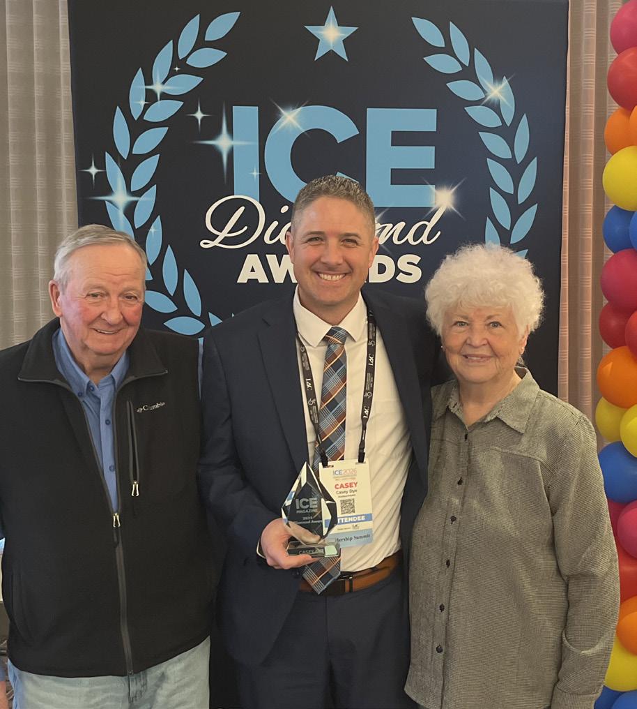
One goal the humble Casey did not have on his to-do list was to be recognized nationally for his leadership and the quality patient care he helps deliver. Yet, that is exactly what happened as a recipient of an ICE Diamond Award.
Casey accepted the award earlier this year at the 2026 Imaging Conference & Expo in San Antonio, Texas. Proud family members, friends, peers and fellow award winners looked on and listened to his passionate acceptance speech.
In his comments, Casey shared the axiom “Teachers open the door, but a student must enter in by themselves.”
It is a statement that reflects his own belief in the power of mentorships having served as both a mentor and a mentee.
“My approach is to lead by example. I must be willing to step in and do things that I ask my team to do,” Casey explained regarding his approach to leadership.
“As a leader, I welcome ideas on how we can improve and progress our departments. Policies set the border for all of us to follow. Within those established borders,
team members are welcomed and encouraged to give the best care possible to our customers. I stress to treat each customer as though they are your family member.”
“When you look at a person, remember they have a family member or members that think they are amazing at their job!” he said. “So, treat them like they are doing an amazing job and they will be amazing, or at least help them be amazing.”
It is an approach he has used to attain goals. Casey says he considers his greatest accomplishment one that continues on a daily basis.
“Managing seven different departments that are growing, progressing and actively working to break down silos towards each other and see each other as all on the same team with the same goal to accomplish,” Casey says when asked about his greatest feat. “Each of these departments having their own unique challenges and trying to keep each challenge small with minimal disruptions to the team and customers. I can be in the middle of one conversation about an area of concern while receiving a text from another person in another department needing urgent attention. I can quickly pause the conversation, address the urgent issue and come back to pick up where we left off without the need to be reminded where we were in our conversation.”
Casey has come a long way since his introduction to diagnostic imaging as a child.
“I was exposed to the radiology world several times throughout my early life. When I was about 10, I was with my uncle who is a radiology technologist. He was called into the hospital while we were out and I was able to go with him and see some of the behind the scenes of what he did,” he recalls. “I then was able to tour and learn more about the radiology department when I was in Boy Scouts of America.”
Later in life, he was hired as a radiology aide and transported patients from the emergency department to the radiology department. His immersion in radiology continued in college when he began developing X-ray film while working on pre-requisites for a radiology program at Idaho State University.
From a 10-year-old boy interested in his uncle’s job to an award-winning imaging professional, Casey continues to love his job because of what it allows him to do.
“I love helping people and want to help them have the best life possible. It takes a team to make this happen,” Casey says. “I learned early in my career when a person comes in from an accident, the trauma has already happened. I get to try and take a Michelangelo X-ray of something that should not look like it does. From these images, the radiologist can quickly help their colleagues to treat the patient.”
He also loves his family.
“My family is my world. I love watching them grow and excel in their passions and interests,” Casey says. “I have three children. A son currently serving a full-time mission for our faith. I also have two daughters, one graduating high school while simultaneously graduating college with her associates in general studies this spring and one finishing up her sophomore year of high school.”
At work or at home, Casey is a humble leader who seeks out the best in those around him. •
CASEY DYE
MHA, RT (R) ARRT, Madisonhealth Executive Director of Diagnostic Services
Who has had the biggest influence on your life?
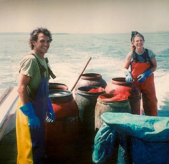
My parents have been the biggest influence on my life. In particular, my father. In high school one year for homecoming week, the student body was asked to dress up as our hero of the day. I chose to dress up like my dad. He is my hero. He served as a medic in the Vietnam War leaving behind my mother and my oldest brother to serve our country. When he returned, they lost the farm they had started before the war. They then moved and started building cabins with his brother in a small town in Idaho close to Yellowstone National Park where we all have called home since. In a time where bitterness could have easily consumed them both, they chose happiness and to stay close to our faith while rising my siblings and me. When someone needed help from a leaking roof or frozen pipes in their cabin, my dad would get a call and off we would go to help someone in need. He is a very selfless and compassionate gentleman. He has passed this trait on to all of his children. When you are truly serving others, you find true happiness that warms your heart. This lesson has carried me throughout my life. He always said growing up, “spare the rod, spoil the child,” then quickly followed it up with “that is a fishing rod of course!” We spent many hours in a boat fishing and talking about everything!
Clock Off THE
JENNIFER COPPERWHEAT, CARDIOVASCULAR SONOGRAPHER, OSWEGO HEALTH
BY MATT SKOUFALOS
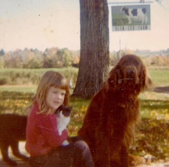
Growing up in Remsen, New York, Jennifer Copperwheat remembers a childhood of seemingly limitless discovery on her family farm. The property was alive with domesticated animals and wildlife, and Jennifer was invested in all of it.
She had an Irish setter, a bunch of cats, a rabbit; even her own cow. The family property extended to a little slate rock waterfall that she and her sisters called “the pretty land,” and the little menagerie would trail behind her to see it, too.
When the veterinarian visited to attend the herd, she would tag along and ask questions. She learned how to deliver calves; how to give shots to the cats with distemper that were left at the end of her quarter-mile driveway, the last stop on her elementary school bus route.
“I just had such a love for animals,” Jennifer said. “I told my father that I hate that when the cow doesn’t give milk you sell it and it gets slaughtered. We had chickens that we’d have to kill for dinner … needless to say, I don’t eat meat anymore.”
“I always told my father, ‘One day I’m going to have a farm, and I’m going to save animals.’” she said.
Jennifer worked as a commercial designer in New England until the early 2000s, when the family moved to South Carolina. She followed her curiosity into a healthcare career with Spartanburg, S.C., (Mary Black Health System at that time), and volunteered to travel to Greenville Memorial Hospital for additional training in advanced ultrasound techniques. Jennifer marveled at the way the
imaging modality unlocked her understanding of the body.
“I got really very interested in going through all the different areas of pediatric ultrasound – brains, lungs –things they didn’t do back then,” she said. “I was floored at how many things you could see, and how you could see it live.”
Life in South Carolina was good, until one day, Jennifer got a call from her father that he was feeling poorly. She ran down his symptoms with him over the phone, and advised him to go in for testing. When physicians diagnosed her father with pancreatic cancer, Jennifer took a leave of absence from work, and drove to New York to take over his chores on the family farm. Suddenly, she was once again milking the cows twice a day and stacking hay in the barn while tending to her dad.
“My husband Marcus had to take on our three kids that summer,” Jennifer said. “They came up and learned a little work ethic – but they didn’t have to chase the headless chicken around the peony bush for dinner.”
A surgeon in Albany extended her father’s life by another seven years via a Whipple procedure. That time was spent with the family, who moved up from South Carolina to be together with him. Jennifer started working at Oswego Health in 2017, and has been a cardiovascular sonographer with the institution ever since. Her family purchased a 14-acre former flower farm nearby, and when her father did pass, she used some of the money he’d left her to install a barn on the property.
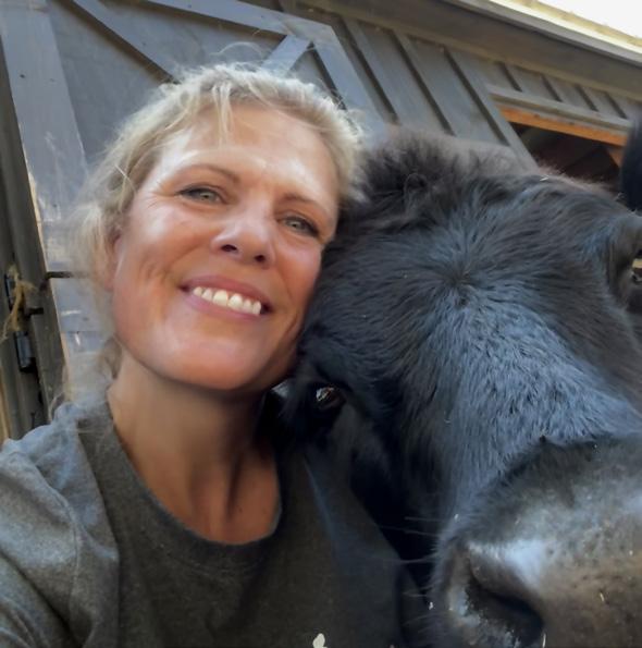
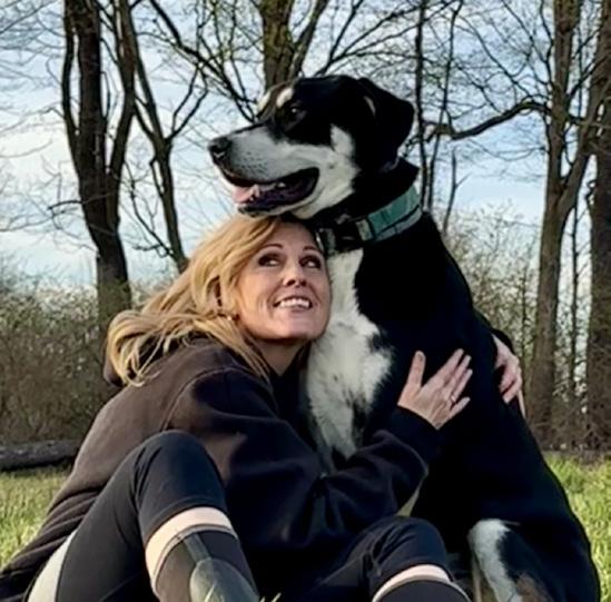
That barn helped Jennifer make good on her childhood oath: today, it is a haven for rescue animals that otherwise wouldn’t be candidates for rehabilitation. There’s Duncan, the Scottish Highland cow, Freya, another cow, and Coo-
per, the rescue dog who serves as their companion. Two pigs, Sheamus and Ferguson, were saved from a feeder lot. Jennifer also cares for two horses, including one with dwarfism, a couple of retired goats who no longer gives milk or bears kids, and two other goats, Remington and Benjamin, who survived a barn fire near Binghamton, New York.
“We have a good time out here,” Jennifer said. “We have a lot of people with kids who could use a place to go pet a goat.”
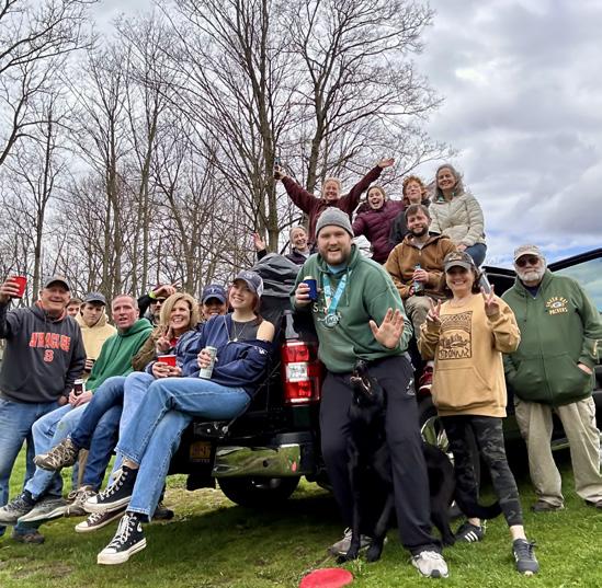
The farm serves mostly as open space for the animals to wander in and out of the barn, and graze the fields. Jennifer gardens there, and whatever she plants seems to take root because “the soil here is magical.” However, as much as the property supports her agricultural curiosity and affords livestock an opportunity to age in place, it also facilitates community gatherings that Jennifer enjoys hosting every April.
“We have an Earth Day party every year, play goofy games, host a lot of bands and serve food,” she said. “We don’t charge money for anything we do. We just want to bring people together because I think it’s important for people to gather and have fun.”
“I love having family around me, and being able to come together,” she said. “I love having gatherings here in the farm; anything to have everybody come over. It’s so much fun to embrace life and have friends and everybody around you.”
The farm hosted her son’s wedding, for which Jennifer planted 240 feet of sunflowers and built an arbor using woodworking skills learned in childhood from her old bus driver. Today, that arbor marks the entrance to her garden.
Jennifer’s newest ambition, aside from travel, is to perfect her sourdough bread recipe, for which she’s already begun grinding her own wheatberries to make flour.
“If I’m interested in something, I immerse myself,” Jennifer said. “That’s the best way to get a taste of life in every area you’re interested in. I get bored easily, so I just keep going and learning other things.”
Being returned in her later years to the life in which she grew up has helped underscore not just Jennifer’s love of animals and farming, but the variety of experiences that come with building community.
“I never say no to an adventure,” she said. “We get this one life. Are you really going to spend it wondering just what it would have been?” •
CORPORATE PROFILE
MAULL
BIOMEDICAL TRAINING
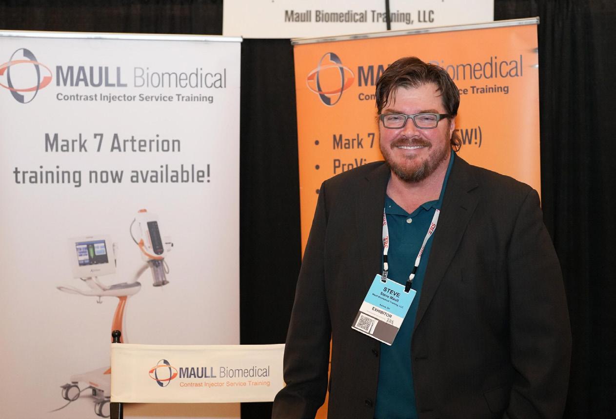
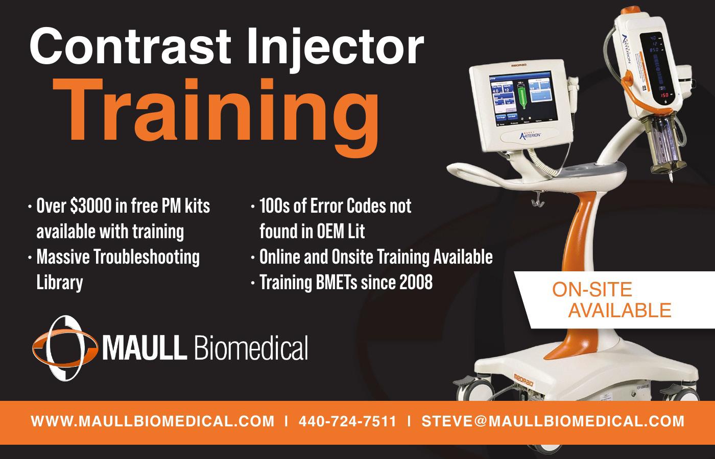
The evolution of contrast injector training for healthcare technology management (HTM) and clinical engineering professionals mirrors the growth of Maull Biomedical Training LLC.
“In 2008, I was doing some industry research into what clinical engineering training was in demand but not being met,” said Stephen Maull, founder and owner of Maull Biomedical Training LLC. “That’s when I came to realize that the contrast injector industry was completely unrepresented in the training sector. I thought it was odd because I had worked on contrast injectors in the military and they didn’t seem that difficult.”
Maull served in the United States Air Force as a BMET and BMET Instructor for 11 years. He realized his expert military training could benefit others and have a positive impact on patient outcomes throughout the nation.
“Nobody, and I mean nobody, was providing training on these devices,” Maull said.
Maull thought it over at length before jumping in with both feet.
“After a couple of months of thinking and wondering and fretting about it I finally thought, ‘Screw it, I’m doing it.’ So, in the summer of 2008 I started Maull Biomedical Training and had a stomachache for a year,” he added.“Eighteen years later, we’re still going strong and growing.
Looking back, Maull recalls that he was not the only person who saw a demand for training on contrast injector service.
“I didn’t run into any challenges as far as convincing the industry this training was needed. It was actually a large organization that came to me asking if I could find or develop training for them for certain modalities and contrast injectors was one of them,” Maull said.
The more industry research he did, the more apparent it became that contrast injector service training was something the HTM field needed.
A reason for the company’s success is that it is on the same page as healthcare providers when it comes to cutting costs
and providing cost savings. Maull Biomedical Training also provides a unique service.
“The OEMs offer training on some of their injectors, but they are much more expensive than we are. Plus, if you go to the OEM for training, you only get trained on their equipment whereas our training encompasses the injector models of multiple manufacturers.”
Maull Biomedical Training provides very in-depth training on various product lines from subject matter experts.
“We train BMETs on the principles of contrast injection (what it is, what it’s used for, and so forth) and then we teach them how to operate, PM, calibrate and repair the contrast injectors,” Maull said. “The students get on actual working contrast injectors where they learn to operate it, then perform a full PM using our custom MIST training software. We also go over the most common malfunctions with each model and the most likely fix. We currently have 13 different models of injectors that we provide training on.”
Maull has another excellent idea when he decided the company would provide in-depth, high-level training online. “We have taken our training to a place no other comprehensive BMET training has ever gone; completely online and computer based. If you go to our website and register (MaullBiomedical.com) you can see the ‘Introduction to Contrast Injectors’ and operational training videos for all the models we provide training on,” he explained. “The first step is to watch the ‘Introduction to Contrast Injectors’ videos and take the in-video quizzes. Once they pass all the quizzes, they receive a training certificate stating they have successfully passed the Introduction to Contrast Injectors Training Course. Then, they can take the Operational Training Courses (online videos) for any particular model they wish. All of this training is free to anyone who registers on our website. Then, they can purchase the PM training software for each model they wish.”
“The PM software is best described as a comprehensive computerized PM check sheet with how-to videos for each PM step. We actually have a free demo version online that anyone can
download,” Maull said. “After they have finished performing their first successful PM on that particular model they email Maull Biomedical a copy of the PM report that gets generated. Then, they get another training certificate stating they have been successfully trained on the PM and calibration/verification procedures for that particular model.”
Maull’s genius idea makes training convenient for everybody.
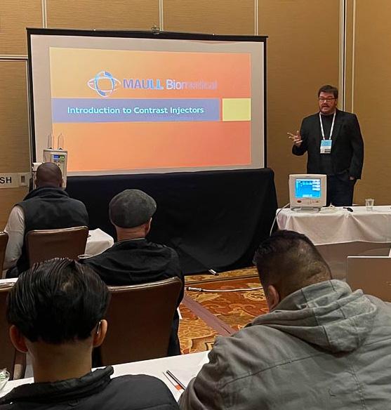
“What this allows is for you to get your contrast injector training when you need it and where you need it. No more travel and being away from your hospital and home for days,” Maull said. “The BMET can download this software and learn at their own pace anytime, anywhere. This is truly the best computer-based modality and model-specific BMET training in the world. If a BMET still wants a classroom setting for the training, we can still arrange that as well.”
The online training was especially helpful during the COVID-19 pandemic when travel to training sites was very limited. Next up is another high-tech way to provide expert training to HTM and clinical engineering professionals around the world.
Maull’s decision to provide training in 2008 has proven to be the right call for him and HTM professionals. The results speak for themselves.
As healthcare technology continues to evolve, the expectations placed on HTM and clinical engineering professionals are also changing. Hospitals and imaging centers no longer seek only reactive service support; they increasingly seek highly trained in-house teams capable of maintaining complex imaging support systems efficiently, safely, and cost-effectively. That shift has created a greater appreciation for specialized education and practical skills training that can be applied immediately in the field.
“The BMET can download this software and learn at their own pace anytime, anywhere. This is truly the best computer-based modality and modelspecific BMET training in the world.”
“Online/remote training is what actually what kept us afloat during the pandemic in 2020. The next step that coincides with our remote training option is to make it virtual reality (VR) training; this will take the remote training option to the next level,” Maull said. “Right now, the remote students watch videos on the proper operation of the injector then they download our maintenance software that takes them through the maintenance procedures and helps with troubleshooting. VR will replace the online quizzes for the operation training and aid in the maintenance training. Instead of the student watching videos and taking quizzes on how to properly operate a contrast injector, they will have a virtual injector in front of them that will allow them to virtually operate a contrast injector just like you would a real one –instead of just watching videos.”
For many healthcare organizations, reducing equipment downtime is a top priority. Contrast injectors play a critical role in diagnostic imaging procedures, and even minor operational issues can disrupt patient schedules, delay diagnoses, and increase operational costs. By equipping BMETs and clinical engineering teams with specialized training, healthcare providers can respond more quickly to service needs while reducing reliance on outside service providers for routine maintenance and troubleshooting.
Maull Biomedical Training’s approach reflects a broader transformation taking place across technical education. Instead of limiting learning to occasional classroom sessions, the company has embraced a continuous-learning model that allows technicians to build confidence over time through repeated exposure, hands-on exercises and model-specific instruction. This type of ongoing education is especially important in imaging environments where equipment configurations and software capabilities continue to advance. •
For additional information about Maull Biomedical Training, visit MaullBiomedical.com.
ICE Debut
REVIVE IMAGING SOLUTIONS
Revive Imaging Solutions is an independent imaging service organization focused on delivering OEM-level expertise with greater responsiveness and cost efficiency. It specializes in GE and Siemens MRI, CT, and PET/CT systems, offering a full range of services including preventative maintenance, emergency repairs, system installations and deinstallations, high-quality parts sourcing, and comprehensive magnet and cryogen support.
“One area we’re especially excited about is our recent investment in equipment for performing passive shimming on newer MRI systems,” President and CEO Jake Donald said. “Historically, this has been a capability limited to OEMs and a small number of third-party providers. Revive Imaging is proud to now offer this advanced service, expanding access to a critical aspect of MRI performance optimization.”
ICE Magazine recently found out more about Revive Imaging Solutions and all that it has to offer the diagnostic imaging community.
Q: HOW DOES YOUR COMPANY STAND OUT IN THE IMAGING SPACE?
DONALD: Three things set us apart.
First is our depth of experience and accountability. With over 25 years of hands-on experience – including military training, a long tenure with GE Healthcare, and years in national technical support – we bring a level of technical precision and discipline that clients immediately recognize. Just as important, we stay personally engaged. When a facility calls Revive, they’re not entering a ticketing system – they’re working directly with an expert who understands the urgency of keeping imaging systems online.
Second is our diagnostic-first approach. We don’t
believe in “throwing parts” at problems. We focus on identifying root causes and resolving issues correctly the first time, minimizing downtime and protecting revenue for our clients.
Third is our ability to provide full MRI magnet and cryogen services – capabilities that very few independent service organizations can offer at this level.
Q: WHAT IS ON THE HORIZON FOR YOUR COMPANY?
DONALD: Our primary focus is expanding direct partnerships with healthcare providers across Texas. While we’ve had strong success supporting other service organizations, our goal is to build long-term relationships directly with the facilities that rely on these systems every day.
We’re also investing in brand visibility, digital presence and local market engagement to ensure that when imaging leaders are looking for reliable service, Revive Imaging is top of mind – a partner who understands what’s truly at stake.
Q: IS THERE ANYTHING ELSE YOU WOULD LIKE ICE MAGAZINE READERS TO KNOW?
DONALD: At the end of the day, imaging equipment is the heartbeat of modern medicine – and downtime is not just an inconvenience, it’s a direct impact on patient care and facility performance.
At Revive Imaging Solutions, we take that responsibility seriously. Our mission –“Bringing your imaging systems back to life” – isn’t just a tagline. It reflects our commitment to restoring performance, extending system life and giving our clients confidence that their equipment is in capable hands.
We’re not trying to be the biggest – we’re focused on being the most reliable, the most responsive and the most trusted partner in the markets we serve. •
For more information, visit reviveimagingsolutions.com
Simplifying The Imaging Equipment Ownership Experience
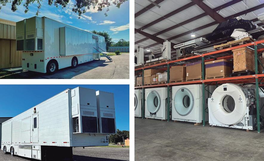
Image One Medical is the only Engineer-Owned medical equipment service group that is fully dedicated to Florida’s amazing base of Imaging Centers, Hospitals, and Cancer Treatment Centers.
We have a mission: Self perform on every aspect of our business. Specialize on specific modalities: Pet CT, CT & MRI, and Focus in a key geographic region.
Fort Myers I Fort Lauderdale I Tampa I Orlando
FLORIDA BASED
Dealer and servicer of PET CT, CT and nuclear medicine
• Equipment service: full coverage plans
• Equipment sales: installation, relocation and project management
• Mobile coach construction, refurbishment, maintenance and management
GUIDE TO AHRA
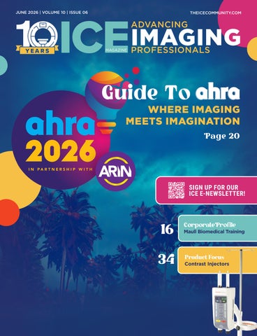
GUIDE TO THE AHRA ANNUAL MEETING WHERE IMAGING MEETS IMAGINATION
The annual meeting of AHRA: The Association for Medical Imaging Management has long been a cornerstone event for imaging leaders seeking education, professional development and networking opportunities. In 2026, the meeting takes on added significance with a new partnership designed to broaden perspectives across the imaging care continuum.
Set for July 12-15, 2026, at the Orlando World Center Marriott in Orlando, Florida, the AHRA Annual Meeting will bring together imaging administrators, radiology leaders, clinicians, educators and industry partners for four days of learning, collaboration and innovation. This year the theme is “Where Imaging Meets Imagination.” The theme underscores the event’s focus on forward-thinking strategies and practical solutions that support high-quality patient care.
One highlight of the 2026 meeting is AHRA’s collaboration with the Association for Radiologic & Imaging Nursing (ARIN), marking a significant step toward interdisciplinary engagement within medical imaging. The partnership introduces new programming and expanded opportunities for collaboration between imaging leaders and imaging nurses, reinforcing the shared goal of delivering safe, efficient and patient-centered care.
Michelle Dossa, director, department of imaging services at University Hospitals Elyria Medical Center, is a longtime attendee who applauds the annual event for many reasons.
“The AHRA Annual Meeting brings together imaging professionals from a wide variety of backgrounds and healthcare organizations (administrators, managers, clinical leaders, imaging nurses, and vendor partners) to focus on leadership development, operational excellence, patient care, and the future of medical imaging,” Dossa explains.
“Attendees have a unique opportunity to learn from industry thought leaders, earn continuing education credits in a variety of imaging topics and innovative ideas, and build relationships with industry peers to support your
clinical and operational success. It is also a great opportunity to learn more about AHRA, what it does for us and our imaging industry, and how you can become involved in its great work.”
Dossa listed her top three reasons to attend the AHRA Annual Meeting as follows:
• High‑value education and leadership development: The Annual Meeting offers high-quality, practical education addressing real-world challenges facing imaging leaders, including workforce development, quality, compliance, technology integration and patient-centered care. The sessions provide actionable strategies that can be immediately applied within imaging departments.
• Relationship building with peers, clinical leaders and vendor partners: One of the greatest strengths of the AHRA Annual Meeting is the opportunity to build meaningful relationships across radiology. Attendees can collaborate with fellow imaging leaders and clinical partners while also engaging directly with vendor partners. AHRA activities outside of the education sessions and exhibit hall are also a great way to get to know individuals who are committed to our industry as much as you are. These connections build trust, shared learning and long-term friendships that support your operational and clinical success.
• Exposure to innovation and future trends in imaging: Through its educational sessions and the exhibit hall vendors, attendees gain firsthand exposure to new technologies, workflow solutions and strategic insights. The exhibit hall is one of the best parts of AHRA and spending time there has resulted in several outstanding vendor relationships.
Traci Foster, FAHRA, CRA, MSRS, RT(R), is the director of outpatient imaging with Memorial Hermann Healthcare System’s Ambulatory Services Outpatient Imaging Division. She encourages everyone within diagnostic imaging to attend the AHRA Annual Meeting.
“AHRA and the annual conference is for anyone, and everyone interested in the field of medical imaging! Leaders, technologists, educators, contractors, vendors; all have a place at AHRA and can benefit from the annual conference,” Foster says. “I am excited that this will be the first year that the Association for Radiologic & Imaging
Nursing (ARIN) is joining us at the conference! AHRA’s mission is to educate, develop, mentor and provide resources to medical imaging professionals and the annual meeting is an incredible way to be exposed to and absorb all AHRA has to offer. At the end, you will walk away feeling more enlightened, inspired and proud of who you are and what you do.”
John Beall, FAHRA, MAA-HA, is a regional director with United Imaging who agrees regarding the value of attendance.
“Any medical imaging professional – regardless of role, tenure or experience level – who wants to grow should attend. Whether you’re just starting out or leading a large department, there’s something here for you,” Beall said. “The biggest benefit, in my experience, is the network you build. These aren’t just contacts – they’re trusted peers you can lean on throughout your career. Beyond that, you gain fresh perspectives, practical tools and renewed motivation to improve not only your own work, but how your entire team delivers care.”
“I’ve attended almost every annual since 2015, and I’ve never left a meeting without bringing something valuable back with me,” Beall added.
PARTNERSHIP EXPANDS THE LEARNING
The 2026 AHRA Annual Meeting represents the first formal partnership between AHRA and ARIN at the annual conference level. The collaboration reflects the evolving nature of imaging services, where teamwork among clinical, operational and nursing professionals plays an increasingly important role in patient outcomes.
AHRA Executive Director Jason Newmark, CRA, FAHRA, emphasized the value of bringing these professional communities together in a single environment.
“This partnership represents an important opportunity to foster deeper collaboration across the imaging care continuum,” Newmark said in an announcement about the event. “By bringing AHRA and ARIN members together in one dynamic environment, we are creating space for shared problem-solving, broader dialogue and innovative thinking that will benefit the entire imaging community.”
Through this partnership, attendees can expect integrated programming designed to reflect the realities of modern imaging departments, where clinical workflow, patient safety and operational efficiency intersect daily. The collaboration also highlights the growing recognition that imaging excellence depends on coordinated teamwork among multiple disciplines.
WHAT TO EXPECT AT AHRA 2026
The AHRA Annual Meeting is widely recognized for delivering practical education tailored to imaging leaders at every stage of their careers. The 2026 event builds on that reputation while introducing new perspectives through joint programming with ARIN.
Attendees will have access to a comprehensive educational program that includes:
• General sessions featuring keynote speakers and industry thought leaders
• Breakout sessions covering leadership, operations and emerging technologies
• Workshops focused on professional development and clinical best practices
• Networking opportunities with peers and industry partners
• Vendor exhibits showcasing new imaging solutions and services
The meeting will also feature a gathering space designed to encourage collaboration and conversation among attendees, vendors and professional organizations. These informal interactions often lead to valuable insights and lasting professional relationships. ICE Magazine will be welcoming attendees at Booth #736.
For imaging leaders navigating workforce challenges, regulatory changes and rapid technological advancements, the conference provides an opportunity to step back from daily responsibilities and focus on strategic planning and professional growth.
DEDICATED ARIN PROGRAMMING
An added component of the 2026 meeting is the integration of ARIN programming throughout the event. These sessions are designed to provide practical insights from imaging nurses while fostering collaboration with imaging administrators and technical staff.
Key additions include:
• Dedicated ARIN sessions scheduled throughout the conference
• Two preconference workshops led by imaging nursing professionals
• A signature ARIN workshop highlighting clinical best practices
• A focused two-hour session on opening day designed for all attendees
These sessions are expected to address topics such as patient safety, contrast administration, workflow coordination and communication among care teams – areas where nursing expertise plays a critical role.
By incorporating these perspectives into the broader educational program, the meeting aims to strengthen collaboration between clinical and administrative leaders while supporting more coordinated patient care.
GUIDE TO AHRA
EDUCATION
The healthcare environment continues to evolve rapidly, and imaging departments face increasing pressure to improve efficiency, manage costs and maintain high standards of patient care. The AHRA Annual Meeting addresses these challenges through education that combines strategic leadership concepts with practical operational guidance.
Common subject areas typically covered at the conference include:
• Imaging department management and leadership
• Quality improvement and patient safety
• Workforce development and retention
• Regulatory compliance and accreditation
• Financial performance and budgeting
• Technology adoption and innovation
Sessions are structured to provide actionable insights that attendees can apply immediately within their organizations. Whether participants oversee large health system imaging operations or smaller community-based departments, the program is designed to deliver relevant and practical knowledge.
NETWORKING & PROFESSIONAL CONNECTIONS
Networking remains one of the most valuable aspects of the AHRA Annual Meeting. The event draws imaging professionals from across the United States, creating opportunities to share experiences, discuss challenges and explore new ideas.
Networking opportunities will include:
• Welcome receptions and social events
• Informal networking sessions
• Vendor-hosted demonstrations
• Peer-to-peer discussions
• Collaborative workshops
A joint celebration event is also planned to recognize the contributions of imaging professionals and highlight the collaborative spirit that defines the field. These gatherings help strengthen professional relationships and foster a sense of community within the imaging profession.
For many attendees, connections formed during the conference become long-term professional resources that support career growth and organizational success.
CONVENIENT DESTINATION
The Orlando World Center Marriott will serve as the host venue for the 2026 meeting, offering extensive conference facilities and convenient access to dining, entertainment
and transportation options.
Orlando remains a popular destination for professional conferences because of its accessibility, family-friendly attractions and wide range of accommodations. The location also supports strong attendance, making it easier for imaging professionals from across the country to participate.
For attendees traveling with family members, the destination provides opportunities to combine professional development with personal time, a factor that often enhances the overall conference experience.
STORIED HISTORY & BRIGHT BEGINNINGS
Since its founding in 1973, AHRA has focused on supporting imaging professionals through education, leadership development and community engagement. The organization continues to play a central role in shaping best practices within medical imaging management.
The 2026 partnership with ARIN reflects a broader shift toward interdisciplinary collaboration within healthcare. As imaging services become more complex, success increasingly depends on coordinated teamwork among clinical, operational and administrative professionals.
By bringing these groups together under one roof, the AHRA Annual Meeting aims to inspire new ideas, strengthen professional relationships and support the continued advancement of imaging services nationwide.
WHY AHRA 2026 MATTERS
For imaging leaders navigating workforce shortages, evolving technologies and rising patient expectations, staying informed and connected is more important than ever. The AHRA Annual Meeting provides a structured environment for learning, collaboration and professional growth.
AHRA 2026 is more than a conference. It is a gathering of professionals committed to improving patient care, advancing imaging services and shaping the future of healthcare.
With its expanded programming, collaborative focus and forward-looking theme, the 2026 AHRA Annual Meeting promises to deliver an engaging and impactful experience for imaging leaders across the country. •
July 12-15, 2026
Orlando, FL
Don’t Just Keep Up. Lead Forward.
• Sharpen your leadership.
• Expand your impact.
• Lay the ground work for what’s next for your imaging department.
From strategy to operations to innovation, gain the tools to lead imaging into the future.
EXPERT OFFERS PLAYBOOK FOR WORKING WITH TECHNICAL SUPPORT
System downtime remains one of the most persistent challenges facing imaging and clinical engineering teams, affecting everything from patient throughput to departmental revenue.
During the recent ICE Magazine webinar, “Reducing Downtime: John Walker’s Guide to Using Technical Support Effectively,” sponsored by AllParts Medical, attendees gained practical, field-tested strategies for minimizing disruptions and accelerating system recovery through better collaboration with technical support teams.
The session focused on a core reality familiar to field service engineers and imaging professionals alike: downtime is rarely caused by a single issue, but it can often be shortened through better preparation, communication and decision-making. Walker emphasized that technical support should not be viewed as a last resort, but rather as a strategic partner in the troubleshooting process.
Walker outlined a structured approach to engaging technical support more effectively, beginning with recognizing the right moment to escalate an issue. Waiting too long to involve support can prolong outages, while calling too early without sufficient information can slow resolution. The key, he explained, is developing situational awareness like understanding when troubleshooting efforts have reached a point where additional expertise is needed.
A central theme of the presentation was preparation. Walker encouraged engineers to gather critical system details before contacting support, including error codes, recent maintenance history, system configuration information and a clear timeline of events leading up to the failure. This level of readiness allows support teams to move directly into problem-solving mode rather than spending valuable time collecting basic information.
Equally important is communication. Walker stressed the value of clearly describing symptoms, documenting troubleshooting steps already performed and avoiding assumptions about root causes. Structured communication, he noted, helps technical support teams quickly narrow down possibilities and recommend targeted solutions.
The webinar delivered actionable tools that attendees can immediately apply in their daily work. Walker shared checklists and documentation practices designed to standardize the troubleshooting process and improve consistency across teams.
Among the recommended best practices:
• Maintain a running log of system performance and maintenance activities
• Capture screenshots or photos of error messages when possible
• Document environmental conditions that may affect system performance
• Establish clear escalation thresholds for contacting technical support
• Conduct post-incident reviews to identify lessons learned These practices help resolve current issues more quickly and build institutional knowledge that can prevent future downtime.
Walker also highlighted the importance of relationship-building between field engineers and technical support personnel. Familiarity with support teams and understanding their processes can streamline communication and create a more collaborative problem-solving environment.
Attendees were asked, “’How would you describe ICE webinars to a colleague?”
“Very informative,” said Edwin Alvarez, an assistant director of radiology at Montefiore Hospital.
“Informative and broadening of perspective,” added Christopher Wheeler, a CT technical support specialist at AllParts Medical.
“Great source of education,” noted Sondra Waltmon, outpatient imaging supervisor and PACS administrator at Baptist Health Paducah.
Attendees were also asked, “Why do you read ICE magazine?”
“The fresh perspective presented within the ICE magazine regarding healthcare continues to keep my passion alive,” said Gail Renata Marlowe, a medical imaging manager at Woman’s Wellness Center in Pennsylvania.
The session is part of ICE Magazine’s ongoing webinar series, which continues to expand its reach within the imaging and healthcare technology management community.
Sponsored webinars remain a key component of ICE Magazine’s educational mission, providing accessible continuing education opportunities and connecting industry experts with frontline professionals. •
For information about upcoming webinars, visit ICEwebinars.live.
Imaging News
A LOOK AT WHAT’S CHANGING IN THE IMAGING INDUSTRY
COVERA HEALTH & MEDMO MERGE
Covera Health and Medmo have announced their combination into a single organization, creating a platform that manages diagnostic imaging for nearly 6 million Americans across Fortune 10 and 100 employers, threeof-the-five largest national health plans, and thousands of value-based primary care physicians.
Covera’s lead investor, global software investor Insight Partners, is committing additional capital to fund the combination and accelerate expansion.
Walmart and Blue Cross Blue Shield of Michigan are among the employer and health plan partners that have built programs on Covera’s platform. The addition of Medmo’s referring physician relationships, including some of the largest value-based primary care organizations across the country, extends the combined company’s reach to thousands of ordering physicians across all 50 states.
“Where a patient gets their imaging done is not a neutral decision. The same study, ordered for the same reason, can produce a meaningfully different diagnosis depending on where it is performed and who reads it,” said Ron Vianu, CEO of Covera Health. “That variation was always there. Covera built the infrastructure to measure it for the first time and proved that routing patients to the right facility produces meaningfully better outcomes. What we could not do alone was ensure patients reliably reached
the right place to begin with, and that the result connected back to the physician and care team who needed to act on it. Medmo solved that. It is the first platform in radiology that closes the loop from the moment a referral is written to the moment a patient is on the right care path.”
“Providers often lose visibility the moment a patient walks out of the office with an imaging order,” said Lucas Takahashi, CEO of Medmo. “Medmo closes that gap, ensuring patients reach the right facility, complete their study, and that results get back to the physician who needs to act on them. Joining with Covera means that every step of that journey now carries clinical intelligence. Together, we’ve built something new for radiology: a platform that delivers quality and coordination as one.”
“Our investment in Covera was grounded in the belief that diagnostic quality is the missing foundation of value-based care,” said Jeff Horing, co-founder and managing director at Insight Partners. “This combination with Medmo closes the critical gap in radiology, from the moment a study is ordered to the moment it drives the right care decision. That’s not an incremental improvement. That’s new infrastructure for the entire system. With clear momentum and a capable team in place, we have strong conviction in this next phase of growth.”
REMBRA PLATFORM EARNS 510(K) CLEARANCE
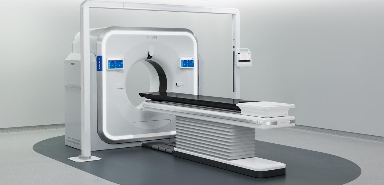
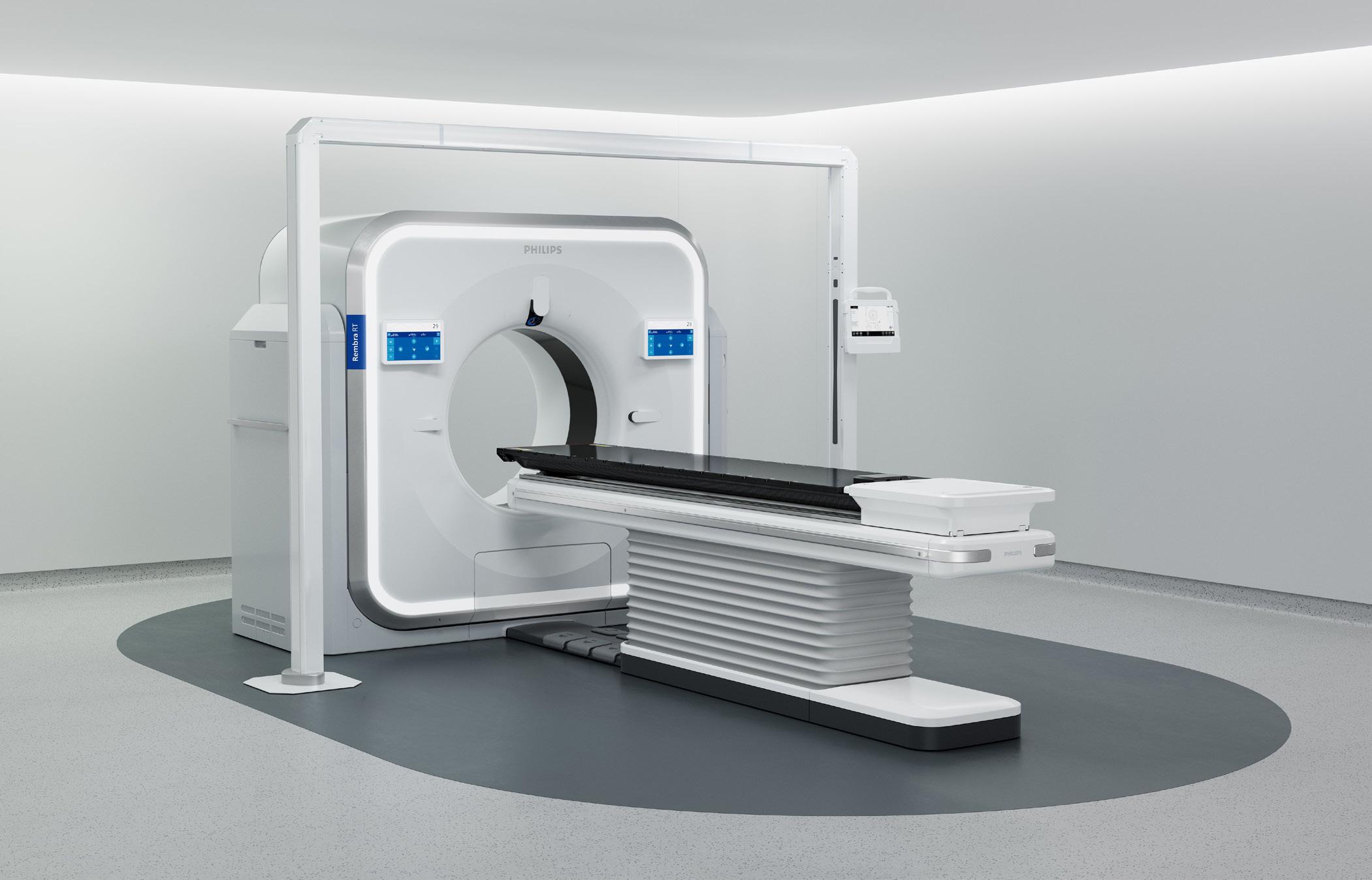
Royal Philips has received 510(k) clearance from the U.S. Food and Drug Administration (FDA) for its Rembra platform of scanning systems, including Rembra CT, Rembra RT and Areta RT.
The newly cleared systems are designed to help healthcare providers respond to growing demand for imaging and radiation therapy planning by helping to improve speed, efficiency and access to high-quality imaging. As imaging volumes and clinical complexity continue to rise, healthcare providers need solutions that can help them work more efficiently while maintaining confidence in clinical decisions.
The Rembra platform of systems also extends the role of CT across diagnostic radiology and radiation therapy, helping to enable more connected workflows and a coordinated approach to patient care – from rapid assessment in acute settings to precise cancer treatment planning.
“As healthcare systems manage increasing demand and complexity, imaging plays a critical role in enabling timely and informed clinical decisions,” said Dan Xu, business leader of CT at Philips. “With the Rembra platform, we are redefining what clinicians can expect from CT, combining speed, scalability, and precision to expand access to high-quality imaging while supporting confident diagnosis and highly
accurate treatment planning.”
With the largest-in-class 85 cm bore, Rembra CT is purpose-built for the front lines of care, enabling ultrafast, high-throughput imaging in demanding clinical environments, supporting up to 270 exams per day in various settings including emergency departments, critical care and interventional settings. Its advanced acquisition and reconstruction capabilities support rapid, high-quality imaging at scale, helping clinicians make timely decisions in critical situations without compromising diagnostic confidence.
Rembra RT and Areta RT bring these performance advantages into radiation therapy, where precision is critical. These systems support high-fidelity imaging for treatment planning with a wide 85 cm extended field of view and next generation 4DCT imaging capabilities, supporting more accurate targeting of tumors while helping protect healthy tissue. By streamlining simulation and planning workflows, they help reduce variability and improve efficiency, supporting more precise, personalized cancer care.
The FDA clearances further strengthen Philips’ comprehensive CT portfolio and reflect its continued focus on integrating advanced imaging technologies with intelligent and connected workflows.
BOSTON IMAGING AND NEUROLOGICA UNIFY UNDER SAMSUNG HME
AMERICA
Samsung Medison recently announced that its U.S. medical imaging businesses, previously operating as Neurologica and Boston Imaging, will unify under a new corporate identity, Samsung HME (Healthcare and Medical Equipment) America. This transition brings the company’s U.S. ultrasound, digital radiography and computed tomography (CT) operations under one integrated structure that reflects the global Samsung brand behind its technology.
The unification underscores Samsung’s continued strategic investment in advancing medical imaging innovation and strengthening its presence in the United States healthcare market. By aligning its brand structure with its expanding portfolio and growing U.S. footprint, Samsung reinforces its long-term commitment to delivering cutting-edge imaging solutions, deep clinical collaboration and sustained operational excellence.
“This milestone represents more than a name change,” said Kyu Tae Yoo, CEO of Samsung Medison and Division Head of Samsung HME at Samsung Electronics. “It reinforces Samsung’s long-term commitment to healthcare providers nationwide and reflects the growth and maturity of our U.S. medical imaging organization. Our growth has been powered by listening closely to our customers and translating real clinical challenges into innovation at a pace rarely seen in healthcare; and we will continue along this path.”
The announcement follows a period of significant growth across Samsung’s U.S. medical imaging portfolio, marked by expanded product innovation, organizational investment and deeper engagement with health systems nationwide. As demand for advanced imaging solutions continues to evolve, the company is scaling its U.S. presence to meet the needs of a rapidly transforming healthcare environment.
“This new chapter as Samsung HME America strengthens how we connect with customers and partners while giving us the flexibility to innovate and advance medical imaging for the future,” said Tracy Bury, chief commercial officer. “With a unified organization, we can deliver greater consistency, deeper collaboration, and a more streamlined experience across ultrasound, digital radiography and CT. This alignment positions us to move faster, support our partners more effectively and continue building long-term relationships grounded in trust and performance.”
Recent advancements within Samsung HME America include the Samsung R20 and Z20 ultrasound systems, designed to deliver enhanced image quality and AI-powered tools that support more confident clinical decision-making. The company has also continued to expand its AI capabilities through the Samsung acquisition of Sonio, a French AI-powered prenatal ultrasound startup specializing in earlystage fetal abnormality detection. In addition, Samsung is advancing and expanding its digital radiography portfolio with innovations focused on dose reduction and workflow enhancements.
Samsung HME America remains the global headquarters and manufacturing center for the CT business, advancing point-of-care platforms and pushing the boundaries of medical imaging with photon counting detector technology.
SECTRA COMPLETES OXIPIT ACQUISITION
International medical imaging IT and cybersecurity company Sectra has completed the acquisition of Oxipit, a pioneer in autonomous AI for radiology and the developer of the first solution with CE Class IIB certification for autonomous chest X-ray analysis.
“This milestone strengthens Sectra’s position as a provider of advanced, regulatory-approved AI solutions for diagnostic imaging,” according to a press release.
“With the acquisition of Oxipit, we’re adding a new dimension to our AI offering: autonomous AI that can genuinely reduce radiologists’ workload in daily clinical practice. Equally important, we’re acquiring the clinical validation and regulatory expertise required to deploy this category of AI responsibly at scale. The acquisition complements our vendor-neutral AI marketplace, which remains a cornerstone of our strategy, and our own rapid innovation such as MCP-powered system administration.
Together, these moves demonstrate clear leadership in AI for diagnostic imaging and position us to help healthcare providers manage rising imaging volumes and address staff burnout,” says Torbjörn Kronander, president and CEO of Sectra.
With the acquisition complete, Sectra now owns and will further develop ChestLink, Oxipit’s flagship product and one of the most clinically validated examples of autonomous AI in radiology, the release states.
Unlike conventional AI decision-support tools, which flag findings or assist prioritization but leave reporting to the radiologist, autonomous AI is developed and validated to independently complete a defined diagnostic task. ChestLink automatically identifies and clears high-confidence normal chest X-rays from the worklist, freeing radiologists to focus on cases with a higher probability of disease.
GE HEALTHCARE EXPANDS COLLABORATION WITH DEEPHEALTH
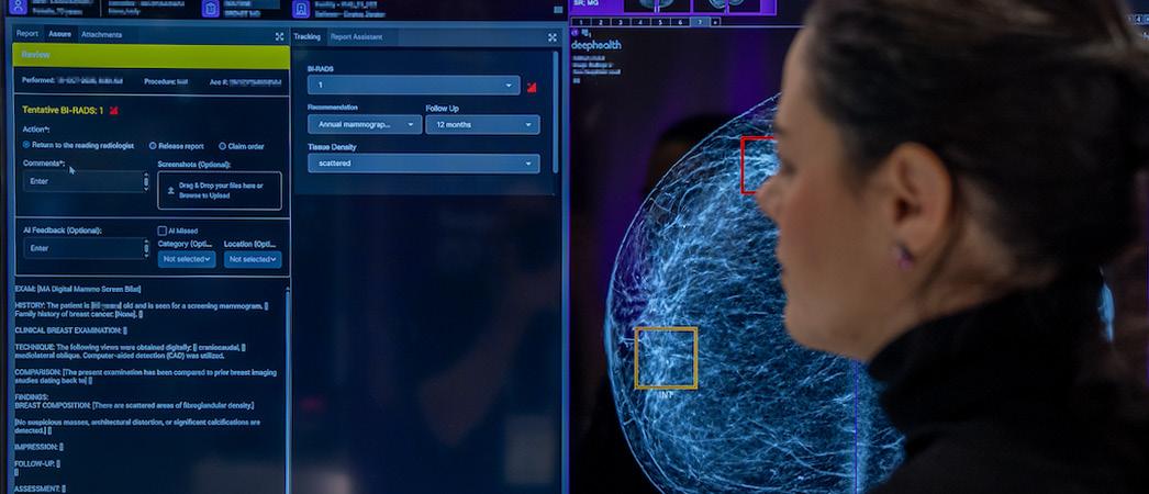
GE HealthCare has announced an expanded collaboration with DeepHealth Inc., a leader in AI-powered health informatics and a wholly owned subsidiary of RadNet Inc., to further the innovation, commercialization and adoption of advanced AI-powered mammography tools.
“The expanded collaboration builds on the joint initiative first announced in 2024, which united DeepHealth’s AI-powered breast cancer screening workflow solution with GE HealthCare’s Senographe Pristina mammography system to enhance image interpretation and improve operational efficiency. This next phase of the joint initiative will extend the collaboration’s relationship for breast cancer solutions internationally, expanding the offering with second reader workflow and extending access to components of DeepHealth’s breast cancer solutions,” a press release states.
With the continued growth in capabilities of DeepHealth’s new Breast Suite2 solution, GE HealthCare is proud to bring to market an advanced, cloud-based suite of modular, interoperable applications that integrate seamlessly into existing breast imaging workflows, it adds.
“Paired with GE HealthCare’s Pristina Via, the combined offering enables scalable, high-efficiency breast cancer screening programs,” it states.
“At GE HealthCare, we’re advancing women’s health through precision care built around the unique needs of women and enhanced by the power of AI,” said Jyoti Gupta, Ph.D., president and CEO, women’s health and X-ray at GE HealthCare. “By integrating Breast Suite AI with our Pristina Via mammography system, we’re helping clinicians detect breast cancer early with greater confidence. These innovations move us closer towards truly personalized prevention and care for women.”
In the largest real-world analysis of AI-powered breast cancer screening in the U.S., a multistage AI-driven workflow from DeepHealth’s Breast Suite has been shown to enable a 21% increase in breast cancer detection rate when compared
to the standard of care. Additionally, Breast Suite’s Timely Alerts feature uniquely integrates with Pristina Via to notify sites of potentially suspicious cases in minutes.
“Pristina Via and Breast Suite have transformed how we approach breast cancer detection,” said Dr. Meghna Krishnan, owner, URPrecious Imaging, Arizona. “The cloud-based Viewer gives us instant access to images from anywhere, and the AI tools, like CAD for lesion detection and automated breast density assessment, can help us deliver fast and consistent results. It’s not just about speed, it’s about confidence. With Breast Suite, our team can make more accurate decisions without workflow disruptions, which ultimately means better care for our patients.”
The existing Breast Suite solution offered by GE HealthCare includes a cloud-first multi-modality viewer, cancer detection, automated density assessment, prioritized worklist, timely alerts, and enhanced reporting. The expanded collaboration will enable GE HealthCare to distribute new Breast Suite applications designed for compatibility with GE HealthCare’s mammography systems, including:
• ProFound Pro, which combines Cancer Detection, a clinical AI solution that offers automatic lesion localization and degree of suspicion that are effective in diverse populations and dense breast tissue, and Automated Density Assessment for consistent, automated density classification with a patient-centric, accurate density assessment of 2D or 3D mammograms to support objective diagnostic decisions.
• Safeguard Review: Optional AI-powered workflow that flags complex cases that may benefit from a secondary review. A large U.S. study demonstrated a 21% increase in cancer detection rate and a 23% increase in cancer detection rate for women with dense breasts with a multistage AI-driven workflow when compared to the standard of care. •
DIRECTOR’S CIRCLE
Contrast Imaging INSIGHTS
Each month, ICE Magazine connects with imaging leaders to explore key trends shaping the industry. This month, experts weigh in on the evolving land‑ scape of contrast imaging, including supply chain challeng‑ es, patient safety and emerging technologies.
This month, the panelists are:
• Noelle Hobbie, executive director of imaging, Central Florida Division, AdventHealth;
• Les Ciancibello, RT(R)(CT), radiology supervisor, Seidman Cancer Center; and
• Jacqui Rose, system director of radiology, UC Health.
Q: WHAT OPERATIONAL OR CLINICAL FACTORS ARE CURRENTLY DRIVING THE GREATEST CHANGES IN CONTRAST IMAGING PRACTICES WITHIN YOUR DEPARTMENT OR HEALTH SYSTEM?
HOBBIE: Several factors are driving change, including the continued growth of cardiac imaging in both CT and MR, emergency department throughput demands, and evolving approaches to renal function screening. Supply chain reliability also plays a major role. We prioritize partnerships with suppliers that can confidently meet the demand of a large, growing organization while controlling cost through economies of scale and enterprise-wide agreements. In addition, we continue to streamline premedication pathways for patients with contrast allergies. Most recently, we are exploring options to electronically send premedication prescriptions to the patient’s pharmacy of choice rather than requiring the patient to pick them
up at an imaging center. We also maintain a strong focus on preventing and managing contrast extravasations.
CIANCIBELLO: I think the biggest change in contrast practice has been the reduction in dose as a result of the global contrast shortage. What we learned from this shortage was that we can produce high-quality diagnostic scans with lower volumes in several routine imaging protocols. I feel it is important to have contrast dosing guidelines; however, they need to be coupled with clinical and patient-based scenarios, allowing for deviation when necessary.
ROSE: Contrast utilization and practice changes are primarily driven by cost and supply availability. To ensure uninterrupted, high-quality patient care, the organization remains flexible and works closely with suppliers to maintain adequate inventory and manage supply chain challenges.
Q: HOW ARE YOU BALANCING PATIENT SAFETY WITH THE NEED FOR EFFICIENCY AND THROUGHPUT IN TODAY’S HIGH-VOLUME IMAGING ENVIRONMENTS?
HOBBIE: We navigate this balance daily – especially in the emergency setting, where CT utilization is highest. Following ACR guidance, we screen appropriately for contrast-induced nephropathy (CIN) risk while avoiding unnecessary renal function testing to support throughput. Where appropriate, we use point-of-care (POC) testing when needed. eGFR results are incorporated directly into the technologists’ EMR worklist for rapid review. In select
high-volume locations, we have dedicated resources focused on readiness for contrast CT (IV placement, CIN screening and POC testing). We also maintain POC testing competency across staff in outpatient settings so patients can complete required testing during the same visit, rather than obtaining lab work before their scheduled appointment.
CIANCIBELLO: Patient safety is always priority number one in my imaging suites. The development and follow-through of solid workflows naturally keep your department running efficiently, and by default, throughput is increased. As an example in my suite, we have a lead technologist who acts as an “air traffic controller” in the space. She has taken the time to assure that all orders are clear, that those outside normal protocols are discussed with a radiologist or attending physician, and that everything is in order for the patient the day they arrive. After that, it is as simple as guiding the patients onto the correct scanner, similar to the way an air traffic controller guides planes onto and off runways. With skilled technologists at the scanners ready for each patient, the scanner — or “runway” — is always clear and ready for the case.
ROSE: To balance patient safety with efficiency and throughput, we consistently review current practices and processes to ensure our efficiency is optimized. We develop future optimization plans via new products or services as those become available and with funding opportunities.
Q: WHAT ROLE DO NEW TECHNOLOGIES PLAY IN DIAGNOSTIC IMAGING?
HOBBIE: New technologies are supporting both safety and efficiency. We are actively evaluating enhancements to power injector technology, particularly safety features that help prevent never-events such as air emboli. We also value time-saving features like automatic priming that create incremental throughput gains. We are also leveraging automated post-processing and image reconstruction, and many of these applications support contrast-enhanced studies, including angiography cases like cardiac CTA. Additionally, we are using technology and decision support to reduce redundant contrast imaging. For example, providers caring for stroke patients may order both CTA head/ neck and MRA when typically only one is needed. We leverage EMR reporting to educate providers and encourage ordering the most appropriate single study.
CIANCIBELLO: I think this is a difficult question to answer. The radiology world is constantly evolving, almost to a point faster than we can absorb what is being offered. Dual-energy CT has been available for 15 or more years, quickly followed by spectral CT and now photon-counting CT. These technologies offer options, opportunities and challenges yet to be understood. Most of us only use these tools and what they have to offer in very limited or niche capacities. It takes time for technologists to adapt to these new technologies. As I often say regarding our new photon-counting scanner, “this isn’t your grandma’s CT
ADVENTHEALTH
NOELLE HOBBIE
SEIDMAN CANCER CENTER
LES CIANCIBELLO
UC HEALTH
JACQUI ROSE
of the old days.” With 0.25-second rotation time, infinite rows of detectors, multitudes of scan modes, reconstruction and filtration options, the combinations are endless. Added to that, the adoption of these technologies by the readers is slow. Like all fields, the radiologist pool is also short-staffed, and they are always being asked to read more data faster. Finding that balance between the two is going to be the challenge.
ROSE: New technologies drive our future planning, whether it’s related to safety, efficiency or throughput. These technologies are critical to enhancing our processes, which will ultimately improve overall care and patient experience.
Q: HOW ARE YOU ENSURING TECHNOLOGISTS AND CLINICIANS REMAIN COMPETENT AND CONFIDENT AMID STAFFING CHALLENGES?
HOBBIE: We rely on a well-structured onboarding plan and ongoing competency validation. Our annual skills fairs include training and competency checks for IV starts, infiltration/extravasation recognition and response, and proper power injector use. We ensure hands-on education is a part of all clinical competency checkoffs.
CIANCIBELLO: Our health system is and has been making a concerted effort to optimize and standardize protocols and technologies systemwide. With 34 scanners, consisting of four different vendors at multiple locations across our region, this takes time. Holding monthly or bimonthly meetings with CT supervisors and leads gives us an opportunity to share what’s new and coming, what’s changing and what’s not working. Keeping an open line of communication between the academic and community departments across our system has been extremely helpful in getting us to a place of consistent practice. Once you’re consistent in what you do, you can more easily share staff from location to location.
ROSE: Maintaining competencies is challenging in today’s environment when we have multiple teammates at many locations using a variety of technologies. Currently, most of our team members rotate between locations, so it is imperative that they remain competent on all equipment and processes to provide the safest possible environment for our patients and themselves. We achieve this by partnering with our vendors to provide top-quality education and training to each member of the team, as well as our clinicians who guide our protocols. This is refreshed and validated annually to ensure nothing falls through the cracks.
Q: WHAT TRENDS OR ADVANCEMENTS IN CONTRAST IMAGING DO YOU BELIEVE WILL HAVE THE MOST SIGNIFICANT IMPACT OVER THE NEXT THREE TO FIVE YEARS?
HOBBIE: One significant initiative will be expanding contrast-enhanced imaging into our mammography services. CEM is a cost-effective, supplemental screening option for women with high risk and/or dense breast tissue. This will provide an additional option for patients who are unable to complete a breast MRI. It will require thoughtful change management for women’s center teams who have not previously supported contrast workflows.
The increasing availability of advanced CT technology, such as dual-source and dual-energy scanners, will continue to improve vascular visualization, even when injection timing or quality is suboptimal. Using these capabilities to compensate for patient-centered challenges can help avoid repeat scans or re-injections. Finally, we expect continued emphasis on routine protocol review to identify opportunities to minimize contrast dose while preserving image quality and diagnostic performance.
CIANCIBELLO: I think for high-volume sites, bulk contrast delivery will continue to be the trend. I feel the focus on contrast reduction will continue to be important from both a cost and patient care perspective. I’d really like to see some advanced connectivity between injectors and RIS, as today this is still a manual process requiring many steps for both technologists and readers alike. Ideally, the injector should know what contrast agent was loaded, capture the lot number, volume and flow, and send this directly to the patient’s chart as well as the dictation system for documentation and billing purposes.
ROSE: New technologies will likely have the greatest impact over the next few years. We are focused heavily on patient safety and will be working closely with our vendor partners to enhance technological advancements that will ensure that patient safety is improved. One key area is improving our extravasation rates. While we are well within the ACR recommendations, our goals are to reduce rates overall and improve patient experience at all locations. •
PRODUCTS
Market Report
FIRMS FORECAST CONTRAST MEDIA MARKET GROWTH
STAFF REPORT
The global contrast media market size was estimated at $7.31 billion in 2025 and is projected to reach $13.86 billion by 2033, growing at a CAGR of 8.39% from 2026 to 2033, according to a report from Grand View Research, a market research and consulting company.
The demand for contrast media is rising owing to the growing need for medical imaging and the rising burden of chronic disorders, the report states.
“High volume of imaging procedures, such as ultrasound and MRI scans, is anticipated to support the market growth,” the report states. “The growing global demand for medical imaging is a key factor driving the expansion of the contrast media market. Medical imaging plays a crucial role in the early detection, accurate diagnosis, and effective treatment planning of various health conditions. Millions of patients undergo diagnostic procedures such as MRI, CT scans, and X-rays every year.”
According to a study published by the National Library of Medicine in October 2023, approximately 40 million MRI scans are performed annually in the U.S. alone. An expected increase in the volume of imaging procedures worldwide is expected to significantly boost the demand for contrast media, essential for enhancing the visualization of internal body structures during these examinations.
In addition, the report highlighted key market trends and insights, including:
• North America dominated the contrast media market with the largest revenue share of 38.92% in 2025.
• By modality, the X-ray/CT segment led the market with the largest revenue share in 2025.
• Based on product, the microbubble contrast media segment is anticipated to grow fastest over the forecast period.
• Based on end use, the hospitals segment led the market with the largest revenue share in 2025.
• By application, the neurological disorder segment led the market with the largest revenue share in 2025.
• Based on route of administration, the intravenous segment is anticipated to grow fastest over the forecast period.
Fortune Business Insights reports that the global contrast media injectors market size was valued at $2.33 billion in 2025. The market is projected to grow from $2.59 billion in
2026 to $5.97 billion by 2034, exhibiting a CAGR of 11.01% during the forecast period.
The report also stated that increased diagnostic imaging will fuel market growth.
“The contrast media injectors market is a vital segment within diagnostic imaging equipment, supporting accurate and controlled delivery of contrast agents during imaging procedures such as computed tomography, magnetic resonance imaging and angiography,” the report said. “These systems are designed to ensure precise flow rates, timing, and pressure control, which are essential for high-quality diagnostic outcomes. Contrast media injectors market analysis highlights increasing dependence on advanced imaging techniques for early disease detection, interventional procedures and chronic condition management. Hospitals and diagnostic centers rely on injector systems to improve workflow efficiency, patient safety, and image consistency. The market is characterized by continuous technological improvements focused on automation, dose accuracy and integration with imaging modalities, reinforcing the clinical importance of contrast media injectors across modern diagnostic environments.”
The report adds that the contrast media injectors market in the United States is driven by high imaging procedure volumes, advanced healthcare infrastructure and widespread adoption of sophisticated diagnostic technologies.
“Healthcare facilities in the United States prioritize injector systems that enhance patient safety, reduce contrast waste and support high-throughput imaging workflows,” it reads. “Contrast media injectors market insights indicate strong demand from hospitals, outpatient imaging centers and specialized diagnostic clinics. The U.S. market emphasizes technologically advanced injector platforms with user-friendly interfaces and safety monitoring features. Continuous upgrades of imaging departments and focus on operational efficiency support steady utilization of contrast media injectors across the United States healthcare system.”
The April 2026 report also states that one trend identified in the contrast media injectors market analysis is a growing preference for dual-head and syringeless injector technologies.
Dual-head injectors support complex imaging procedures by allowing saline flushing alongside contrast delivery, improving image clarity and reducing contrast usage. Syringeless injectors are gaining attention due to reduced setup time, lower waste and enhanced infection control. •
Product Focus
Contrast Imaging
BAYER
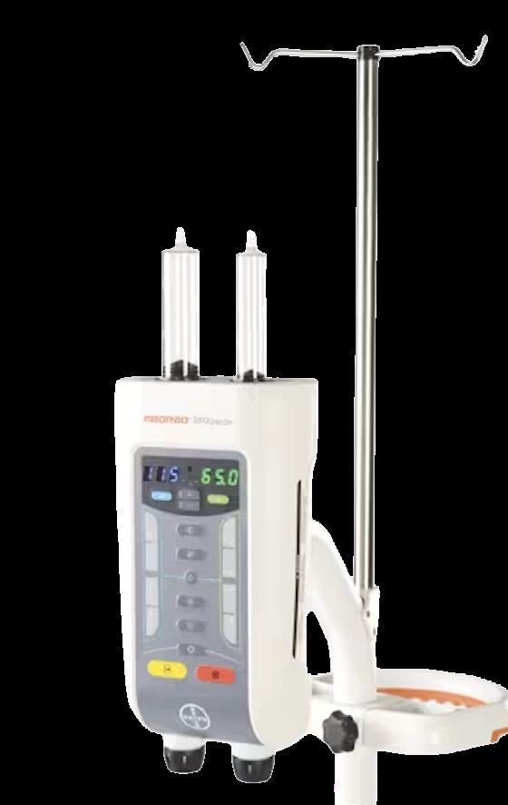
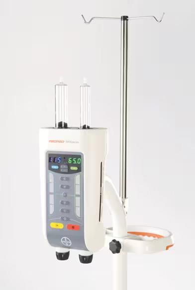
MEDRAD MRXperion
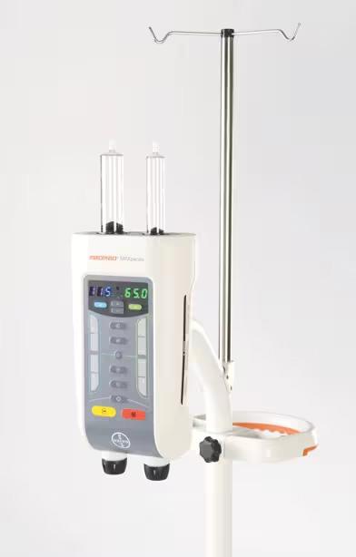
MEDRAD MRXperion is a piston-technology power injector used to deliver contrast agents and saline during contrast-enhanced MRI procedures. The system supports streamlined workflow, point-ofcare efficiency and injection data management. MR systems are deployed across healthcare settings with differing technical and workflow requirements. MRXperion is engineered to support this full spectrum of use, from routine clinical imaging to advanced research applications. A recent 510(k) clearance further enhances Bayer’s integrated MR portfolio spanning contrast media, injection systems, software and workflow solutions.
*Disclaimer: Products are listed in no particular order.
ROYAL PHILIPS Transcend Plus 2 3 GE HEALTHCARE
CT motion Syringeless Power Injector
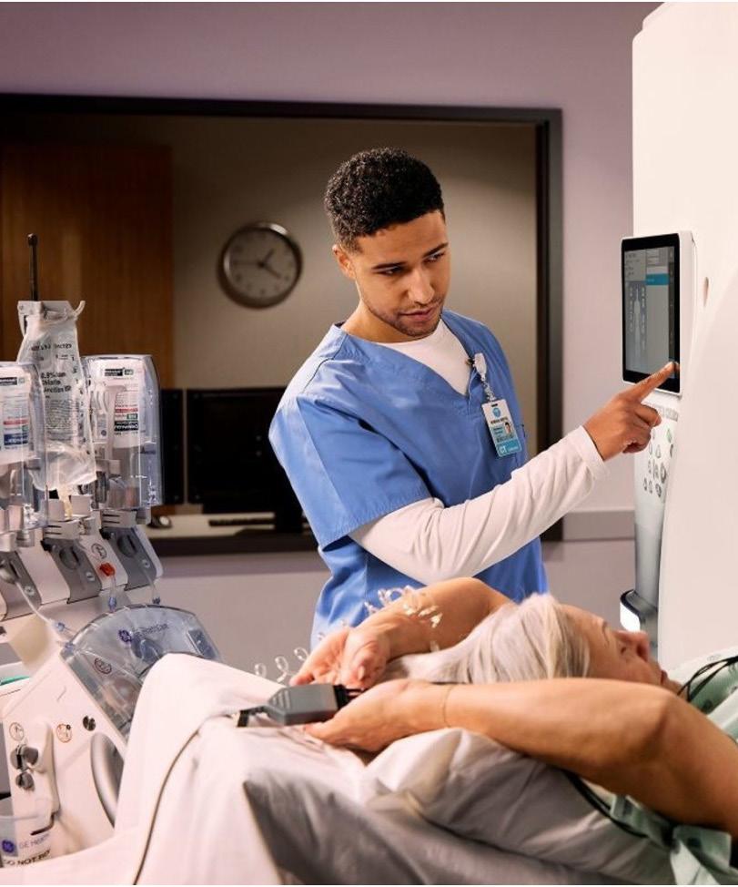
Designed for use with iodinated contrast media, the CT motion injector reflects GE HealthCare’s focus on reducing plastic and contrast media waste in CT imaging settings. The technology has been featured in GE HealthCare sustainability communications, including at RSNA, as part of a broader effort to support more environmentally conscious practices in radiology departments.
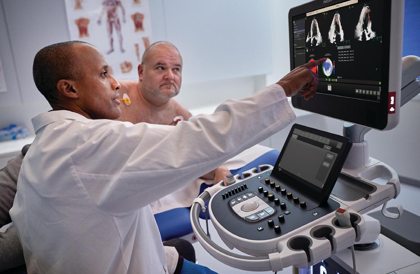
Transcend Plus delivers major advancements in image quality and artificial intelligence (AI), including FDA-cleared 2D and 3D image quality enhancements and a growing suite of AI-enabled clinical applications – all designed to meet the growing challenges of cardiac care delivery. Transcend Plus is designed to help cardiology teams work smarter, not harder – offering fast, reproducible results with uncompromising image quality and decision-making support. Transcend Plus visibly improves
sharpness, contrast and detail in 2D and 3D imaging, featuring the latest FDA-cleared enhancements for EPIQ CVx and Affiniti CVx. These upgrades support superior visualization of cardiac anatomy and function, enabling diagnostic confidence even in the most complex or technically challenging cases. The update also introduces the new 2D Auto EF Advanced feature, expanding AI capabilities for contrast images – essential for accurately assessing cardiac function.
SIEMENS HEALTHINEERS myExam Companion 4
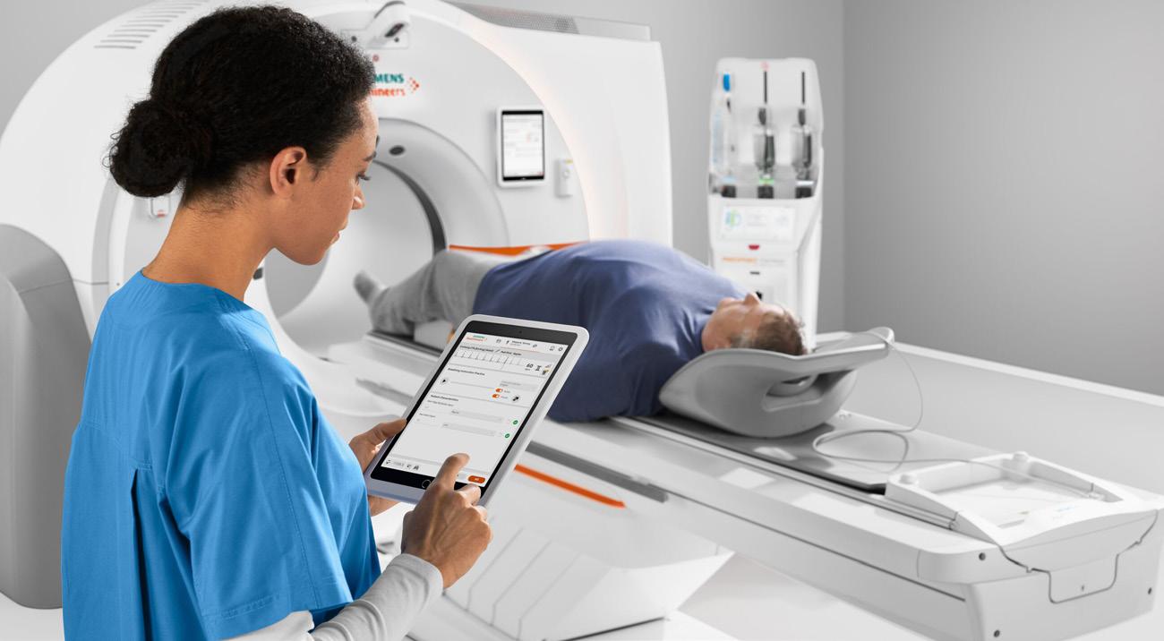
Computed tomography scanners from Siemens Healthineers, including the Somatom go.platform, the Somatom X.platform, and the Naeotom Alpha class of photon-counting CT scanners, feature myExam Companion. This intelligent user interface uses clinical language and easy-to-follow visuals to enable even inexperienced users to find the optimal combination of parameters for most patients and procedures. Together with GO artificial intelligence technologies, these CT systems deliver personalized imaging for precise dose and contrast media optimization. The combination of radiation dose-minimizing CARE Contrast III and pre-defined default contrast media protocols¹ with myExam Companion
clinical decision trees offers a holistic contrast media management solution, increasing process efficiency and standardizing quality in contrast enhancement. The Check&GO intelligent algorithm flags problems with scan coverage and contrast distribution. The FAST ROI (Region of Interest) feature automates aortic detection to help the technologist identify the optimal moment to begin contrast administration. Additionally, an integrated, gantry-mounted injector arm addresses the cost and complexities of traditional pedestaland ceiling-mounted injector solutions. •
¹ The FDA does not endorse the factory contrast protocols over the range of drug administration options provided in the drug label.
ADVOCACY IN ACTION
BY MATT SKOUFALOS
RECOGNIZING WHEN AND HOW TO SPEAK UP ON BEHALF OF PATIENTS, COLLEAGUES AND CAUSES
In any complex structure, be it a workplace, a professional association or a segment of an entire industry, resources are finite. Not every need will be met; moreover, not every need will necessarily be understood by the entity within which it has arisen. How do such issues come to the attention of decision-makers who can address them? Unless the structure of the system in question allows for rigorous, topdown evaluation, most challenges bubble up after something has already been broken. In the healthcare space, however, issues that lack attention may require the mechanisms of advocacy to be addressed.
Caitlin Donovan, senior director of the Hampton, Virginia-based Patient Advocate Foundation, said that patient advocacy in healthcare is rooted in “fundamental ethics of respect for people who are not getting equitable treatment and equitable access.” From the standpoint of those circumstances, the most effective advocacy approaches align various parties who are close to the issue with “assertiveness, not aggression, and diplomacy,” Donovan said.
“The people who are most passionate [about such issues] are less often the patients themselves, but people who love someone who has been diagnosed,” she said, “and that type of fervor carries them through decades of advocacy work.”
In the United States, the modern template for patient advocacy comes from approaches that first brought diseases like breast cancer and HIV/AIDS to the forefront of public discussion after years of being minimized or dismissed.
“During most of the 20th century, a woman diagnosed with breast can-
cer underwent a radical mastectomy,” wrote Bob Riter of the Cancer Resource Center of the Finger Lakes. “Decisions were made by physicians, and women often learned of their cancer diagnosis when waking up from surgery, absent one breast. A public discussion about breast cancer – especially your breast cancer – was unthinkable.”
Then, individuals with prominent political and social standing – like First Lady Betty Ford and journalist Rose Kushner, a Vietnam War correspondent who wrote about her breast cancer diagnosis in the 1970s – brought the issue to light, encouraging women to be more actively engaged in their diagnosis and treatment. Within 20 years, the National Breast Cancer Coalition had been established, which Riter noted “led to Congress channeling more than $1 billion for breast cancer research through the Department of Defense.”
From there, breast cancer advocacy shifted from raising awareness of the issue to increasing access to medicine, clinical trials and the affordability of both, Donovan said. But it took coordinated efforts among those diagnosed with the disease, people closest to them, and others with a degree of celebrity to step into the public eye and de-stigmatize the conversation.
Similarly, in the 1980s, stigmatization was the biggest initial hurdle to clear for those suffering from HIV/AIDS. Activists first took on the concept of “social death” that people battling the virus experienced, as the disease first emerged among communities of gay men in North America, who were fighting for recognition of their plight.
In the context of the 1980s HIV/AIDS crisis, “social death” refers to the process by which individuals were deemed “as good as dead” and socially ostracized
long before they actually died from the physical illness. It was a state of being socially isolated, cast out from families, and stripped of personal identity and social roles due to intense stigma and fear.
When “undiagnosed gay men also faced the stigma of the disease,” Dr. Joseph Wright wrote in a 2013 analysis published in the American Journal of Public Health, “gay community political leaders soon began trying to combat the problem of stigma on behalf of the whole community.
“But only people with AIDS faced the problem of social death, and that challenge caused gay men with AIDS to see their agenda as distinct from that of the gay men around them,” Wright wrote. “In the process, they invented the idea of AIDS activism by and for people with AIDS.”
The stigmatization of those suffering from an illness like HIV/AIDS led to a “social death” that marked the anticipation of their physical death, and which in many ways compounded their suffering.
“Some cultures simply avoid discussion of life-threatening illness as a means of preventing social death,” Wright wrote. “Premature social death can be hastened by institutionalization, poverty, or stigma, which can push the socially dead into ‘zones of social abandonment’ – physically segregated spaces in which the socially dead await their biological deaths.”
The response from the gay community, an already marginalized group that had been mostly left to fend for itself in the wake of AIDS, was to organize around a founding document they called “The Denver Principles.” Its statements of intention included being referred to, not as “victims” or “patients” but “people with AIDS,” a phrase that emphasized their intentions “to live as full and satisfying sexual and
The key to advocacy at the legislative level is sustained pressure and a focus on driving incremental change. When big wins are elusive, focusing on smaller victories helps to keep the issue front and center in conversations about resource allocation and policy changes. — Victor Seghers
COVER STORY
emotional lives as anyone else.”
Those organizing efforts are some 50 years old, and yet, as medical treatments have advanced to include pharmaceuticals that make life with the virus much more manageable, some of the underlying issues around insurance coverage and industry regulations still persist.
“The struggles are ongoing, and that’s why advocacy for research is necessary all the time,” Donovan said. “We’re not even close to solving these problems.”
In the medical imaging space, similar challenges still persist for patients who need coverage for preventive care, diagnostic services, and closing loopholes that freeze out segments of the population who are already marginalized because of their circumstances. Even hard-won victories can be erased by changes in private sector corporate policy, previously established law and other complicating factors.
“We are constantly in danger of backsliding,” Donovan said.
She also spoke about the various roles that can be undertaken when organizing on behalf of people battling medical conditions, whether it’s fundraising, storytelling, legislative interactions or establishing groups to help drive local change around the issues. What’s most important, however, is making sure that none of that overtakes the voices of the people at the center of the issue – those in need of broader support.
“I’ve been here for 14 years and one of our biggest pet peeves has been this idea that you would see gatherings where people would say, ‘We’ve put all the stakeholders in the room,’ and there wouldn’t be any patients,” Donovan said.
“Taking the time to treat each patient like an individual person goes such a
long way,” she said.
“If there’s a shared thread among people who are frustrated, it’s that they don’t feel respected. Injecting predictability into their experience from the moment they enter the [physician’s] office [is a critical goal]. Healthcare is intensely personal, and we’re at a point where it’s also uniquely expensive and complicated,” Donovan said.
Other times, professional advocacy can take the form of inviting outside perspectives into a space that’s often siloed. When radiologist Elliot Fishman was exploring ways to improve patient experiences in the imaging space, he established a monthly lecture series intended to help imaging professionals evaluate their approaches to their jobs.
“Taking the time to treat each patient like an individual person goes such a long way.”
— Caitlin Donovan
Fishman, a professor of radiology, surgery, oncology and urology at Johns Hopkins University Hospital in Baltimore, Maryland, knew that the professionals in his department attended numerous lectures about imaging modalities and specific disease states with regularity. But he believed they could benefit from the insights of highly successful people from outside the medical field.
Since the lecture series launched, speakers have included NVIDIA CEO Jensen Huang, PIXAR founder Edwin Catmull, former Johns Hopkins President William Brody, who also became president of the Salk Institute; and even Lynda Carter, the actress famous for playing Wonder Woman.
Speakers delivered addresses on a variety of topics, like the future of work or AI as a public service. Cindy Wolf of The Charleston spoke about her expertise in delivering hospitality at a prominent Baltimore restaurant and the insights it could offer to patient care that makes them feel as important as guests at a high-end dining establishment. Their talks were translated into white papers that were then published by the Journal of the American College of Radiology, reaching a broader audience than those attending in person and including takeaways for radiology professionals.
But Fishman also noticed how well attended the talks were, an observation he attributed to the non-hierarchical nature of the interac-
“What people need to focus on is looking at the best of the best outside of medicine,” Fishman said. “You need to look at what other people are doing who are successful.”
“If we’re going to have someone
COVER STORY
medical from Mayo or NYU come and speak, I think people get a little resentful; a little defensive,” he said. “If you have someone who comes from a company like Google or Amazon, we’re not competing with them. They’re just telling us a story. No one’s getting up there and saying, ‘Here’s the right answer.’ They say, ‘Here’s what I find worked for me.’ I look at it as a conversation between the speaker and you.”
“I think people need to listen to other people, what challenges they’re facing in other industries, and what they’re doing about it,” Fishman said. “Everybody wants the best; you want to do whatever you can do to make things right.”
“If you have people who speak about it – how do you run things, how do you think about things – to me, that’s what I think may be important,” he said. “At the end of the day, what works for a company like NVIDIA or PIXAR, you should find out.”
From helping patients push for better care and broader recognition of their circumstances, to developing non-hierarchical insights to benefit imaging professionals, approaches to advocacy take several forms. At the highest levels, professional organizations lean on advocacy efforts in interactions with lawmakers who have the ability to direct funding, offer support and carry forward issues that can positively affect a broad spectrum of outcomes.
Victor Seghers is a pediatric radiologist and nuclear medicine physician at Texas Children’s Hospital in Houston and vice chair of the Committee on Government Relations for the Society of Nuclear Medicine and Medical Imaging. (SNMMI). For Seghers, one of the biggest hurdles to clear when presenting his platform to government officials (or even the general public) involves educating his audience about his professional specialty and the opportunities it can drive for people suffering from cancers.
Theranostics — a portmanteau born of radiotherapy and diagnostic imaging — describes the field of novel radiopharmaceutical treatments designed to identify, bond with, and then destroy
cancerous cells in patients’ tumors. The technology behind the drugs is complicated, involving infrastructure like cyclotrons, which generate high-energy particle beams to create radioisotopes.
The product of that process can be short-lived, and often requires a high-end courier to deliver it in time for treatment. Its applications can be highly effective, but also highly specific, and, while proving their use cases over time, require further investment to maximize their impact. In addition to explaining the value of the technology and the process it supports,
“I think people need to listen to other people, what challenges they’re facing in other industries, and what they’re doing about it.”
— Elliot Fishman
advocates like Seghers also have to ask lawmakers to help make sure practitioners can be paid for their efforts — and that patients can afford the treatments.
In the past year, SNMMI has advocated for changes in reimbursement calculations at the congressional level, moving toward an average sale price rather than the mean unit price by which the Centers for Medicare and Medicaid Services has calculated it.
“They are very different methodologies,” Seghers said. “This newer methodology has much better data to track actual costs, and therefore makes it easier for us to get reimbursement from the government so that we can ensure that these medications are paid for. Hospitals are therefore willing to provide the service, and then patients can get access.”
“As a physician, I see the patient, so I can see the benefit that the patient gets,” he said. “That’s really what galvanizes us, when we see what this is doing for this patient.”
The key to advocacy at the legislative
level is sustained pressure and a focus on driving incremental change, Seghers said. When big wins are elusive, focusing on smaller victories helps to keep the issue front and center in conversations about resource allocation and policy changes. Consistent messaging over time is the only tool that helps to overcome changes in leadership, priorities and focus.
“We’re constantly doing advocacy, whether it’s patient advocacy or governmental advocacy, front and center,” Seghers said. “That’s the only way that you can do this, because you’re always speaking on the same topics to different people.”
“We do not think that we’re going to be successful this year, necessarily, so, it’s really just coming back over and over and over again over the next three, four, five years, with the same, consistent message, and then, hopefully, we can start to make changes,” he said.
“You have to demonstrate value. You have to demonstrate need. You have to explain the situation. The situation can be, ‘something’s expensive,’ but why is it expensive? What is the benefit of it?” he added. “You’re constantly having to provide the education to explain the value proposition for this, and the benefit for patients, who are ultimately the citizens, and paying for this with our tax dollars.”
For people who aren’t certain where to begin working on issues that require sustained pressure of coordinated advocacy, Seghers encourages them to simply find a level of engagement that works for them, whether at work, in the local community, or within an organization to which they belong.
“Just get involved,” he said. “Do anything to jump in. Exercise that muscle. Get used to having these conversations, build rapport, become better at delivering your talk when you have to.”
“If you throw out a thousand seeds, maybe only two seeds grow, but those two seeds can be very powerful,” Seghers said. “It’s a very inefficient process, and that is the difficult thing for people to get their head around. But advocacy just needs to be done.” •
THE MAGIC WAND LITMUS TEST
DIRECTOR’S CUT
BY NICOLE DHANRAJ
Iwant to tell you a quick story and I have a feeling you’ll recognize the “weight” of it.
I’ve been in one of those stretches lately where you’re just … tired. Not the “I need a long nap” kind of tired, but the kind where the workload starts to blur, and you can’t tell if you’re moving forward or just surviving. In leadership, you are often the “fixer.”
The one everyone relies on. You can be the pillar of the department and still be quietly running on fumes. Then, out of nowhere, I was given a gift. It was a magic wand. A literal wand that lights up when you turn it on. The note said: “This to help you create magic for our department.”
I just stared at it. Because if you run imaging, you know that “creating magic” is actually in your job description. You create magic with staffing gaps, broken magnets and overflowing ER waiting rooms. You do it while keeping techs safe and patients moving, and quality from slipping. But here is the thing: It wasn’t the wand that changed my mood.
It was the fact that I was seen. Not a generic “good job.” Seen like, “We actually notice what you carry.”
And it did something immediate. My shoulders dropped. My brain got quieter. I felt like myself again. That moment taught me something every imaging director needs
to hear. Sometimes what you think is “I’m done” is actually “depletion.” And the distinction is life-saving, because the fix is different.
DIAGNOSTIC DIFFERENCE
Here is the simple way I can explain it: Depletion means you’re drained, overloaded and worn down but you still care. You’re tired, but you haven’t checked out. It sounds like:
• “I can’t keep up like this … but I still want to.”
• “I still care, I just don’t have any fuel.”
• “If I could catch my breath, I’d be okay.” Depletion is a fuel problem. The answer is oxygen: support, recognition, fewer interruptions, fewer open loops and a little pressure relief.
DISCONNECTION
This is the one people hesitate to name. It also can be named burnout. It’s when something deeper starts breaking, your sense of meaning, hope, ownership or belief that the game is winnable.
It sounds like:
• “Even when things go right, I don’t feel it.”
• “Nothing changes here.”
• “I’m tired of caring more than everyone else.”
• “I don’t know if it’s worth it anymore.” Disconnection is a system problem. The
answer isn’t “try harder.” The math has to change: authority, resources, staffing strategy, expectations, support structure.
LITMUS TEST
Think about the last time someone genuinely supported you. Not a drive-by “thanks.” Not a generic “good job” email. I mean a moment where someone truly saw the weight you carry and showed up in a real way. If that moment would bring you back to life, even a little bit … you’re probably not “done.” You’re depleted.
Depleted people rebound when they get oxygen. Disconnected, burntout leaders don’t rebound with a “win” because the win doesn’t mean anything anymore.
REFILL THE TANK
If you realize you’re depleted, you don’t need a career change; you need an infusion.
Here are three things that actually help:
1. Close three small loops: Pick the three nagging tasks that follow you home and keep tapping your brain at night. Close them or move them forward enough that your brain stops refreshing them.
2. Stop being the department’s emergency button: If every minor hiccup is escalated to you, your nervous system never resets. Pick one category this week that gets routed through a lead first. One boundary can change your whole week.
3. Protect a “no ‑ guilt” block: Create one hour a week where you aren’t a director. You’re just a person. No optimizing, no catching up, no “just one more thing.” Because you can’t create magic on empty forever.
WHY I’M TELLING YOU THIS
I know there are directors reading this who are quietly asking:
“Am I losing my passion?”
“Do I need to quit?”
“Why can’t I shake this?”
I’m not here to diagnose you in a newsletter. I’m just here to tell you that if one genuine moment of support makes you feel like “roaring” again, you aren’t broken. You aren’t weak. You’ve just been “creating magic” for a long time without anyone replenishing your wand. You deserve fuel.
Sometimes just saying “I’m running on empty” to someone who understands the industry is the first step to refilling the tank.
If you’re feeling more “burnt” than “depleted” and don’t know the next step, reach out. Let’s look at the math together and see where we can find you some oxygen.
And if you don’t want to share a story, here’s a simple challenge instead: This week: close one loop, set one boundary, protect one hour. That’s the start of refilling the tank. •
If this story resonated with you, I’d love to hear your “Magic Wand” moment. Message me at nicoledhanraj@gmail.com
Nicole Dhanraj, is a radiology administrator and workforce strategist focused on military-to-healthcare transition pathways. Radiology leaders interested in exploring this approach are encouraged to connect with her directly at nicoledhanraj@gmail.com.
A CELEBRATION OF EXCELLENCE
Each year during the ASRT Annual Governance and House of Delegates Meeting, the society proudly celebrates members whose passion, leadership and service have helped advance the medical imaging and radiation therapy profession. Through their commitment to excellence, service and patient care, these individuals embody ASRT’s mission to advance the profession and ensure the highest standards of safe, quality care. Their work doesn’t just strengthen ASRT – it shapes the future of the profession. The society is honored to recognize their extraordinary contributions with prestigious awards such as the ASRT Award for Advocacy, Student to Leadership Development Program Alumni of the Year, distinguished author awards and the BeRAD Professionalism awards. In addition, the society recognizes its newest Life Members at the annual meeting. This year, ASRT selected three outstanding members for life membership. To be eligible for the honor, a member must have 30 or more years of consecutive membership and have dedicated their career to supporting the radiologic technology profession. The 2026 Life Members are Donna Newman, B.A., R.T.(R), CNMT, NCT, PET, FASRT, of Fargo, North Dakota; Marilyn
Sackett, M.Ed., R.T.(R), FASRT, of Missouri City, Texas; and Joseph Whitton, M.S., R.T.(R)(MR)(CT), FASRT, of Mount Sinai, New York. Together, these honorees exemplify the integrity, leadership and lifelong commitment that continues to advance and elevate the profession.
The Award for Advocacy highlights advocacy work achieved in the previous calendar year by an individual and an ASRT affiliate society. Winners of the award have advocated on behalf of medical imaging and radiation therapy professionals and the patients they serve by engaging with lawmakers and regulators. The 2026 ASRT Individual Award for Advocacy was awarded to Wendy Weaver, B.S., R.T.(R)(M), of Benson, North Carolina, for demonstrating outstanding legislative or regulatory advocacy efforts on behalf of the radiologic sciences community. The 2026 Affiliate Award for Advocacy was awarded to the Louisiana Society of Radiologic Technologists in part for its work in actively monitoring house bills which could negatively impact the profession and state licensure board.
The SLDP Alumni of the Year award honors a member who is new to the profession and demonstrated exemplary dedication while participating in the Student to Leadership Development Program, which is a three-year program offering participants a chance to get to know the ASRT, attend educational sessions and
MARILYN SACKETT
DONNA NEWMAN
JOSEPH WHITTON
network with medical imaging and radiation therapy professionals. This year’s winner was TJ Gardner, B.Arch., B.S., R.T.(T), of Gilbert, Arizona. TJ spent 25 years as a licensed architect in the healthcare profession before transitioning to study radiation therapy. He graduated from the MD Anderson School of Health Professions in Houston and is an active volunteer nationally and locally, committed to elevating the profession.
The distinguished author awards recognize the best peer-reviewed articles published in ASRT’s scientific journals the previous calendar year. The awards are chosen by members of each journal’s respective editorial review board. The 2025 Radiologic Technology Distinguished Author Award recipient, in honor of Jean I. Widger, is Kevin R. Clark, Ed.D., R.T.(R)(QM), FASRT, FAEIRS. His article, “Comparative Analysis of LLMs’ Performance on a Practice Radiography Certification Exam,” was published in the May/June 2025 issue of Radiologic Technology. The 2025 Radiation Therapist Distinguished Author Award recipient, in honor of Harold Silverman, is Danielle McDonagh, D.H.Sc., R.T.(T); Clodagh Starrs, M.Sc., PgC; Samantha Skubish, M.S., R.T.(R)(T); Kavita Dharmarajan, M.D.; Hulya Kocyigit, Ph.D.; Márcio A. Diniz, Ph.D.; and Maria Dimopoulos, Ph.D., R.T.(T), for their article, “Effect of APRT Intervention on Inpatient Radiation Therapy Throughput,” which was published in the spring 2025 issue of Radiation Therapist.
Finally, the BeRAD Professionalism Award recognizes health systems, imaging centers, hospitals, clinics and other healthcare facilities in which medical imaging and radiation therapists demonstrate a culture of professionalism. The 2026 recipients are the radiation therapy team at The Ohio State University Wexner Medical Center, The James Cancer Hospital and Solove Research Institute and Mount Sinai Health System Department of Radiation Oncology in New York. The two institutions demonstrated their efforts to support medical imaging and radiation therapy professionals by offering a team approach to patient-centered care, opportunities to earn credentials, continuing education, advanced degrees and opportunities to be active in professional associations as well as local community outreach events. In addition, they detailed processes or programs that deliver equitable and accessible patient care, continuous quality improvements and safe patient care.
These honorees reflect the depth of talent, dedication and leadership within the ASRT community. Their achievements underscore the vital role that service, advocacy, scholarship and professionalism play in advancing medical imaging and radiation therapy and improving patient care. ASRT is proud to celebrate these exemplary members and organizations whose contributions continue to inspire colleagues, strengthen the profession and shape its future for generations to come. •
USING AI IN HEALTHCARE DOES NOT FIX HEALTHCARE
PACS/IT/AI
BY MARK WATTS
If a health system applies AI to accelerate patient access but provides the same care delivery model built upon perverse incentives, you’ll accelerate all the broken consequences after that. More patients seen, same suboptimal outcomes.
One thing that’s always been true about technology, and that remains true in the age of AI, is that tech mostly makes the existing system move faster. This will happen even faster with AI. And, on the rare occasions tech restructures a system, it takes decades. However, we don’t have that kind of runway here.
The problem is that if a current system is full of fundamentally flawed incentive models and dumb processes, the AI and tech just churn out more of it.
We see it already with clinical documentation: AI scribes don’t eliminate having to document low ROI/nonsensical things. It just hides the work from clinicians. Which is OK, it’s not like that will impact patient outcomes in an obvious way.
But when we look beyond administrative/ operational workflows, and into actual clinical care, we start to see all kinds of ways that AI-ing the current world accelerates stuff that doesn’t make sense.
In a fee-for-service world, maybe fully optimized AI scribes and co-pilots let more patients have primary care visits in a day. Cool, I like that. Yet, it doesn’t magically make the healthcare system care about population health.
AI will get good at screening surgical patients and predicting risk, but that also means
those organizations who are cherry picking healthier patients will just get much better at that – and make it more likely that complex, sicker patients have a harder time getting care.
Even in a sort of value-based payment world where readmissions are penalized, AI will help hospitals even further optimize keeping readmissions at the lowest possible rational rate. At some point, the marginal cost of preventing the next readmission exceeds the penalty you’d avoid, and AI will be used in real-time to even better toe that line.
Whatever rules you put in place, whatever system you design, whatever incentive models you create – AI will help health systems optimize the heck out of it. While most people entered healthcare to help patients, the reality is that health system boards carry lots of fiduciary obligations. If a CXO is not willing to stay in line to hit the KPIs, the board will find someone who will.
Which means short of fixing its own perverse incentives, what healthcare will always need more of is courage to keep doing what’s right for patients even when it’s not the most profitable decision.
Yes, we will need payment models, regulation, etc. to change too. We all know how long that will take.
So, in the age of AI, where courage is already in short supply and the ability to optimize the existing system gets only better, a culture of courage is increasingly important. •
Mark Watts is an experienced imaging professional who founded an AI company called Zenlike.ai.
TRUST IS BUILT IN THE ROOM: HOW IMAGING ENVIRONMENTS
ACCELERATE (OR
IBY NICOLE DHANRAJ DEI
BLOCK) COOPERATION
n imaging, trust isn’t a vibe. It’s the difference between a clean study and a repeat scan.
If a patient is tense, your images will show it.
In my previous column, we broke down the environment into three layers: physical, social and symbolic.
Today, we’re looking at how those layers combine into one critical operational outcome: therapeutic trust.
The Constructing Trust Model reminds us that trust isn’t just a feeling. It’s a nervous system state. When people feel unsafe, the brain shifts into protection mode: attention narrows, breathing changes and muscles tense.
In imaging, that doesn’t stay “emotional.” It becomes motion artifacts, interruptions and repeats.
And when trust shrinks, patients don’t always complain. They quietly disengage.
THE PARADOX: WHY ‘TOO MODERN’ CAN SIGNAL RISK
When patients describe a space as “too modern,” they aren’t critiquing your architect’s taste. They’re describing a trust barrier.
To leadership, “modern” means investment, efficiency, and excellence. To a stressed patient, “modern” can mean cold, impersonal and designed for the system –not the person.
In trust-sensitive care, unfamiliarity feels like risk. When patients perceive risk, they go on guard. This isn’t a customer service issue. It becomes a clinical bottleneck:
• No-shows and last-minute cancellations
• Motion artifacts and repeat sequences
• Longer room times and delayed throughput
• Technologist burnout from the emotional labor of “talking down” anxiety
We often talk about staff as “trust advocates.” We want our front desk teams and technologists to build rapport quickly. A poorly designed environment can prevent even the best staff from building trust.
If a technologist has to explain a procedure while hallway noise bleeds into the room, the patient doesn’t hear confidence, they hear chaos. If a patient has to navigate a confusing maze while wearing a gown, the interaction starts from a trust deficit.
Inclusive design asks a simple question: What should the environment make easy, so trust forms faster?
FOUR ENVIRONMENTAL CAPABILITIES THAT CONSTRUCT TRUST
To move from “modern” to “trusted,” imaging environments need to deliver four capabilities.
1. Predictability: “I know what’s next”
Anxiety thrives on the unknown. Trust grows when the environment makes the process intuitive.
• · The Fix: Minimal signage using patient-first language and clear transitions from public zones to clinical areas.
• · The Goal: Eliminate the “Where do I go now?” moment of panic.
2. Dignity protection: “I am safe here”
Dignity isn’t a policy. It’s a physical sensation. Support looks like:
• Changing areas that are truly private
• Secure, obvious storage for personal items
• A gown workflow that prevents “public vulnerability” moments (including routes that don’t force patients past crowded waiting areas)
The goal: ensure a patient never feels on display while vulnerable.
3. Refuge: “I can regulate here” Imaging is sensory-heavy. Patients need micro-mo -
ments to reset their nervous system before the scan.
Support looks like:
• Quieter sub-waiting zones or a defined quiet corner
• Seating that preserves personal space (not “airport-style” rows)
• Visual barriers that reduce the feeling of being watched
The goal: reduce agitation before the patient enters the scanner.
4. Agency: “I have a say”
The biggest threat to trust is a total loss of control.
• The Fix: Visible “pause points” where permission is asked, and a clear signal (like a sign or script) that they can pause at any time.
• The Goal: Shift the patient from a “subject being processed” to a partner in care.
THE PRE-SCAN TRUTH
Trust is won or lost before the magnet ever ramps up. It’s built at check-in, in the waiting room and in the changing area. By the time the patient is on the table, the emotional tone is already set.
That’s why imaging leaders can’t treat trust as an “extra.” It’s part of the exam.
WHAT YOU CAN DO THIS WEEK
1. Map your trust breakers: Walk the patient journey today. Identify three points where an anxious person would feel lost, exposed, rushed or unsure what happens next.
2. Add one agency cue: Make it visible and consistent: “You can pause at any time.” Then, train staff to reinforce it.
3. Perform a “Tone Audit” of your signage: Walk through and read every sign as if you were a guest. Replace one “Institutional Command” (e.g., “No Cell Phones Beyond This Point”) with a “Partnership Invitation” (e.g., “To protect patient privacy and clinical equipment, please put away phones before entering.”).
THE BOTTOM LINE
Modern imaging doesn’t just need modern equipment. It needs spaces that construct trust especially for patients who walk in guarded or overwhelmed.
When the space feels safe, care becomes easier to deliver. Quality improves. Throughput stabilizes. And patients come back.
NEXT IN THE SERIES
Next, we’ll get extremely practical with trauma-informed imaging design, how to build safety, choice and control into the patient experience without a remodel. Because when you reduce perceived threat in the environment, you don’t just create comfort. You reduce interruptions, protect quality, and make trust easier for your staff to earn. •
Nicole Dhanraj, is a radiology administrator and workforce strategist focused on military-to-healthcare transition pathways. Radiology leaders interested in exploring this approach are encouraged to connect with her directly at nicoledhanraj@gmail.com.
PUTTING TIMELESS WISDOM TO WORK
EMOTIONAL INTELLIGENCE
BY DANIEL BOBINSKI
Throughout recorded history, brief phrases packed with truth have been handed down from one generation to the next. As far back as 1500 B.C., the Babylonian Counsels of Wisdom gave us useful guidance such as “Do not say evil things; speak well of people,” and “Reverence begets favor.” A few centuries later, King Solomon added to the treasury with “A gentle answer turns away wrath, but a harsh word stirs up anger,” and “Plans fail for lack of counsel, but with many advisers they succeed.” And one of my personal favorites from Solomon is “He who walks with the wise grows wise, but a companion of fools suffers harm.”
Stepping forward in time, the Greeks and Romans kept the tradition alive. Aristotle wrote, “Misfortune shows those who are not really friends.” Cicero counseled, “Never go to excess, but let moderation be your guide.” And around 600 B.C., Aesop gave us “Better to be wise by the misfortunes of others than by your own.”
Although each of these sayings is just a few words, books could be written around any one of them to expound upon their truths.
One modern joy of these ancient morsels of wisdom is that any of them can also be made into a social media post or be shared as an online meme. While social platforms let us flood the world with insightful axioms, they’re only valuable if
we incorporate their truth into our lives.
Thus, we need to do more than read a meme, then nod and say, “Yep, that’s true.” These wisdom morsels become valuable when we consider them in context of our own lives and find ways to apply them.
For example, think about the imagery that accompanies the phrase, “A turtle only makes progress when it sticks its neck out.” We can learn much from that symbolism. First, it’s a turtle. This means we can make forward progress, even if it’s done slowly. Then, by sticking out our necks, we can equate that to taking calculated risks and moving steadily forward to achieve our goals.
This axiom about the turtle can be paired with other similar sayings, such as “No risk, no reward,” or “Nothing ventured, nothing gained,” or “A journey of a thousand miles begins with a single step.” These are all great sayings, but if we don’t leverage their wisdom, we get nothing from them.
Therefore, to really make these axioms useful, we must think back and identify situations in which their concepts helped us move ahead or get out of a predicament. Said another way, the more one can associate any statement of truth with one’s own experiences, the more useful and valuable the statement will be.
Strong associations are best built through two channels – frequency and intensity. In other words, if you can associate an axiom with multiple events in your life, then your brain can make a solid connection with the value of that axiom. A similarly solid connection can
be established if you can connect the axiom to an event that held a strong emotion for you.
Over the years, I’ve come up with several of my own truisms to help people succeed. What follows are some of my favorites, along with a bit of explanation to help you find connections with your life experiences.
You’ll go where you’re focused. If you’ve taken a safe driving course, you’ve learned that whatever vehicle you’re driving or riding tends to go in the direction you’re looking. The same principle applies in just about every aspect of life. You will move in the direction of your focus. If you’re looking only at the trouble spots, you’re likely to stumble. But if you’re focusing on potential solutions and paths that lead to success, you’re likely to achieve success. Think about it. When has this been true for you?
Slower up front equals faster down the road. This is a helpful phrase for Type A personalities to memorize. Some people believe that the shortest distance between two points is a straight line, but that is not necessarily the case. When dealing with “slower moving people” (about 65% of the population), faster-moving people benefit by slowing down and working with others to create harmony, not pressure.
It’s counterintuitive, but it’s frequently true – moving slower up front often brings faster results! I’m confident that with some thought, you will find situations from your own life that are good examples of this axiom.
Acknowledgment does not equal agreement. Good communication skills include being able to acknowledge someone else’s point of view even if disagreement exists. If you’re like me, you can think of some talk radio hosts you dislike because they fail miserably at this. It’s unfortunate,
but many people believe that acknowledging an opposing point of view is the same as agreeing. It’s not true!
Let’s say you believe the moon is made out of blue cheese. I am quite capable of acknowledging your belief in a respectful way without agreeing with you. Think about it. Has anyone ever respectfully acknowledged your point of view even though you knew they didn’t agree with you? If so, hang on to that. The power associated with this axiom will help you be a better communicator.
Only make new mistakes. This saying pairs well with something Theodore Roosevelt once said. “The man who never makes a mistake is the man who never does anything.” The idea is that everyone makes mistakes. Unfortunately, some people keep making the same mistakes over and over. When have you chosen to learn instead of staying stuck in a rut?
By the way, the principle of learning from mistakes can also be tied to another one of my favorite sayings, “Good judgment comes from experience; experience comes from bad judgment.”
Bottom line, whatever sayings permeate your mind will influence your life. Therefore, knowing that you will go where you’re focused, may I suggest selecting phrases to remember that will guide you in useful, productive ways. •
Daniel Bobinski has a doctorate in theology and is a best-selling author and a popular speaker at conferences and retreats. For more than 30 years he’s been working with teams and individuals (1:1 coaching) to help them achieve excellence. He was also teaching Emotional Intelligence since before it was a thing. Reach him by email at DanielBobinski@ protonmail.com or 208-649-6400.
MEDICINE FOR THE SOUL BALANCE YOUR PLATE WITH PRODUCE, PROTEIN & WHOLE GRAINS
BY FEATURE IMPACT
Ma king health-conscious decisions with your at-home menu doesn’t have to be complicated, but it does require commitment and a little more foresight at the grocery store. Simple adjustments to overall eating patterns, like building your plate around produce, powerful proteins and whole grains, can encourage better health.
Nutritious meal-planning can be easy, affordable and delicious – and it all starts with a little balance. Consider this advice from the American Heart Association’s Healthy for Good initiative, nationally sponsored by the Egg Nutrition Center, to adequately round out a healthy plate.
FRUITS AND VEGGIES
Loaded with nutrients, fresh produce is a perfect place to start each plate. Fill about half your plate with combinations of bananas, strawberries, blueberries, sweet potatoes, peas, carrots and more of your favorites. To boost variety with each meal, look for fruit and veggie blends or step out of your usual routine and try something new.
• Grocery tip: Frozen fruits and veggies are often cheaper and just as delicious as their fresh counterparts. It’s frozen right after it’s picked, locking in freshness for a longer shelf life. Just be sure to avoid products with added salt or sugar.
POWERFUL PROTEINS
Healthy food isn’t just fuel. It should be easy and nourish you, too. Look for versatile solutions that provide a protein power-up like skinless chicken breast, no-saltadded beans, low-sodium tuna, tofu or eggs. High-quality protein from foods, along with a balanced diet and regular exercise, can help support the muscles in your body. Eggs are an example, along with
lean animal protein and some plant-based proteins. According to the American Heart Association’s guidelines, healthy people can include up to one whole egg daily, and up to seven per week, as part of a heart-healthy diet. For older adults with normal cholesterol, two eggs per day is acceptable.
Eggs deliver protein and choline, nutrients that help support growing brains and bodies, especially early in life. In fact, many Americans don’t get enough choline, a critical nutrient for supporting brain development, memory and mood.
• Grocery tip: Stock up on proteins that offer both versatility and affordability, helping you cook multiple meals per week without putting a dent in your wallet. Eggs are a perfect example as they can be used at any meal, not just breakfast – try them in wraps or omelets in the morning, hard-boil them to go with a lunchtime salad or add to a frittata at dinner.
WHOLE GRAINS
Finally, fill that last quarter of the plate with ready-togo whole grains, many of which provide dietary fiber that can support a healthy heart and healthy digestion. Conversely, most refined grains contain little or no fiber.
Examples of whole grains include barley, brown rice, corn, oatmeal, rolled or steel-cut oats, popcorn, quinoa, sorghum and wild rice, along with products labeled “whole grain” or “whole wheat” like bread or pasta.
Grocery tip: Look for whole grains that can make mealtime a cinch. Corn tortillas, instant oats and even popcorn can help you create nutritious plates and better-for-you snacks with the snap of your fingers. Try keeping packets of pre-cooked brown rice on hand to bulk up favorite dishes or toss with leftovers to round out a healthy plate.•
Visit Heart.org to discover more nutrition guidance for heart ‑ healthy meals.
3 TIPS TO COMPLEMENT A HEALTHY, BALANCED PLATE
Nutrition isn’t just about what’s on your plate – it’s also about what’s next to it and how much is on it. Pair a balanced meal with this advice to help take healthy eating to the next level.
STAY HYDRATED
If it’s not already a habit, add a glass of water to each meal. Staying hydrated helps your heart pump blood throughout the body, get rid of waste and regulate body temperature. While water is the best source of hydration, water-rich foods like fruits and vegetables can also play a role.
WATCH PORTION SIZES
Pay attention to suggested servings from your favorite food groups. For example, one slice of whole-grain bread, 1/2 cup of cooked pasta and two ounces of cheese (about the size of a domino) are the suggested serving sizes for these popular foods.
You can serve and eat smaller portions by:
• When cooking at home, offer the proper serving size to each family member then put extra food away for leftovers.
• When dining out, skip appetizers and split your meal with a friend or partner.
• Avoid eating while watching TV or at your computer. It may be harder to control how much you’re eating if you’re not paying full attention.
• Mindfully measure out your snacks – even fruits and veggies – into appropriate portions before serving instead of helping yourself directly from the bag or box.
LIMIT SALT
While sodium supports vital functions like controlling your body’s fluid balance, excess sodium in your bloodstream pulls water into the blood vessels, increasing the amount of blood inside them and thus increasing blood pressure. More than 70% of the sodium people eat comes from packaged, prepared and restaurant foods due to salt added for flavoring, stabilizing, preserving and reducing bacterial risk, according to the American Heart Association. Cutting back on sodium can help reduce the rise in blood pressure that occurs as you age, even if you don’t have high blood pressure now. It may also help reduce the risk of heart attack, heart failure, kidney disease and more.
Try cooking more meals at home where you can control sodium intake, flavoring foods with herbs and spices instead of table salt and rinsing canned beans and vegetables to wash away excess sodium.
ICE Break
“A true leader has the confidence to stand alone, the courage to make tough decisions, and the compassion to listen to the needs of others.”
Douglas MacArthur
and
Featuring Non-Magnetic Products for your MRI Environment:
* Wheelchairs, Walkers & Stretchers, including Bariatric Models
* Complete Non-Ferromagnetic line that will pass Ferromagnetic detection systems
* Breast Biopsy Trays and Draping
* Coil Carts
* Oxygen Equipment
* MRI Safe Lights
* Safety Signs and Stickers
* Full Line of Maintenance Products to keep your MRI suite Clean and Sanitized
ICEDEX
ICE MAKER PREFERRED VENDORS
• $395 a month
• 1/6 page ad included in the marketplace
• Up to 5 categories in our ICEDEX
• Your company name, website and phone included in your ICEDEX listings
• Your company name in the alphabetical business index