







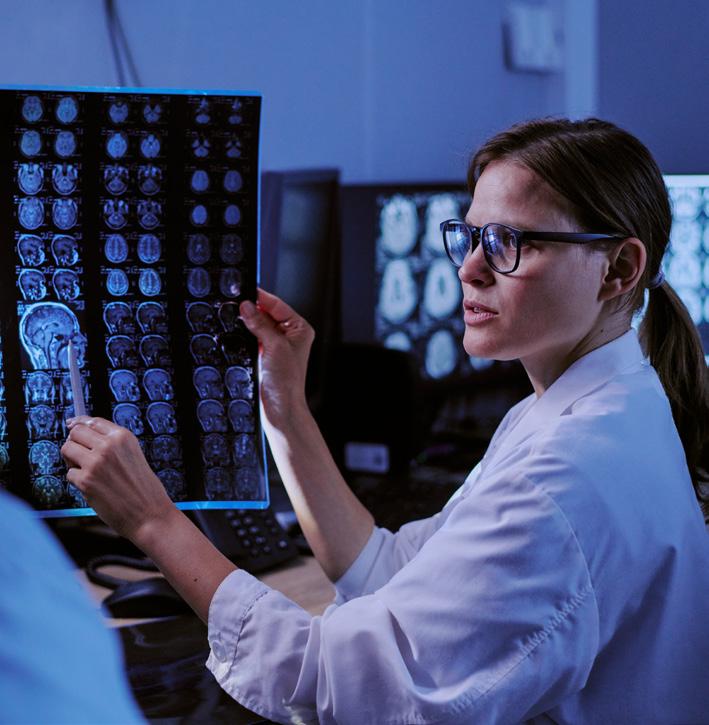
One pathway that radiology has not fully evaluated is the Department of Defense SkillBridge program.
In the do-more-withless era of healthcare, sustainability has transitioned from an aspirational public relations tool into a financial strategy.
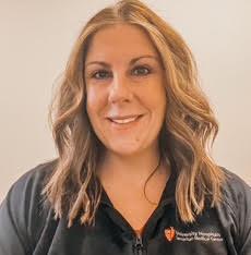
Carolyn Miller, CNMT, RT(N), is a nuclear medicine team leader at UH Samaritan Medical Center.
APRIL 2026
Catch up on the latest news from around the diagnostic imaging world.
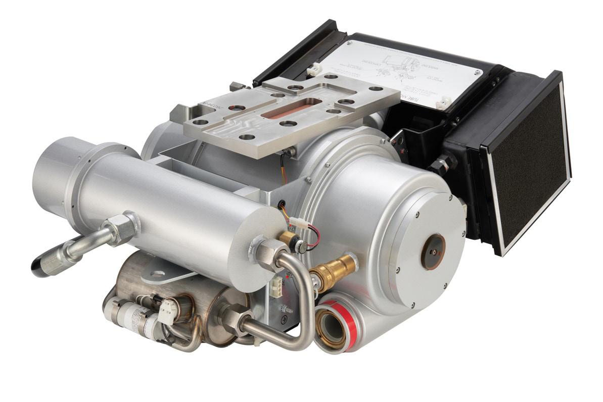
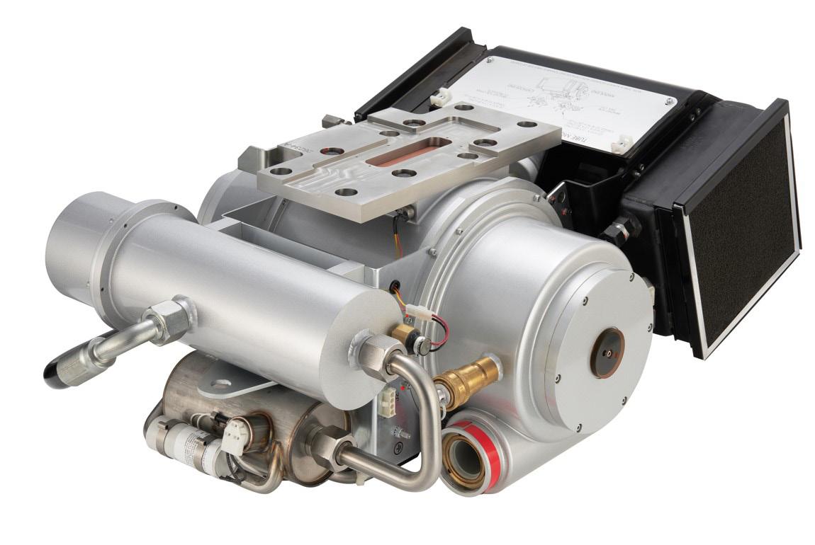
A look at imaging parts that keep devices working. 32
PRODUCT FOCUS

46
DEI
Hospital and imaging center design must factor in the patient experience.
President
Vice


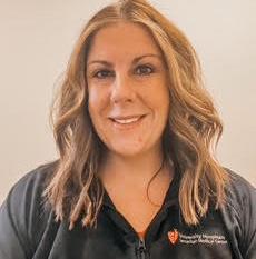
Growing up, Carolyn Miller had injuries that required X-rays and that experience sparked her interest in imaging.
Ca rolyn Miller, CNMT, RT(N), is a nuclear medicine team leader at UH Samaritan Medical Center. ICE Magazine recently interviewed her to find out more about her imaging career path and her plans for the future.
Q: WHERE DID YOU GROW UP?
A: I grew up in Northeast Ohio (Wickliffe and Willoughby Hills). After college, I moved to Ashland, where I still reside currently.
Q: WHERE DID YOU RECEIVE YOUR IMAGING TRAINING/ EDUCATION?
A: I attended the University of Findlay’s Nuclear Medicine Institute and graduated with a Bachelor’s of Science. Currently, I am certified through both the Nuclear Medicine Technologist Certification Board (NMTCB) and the American Registry of Radiologic Technologists (ARRT).
Q: HOW DID YOU FIRST DECIDE TO START WORKING IN IMAGING?
A: From a very young age I took an interest in imaging. Growing up, I had frequent injuries. With all the injuries
and broken bones came multiple X-rays and MRIs. As I grew up, I knew I wanted to get into the field but was unsure about what specific area. My high school guidance counselor first introduced me to nuclear medicine. After doing my own research and job shadowing, I knew this was the career I wanted to pursue.
Q: WHAT IS THE MOST REWARDING ASPECT OF YOUR JOB?
A: The most rewarding aspect of my job is seeing the impact that I can positively make on my patients. Working at Samaritan for 20 years has allowed me to form relationships with my patients. So many of them come to see me and are facing so many uncertainties about their health. They are scared, worried and fearful of what might be going on. I truly strive to provide the best care and patient experience I can while they are with me. I show them compassion, empathy and understanding. I take time to listen to them and ease their concerns. So many patients have told me through the years that I have made their experience easier and lightened their burden. Their feedback makes this job so rewarding!
Q: WHAT DO YOU LIKE MOST ABOUT YOUR POSITION?
A: What I value most about my role as a team lead is being a reliable source of support for my coworkers. I strive to encourage and guide my coworkers so that we can work collaboratively and be successful both as a department and an organization. I also believe strongly in leading by example, demonstrating empathy and compassion – not only towards my team, but also towards the patients that we take care of.
Q: WHAT INTERESTS YOU THE MOST ABOUT THE IMAGING FIELD?
A: What interests me the most about the imaging field is the direct impact it has on patient care, and the continuous advancement there is with technology. Nuclear medicine plays a unique role in both diagnosing and treating diseases at a functional level, which can be critical for early detection and accurate diagnosis. The imaging field is always evolving with new software and techniques. Those advancements keep the work engaging, and encourage ongoing learning, training and professional growth. It is very rewarding to know that the images I take will help guide clinical decisions and can significantly effect patient outcomes.
Q: WHAT HAS BEEN YOUR GREATEST ACCOMPLISHMENT IN YOUR FIELD THUS FAR?
A: Becoming a team lead has been a great accomplishment. Being able to guide, motivate, support and
help coworkers’ problem solve is extremely rewarding. Through the years I have been recognized by senior leadership and won awards for patient experience and a certificate of excellence for clinical support. Knowing that I make a positive impact with patients and staff assures me I am exactly where I should be.
Q: WHAT GOALS DO YOU HAVE FOR YOURSELF IN THE NEXT 5 YEARS?
A: My goal over the next 5 years is to continue to grow as a leader in radiology. I would love to strengthen my leadership skills, continue to mentor staff, and help drive improvements directly related to workflow, quality and patient care. Ultimately, I want to be viewed as a trusted leader who helps shape the future of radiology through innovation, accountability and a strong team-focused culture. •
FAVORITE HOBBY: Reading
FAVORITE SHOW: “Call the Midwife”
FAVORITE FOOD: Mexican
FAVORITE VACATION SPOT: Destin, Florida
1 THING ON YOUR BUCKET LIST: Take my mom to Ireland
SOMETHING YOUR CO-WORKERS DON’T KNOW ABOUT YOU: I collect Polish pottery.

Un iversity Hospitals Radiology Informatics Supervisor Tonisa Bermudez is a private person who overcame childhood poverty in a quest to fulfill her potential. Her drive was sparked, in part, by a high school teacher whose sage advice helped an unpolished gem begin to shine.
Tonisa recalls the moment as the greatest advice she ever received.
“My high school geometry teacher, Mr. Ferguson, once pulled me aside to tell me that I had great potential, that I needed to straighten up and live the life I was meant to live not the one I was born into,” she shares.
“I grew up in a house that was well below the poverty level. Today I have a great job, home and family. Overcoming the struggles and roadblocks of my youth to build what I have today is my greatest accomplishment,” Tonisa adds.
A geometry teacher who believed in her paired with an interest in the field fueled her plans for the future.
“I was always interested in healthcare, and I happen to have had a friend who was a radiology tech which led me to the technologist career path. Eventually, I transitioned to the IT side and have loved where this road has taken me,” Tonisa says.
She loves her job and one reason is because, “Every day
I get to learn something new.”
“My role puts me on the front line for new technologies and implementations that have systemwide impact for end users and, most importantly, patients,” she adds.
As a leader, Tonisa gives her all and wants the same from her team.
“I don’t expect more from my employees than I expect from myself,” she says. “I always believe that honesty is the best approach even though it can, at times, make my day more difficult. I believe that by holding everyone to the same standards creates a fair environment which contributes to strong relationships between all members of the team.” Tonisa has benefitted from positive leaders and hopes to pay it forward.
“Beverly Rosipko showed me that hard work pays off and taught me not to let the roadblocks stop the progress. There’s almost always another path that can be taken,” she adds. “I like to think I mentor all members of all my teams, though I do place the greatest focus on the team leads.”
A feature article in a national publication is not something Tonisa expected.
“I am a very private person, so the word sharing is difficult for me. I would like to say how grateful I am for the wonderful people in my life and how proud I am to be in a position where I have the potential for positive impact on so many people every day,” she says. •
MBA,
ARRT (R),
CRT
(M),
University Hospitals Radiology Informatics Supervisor
1. What is the last book you read? “The Infinite Game”
2. Favorite movie? “Braveheart”
3. What is something most of your coworkers don’t know about you? I never learned how to swim.
4. Who is your mentor? Beverly Rosipko my director and Mr. Ferguson my high school geometry teacher.
5. What is one thing you do every morning to start your day? Every day I kiss my dog Nala. When I wake up and just want to roll back over to sleep, she gives me the get up and moving pep talk I need.
6. Who has had the biggest influence on your life? My daughter Sierra. I have always wanted to give her all the things I could never have as a child and wanted to make sure she never wanted for everything. On the hardest days she is my motivation to push through.
7. What would your superpower be? I would want the ability to read minds. Not because I am nosey, but I believe people hold back because they are afraid of what others will think of their opinions. With employees especially, I want to know what they are really thinking and struggling with so I can help them.
8. What are your hobbies? Spending time with family, especially my granddaughter Addy.
9. What is your perfect meal? I consider anything my husband makes to be the perfect meal. He is an amazing cook.

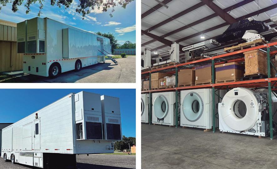
Image One Medical is the only Engineer-Owned medical equipment service group that is fully dedicated to Florida’s amazing base of Imaging Centers, Hospitals, and Cancer Treatment Centers. We have a mission: Self perform on every aspect of our business. Specialize on specific modalities: Pet CT, CT & MRI, and Focus in a key geographic region. Fort Myers I Fort Lauderdale I Tampa I Orlando
FLORIDA BASED
Dealer and servicer of PET CT, CT and nuclear medicine
• Equipment service: full coverage plans
• Equipment sales: installation, relocation and project management
• Mobile coach construction, refurbishment, maintenance and management


BY MATT SKOUFALOS
As an outreach liaison for teleradiology company Excalibur Healthcare of Moorestown, New Jersey, Betsy Cook connects its radiologists with healthcare systems and hospitals who need their services. As a component of her work in service delivery operations for Excalibur, she also produces the RadCentral podcast.
“At Excalibur, whether you’re the person answering the phone, or you’re the person doing IT, we’re all supposed to be informed about radiology,” Cook said. “It’s part of our company culture.”
“The podcast idea came out of this idea that we’re sharing this information, and we want to extend that conversation out into the public,” she said. “It’s one of the reasons why I really like working here, because I get to have these conversations with enlightening people about healthcare and subject matters that, five years ago, weren’t even on my radar – because I wasn’t doing radiology; I was making clothing.”
Cook came to Excalibur after her own entrepreneurial venture, National Picnic, was winding down as a full-time endeavor. She established her brand with the motto, “creatively casual clothing for adult women,” and a desire to parlay her love of sewing into a small business.
Cook traces her earliest days as a clothier to a junior high home economics class, in which the teacher who put a needle and thread in her hands unlocked a lifelong
passion for the physical sensation of sewing. She returned to the elective again and again throughout high school.
“I’ve wanted to sew things ever since, even if it’s not for business,” Cook said. “I just love to sew. It’s just in me, and if I don’t do it, then I feel like I have to get back to it.”
“I have a muscle memory for sewing the way someone else might have a muscle memory for shooting a three-pointer,” she said. “It’s what I want to do, and I’ve done it enough that it comes really easy.”
Cook’s first sale came after she had made her own pair of Jams: drawstring Hawaiian board shorts that hit national popularity in the late 1980s. After seeing her wearing them, a field hockey teammate complimented them, and commissioned a pair of her own. Cook remembers asking all of $2 for the work.
“I should have charged a lot more,” she laughed. Whether or not it was lucrative, the experience taught her, “If you’re good at it, you start to realize how quickly you can make something.”
For as much as she enjoyed sewing, Cook put the hobby on hold throughout college, and didn’t really return to it until she’d purchased her home. She realized quickly that “there’s suddenly all these projects that a sewing machine would really come in handy for.” She retrieved her mother’s old sewing machine, and began making things she needed for the household.
“It started to get where the machine was in the house, so I just started making clothes again, because that’s what I wanted to make,” Cook remembered.
Eventually, she splurged on a $1,200 computerized


sewing machine from the Sew and Vac in her hometown of Collingswood, New Jersey. She’d been making things for herself and her daughters, and realized she wanted to create her own designs. When enough people started trying to commission her work, Cook began to believe she could start a business venture.
“There’s a moment that you get to when you’ve sewn so much, and you’ve stared at so many patterns, that you start to realize that you don’t need the patterns, and you can start making your own patterns,” she said.
“I researched industrial sewing machines, and joined a forum for small business entrepreneurs that make products that are stitched,” Cook said. “I found out that I needed to get myself a couple industrial sewing machines, and go to trade shows to buy fabric, so that’s what I did.”
“Then, I decided, ‘I’m launching a brand, and I’m going to try to make this my livelihood.’”
Unfortunately, Cook soon grasped the size of the gulf between being a neighborhood seamstress and a department-store wholesaler. At trade shows, she couldn’t get retailers to take a chance on ordering her products against competitors that were infinitely bigger, better resourced, and delivering garments with a bigger profit margin.
“I thought I was going to be a mogul, and I didn’t know how to go about it, and I quickly learned that it was a game that I was too inexperienced to play,” Cook said.
“I wasn’t interested in sewing clothing to someday hand it off to a factory in Vietnam, because then I wouldn’t be sewing the clothes,” she said. “It’s a completely different thing if you love it because you’re physically handling the stuff. I actually liked manufacturing more than being a design house.”
Instead, Cook took her business to her fans. She retailed items at local craft fairs and markets, trunk shows and pop-ups, and built a customer database to whom she could sell directly through her online presence. By turns, she operated storefronts in the textile district of Center City Philadelphia, suburban Haddonfield, New Jersey, and a multivendor maker space in her Collingswood hometown.
“If a buyer doesn’t like my things, I know my customer’s out there; I just need to get to them directly,” Cook said. “You develop a fan base, and they start looking you up online. I’m like a secret-menu maker.”
These days, when Cook sews, it’s for herself, which is something she never got to do when she was trying to make it work as a business. It’s brought her even closer to the thing she loves, giving her the freedom to follow her inspiration, and know that she’s got no regrets at having tried her hand at it professionally.
“Now I go assist with radiology, and then I come home, and I can just do what I want with my fabric,” she said. •

Espin Medical was founded with a clear purpose: to help healthcare organizations get the most value from their surplus and end-of-life imaging equipment. It specializes in purchasing all modalities of used medical imaging systems and often provides a strong alternative to traditional trade-in options. In many cases, a second opinion helps facilities recover significantly more from their existing assets.
As part of Espin Medical’s continued growth, the company expanded its services to include nationwide equipment removal and environmentally responsible disposal. This allows facilities to clear valuable space by removing outdated or non-operational equipment, lower overall project costs and simplify the process by working with a single partner.
“We work with hospitals, OEMs and imaging centers that need equipment changes handled efficiently, safely, and with minimal disruption,” Espin Medical President Dr. George Gedevanishvili, or as many in the industry know him as just “Dr. George” said.
ICE Magazine recently found out more about the company and its plans in a Q&A with Dr. Gedevanishvili.
Q: HOW DOES YOUR COMPANY STAND OUT IN THE IMAGING SPACE?
Dr. Gedevanishvili: Imaging equipment projects involve tight timelines, multiple teams and very little room for error. Our focus is simple: execute well, communicate clearly and take full responsibility for the outcome. Our approach is built around ownership and simplicity. Each project is managed through a single point of contact who supports the customer from initial planning through final completion, eliminating handoffs and providing clear accountability at every stage. We work closely with each facility’s team to align schedules with clinical operations and hospital workflows, completing work safely, efficiently and on time while minimizing disruption to patient care. To deliver consistent results nationwide, Espin Medical
works through a carefully vetted network of specialized partners who meet our standards for safety, insurance and hospital compliance. Every project is directed and overseen by Espin, ensuring a coordinated process and a clear line of responsibility from start to finish. That combination of single-point accountability, operational alignment and nationwide execution is what sets Espin Medical apart.
Q: WHAT IS ON THE HORIZON FOR YOUR COMPANY?
Dr. Gedevanishvili: One of our key initiatives is the upcoming launch of Espin Connect, our proprietary mobile solution that allows healthcare organizations and OEMs to track their equipment projects in real time and stay informed without constant emails or calls. The goal is to simplify coordination and give project teams and facility leaders clear visibility at every stage, reducing the need for follow-up and manual status requests. We believe this level of efficiency and transparency will become the new standard for managing complex imaging projects, especially for multi-site organizations and large-scale programs. At the same time, we are scaling our operations in a disciplined way – strengthening processes and consistency so we can grow while maintaining the responsiveness, accountability and hands-on service our customers expect.
Q: IS THERE ANYTHING ELSE YOU WOULD LIKE ICE MAGAZINE READERS TO KNOW?
Dr. Gedevanishvili: Equipment changes don’t have to disrupt daily operations. With the right planning, communication and real-time visibility, these projects can move forward smoothly and with minimal impact. Tools like Espin Connect help reduce uncertainty and keep everyone aligned throughout the process. At Espin Medical, our goal is to bring structure and clarity to every project so our customers can move forward with confidence and keep their attention where it belongs – on patient care. •
For more information, visit espinmedical.com



The 2026 Imaging Conference & Expo featured a record-setting number of exhibitors and attendees at the San Antonio Marriott Rivercenter in San Antonio, Texas. Total attendance reached 544 individuals with 84 exhibits. A packed slate of education offered continuing education approved by ACI, AHRA and ASRT.
The three-day event also featured networking events including an “ICE Break” lunch and signature evening functions. In addition, ICE 2026 delivered specialty gatherings such as the invitation-only Leadership Summit and Reverse Expo.
ICE is the only industry event dedicated exclusively to imaging directors, radiology administrators, radiologic technologists and imaging engineers from hospital departments, freestanding imaging centers and group practices.
Imaging professionals and company representatives praised the conference.
Jordan Hermiller, a technical manager with Agfa Radiology Solutions, enjoyed presenting at the conference and manning the company’s booth in the exhibit hall.
“ICE brings together the people who are making purchasing and operational decisions in imaging departments, including managers, directors and lead technologists who understand what their teams need,” he said. “For Agfa, those are exactly the conversations we want to be part of. Beyond the business side, what I personally enjoy most is the engaging dialogue that happens around radiology and healthcare more
broadly. You get into real conversations about where the profession is headed, the challenges departments are facing and how technology fits into the bigger picture. That’s hard to replicate anywhere else.”
“The conversations around AI and automation felt much more grounded than they have at other conferences, with attendees asking practical, informed questions,” he added. “It was a fun experience and a reminder of why ICE holds a unique place in the imaging community.”
First-time attendee Melissa Ramos, outpatient radiology assistant director with Shannon Health, shared her unique perspective.
“As a first-time attendee, it really did have the look and feel of the annual large AHRA conference. It felt similar but also more personable. The reverse expo was a new experience for me. I found it useful and fun!” she said.
Advanced Ultrasound Systems Director of Inside Sales Adrianna England enjoys the annual conference because it provides an opportunity to network.
“AUS loves to support HTM and ICE gives us a positive environment, with lots of opportunities to showcase our training, staff and products,” she said. “For me personally, I think every ICE show is fantastic, but this show was physically beautiful. I loved the bright colors, the fun food signs, and of course, anywhere I looked near the exhibit hall, I could easily identify your staff.”
“ICE is where the imaging community comes together to connect, unite and build a better
tomorrow,” John Bowser with Downtime Trace added.
Innovative Radiology’s Michael Hickman agreed.
“A hyper-focused show catered to the imaging world, with energetic atmosphere. Put this one on the books every year,” Hickman said.
Intelas was well represented at the conference.
“ICE is an opportunity to meet the vendors that we are working with and make connections to new ones, as well as an opportunity for classes to further enhance me professionally,” said Adrienne Trapnell, a senior strategic sourcing specialist with Intelas.
“Great opportunity to meet professionals, learn through shared experiences and network to maximize your own service delivery plan,” Randy Willis, a resident regional manager, added.
HealthLevel Director of Marketing Hijinio Reynoso also mentioned the networking aspect of the event.
“A conference that brings people together from throughout the industry while still offering a 1:1 connection,” is how he described the Imaging Conference & Expo.
Orlando Health Melbourne Director of Radiology Rich Egan ranks the conference high on his list.
“One of the better radiology conferences that I attend each year. I like it because you have CEs co-mingling in attendance,” he said.
Hermiller left San Antonio excited about everything the Imaging Conference & Expo offers the diagnostic imaging community.
“What makes ICE special is the sense of community,” he said. “It’s a conference where people genuinely care about advancing the profession and supporting each other, and that came through in every conversation I had this year. I also appreciate that the conference recognizes imaging leaders through the Diamond Awards. It’s important to celebrate the people who are driving this profession forward. I’m proud to be part of the ICE community.” •
Find more at Attendice.com.




The recent Imaging Community Exchange (ICE) Magazine webinar “An Overlooked Talent Pipeline” presented by Nicole Dhanraj provided advice on uncovering a wealth of options for imaging leaders facing staffing shortages.
Imaging leaders are being asked to do more with the same teams, the same workflows and the same limited hiring options. The pressure shows up everywhere. The staff is stretched thin. Imaging leaders are stepping in to cover gaps. Departments must shift their focus to getting through the day instead of building for the future.
Dhanraj’s insightful webinar explored an overlooked talent pipeline that rarely presents itself in imaging workforce conversations. However, this untapped pipeline has the potential to support operations, ease workload strain and expand how imaging leaders think about staffing and workforce planning.
Rather than another hiring pitch, Dhanraj provided a practical, down-to-earth discussion about where additional support can come from. She shared how to evaluate it without disrupting patient care, compliance or team culture. The webinar delivered a fresh perspective for radiology and imaging leaders, managers and administrators when it comes to workforce strategy, operational support and long-term stability. The webinar was especially helpful for departments feeling the weight of ongoing staffing and workload challenges.
After the webinar, Dhanraj left attendees able to:
• Identify gaps in traditional imaging staffing and workforce planning approaches
• Explore an overlooked talent pipeline outside standard imaging recruitment
• Assess how alternative workforce pathways may support imaging operations without increasing risk
• Apply practical considerations to decide whether this approach fits their department
The presentation was popular with a high rating from attendees via a post-webinar survey. The survey included the question, “How will this webinar help you do your job better?”
“Will take this information into more client discussions,” said Carol Davis-Smith, president of Carol Davis-Smith & Associates.
“Provided an innovative method of creating a job pool of talent,” said Jeralynn Scott, a retired manager.
“This opens new doors that I did not know were available,” said Stephanie Benavidez, radiology manager with Oswego Health.
“Looking to the military for talent,” said Stephanie Voigt, director women’s imaging & practice support with Consulting Radiologists Ltd.
“It helped open my eyes to different perspectives,” said Jeffrey Fletcher, a radiology manager with Texas Health Resources Dallas.
“This is a pipeline I have never heard about. I am excited to explore it further!” shared Lisa Walling, a director with Oklahoma City Indian Clinic. •
For information about upcoming webinars, visit ICEwebinars.live.
Register now for the May 5th webinar “Mastering the Use of a Tissue Mimicking Phantom for Ultrasound”.
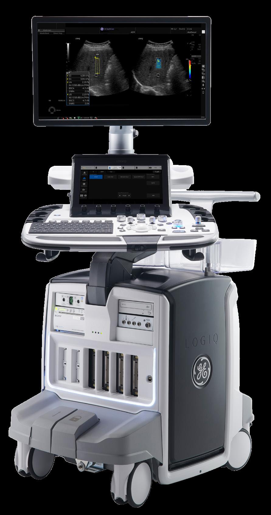
GE HealthCare has announced the next generation of LOGIQ general imaging ultrasound systems – an intelligently designed portfolio built to elevate clinical imaging, accelerate workflows and unlock deeper diagnostic insight.
A news release states, “Equipped with advanced imaging capabilities, enhanced AI-powered automation, and an expanded open digital platform, the latest LOGIQ systems are engineered to simplify daily practice while supporting more confident, informed clinical decisions.”
To support imaging across a broad range of applications, the newest LOGIQ portfolio introduces advanced imaging capabilities, enhancing clinical confidence across a variety of care areas, and expanded transducers, including specialty and multi-purpose probes and the wireless Vscan Air, delivering exceptional image quality and diagnostic precision.
“Clinicians need solutions that remove complexity and enable them to perform at their best every day,” said Sally Lee, general manager, general imaging, primary care and
automated breast ultrasound (ABUS), CCU, GE HealthCare. “The latest LOGIQ systems combine advanced imaging with comprehensive tools and AI-driven innovation for a next-level ultrasound experience – elevating efficiency, streamlining system interactions and empowering confident clinical decisions.”
To further accelerate clinical workflows, new AI-powered tools help detect and segment anatomy, enhance measurement accuracy, and reduce system interactions. New tools such as Auto Abdominal Suite 2.0 with SmartEdit capabilities add Auto Abdominal Color Assistant 2.0, Auto Aorta Measure Assistant, and Auto CBD Measure Assistant.
The expansion of the LOGIQ open digital platform unlocks new system potential by enabling seamless integration of third-party applications alongside Verisound Digital offerings. This platform delivers streamlined access to AI tools and integrated on-scanner ViewPoint reporting – supporting more connected workflows, personalized system customization, and consistent, datadriven decision-making and reporting.
AGFA HealthCare continued its strong business momentum across the U.S. market, as health systems expand their use of scalable, cloud-based enterprise imaging solutions that support clinical efficiency, clinical collaboration and organizational growth, according to a press release.
A key highlight of this momentum is a new agreement with a rapidly growing, multi-state radiology group delivering diagnostic imaging and professional reading services across 11 sites in four states. To support its continued expansion, the organization selected AGFA HealthCare’s Enterprise Imaging Cloud Teleradiology solution to unify radiologist workflows across its network. The group will transition from multiple PACS and dictation systems to a single, cloud-based diagnostic platform designed to improve efficiency, reduce complexity and scale without added IT infrastructure. The agreement reflects a broader pattern of adoption among radiology groups seeking scalable, cloud-based platforms to support growth and clinical performance.
AGFA HealthCare also expanded its Enterprise Imaging footprint with Tampa General Hospital (TGH),
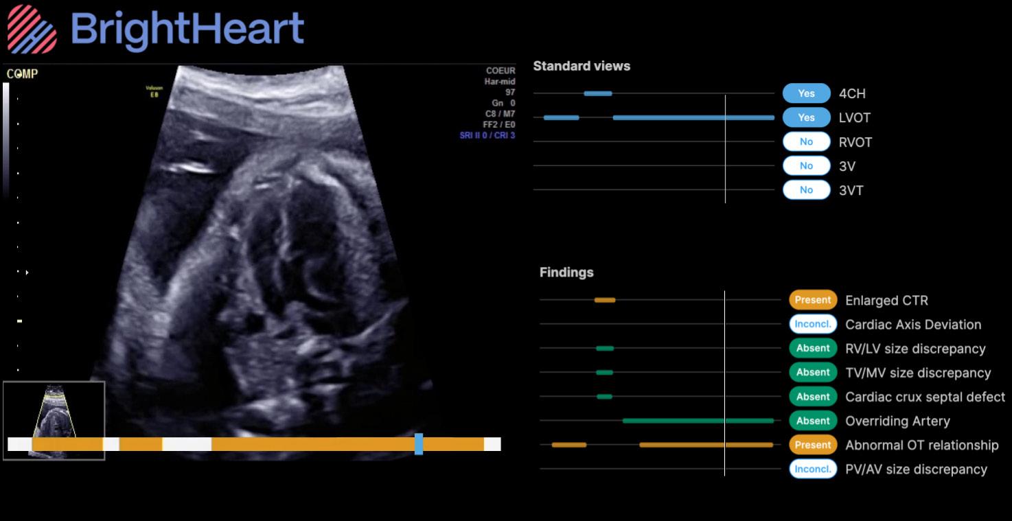
BrightHeart, a global leader in AI-driven prenatal ultrasound, recently announced the availability of its B-Right AI Platform through GE HealthCare’s Voluson Solution Store, bringing advanced AI support directly into routine prenatal care. The addition of this AI platform to the Voluson Solution store is poised to improve early detection of congenital defects, reduce diagnostic uncertainty, and ultimately support better outcomes for clinicians, expectant parents and their babies. This collaboration combines BrightHeart’s innovative AI technology with GE HealthCare’s trusted Voluson ecosystem through the Voluson Solution Store – the first digital marketplace for women’s health ultrasound, available in the U.S. and Europe. Together, the companies are helping to democratize access to expert level prenatal care by making clinically validated AI for congenital defect screening and full scan support available to Voluson ultrasound users. Supported by strong clinical evidence — including two recent publications in The Green Journal — and developed with leading fetal cardiology and maternal and fetal medicine
which extended its Enterprise Imaging Cloud SaaS deployment across multiple facilities, including TGH Imaging, TGH Main, and the University of South Florida. The expansion supports more than 1.5 million annual imaging studies and replaces fragmented departmental systems with a unified, enterprise-wide imaging platform.
These enterprise imaging (EI) selections reflect the strength of AGFA HealthCare’s established reputation for delivering imaging platforms that support clinical workflows, operational efficiency, and enduring customer partnerships. The AGFA HealthCare EI Platform has been recognized multiple years running by KLAS, including Best in KLAS awards for Enterprise Imaging for Radiology (small-under 300k studies) (in 2026), XERO Universal Viewer (2024, 2025 and 2026), and Enterprise Imaging VNA (2025 and 2026).
In addition to these agreements, AGFA HealthCare closed multiple Enterprise Imaging and Cloud deals across the U.S., spanning health systems and radiology providers focused on consolidating imaging environments, modernizing infrastructure, and enabling enterprise-wide access to imaging data.
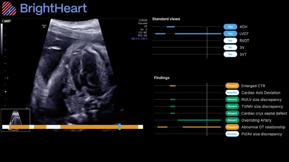
(MFM) experts, BrightHeart goes far beyond basic quality checks, delivering reliable, evidence backed insights that reduce diagnostic errors and improve the detection of congenital defects while strengthening workflow efficiency. BrightHeart’s AI Platform enhances Voluson’s offering with a simple, intuitive, cloud-based solution that supports enhanced care spanning acquisition, interpretation, and review across care settings and workflows.
BrightHeart has already demonstrated significant benefit with its platform, not only improving screening capabilities and reducing errors, but also improving workflow by improving scan completeness, saving time, and increasing confidence.
Two medical ultrasound organizations recently announced their endorsement of expanded training in contrast enhanced ultrasound (CEUS) under the newly updated National Education Curriculum (NEC) for sonography programs.
“CEUS has transformed how we diagnose and monitor disease, and expansion of CEUS training for sonography students is a critical step forward in ensuring broader patient access to one of the most innovative and patient friendly advances in diagnostic imaging,” according to a joint statement issued by the International Contrast Ultrasound Society (ICUS) and Inteleos.
The NEC is a consensus-based curriculum guide developed by a coalition of sonography professional organizations as a framework for sonography training programs across the United States. This guide can also be used by sonographers who are preparing for credentialing examinations.
The new NEC updates follow the recent expansion of the sonographer scope of practice to include a more active role for trained sonographers in the administration of CEUS examinations. The “Scope of Practice and Clinical Standards for the Diagnostic Medical Sonographer” was updated in 2024 by the Society of Diagnostic Medical Sonography (SDMS).
“Expanded CEUS training will help prepare the next generation of sonographers to deliver safer, more effective and more patient-centered care,” according to Maria
Stanczak, a member of the ICUS board who was instrumental in developing the new CEUS content for general imaging concentrations.
The earlier version of the NEC, adopted in 2016, contained little CEUS training information, which helped slow the adoption of CEUS in the United States compared to Europe and Asia, according to Dr. Jordan Strom, associate professor of medicine at Harvard Medical School, director of the echocardiography laboratory at Beth Israel Deaconess Medical Center, and an officer of ICUS. Strom played a leading role in developing the new CEUS content for cardiac concentrations.
UCAs do not contain iodine or gadolinium, and have not been shown to have an effect on thyroid or renal function, he said.
In addition, CEUS does not expose patients or staff to ionizing radiation, and CEUS exams provide highly accurate clinical information in real time so that appropriate therapy can be initiated without delay.
“Training in CEUS during formal sonography education enables students to graduate with the knowledge and skills necessary to meet the evolving demands of modern healthcare,” said Hannah Simmons, director of global business growth at Inteleos.

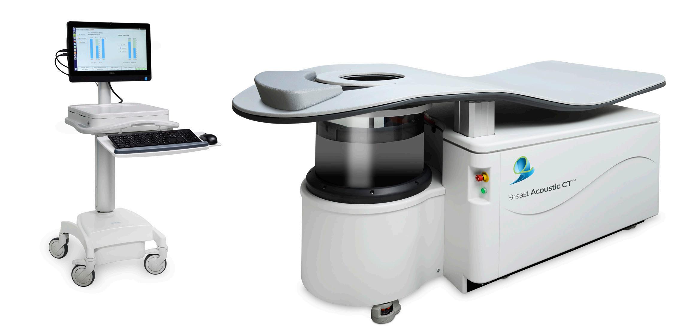
QT Imaging Holdings Inc., a medical device company dedicated to transforming breast health management through innovative, radiation-free imaging technology, has announced that the American Medical Association (AMA) CPT (Current Procedural Terminology) Editorial Panel has approved a new Category III CPTTM code for 3D quantitative transmission volumetric ultrasound tomography of the breast.
The new Category III code, X579T, represents a significant milestone in the clinical and commercial advancement of QT Imaging’s technology, recognizing the distinct clinical service enabled by its radiation-free, compression-free, 3D breast imaging platform. The new CPT code ensures standardized reporting of this emerging imaging service, supporting data collection, utilization tracking and continued evaluation by clinicians, payers and health systems. The code is scheduled for release on July 1, 2026, with an effective date of January 1, 2027.
“Approval of our first CPT code is a meaningful validation of both the technology and its clinical foundation and significantly enhances our commercial strategy in particular to prospective hospital accounts,” said Dr. Raluca Dinu, chief executive officer of QT Imaging. “This milestone
reflects years of rigorous development, clinical engagement, and collaboration with the medical community. It is an important step toward expanding access to a safer, more comfortable breast imaging option that complements existing modalities and supports better-informed clinical decision making.”
Category III CPT codes are established by the AMA to track emerging technologies and services that demonstrate clinical relevance and growing adoption. Acceptance of a Category III code signals acknowledgment by the CPT Editorial Panel that a technology represents a distinct and reportable clinical service and is a major step on the pathway toward broader reimbursement and adoption.
“We plan to continue working closely with clinical partners, professional societies and regulatory stakeholders to advance adoption of our technology, as well as to pursue additional CPT codes,” Dinu added.
QT Imaging’s system uses advanced ultrasound tomography to produce true 3D volumetric images of the breast without ionizing radiation or painful compression. The technology is designed to provide quantitative, reproducible information that can be particularly valuable in women with dense breast tissue, while improving the overall patient experience.
Royal Philips has announced the availability of the InkSpace Imaging Snuggle pediatric body array coil for Philips 3.0T MRI systems. Designed specifically for pediatric patients, the Snuggle coil has been optimized and validated for use with Philips 3.0T MRI systems, bringing enhanced comfort for children and efficient, high-quality imaging for clinicians, according to a press release.
Young patients often find MRI exams challenging, and the Snuggle coil was created to make the experience more comfortable and less intimidating. Its light, blanket-like design and soft, flexible structure gently wrap around the patient.
The Snuggle coil’s high-density array and flexible design allow sharp, high-resolution images and more efficient exams across a range of pediatric anatomies. These advantages can reduce the need for additional imaging or follow-up procedures while helping technologists work more efficiently through easier handling, faster setup and shorter scan times, helping to improve throughput and reduce repeat exams.
“This collaboration combines Philips MRI leadership with InkSpace Imaging’s focus on pediatric design,” said Ioannis Panagiotelis, Ph.D., business leader, MR at Philips. “The Snuggle coil shows how thoughtful design can make MRI scans less stressful for children while giving clinicians the image quality and workflow efficiency they rely on.”
Recently cleared through the U.S. Food and Drug

Administration (FDA) 510(k) process, InkSpace Imaging developed the Snuggle coil to provide a more child-friendly alternative to traditional rigid devices.
“Snuggle was designed and built by InkSpace Imaging to solve a very specific pediatric challenge: comfort and performance without tradeoffs,” said Peter Fischer, CEO, InkSpace Imaging. “By enabling compatibility with Philips 3.0T MRI systems, we can bring that child-centered design to more hospitals and help care teams get the scan right the first time.”
The Snuggle pediatric coil for Philips 3.0T MR systems is available in the United States, with rollout to additional regions planned in the future. Philips and InkSpace Imaging continue to explore opportunities to expand comfort- and efficiencyfocused coil technology, supporting radiologists, technologists, and families with safer, faster and more effective MRI exams.

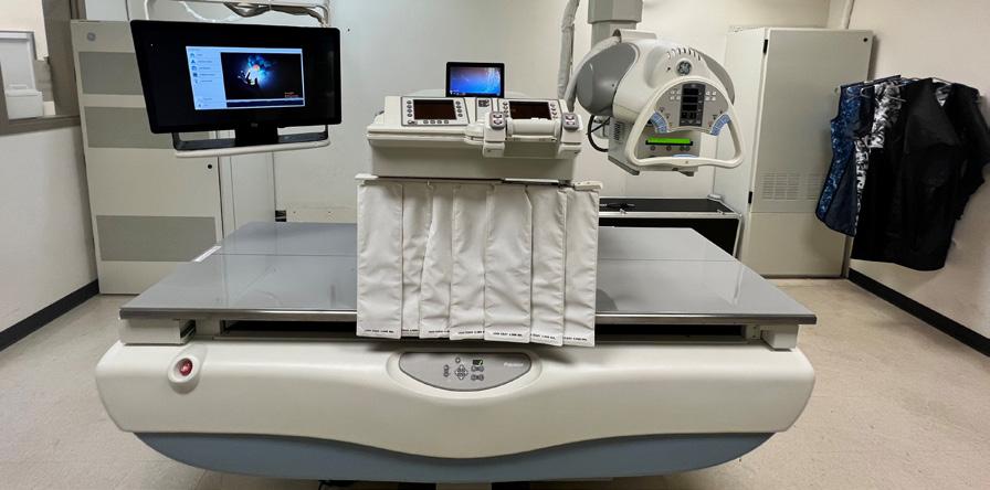
- New Windows 10 Platform with Dynamic Digital DR Panel
- Eliminates the OEM Saturn Windows 95 Platform
- Dose Reduction
- HIPAA Compliant, 510k approved.
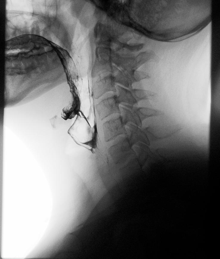
- Fluoroscopy digital recording loops for MBS studies
- Upgrade Provided to your Proven R/F System in 4 days.
- Complete Refurbished Systems Available

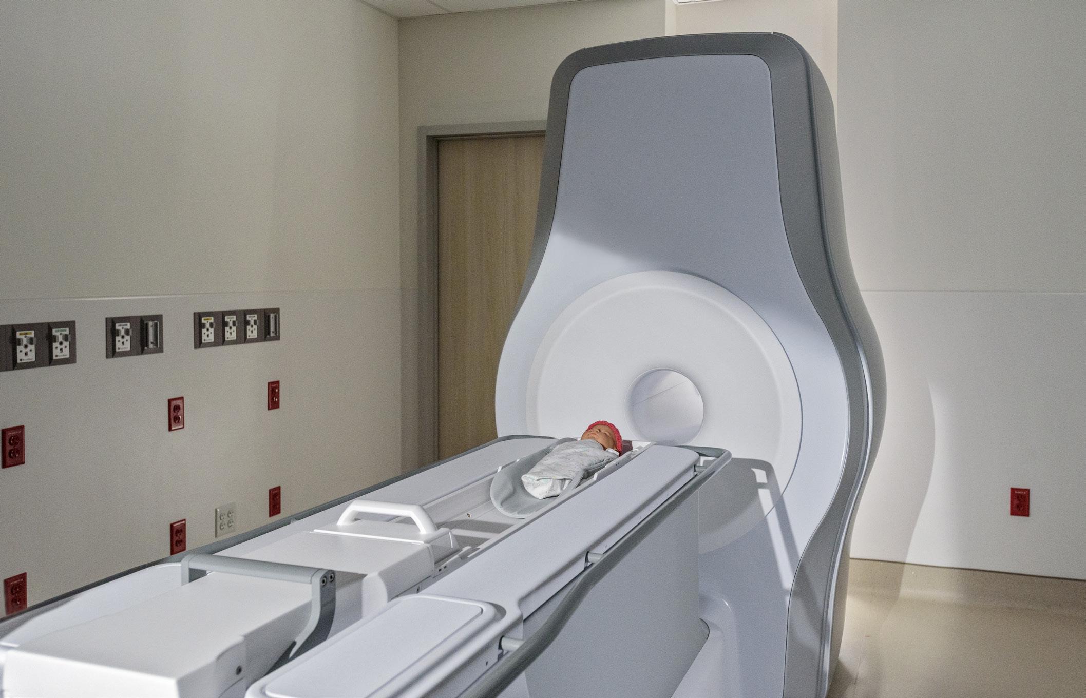
Eyas Medical Imaging Inc. has received U.S. Food and Drug Administration (FDA) 510(k) clearance for its Ascent3T Neonatal Magnetic Resonance Imaging (MRI) System, a whole-body magnetic resonance scanner designed and optimized specifically for neonate and infant anatomy, including head, body and extremities.
“510(k) clearance marks a significant milestone for the company and reflects our mission to enable healthcare professionals to save more babies’ lives with state-of-the-art, precision imaging,” said Matt Storer, president and CEO of Eyas Medical Imaging.
The Ascent3T is the first high-field, 3 Tesla (3T) dedicated neonatal MRI system in the world, according to a press release. It utilizes a 3T magnet, enabling a more comprehensive and precise diagnostic tool that provides healthcare professionals with detailed imaging of vital anatomy, including the brain, lungs, heart and abdomen. The Ascent3T addresses the technical limitations of using an adult-size MRI system to image babies and provides clinicians with improved ability to visualize and diagnose disease in the neonatal patient population.
A significant barrier to using MRI for neonates has
been the risk of transporting fragile newborns from the neonatal intensive care unit (NICU) to alternate MRI locations within the hospital. Eyas’ innovative engineering solves that problem by delivering a whole-body 3T neonatal MRI system with a compact footprint that allows direct installation in the NICU without sacrificing image quality. Unlike most conventional MRI systems, the Ascent3T is virtually helium-free and does not require a quench pipe or outside venting.
The Ascent3T was conceived at one of the world’s top children’s hospitals, Cincinnati Children’s, incorporating learning from their over 1,700 infant MRI scans on prototype systems. It includes features, such as a detachable patient table that can serve as a patient transport device, providing flexibility in NICU workflow based on hospital needs and preferences. The system also leverages advanced electronics, operating software and pulse sequences from Philips Medical Systems Nederland B.V.
The company is currently scaling up operations and expects to be in commercialization in the United States later in the year. The device is not yet commercially available in other countries.
Cairn Surgical Inc. announced that a study in the Journal of Breast Imaging showed that a single imaging session combining prone and supine MRI performed prior to breast conserving surgery provides highly accurate and efficient imaging of breast tumors that could contribute to significantly reducing positive surgical margins.
Prone MRI – with the patient face down – is sensitive in assessing the extent of breast cancer and potentially identifying additional cancers missed by other methods, but has not definitively helped decrease the positive margin rate for breast cancer surgery. Supine MRI – with the patient face up in the surgical position – is a potential strategy to enhance the precision of breast cancer surgery but is not commonly used today.
Cairn Surgical developed the combined, single-session approach outlined in the paper to seamlessly capture more precise and comprehensive tumor data while keeping the patient and tumor in the same supine position used in surgery. Supine imaging integrated into conventional prone imaging serves as the foundation for the company’s Breast
combined approach displayed clear tumor to fibroglandular contrast, similar to that resulting from separate imaging sessions, allowing for excellent visualization of tumor margins,” said Roberta M. diFlorio-Alexander, MD, lead author of the paper and associate professor of radiology at Geisel School of Medicine at Dartmouth. “Prone and supine MRI performed in one imaging session allows for accurate assessment of disease extent, which can provide important navigational guidance for innovative technology that optimizes the surgical approach.”
In the prospective study of 74 breast cancer patients, tumor-to-fibroglandular tissue contrast ratios (TFRs) from a single-session prone-to-supine breast MRI were compared to those from separate-day prone and supine breast MRIs. One standard bolus dose of contrast was delivered prior to each MRI in the session — consistent with standard cardiovascular MRI practice — and showed no adverse events. TFR values extracted from images segmented by an experienced radiologist and confirmed by an independent breast radiologist were compared to confirm the ability to precisely identify tumor margins and determine the feasibility of scaling the single-session technique for wider

The Breast Cancer Locator is considered an investigational device in the U.S. and is limited by U.S. law to
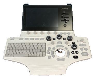

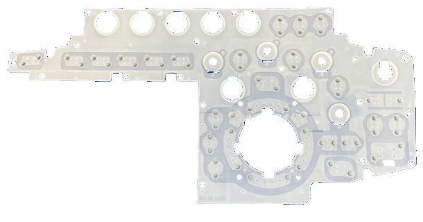
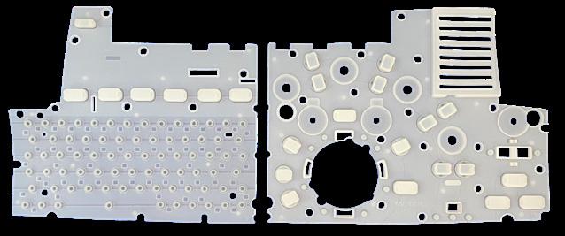
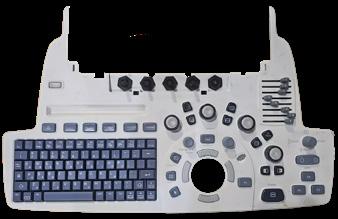

This month ICE Magazine asked imaging leaders to share their insights regarding imaging parts. What are the challenges they face? What process is used to find and acquire imaging parts? ICE Magazine asked these questions and more to share their knowledge with the ICE community. Participants in the Director’s Circle on imaging parts are:
• Intelas Regional Director of Operations Eric Massey;
• Shannon Health Assistant Director of Outpatient Radiology Melissa Ramos;
• Sodexo Healthcare Imaging Director 1-HTM Josh Nice; and
• UC Health, Cincinnati Ohio System Radiology Director Jacqui Rose.
Q: WHAT ARE THE MOST SIGNIFICANT CHALLENGES YOUR FACILITY FACES WHEN SOURCING IMAGING PARTS FOR REPAIR AND MAINTENANCE OF DIAGNOSTIC IMAGING DEVICES?
MASSEY: I oversee imaging service in multiple hospitals throughout the U.S., and the most significant challenge we face when sourcing imaging parts is time to availability. In major metropolitan areas, this is easier to manage. Vendors often have multiple customer sites clustered together, which makes it more practical to establish forward stocking locations. Costs can be shared across facilities, logistics are simpler and proximity to major airports allows for faster replenishment when parts are used. The challenge increases significantly in remote or rural regions. These sites are often far from major airports and distribution hubs, which can add substantial delays to part delivery. In those environments, same-day or nextday recovery becomes much harder without intentional planning. To mitigate this, we have to be strategic about placing forward stocking locations in these regions and focus on keeping high-failure and mission-critical parts
readily available. That planning is essential to restoring imaging systems within the same day or within a 24-hour window, which directly impacts patient care, throughput and clinical confidence.
RAMOS: Location, location and very few engineers in the area. We are located in west/central Texas and most of our service team comes from the bigger cities – Dallas, San Antonio, Lubbock, Oklahoma. Delays happen while we wait for service to come triage equipment and wait longer for parts.
NICE: The availability of parts for aged systems. In many cases they are refurbished as new ones are not being produced. Some come dead on arrival (DOA) and some cannot be sourced at all.
ROSE: Our most significant issues with sourcing parts lies in the issues with age of our equipment. When our units are end of service life, the parts are very difficult to obtain and, of course, cannot be guaranteed by the vendor. We always try to get OEM parts, however, we often run into issues with those parts being on back order or simply unavailable. Even if they are available on older units, it can take days to get them on site.
Q: CAN YOU DESCRIBE THE PROCESS YOUR ORGANIZATION USES TO OBTAIN IMAGING PARTS, INCLUDING DECISION FACTORS FOR CHOOSING OEM VERSUS THIRD-PARTY SUPPLIERS?
MASSEY: We work with a defined set of approved vendors for each imaging modality, and all parts orders are routed through our technical resources group. That team is responsible for sourcing components through our preferred suppliers and coordinating availability and logistics. The
final decision on whether to use an OEM or a third-party supplier is made collaboratively by our onsite engineers and our national support team. That decision is driven by a combination of technical risk, expected part life, system criticality and total cost of ownership. Longevity is a major factor. For example, certain cath lab X-ray tubes can last eight to ten years. In those cases, it often makes more sense to invest in a new OEM tube, even at a higher upfront cost, because the long service life and reliability can outweigh the savings of a lower-cost third-party option that may only last two or three years. Our goal is to balance cost, reliability and clinical uptime so the equipment performs consistently over its full life cycle.
RAMOS: It depends on the age of the system. New equipment is generally under the OEM especially for PET, MRI, C-arms and mammography. For X-ray and ultrasound generally we use third party. We are open to third party – we would look at terms of contracts, pricing and engineer availability.
NICE: We have a procurement team who has access to multivendor support. They are able to search for part availability from all vendors, OEM and third-party, and then provide my team pricing for outright purchase or exchange as well as warranty details so that we can choose what fits our needs.
ROSE: We always start with OEM parts and go through our clinical engineering for those units which are under contract. For those units not covered by a contract, we receive help from our clinical engineering teammates to assist with finding parts from our vendors.
Q: HOW DO YOU EVALUATE AND ENSURE THE QUALITY AND RELIABILITY OF PARTS? WHAT CRITERIA MATTER MOST TO YOUR TEAM?
MASSEY: Quality and reliability start with the vendor’s harvesting and testing practices. We closely evaluate whether a supplier has disciplined processes in place for how parts are removed, inspected, tested and packaged before shipment. Electrostatic discharge (ESD) protection is one of the most critical criteria we look at. ESD is a leading cause of DOA parts in imaging systems, so we expect proper ESD controls to be used from the moment a part is harvested through testing, storage and shipping. That same discipline must continue on our side during handling and installation. Ultimately, the part must be protected from
static electricity throughout its entire life cycle, from removal to installation. Vendors and technicians who follow consistent ESD best practices significantly reduce failures, rework and unnecessary downtime, which is why this is such a high priority for our team.
RAMOS: I would have to say we trust the vendors to get us quality parts. I would assess the reliability on the repairs and uptime.
NICE: In cases where the OEM parts are viable options, we lean that route. We also have a few preferred third-party vendors who we have used and will continue to use that have been vetted and have provided quality parts in the past. In the event that we go with a new vendor, we select a greater warranty package to ensure that if a part does arrive DOA, we can get another quickly.
ROSE: The quality and reliability of parts is evaluated over time along with the quality of service and reliability of vendor services. Any deviation of service or parts reliability is noted and tracked over time to identify trends which may identify a need for a change in contracting or need for a new contract.
Q: TELL US ABOUT A SITUATION WHERE PARTS AVAILABILITY OR QUALITY DIRECTLY IMPACTED EQUIPMENT UPTIME OR PATIENT CARE AND HOW YOUR TEAM HANDLED IT.
MASSEY: We were asked to help a customer with a radiology room that had been down for nearly 50 days while under an OEM contract. The system was out of service due to a tabletop component that was being sourced from overseas, and the extended lead time was significantly impacting equipment availability. The customer reached out to see if there were any alternatives to get the room operational sooner. Based on our experience, we knew that one of our approved third-party partners had a nearby warehouse that supported this specific platform and often stocked components for older systems. After a quick call, we confirmed the part was available locally. The vendor was able to provide and install the component, allowing the room to be returned to service within a couple hours of them placing the call to us. This situation highlighted the value of having multiple sourcing options. While OEMs play a critical role, especially for newer technology, independent service organizations like Intelas can often leverage broader supplier networks for legacy systems. That flexibility



can significantly reduce downtime and help restore access to imaging services when patient care and throughput are at stake.
RAMOS: Particularly in mammography, we have had countless hours of downtime with 5 systems for detectors, and various other issues. We perform over 23,000 exams per year and rely heavily on these 5 machines. When we are down, even with one room, we have significant delays to our patients. Oftentimes rescheduling and pushing their annual or diagnostic appointments out weeks.
NICE: There was a time last year where there was a major run on detectors, flashpads to be specific. We had gone through a number of them, failures and use error, but even the OEM was out of stock. Both new and refurbished detectors were being requested with very long lead times. My team and I kept in contact with the OEM and their parts team daily. Our procurement team kept reaching out to the third-party vendors daily as well. As soon as we were able to locate the desired flashpad, we purchased and replaced. Keeping the client, UH, informed the entire time and providing them timelines along the way. Clear communication is key in times of backorders.
ROSE: We are currently experiencing a backorder of parts that is delaying repair of a unit that directly impacts the ability to provide patient care. This has caused us to place this unit on the list for emergency replacement. The unit is an older unit, and parts are difficult to source.
Q: WHAT TIPS, PROTOCOLS OR BEST PRACTICES DO YOU RECOMMEND TO IMPROVE PARTS MANAGEMENT?
MASSEY: Strong parts management starts with understanding your supply chain. It is important to know where your vendors’ parts are coming from and to evaluate how those parts are harvested, tested and handled before shipment. Those processes directly affect reliability and reduce the risk of repeat failures. Logistics matter just as much as quality. Vendors that offer courier services and maintain forward stocking locations can significantly shorten delivery times, especially for critical components. Establishing preferred vendors by OEM and imaging modality helps standardize expectations, pricing and performance. Finally, teams should have a clear understanding of expected part life. Knowing how long specific components typically last allows for better decisions on whether to purchase new or refurbished parts, balancing upfront cost with long-term reliability and uptime.
RAMOS: I like the model United Imaging has and they have a parts warehouse in Houston. Sadly, we don’t have any of their equipment yet!
NICE: Always cast a wide net. There are a number of good
third-party sources who provide great parts and service. Do not be afraid to utilize them. Also, try then to incorporate part pools within your own organization and replacement pools with vendors. Being able to have extra parts on-hand for hot swaps ensures quicker turnaround and reduces downtime. Building in replacement pools with vendors directly allows for priority selection as well which helps reduce backorder time when you are at the top of the list.
ROSE: Creating relationships with parts vendors and OEMs to ensure parts are available as needed. We provide space on site to store key parts to ensure availability and reduce transport time. Working closely with all resources to source parts increases the likelihood of improving the turnaround time for getting equipment up and running.
MASSEY: Over the next three to five years, I expect artificial intelligence to play a much larger role in how imaging parts are sourced and managed. AI will be used to analyze historical data on parts performance, failure rates and life cycle trends, helping teams make more informed decisions on whether an OEM or third-party option is the best fit for a given repair. AI will also support smarter inventory strategies by identifying high-failure and high-risk components that should be forward stocked based on utilization, geography and system criticality. That level of insight will allow organizations to be more proactive rather than reactive when it comes to uptime. In addition, AI will help streamline the parts ordering process by automating sourcing recommendations, flagging potential delays, and improving coordination between service teams and suppliers. Overall, these tools will enable faster decisions, reduced downtime and more consistent maintenance outcomes across imaging fleets.
RAMOS: Great question as it sounds like changes are coming. More to come! I will look to ICE to keep us informed.
NICE: I believe that we will see a shift in the use of multivendor and third-party options, especially for aged equipment that is no longer covered by the OEMs. Inhouse teams will work to obtain more training and try to handle these devices on their own.
ROSE: We will continue to create opportunities and creative solutions for vendors to utilize space to store key parts to ensure part availability and decrease downtime. This may extend to storing parts for other customers as well. As equipment ages, we must partner with our vendors to develop creative solutions that bring value to our patients through equipment uptime. •

Th
e global medical imaging market size was valued at $43.5 billion in 2025 and is projected to reach $64.7 billion by 2033, growing at a CAGR of 5.1% from 2026 to 2033, according to a Grand View Research report.
“The increasing prevalence of chronic diseases, surging demand for early disease diagnosis and detection tools, growing geriatric population, rising investments by key players, product innovations, and technological advancements such as integration of artificial intelligence (AI) in medical imaging, and development of point-of-care medical imaging equipment are expected to drive market growth during the forecast period,” the report states.
The global diagnostic imaging market size is projected to surpass $50.12 billion in 2025 and is anticipated to reach around $76.69 billion by 2034, according to a report from Precedence Research. The rising awareness of early disease detection is expected to boost the market.
Reports of growth for the diagnostic imaging market mean growth of the imaging parts and service markets.
AI-driven diagnostic imaging has opened up new possibilities and is transforming diagnostic imaging in the healthcare industry by assisting healthcare practitioners and doctors to provide more accurate, quick and efficient diagnoses. AI can enhance the accuracy and efficiency of interpreting medical images such as MRIs, X-rays and CT scans. By leveraging AI, diagnostic imaging technology allows healthcare professionals to identify abnormalities and detect diseases with higher precision and speed. AI-powered diagnostic tools can speed up the interpretation of complex images and improve efficiency, resulting in the early detection of chronic diseases. Moreover, AI can enhance the accuracy of diagnosis and treatment efficiency, improving overall patient care.
AI may also be used in the future to help predict imaging devices failures before they happen thus supporting the imaging service parts and service markets.
Diagnostic imaging market growth factors, according to Precedence Research, include:
• Rising demand for advanced imaging technologies drives
the growth of the market.
• The growing geriatric population significantly fuels the growth of the global diagnostic imaging market.
• The rising prevalence of chronic diseases is expected to propel the market’s growth.
• The rising demand for early detection of diseases is anticipated to contribute to the expansion of the market during the forecast period.
• The increasing awareness among the population regarding the availability of advanced diagnostic devices positively impacts the market.
The report does suggest that the high cost associated with advanced diagnostic imaging equipment could discourage small healthcare organizations from adopting them, thereby hampering market growth. Moreover, the lack of skilled radiologists, especially in underdeveloped countries, limits the usage of advanced imaging technologies.
Rising technological advancements in imaging technologies create immense opportunities in the market. Advanced imaging technologies play a vital role in the accurate and rapid diagnosis of diseases. Thus, market players are continuously launching new products to cater to consumers’ varying needs.
The ultrasound segment accounted for the biggest market share of 34% in 2024. This can be attributed to the increased adoption of the ultrasound equipment in the healthcare sector owing to its low cost and enhanced features such as instant and accurate ultrasound results, safety, improved radiation-free imaging and non-invasiveness. It is one of the most popular and extensively used diagnostic imaging devices all over the globe.
Parts for ultrasound systems and transdcuers is expected to see growth in the coming years.
On the other hand, the computed tomography (CT) sector is estimated to be the most opportunistic segment during the forecast period. The CT segment witnessed a strong growth in 2022 owing to the increased adoption of the CT for diagnosis of the COVID-19 patients. The high-precision CT scanners and its integration with the technology like artificial intelligence is anticipated to further drive the growth of this segment. •
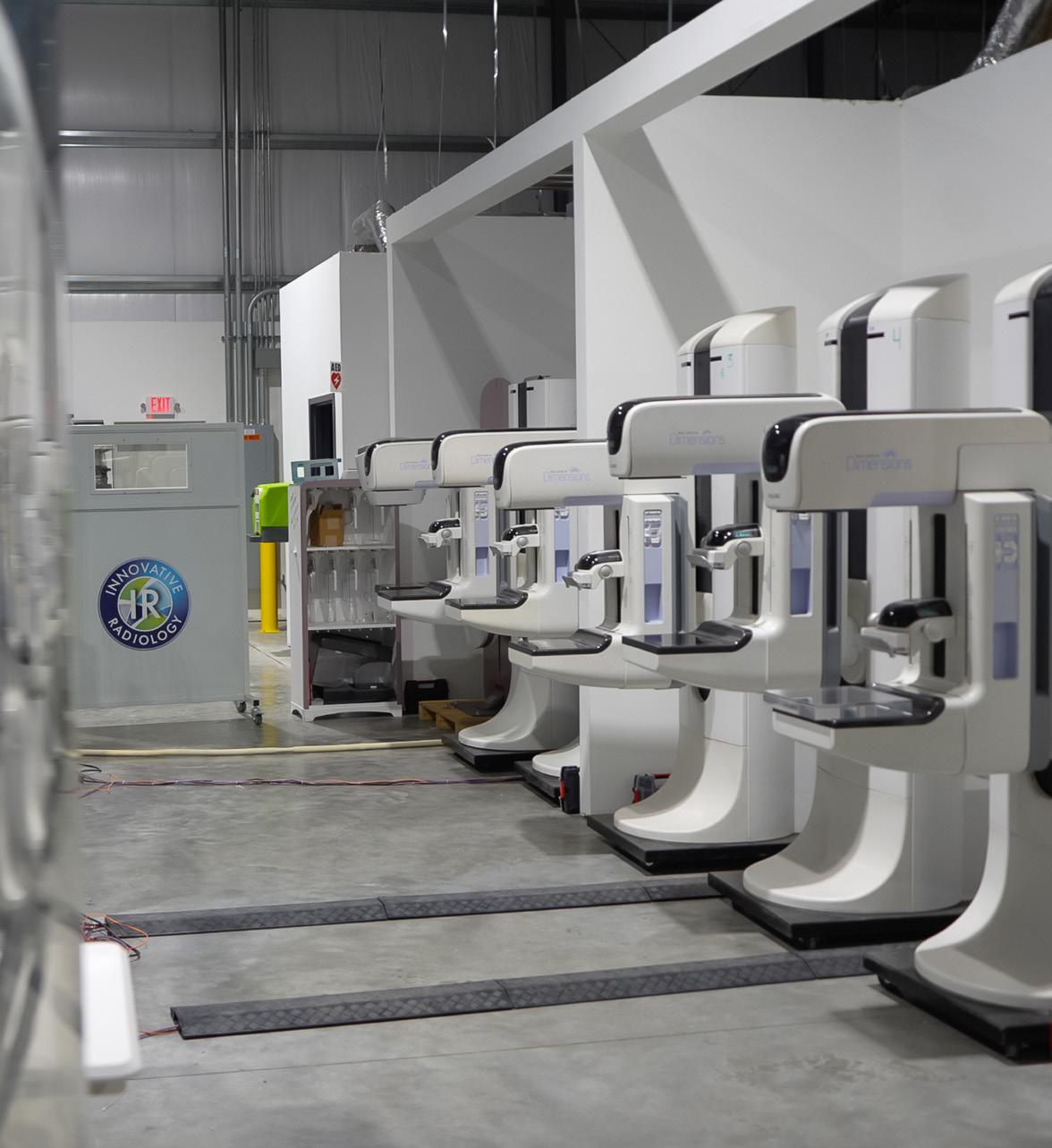
Innovative Radiology has established itself as a trusted leader in delivering high-quality Hologic Dimensions parts and custom-built systems that help imaging providers maximize performance, uptime and patient care. With a deep understanding of breast imaging technology and clinical workflow, Innovative Radiology offers solutions designed to extend the life of mammography equipment without compromising quality or reliability. Every component, including detectors, X-ray tubes, paddles, compression assemblies, power supplies, and key system electronics, undergoes a rigorous multi-point inspection and functional testing process. This ensures each part meets strict performance standards before reaching the field, giving providers confidence in both reliability and image quality. Beyond parts, Innovative Radiology specializes in custom-built Hologic Dimensions systems, tailored to meet facility needs, budget goals and workflow demands. These systems are carefully configured, tested and prepared for seamless integration, allowing providers to expand capacity, create backup coverage or replace aging equipment with confidence. By combining technical expertise, meticulous quality control and responsive customer support, Innovative Radiology continues to help healthcare organizations reduce downtime, control costs and maintain exceptional mammography performance, reinforcing its position as a go-to partner for dependable Hologic Dimen sions solutions.
*Disclaimer: Products are listed in no particular order.


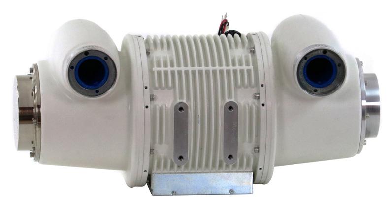
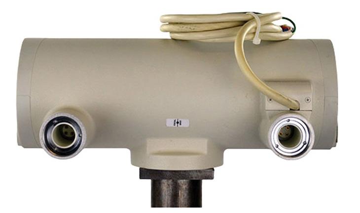
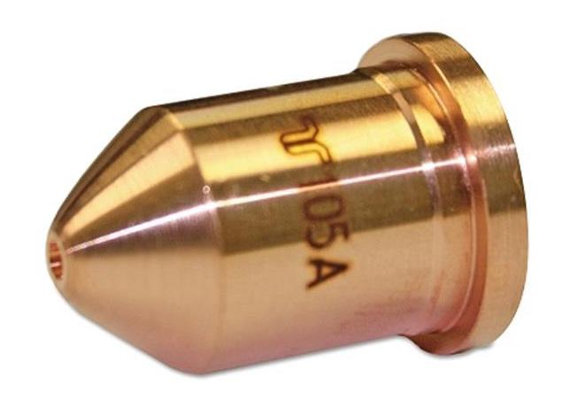
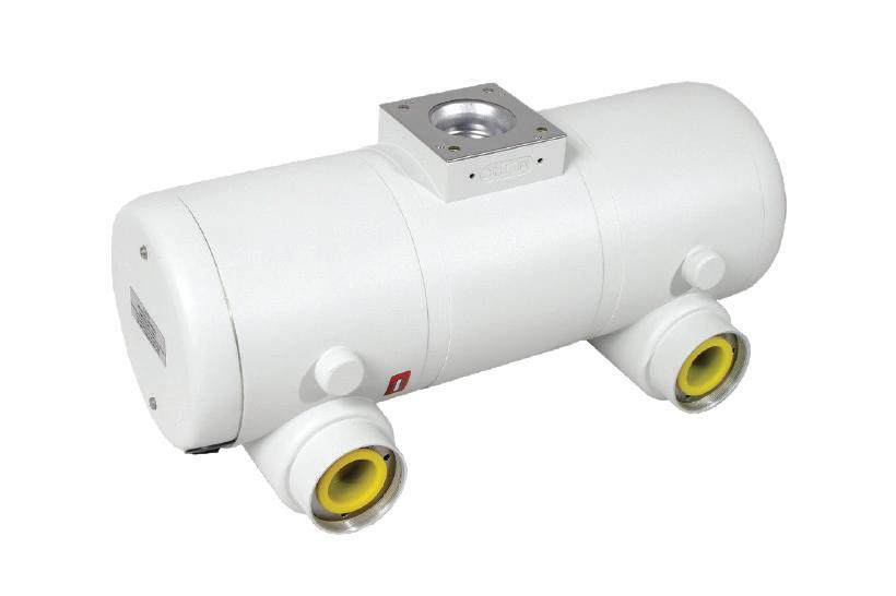
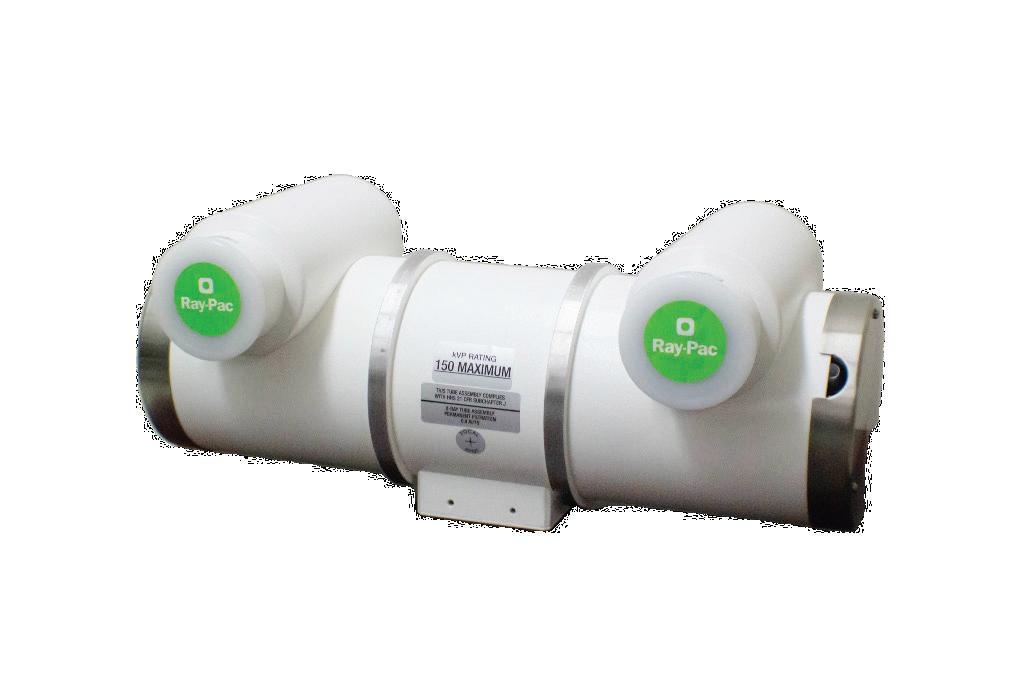
Founded in 1985, North American Imaging (NAI), a PartsSource Company, has built a reputation for providing the highest quality replacement parts for diagnostic imaging equipment. With decades of industry expertise and ISO 13485:2016-certified manufacturing, the company supports radiography, R/F, C-arm, portable X-ray tubes and other imaging systems with reliable replacement solutions. Now, NAI has re-engineered its entire operation to raise the bar for replacement
X-ray tubes, delivering premium-quality solutions designed for performance, reliability and longevity. Their tubes are backed by the longest warranty in the industry: a comprehensive two-year warranty, providing unmatched protection and peace of mind. With product in-stock and ready to ship, NAI helps minimize downtime and keeps imaging departments operating at full capacity.

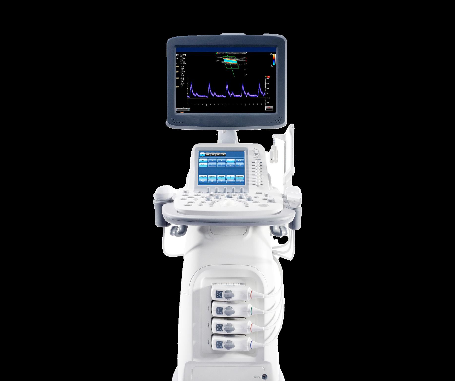
Probo Medical is your complete provider for ultrasound parts sales. Probo Medical’s parts follow stringent Quality Management System requirements. Our parts are harvested in our multiple ISO 13485 certified facilities to provide industry-leading quality. We have new and refurbished ultrasound parts in-stock and ready to ship same day for when you need them most. With a six-month warranty included on all parts, our goal is to expedite repairs, eliminate machine downtime, and help you save over OEM pricing. Our expert technical support team is here to help you source the right ultrasound parts for your practice so you can deliver the best patient care.
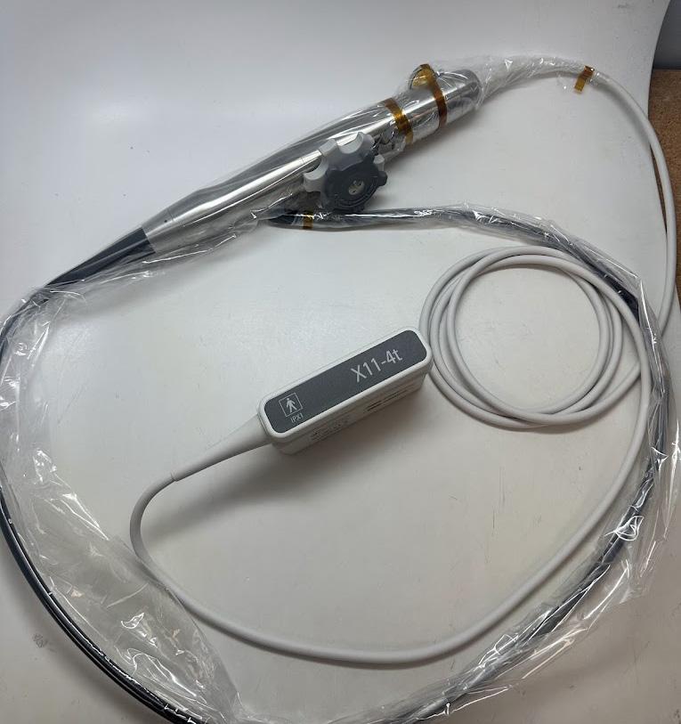
The Philips X11-4t mini 3D TEE was designed for the toughest cases. With a more compact design and advanced 3D imaging, it helps you see clearly even in smallest of spaces, whether you’re working with small children, older adults, or anyone in between. It’s easy to use, familiar if you’ve worked with the X8-2t and X7-2t. The X11-4t gives you the reach, angles, and confidence to take on more cases with less stress. When precision matters, the X11-4t is the tool that fits your patients, your procedures, and your practice. MW Imaging offers a standard 6-month warranty on all X11-4t exchanges for end users.
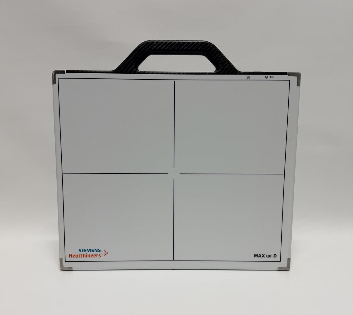
The Siemens Max WI-D Wireless Detector (35x43 panel), PN: 11105032, is a high-quality digital radiography detector designed for reliable wireless imaging performance. Manufactured in 2017, this 35 x 43 cm panel is ideal for a variety of medical imaging applications, providing consistent image capture and dependable operation. This unit has been fully tested and is confirmed to be in good working condition and is ready to ship.
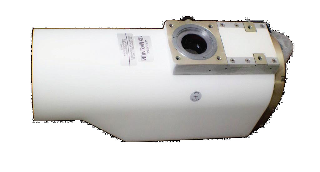
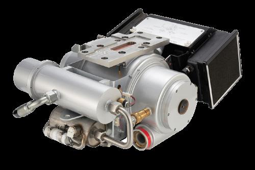
Chronos Imaging has developed an all new, formfit-function replacement tube for the GE Performix 40 Plus. This new 7.0 MHU liquid metal bearing CT tube is manufactured by Chronos Imaging at its factory in Illinois. It is fully compatible with all OEM system requirements and is designed to meet or exceed the performance of the OEM tube. The AU200-C40+LMB is backed with a 1-year full manufacturer’s warranty, and a 90-day risk-free money-back trial period. The liquid metal bearing technology allows for less friction and smoother operation, virtually eliminating bearing-related failures and allowing for quieter operation to improve patient satisfaction. The AU200-C40+LMB is used in OEM CT systems like the GE Revolution EVO and GE Optima 660. •

BY MATT SKOUFALOS

In the do-more-with-less era of healthcare, sustainability has transitioned from an aspirational public relations tool into a financial strategy that means even more to the bottom line as it does
to marketing messaging. As broad a concept as the business cases to which it’s applied, the first steps towards sustainability must necessarily employ efficiency in the act of driving at system efficiency.
In the medical imaging space, both kinds of improvements are sought, as study volumes and patient wait times continue to increase amid a staffing shortfall. Addressing the totality of these issues to yield the most significant impact requires innovation and
practicality, as decision-makers must select the variables that can yield the greatest amount of change before acting upon them.
“You can push the ceiling on innovation, but you have to raise the floor if you want to move the needle in the field,” said Vibhas Deshpande, vice president of sustainability innovation and strategic research at Siemens Healthineers Americas.
For the manufacturers of medical imaging devices, increasing equipment sustainability starts with contemplating the entire life cycle of a product, from its component materials to its end-of-life removal from service. Identifying the biggest impact of its carbon footprint can start with assessment of how raw materials are sourced, and in what quantities; the process by which those materials are transformed into devices or their component parts; and how the final products are delivered to customers.
Once a device reaches the end of its useful life, there’s additional considerations to be made about how it’s retired, refurbished, repurposed, or upgraded in the field, which can stretch the useful length of the resource well into a decade and a half or longer.
“The actual product stays operational for a long period of time,” Deshpande said. “Then you come to end-of-life, and that’s when the materials can go to recycling.”
On most consumption timelines, medical imaging devices make their heaviest impact during the 10-plus years of their useful life because they require significant amounts of energy to operate, and derive the most value for their purchasers by operating steadily, if not continuously.
“This is the biggest bucket we need to focus on,” Deshpande said. “We are doing what we can in our factories with new energy-efficient technologies, and new system designs, but use-phase energy consumption depends on utilization
patterns during a scanner’s lifetime”
Among the improvements that manufacturers seek to drive in device efficiency are performance functions –such as the capacity of a device to be switched into an economy or low-power mode, and the efficiency with which a device can enter or exit such modes – followed by onsite utilization. In that sense, Deshpande said, improvements aren’t always driven by a strict reduction in energy use as much as by efficiently generating the desired clinical outcomes. Facilities can improve their resource efficiency by addressing everything from wait times to ease of scheduling to patient access.
“Making small tweaks to the protocols to make them more efficient, we can increase access to the facility, and then that efficiency itself is more understood.”
- Dr. Michael Markl
“I always like to think of it as an efficiency metric,” Deshpande said. “If I only scanned 10 patients in a 24-hour period, it’s one thing. If I increase that by 10 patients in that same period, I use marginally more electricity but generate far more clinical outcomes.”
“It’s not only the product and product features, but machines have gotten so advanced that patient exams are not as long as they used to be,” he said. “There’s a lot of technological advancement: the longer the exam, the more energy it will use.”
“But utilization is also driven by how much time it takes to swap patients,” Deshpande said. “How long
is the machine sitting idle, consuming energy that is not being used to drive an output? How is the patient workflow through the imaging suite? There are several ways you can speed up this entire process, thereby increasing your utilization rate.”
“Even in designing all these things, are you taking into account the technologist experience?” Deshpande asked. “What staffing models do you need for maximum patient workflow?”
Many of these improvements can be retrofitted to an existing operational structure, “but when you’re building a new facility, that’s the best time to do this,” Deshpande said. Such moments often offer points of entry into efforts that can wring efficiency improvements from the least likely of areas. As much as workflow can be enhanced by artificial intelligence (AI)-powered computing technologies, seemingly lower-tech questions – like the width of a hallway as it relates to rotating a patient table – also can have a significant impact.
Deshpande points to such a collaborative project between the Northwestern Medicine health system and Siemens Healthineers, where decision-makers conceptualized a new medical imaging facility by creating a digital twin of the space and simulating the patient experience there before fitting out the physical space.
“In the digital world, you can easily change, and add one more tech, one more table; close a wall off, and say, ‘Did this relieve my bottleneck?’” Deshpande said. “All of this operational optimization in the digital world before you start building the facility saves enormous costs downstream.”
“The environment in which an imaging device is located has an impact on utilization, so facilities should be designed to maximize patient throughput.”
Dr. Michael Markl, vice chair for research in the department of radiology at Northwestern University Feinberg
School of Medicine, said the digital twin modeling experience helped visualize every detail of the patient journey virtually before fitting out the space. Those efforts are expected to reduce per-patient energy consumption by some 30 percent.
Through the comprehensive digital twin process, “you could really mimic or simulate things with the patients, staffing, techs, nurses, physicians,” Markl said, “and that, to me, is the real-world scenario.”
“Making small tweaks to the protocols to make them more efficient, we can increase access to the facility, and then that efficiency itself is more understood,” he said. “That simulation was done before the facility even opened, and that informed the construction of the facility.”
The facility has been operational for two months, and Markl’s team is in the process of gathering data to validate its predictions, having specifically purchased MRI equipment that can measure the energy consumption to track its impact on patient flow, and compare it across its other facilities.
Philosophically, the impact of simply taking those measurements – called “the observer effect” in physics – alters the outcome of how systems operate. Practically, it could also help communicate the significance of efficiency and sustainability to imaging facility staff, much in the same way that power consumption readouts on other appliances does.
“Your MRI tech and your assistant is supposed to care about the patient, and not really how much energy is being consumed,” Markl said. “If the scanner said where you’re at with the protocol you’ve selected, and how it changes if you drop something out, I think that would create a lot of awareness.”
Driving greater efficiency improvements through broader awareness includes acknowledging that health-
care is itself, as an industry, one of the leading carbon emitters on the planet.
“If healthcare was a country, it would be fourth or fifth in global emissions,” Markl said. “Radiology owns a large chunk of that emissions because we run and operate imaging equipment that consumes a lot of energy.”
By modality, the biggest energy consumer is MRI, Markl said; operating one MR machine generates the power consumption equivalent of 25 single-family households; scaled up, the imaging infrastructure of a facility like Northwestern’s equates to powering a small village. Simply powering down the devices when they’re not in use, or switching to a low-power option, would cut that consumption by a quarter to a third, but this is often not done out of convenience, or necessity to conduct an imaging study on an emergency basis, he said.
“The other option is just running the system more efficiently,” Markl said. “Can you optimize the scan pro-
tocol to make it more efficient? If you could shorten the scan, the power consumption would be lower. Then, can you minimize idle time between patients?”
Beyond facility and institutional efficiencies, Markl supports the expansion of MR life cycles through component and system refurbishment programs, which not only save money, but help keep systems in service longer, which has a downstream effect on patient access.
“That’s a good incentive for the organization or the vendors,” he said.
Markl also spoke about the less-contemplated notion of improving sustainability efforts by patient education efforts that would increase awareness about imaging procedures.
“If you’re getting an MRI, you want the MRI because you want to know what’s going on,” he said. “Nobody really talks to the patient about it. A lot of the conversations have been between the vendors and the societies, and the stakeholders and patients are not as involved as they could be.”
Dr. Susie Yi Huang, a radiologist at Mass General Hospital and associate professor at Harvard Medical School, supports the variety of efforts undertaken system-wide “to try to find common ground for people to understand what sustainability means” in the medical imaging space.
“Many people, especially in this generation, are extremely cognizant of the environmental impact of medical imaging technology,” Huang said. “High-efficiency MRI – getting scan slots to be shorter – also speaks to radiology across the board. As medical demand grows with our aging population, our need to provide timely and quality care with our patients really has come into focus.”
In a healthcare industry that’s broadly tasked to wring as much value as possible from every process, environmental benefits typically are
discussed secondarily to economic incentives. Yet Huang said that her examinations of the topic have illustrated the greater degrees of operational freedom energy consumption mitigation strategies derive from simply scheduling and scanning patients more efficiently.
“In addition to improving revenue by doing more scans per day, you may be able to show improvement in energy efficiency,” she said. “Here at Mass General, we’re embarking to get all of our scan slots down to 20 minutes in the next year.”
To get there, the hospital is thinking through both physical and virtual improvements to its operations. This includes AI-optimized image acquisition and reconstruction methods, which Huang said “allow us to scan faster with more robust processes than we had immediately.” Meanwhile, on the reporting side, AI-powered computing processes help synthesize results through large-language models embedded in dictation software. Such models can also be used to summarize reports, which also helps save time in the interpretation and delivery of study results.
At Assembly Row, a mixed-use development just outside of downtown Boston, Mass General is working through some of its top-to-bottom efficiency analyses in an ambulatory imaging setting designed to hit that 20-minute study window.
By setting up three identical 3T Siemens MRI systems in sequence, Huang’s team could prep one patient while another was being scanned, and wheel the next patient in while the previous patient finished up. Leveraging these identical configurations allowed staff to catch up on time at a facility-wide level, with the aim of showing cost savings by virtue of the revenue increase that patient throughput improvements generated. Power meters installed on the systems helped show the path of energy consumption at every step of the most common outpatient imaging studies, quantified it, and compared that consumption against a control facility operating four MRI systems on a single-scanner model.
“We showed that we could reduce turnaround times from seven to 10 minutes down to three to five minutes,” Huang said, “and when you aggregate that over multiple exams and patients, you can eke out more scan slots.”
“That shows the opportunities that cost savings can actually bring about energy savings,” she said. “We were doing the same patient volumes with the three scanners [at Assembly Row] as compared to the four at the control. It is a bit more resource intensive, but you are gaining in terms of efficiency.”
Demonstration projects like the Assembly Row ambulatory center or the Northwestern digital twin power consumption modeling study illustrate how collaborative approaches to medical imaging sustainability can yield the greatest results. Institutional partnerships underscore the shared responsibility of tackling the issue as well as how they can yield dividends enjoyed by stakeholders across the board.
“The moment we think of this as someone else’s problem, we’re not going to solve the problem,” Deshpande said. “In all my interactions with institutions that own our equipment, I never had the sense that they were suggesting [that sustainability] was only the manufacturer’s responsibility.”
“This is one space where, if we do not work together, we are very unlikely to succeed,” he said. “We all need to be working closely if we want to move the needle. If we say it’s someone else’s responsibility, we will not make meaningful progress.” •

VIBHAS DESHPANDE
Vice President of Sustainability
Innovation and Strategic Research at Siemens Healthineers Americas

DR.
SUSIE YI HUANG
Radiologist at Mass General Hospital and associate professor at Harvard Medical School

DR.
MICHAEL MARKL
Vice Chair for Research in the Department of Radiology at Northwestern University Feinberg School of Medicine


DIRECTOR’S CUT
BY NICOLE DHANRAJ
Radiology leaders are not short on effort. They are short on options. Across the country, imaging departments are competing for the same limited pool of credentialed technologists. Recruitment budgets increase. Travel contracts fill urgent gaps. Internal teams stretch to maintain coverage.
The cycle feels familiar. Recruit harder. Pay more. Hope retention improves.
But what if the constraint is not effort? What if it is access?
One pathway that radiology has not fully evaluated is the Department of Defense SkillBridge program, a federally supported transition model that allows service members to gain civilian work experience during their final six months of military service.
While industries like aerospace and cybersecurity have used SkillBridge strategically for years, healthcare and imaging specifically have been slower to engage. For radiology, that hesitation may represent a missed opportunity.
The U.S. military trains and employs imaging professionals across radiography, CT, MRI,
nuclear medicine and interventional environments. These technologists often work in high-acuity settings that demand strict protocol adherence, disciplined workflow and regulatory awareness.
They are not unfamiliar with structured systems. They live in them.
When these professionals transition out of military service, most healthcare organizations encounter them only after they enter the open civilian labor market. At that point, competition begins.
SkillBridge creates the opportunity for earlier engagement.
Imaging departments can establish structured transition periods that allow:
• Alignment with ARRT credentialing requirements
• Evaluation of modality competencies
• Cultural and team fit assessment
• Continuity planning before separation Civilian credentialing requirements remain intact. It is a pipeline conversation. And radiology has not historically framed it that way.
Imaging departments depend on more than scan volume.
• Documentation integrity
• ACR and Joint Commission readiness
• Compliance tracking
• Workflow coordination
• Quality assurance oversight
These are operational layers where strain accumulates quickly.
SkillBridge participants, whether clinically trained or operationally specialized, can support defined areas within clear supervision parameters. When thoughtfully designed, this strengthens infrastructure without compromising clinical standards.
Radiology administrators are right to ask hard questions about scope, supervision and regulatory compliance. SkillBridge does not change licensure standards. It does not redefine scope of practice. It does not eliminate supervision.
Successful integration requires structured role definition, coordination with compliance teams and alignment with modality-specific credentialing pathways. In many cases, the barrier is not regulation. It is unfamiliarity.
If radiology continues to compete exclusively in the same hiring pool, the pressure will persist. Departments that begin thinking in terms of pipeline development rather than episodic hiring will position themselves differently over time.
SkillBridge is not a quick fix. It is a structured lever, one that allows imaging leaders to engage CT, MRI and radiography professionals before they enter a competitive civilian marketplace. Radiology does not need to lower its standards to address workforce strain. It needs to widen its lens.
For departments willing to evaluate alternative pipeline strategies, the opportunity is not theoretical. It is already embedded within a federal system that many industries have learned to navigate. The question is whether radiology is ready to do the same. •
Nicole Dhanraj, is a radiology administrator and workforce strategist focused on military-to-healthcare transition pathways. Radiology leaders interested in exploring this approach are encouraged to connect with her directly at nicoledhanraj@gmail.com.

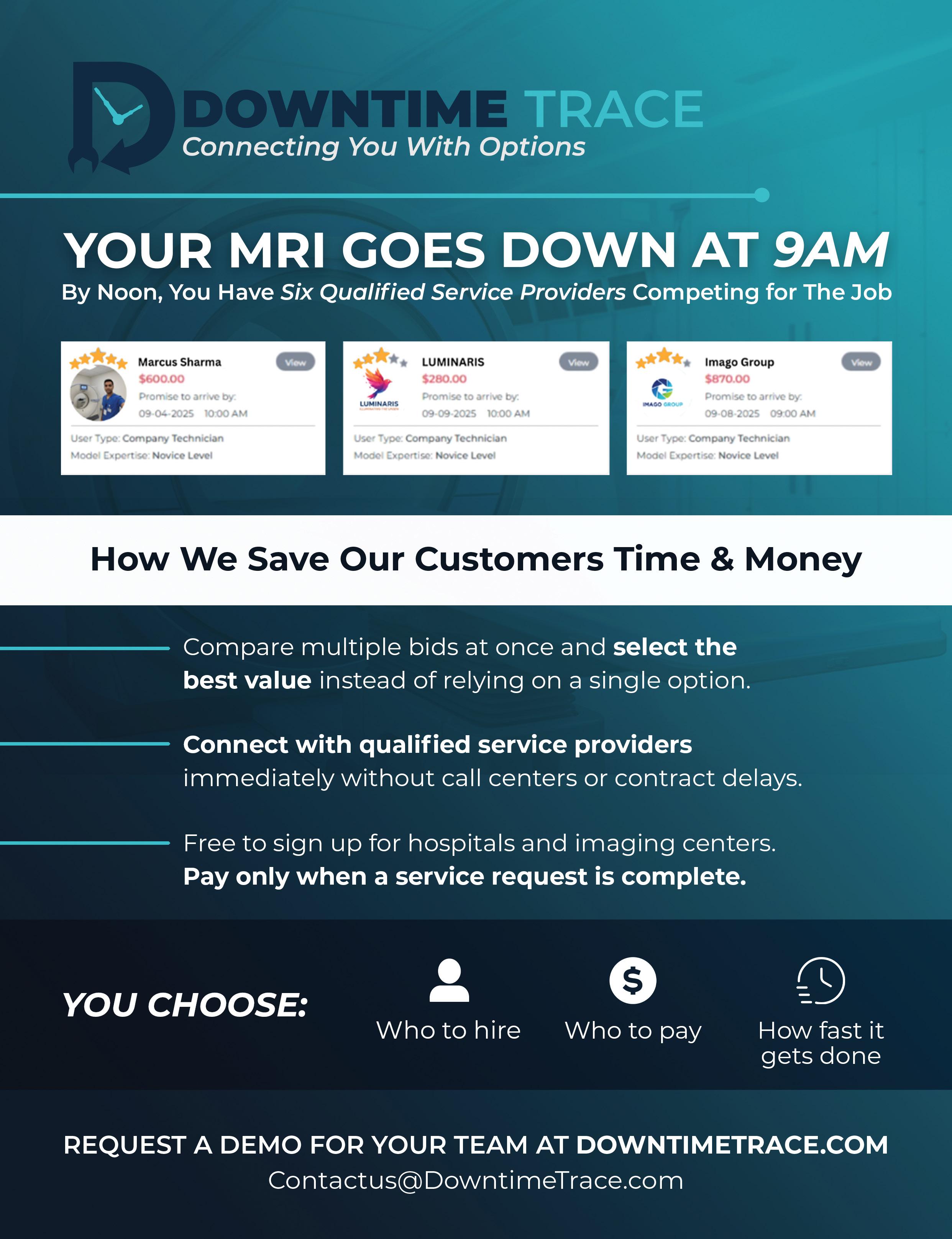

BY MARK WATTS
Infusion pumps sit in a special place in healthcare. They are everywhere, they touch medication delivery, and they run quietly in the background while clinicians focus on the patient. Because they are so common, people sometimes forget how much technology is inside them. Modern pump fleets depend on software, network connections, drug libraries, central management servers, wireless coverage, user logins, remote support tools, and a steady flow of updates and configuration changes.
Infusion pump security is not an “IT side project.” It is part of safety and reliability, right alongside preventive maintenance, calibration and battery checks. When infusion pump cybersecurity is weak, the risk is not just stolen data. The risk is downtime, delayed therapy and a hospital scrambling to keep medication delivery running while systems are unstable.
A well-run Security Operations Center (SOC) helps stop that scramble before it starts. A good SOC feels like a fire station crossed with a quiet reading room. There is always urgency in the background, but most of the work is slow, careful and methodical. SOC staff watch for trouble the way a maintenance team watches for early signs of failure. They look for small shifts in behavior that suggest something is drifting out of control. In the infusion pump world, those shifts can show up as unusual network traffic between pumps and a server, strange login patterns in the pump management console, unexpected changes to drug libraries, or a vendor remote session that starts at an odd hour. The core practices sound simple: watch everything, log everything, verify everything, and never assume you have seen the last weird pattern. In a busy hospital with thousands of devices,
those simple ideas turn into daily discipline.
Most attackers do not wake up thinking, “I want to hack an infusion pump.” They wake up thinking, “I want access.” They want a foothold inside a hospital network. They want credentials they can reuse. They want systems they can disrupt for ransom. Pumps and their management systems can become part of that story because they are widely deployed and often connected in ways that are hard to redesign quickly. Pump ecosystems may include legacy components, shared service accounts, older operating systems in management servers, and vendor tools that are difficult to replace. Even when a pump itself is locked down well, the supporting systems can be exposed if patching is delayed, access is shared or logging is thin.
Hospitals also run on speed and trust. If a device “usually works,” people rely on it and move on. That is normal in clinical care, but it can create security gaps. In real life, small gaps pile up. A password that is the same across multiple devices, a remote access rule that is too wide, an out-of-date certificate, or a drug library workflow that is not tightly controlled can create openings. Security work is about closing those openings without breaking clinical flow. That is where the SOC, clinical engineering, IT, pharmacy leadership, and vendors need to operate like one team instead of separate silos.
Most SOCs divide work into tiers, and the tier approach matters in infusion pump security because speed and accuracy both count. Tier 1 analysts are the first set of eyes. They watch alerts and signals, and they sort noise from the few events that truly matter. In a hospital, noise is constant. Pumps connect and reconnect. Firmware checks happen. Wireless roaming looks “busy” even when it is normal. A Tier 1 analyst learns what normal looks like for a pump subnet at 2 a.m. They learn
what normal looks like when pharmacy publishes a new drug library. They learn how many management console logins are typical during shift change. They learn the difference between planned vendor support and a remote access session that is not on the schedule.
Tier 2 analysts take cases that look real or confusing and work them deeper. In pump security, Tier 2 might investigate why a management server is pushing unusual commands, why a set of pumps is beaconing to an unfamiliar destination, or why a service account tied to pump management suddenly shows activity from a workstation that should never use it. Tier 2 work is about building a story with evidence, not guessing. A good Tier 2 analyst looks for the “shape” of the event. Is it a misconfiguration, a workflow change that was not communicated, or does it match a known attack pattern like credential misuse and lateral movement? In infusion pump ecosystems, that story often includes clinical context. For example, if pharmacy is updating a library and clinical engineering is testing a new configuration, the system may legitimately look noisy. The SOC must know how to validate that quickly without slowing care.
Tier 3 work begins when cases get technical and high risk. Tier 3 might analyze suspicious files found on a pump management workstation, dig into a network capture to see what a device is really talking to, or hunt for hidden persistence on a server that coordinates pump updates. Tier 3 staff also tend to be the bridge builders. They work with clinical engineering to understand the normal behavior of the pump fleet. They work with IT infrastructure to confirm segmentation and firewall
rules. They work with vendors to validate patch plans and remote access methods. This is the kind of work that needs quiet focus, patience and strong documentation.
In infusion pump security, the SOC cannot succeed on skill alone. Leadership sets policies and expectations, and those expectations decide whether the SOC is reactive or prepared. Leaders define what “normal” looks like for the hospital and what “not normal” requires immediate action. They decide response targets, who gets called after hours, and how to coordinate containment actions so patient care is protected. They also define evidence handling rules, because in serious incidents, the details may matter legally and operationally. If a pump management server is compromised, the hospital may need to prove what was touched, what was not touched, and what actions were taken. Without clear leadership guidance, people hesitate. In security, hesitation is expensive.
A strong SOC, backed by clear policies and smart automation, can protect infusion pump fleets in a way that supports care instead of disrupting it. It can catch early signs of trouble, connect the dots across systems, and bring the right people together quickly. The end goal is not perfect security, because no environment is perfect. The goal is resilience. It is keeping medication delivery steady, keeping systems trustworthy, and keeping patients safe when the digital world gets noisy. •
Mark Watts is an experienced imaging professional who founded an AI company called Zenlike.ai.



BY NICOLE DHANRAJ
Irecently walked through a newly built hospital that was, by every visible measure, impressive. The imaging department was sleek and efficient. The technology was state of the art. The space reflected careful planning and significant investment.
When I asked the team how things were going, the answer surprised me. Volumes were low. Patients were not using the facility the way leaders had expected. Many were traveling to other locations, sometimes much farther away.
When staff asked why, the feedback was consistent. The space felt too modern for the patients they serve. Too unfamiliar. It didn’t feel like it was built for them.
That moment reframed how I think about healthcare design. A space can be beautiful, compliant, and technologically advanced, and still fail the patients it was intended to serve. I must say I hadn’t considered this aspect before – especially through a cultural lens.
Healthcare facilities across the country are investing in modernization. Imaging department design often emphasizes standardization, digital workflows and high-
tech aesthetics. These decisions are meant to improve efficiency and safety.
But patients do not experience design through blueprints or equipment specifications. They experience it through culture, memory, stress and trust.
Inclusive healthcare design recognizes that space is never neutral. What feels clean and advanced to leadership may feel cold, overwhelming or unwelcoming to a patient whose past experiences with healthcare involved fear, marginalization or loss of control.
When patient-centered design is missing, people disengage quietly. They delay imaging, miss appointments, or seek care in spaces that feel more familiar, even if those spaces are older or less advanced. This matters deeply in imaging environments. Imaging requires vulnerability. Patients are asked to undress, lie still, enter enclosed machines and trust staff they may have just met. When the environment feels impersonal or overly technical, anxiety rises.
That anxiety shows up operationally through motion artifacts, incomplete exams, repeat scans, longer appointment times and increased strain on technologists. Stress in imaging is not an abstract concern. It directly affects quality,
throughput and outcomes.
Culturally responsive healthcare spaces reduce that stress by building therapeutic trust. Familiar materials, intuitive wayfinding, respectful privacy and visible human presence help patients feel grounded. Trauma-informed and inclusive design choices – such as quieter waiting areas, softened lighting, acoustic control, and spaces that allow family support – give patients a sense of agency at a moment when they have very little.
Standardization alone cannot accomplish this. There is no one-size-fits-all imaging department. Every design choice reflects assumptions about who the patient is and how they should move through care. When those assumptions don’t match the community being served, the space becomes a barrier, even when intentions are good. I saw this first hand and I don’t think the hospital leaders understand how to fix it.
Many leaders are constrained. They inherit buildings, vendor-driven layouts and system-wide design standards. Leadership, however, shows up in the questions we ask. Who might feel uneasy here? Where does efficiency override dignity? Where does technology replace human connection too quickly? How do we invite when our space
feels different/unfamiliar?
True modernization is not just about advanced equipment or clean lines. It includes cultural intelligence. It considers how design influences trust, stress and belonging.
A successful imaging department is not defined solely by how it functions. It is defined by whether patients choose to enter it, feel safe within it and return when care is needed.
When inclusive design places patients at the center, new spaces don’t just look modern. They earn trust.
This article begins a series on inclusive healthcare design and how physical and technological spaces influence patient trust, stress and access to care.
Future pieces will explore these dynamics in imaging environments and other clinical settings, with a focus on the lived experience of the patients we serve.•
Nicole Dhanraj, is a radiology administrator and workforce strategist focused on military-to-healthcare transition pathways. Radiology leaders interested in exploring this approach are encouraged to connect with her directly at nicoledhanraj@gmail.com.

Almost every organization has that one colleague who drains the energy from every meeting. It’s often someone who resists collaboration and acts as if their way is the best way. One phrase that could be applied to such individuals is “workplace disruptors,” because not only do they frustrate their peers, they erode trust. They drive even talented people to the exit. Left unchecked, a single such team member can unravel even the most productive company culture.
You’ve probably encountered such a person. Not only do they believe their way is the right way, they resist even considering alternative perspectives. Complicating the situation, their mantra is usually, “This is who I am – deal with it.” Even one person displaying such an attitude can cause a company to crumble from within, especially if that person is in senior management or a leadership position.
Let me share a real-world example. We’ll call this gentleman Bill (not his actual name). Bill was actually quite talented. He grasped the big picture with ease and knew how to identify obstacles in the way of success. But despite that, he was seen as an uncooperative know-it-all. He was not just a disruptor, he was considered abrasive.
Bill had received several promotions, although those advances happened in spite of his interpersonal style, not because of it.
How do I know these details? Because Bill’s supervisor brought me in to work with him. The company’s president wanted Bill to smooth out his rough edges and embrace collaboration because too many other valuable employees had reached their breaking point and were ready to quit.
“Bill is super valuable,” the president told me, “but if he doesn’t start working better with others, I’m going to lose the whole team.”
The main issue is that Bill saw problems in everyone else’s workstyle except his own. For example, he was quick to say the office manager needed to overhaul her workflow and he openly stated that the purchasing manager should’ve shown more initiative.

BY DANIEL BOBINSKI
One could talk with Bill for any length of time and you’d hear him dissect nearly all of his coworkers’ shortcomings. However, if anyone asked Bill about his work or his department, he’d usually respond by saying he needed more sleep because he was working so incredibly hard.
Stated plainly, Bill was oblivious to the fact that his direct reports bit their tongues when he was around. They were afraid to talk about their work just to protect their paychecks. Bill also didn’t see how his attitude impacted his peers. As one of Bill’s colleagues confided to me, “Nobody wants to be in the office when Bill makes his rounds.” Another told me, “We all maintain our professionally polite masks, but we don’t engage with Bill except when necessary, because we don’t think he appreciates what we do.”
You’ve likely heard it said that employees don’t quit companies; they quit difficult managers. Ironically, people like Bill struggle to understand why people around them are leaving. They point to every possible factor except their own behavior.
If you’re dealing with such people in your organization, the solution should always include human resources, but it often boils down to a direct ultimatum. This usually needs to come from someone higher on the organizational chart, and might sound like, “You need to make substantive changes, or other changes will be necessary.”
It was only after such a line was drawn that Bill agreed to work on himself.
I should point out that these situations become especially problematic if the offending party serves at the top of an organization, as there’s nobody with the authority to create an ultimatum. But that’s a topic for an entirely separate discussion.
If there’s someone in your organization whose eyes need to
be opened to the negative impact they’re having on others, let me offer this advice: don’t wait. The longer a person operates with an abrasive style, the greater the probability that the person’s self-defensive walls will grow higher when challenged.
Their mindset becomes: “These methods brought me this far – why abandon them now?”
Still, at the heart of the problem, it helps to have empathy for the person who has little empathy. Laura Crawshaw, founder of the Executive Insight Development Group and author of “Taming the Abrasive Manager,” once said, “Abrasive bosses are driven by fears of being perceived as incompetent and they are blind to their impact on others.”
Thus, enrolling someone in a training class might provide marginal benefit, but it’s seldom the complete answer. The same defensive patterns remain as they adopt the attitude that says, “This soft-skills training is meant for other people, not for me.”
The optimal solution for helping abrasive or uncooperative individuals is personalized one-on-one mentoring or coaching. Why? Because problematic behavior that originates from insecurities need personalized attention. Fears and an underdeveloped ability to connect interpersonal dots are not going to get addressed with other people around.
Through one-on-one mentoring or coaching, specific issues receive direct attention. Troublesome behaviors can be evaluated and discussed carefully. Without such focused ac-

countability, old patterns persist, particularly if the offending party holds significant organizational authority. Remember, abrasive managers may genuinely believe their current style propelled them to their present position.
As a former executive coach for 35 years, I can tell you that if you’re dealing with an abrasive or uncooperative manager in your workplace, know that many certified management and executive coaches have extensive experience resolving these challenges.
So, if you have an uncooperative or abrasive coworker, may I suggest consulting with HR about engaging a coach to assist that individual. Remember, the root cause of their conduct usually traces back to nobody ever teaching them what they needed to learn.
I’m a firm believer that coaching works. My client Bill was reluctant to start coaching sessions, but had been given an ultimatum. Eventually he saw for himself the value of adapting his approach, and after six months his supervisor and colleagues reported that Bill was considerably easier to work with. •
Daniel Bobinski, who has a doctorate in theology, is a best-selling author and a popular speaker at conferences and retreats. For more than 30 years he’s been working with teams and individuals (1:1 coaching) to help them achieve excellence. He was also teaching Emotional Intelligence since before it was a thing. Reach him by email at DanielBobinski@ protonmail.com or 208-649-6400.
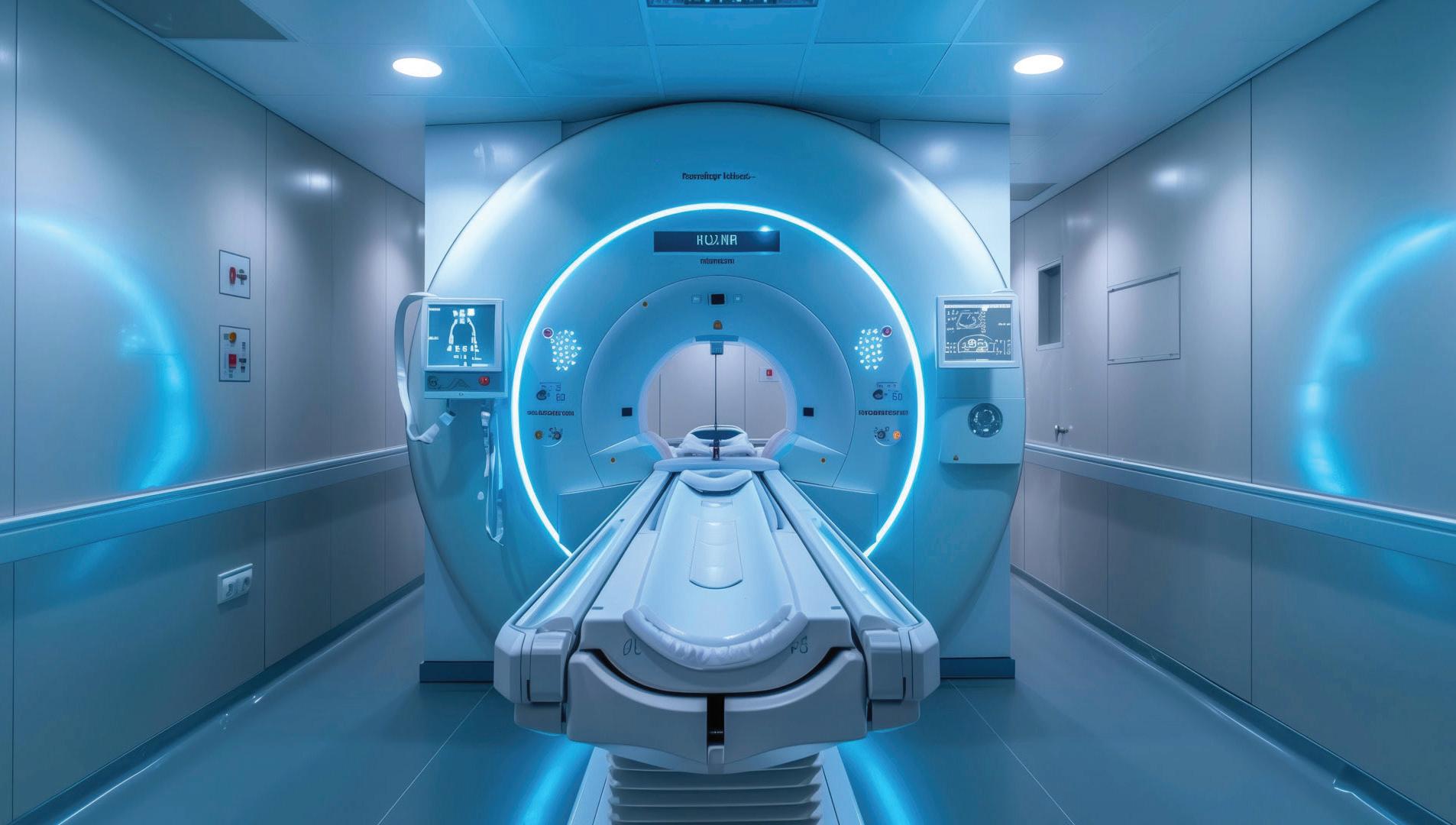
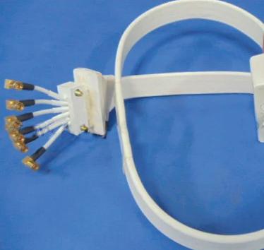

Researchers have found that a specific body profile – higher muscle mass combined with a lower visceral fat to muscle ratio –tracks with a younger brain age, according to a study presented at the annual meeting of the Radiological Society of North America (RSNA). Visceral fat is hidden deep within the abdominal cavity, surrounding vital internal organs.
“Healthier bodies with more muscle mass and less hidden belly fat are more likely to have healthier, youthful brains,” said senior study author Cyrus Raji, M.D., Ph.D., associate professor of radiology and neurology in the department of radiology at Mallinckrodt Institute of Radiology at Washington University School of Medicine in St. Louis, Missouri. “Better brain health, in turn, lowers the risk for future brain diseases, such as Alzheimer’s.”
Brain age is the computational estimation of chronological age from a structural MRI scan of the brain. Muscle mass, as tracked by body MRI, can be a surrogate marker for various interventions to reduce frailty and improve brain health, and brain age predicted by structural brain images can lend insight to Alzheimer’s disease risk factors, such as muscle loss.
“While it is commonly known that chronological aging translates to loss of muscle mass and increased hidden belly fat, this work shows that these health measures relate to brain aging itself,” Raji said. “It shows muscle and fat mass quantified in the body are key reflectors of brain health, as tracked with brain aging.”
For the ongoing study, 1,164 healthy individuals (52% women) from four sites were examined with whole-body MRI. The mean chronological age of the participants was 55.17 years. The researchers combined MRI imaging with T1-weighted sequences, a technique that produces images where fat appears bright and fluid appears dark. This allows for optimal imaging of muscle, fat and brain tissue. An artificial intelligence (AI) algorithm was used to quantify total normalized muscle volume, visceral fat (hidden belly fat), subcutaneous fat (fat under the skin) and brain age.
The researchers found that a higher visceral fat to muscle ratio was associated with higher brain age, while subcutaneous fat showed no significant association with brain age.
“The participants with more muscle tended to have younger-looking brains, while those with more hidden belly fat relative to their muscle had older-looking brains,” Raji said. “The fat just under the skin wasn’t related to brain aging. In short, more muscle and a lower visceral fat to muscle ratio were linked to a younger brain.”
Building muscle and reducing visceral fat are actionable goals, he added. Whole-body MRI and AI brain-age estimates provide objective endpoints to design and monitor interventions, including programs or therapies under study that lower visceral fat while preserving muscle.
Raji noted that this work demonstrates how body and brain health are closely linked.
“This research has validated widely held hypotheses about the association between body composition bio-
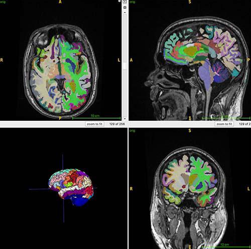
markers and brain health and provides a foundation for those biomarkers to be included in future trials of various metabolic interventions and treatments,” he said.
While widely prescribed glucagon-like peptide-1 (GLP-1) weight loss drugs, such as Ozempic, are powerful at inducing fat loss, they may also be related to a higher burden of muscle loss. According to Raji, the findings of this study could inform the design of future therapeutics, such as GLP-1 medications that target visceral fat more than subcutaneous fat and minimize muscle loss.
“Losing fat – especially visceral fat – while preserving muscle volume would have the best benefit on brain aging and brain health based on insights from our work,” he said. “Thus, our study can inform future treatments by promoting research that quantifies MRI of body fat, muscle and brain age, which can help determine the optimal dosing regimens for GLP-1s to achieve the best outcomes in body and brain health.” •
Co-authors are Somayeh Meysami, M.D., Soojin Lee, Ph.D., Saurabh Garg, M.Sc., Nasrin Akbari, M.Sc., Rodrigo Solis Pompa M.D., M.H.Sc., Ahmed Gouda, M.Sc., Thanh Duc Nguyen, Ph.D., Saqib Abdullah Basar, M.B.B.S., M.P.H., Yosef G. Chodakiewitz, M.D., David A. Merrill, M.D., Ph.D., Alex Exuzides, Ph.D., M.D., Amar P. Patel, M.D., Daniel J. Durand, M.D., M.B.A., and Sam Hashemi, M.Sc

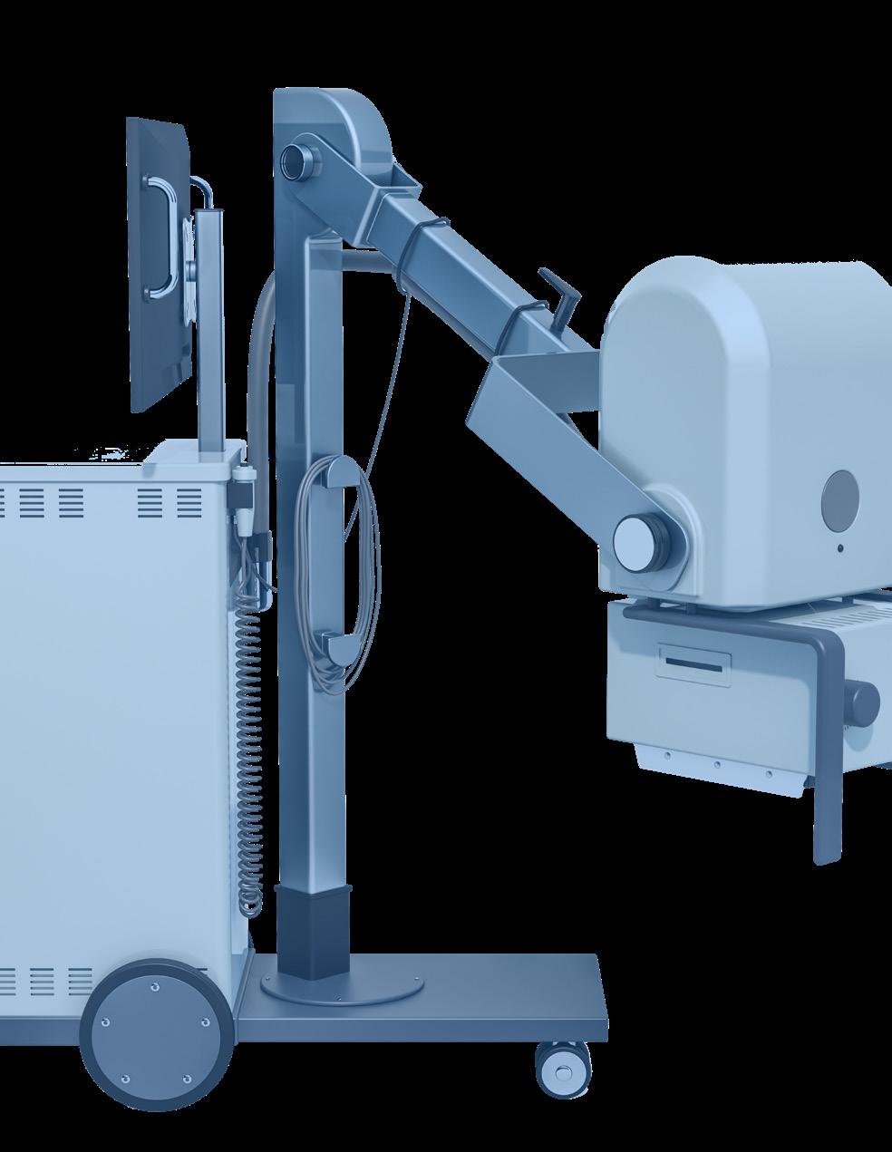


1 Recent X-ray technology improvement: _____ radiology
6 Biol. or chem., for short
9 Under the weather 10 Reciprocal piece in a machine
11 Evaluate vendors based on ____ support, training programs and technology roadmaps
13 Fundamental safety principle in radiation protection, acronym
14 Doctors, for short
15 Make fine adjustments to ensure optimum functioning of imaging equipment, e.g.
17 Soft food
18 Experts knowledgeable in equipment and software, abbr.
20 There’s a been a recent important step forward in the _____ of images in x-ray technology
22 Egg centers 23 The S in PMS

27 X-ray imaging technique showing continuous moving images
32 One of ten digits
33 Public relations, for short 34 Unit of radioactive activity
35 Measuring unit intended to represent the stochastic health risk of ionizing radiation
36 Add together, 2 words
Applaud 15 They are documented in patient records 16 Hosp. section, abbr. 17 Measure of acidity, abbr.
19 Imaging companies have a ____ of care
20 Key positions in the NFL, abbr. 21 Nitrogen and oxygen, mainly
Catchall abbr.
25 Provide needed devices and accessories
26 Way in or out
Musical scale notes
Highway, abbr.
Harden
Choose


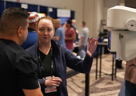
The 2026 Imaging Conference & Expo featured a packed exhibit hall, signature networking events and a record high number of attendees. 1 2 3 5 4


1. The Reverse Expo provides “speed dating” for companies and healthcare leaders to break the ice.
2. A mariachi band filled the exhibit hall with music.
3. Colorful balloons, especially this cactus, were popular photos spots at ICE San Antonio.
4. Exhibitors shared the latest solutions with attendees in the exhibit hall.
5. The ICE18 Leadership Dinner included a gourmet meal and boat tour of the popular San Antonio Riverwalk.
6. The keynote address “Advocacy in Action: What’s Happening in Radiology” provided great insights.
7. Rio & Rhythm: A Riverwalk Fiesta capped off an excellent conference.
8. The popular door prize drawing had many attendees leaving the exhibit hall with valuable prizes.


9. The exhibit hall was the place to be for networking and knowledge.
10. Colleagues always enjoy meeting up at the Imaging Conference & Expo.
11. Everyone was all smiles at the final night party.
12. The Leadership Summit provided a forum for imaging leaders form across the nation to come together and discuss common challenges and solutions. 8 9 10 12 11 7 6





SalesMaker
Espin
ImageOne
LASO Imaging Solutions lasoimaging.com
X-Ray America, LLC x-rayamerica.com • 854-999-6888
Maull Biomedical Training maullbiomedical.com
Diagnostic Imaging A+ Medical Company, Inc aplusmedical.biz/ •


•
•
•
•
•
•


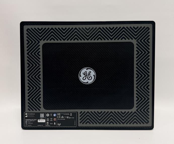
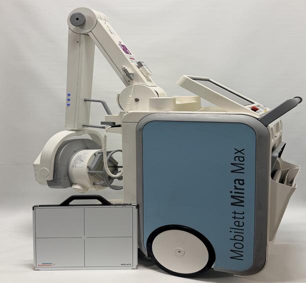
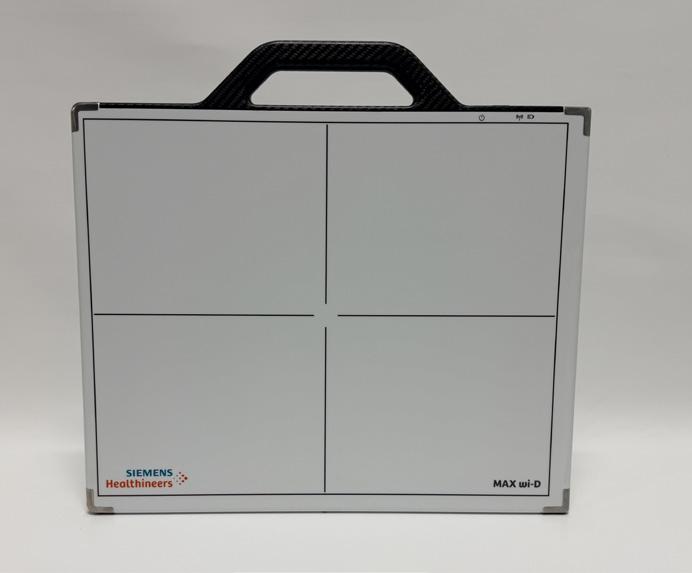


Scan the QR code to cast your vote