LEADING THE WAY
SPRING 2026 | VOLUME 18, NUMBER 1
4
6 Implementing Trauma Video Review in a Pediatric Trauma
4
6 Implementing Trauma Video Review in a Pediatric Trauma
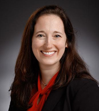
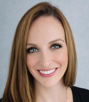
In this edition of Leading the Way, Dr. Gwen Lomberk provides her final reflections on the Division of Research, which she started nine years ago! We are so fortunate to have Dr. Tammy Kindel continue this invaluable resource for faculty and resident academic success. The Division of Research has grown to encompass a wide range of academic achievement which is so critically important to career development. Academic achievement makes everything else possible – it is the key to clinical program growth, the bridge to scientific collaborations, an inspiring advocate for the patient of tomorrow through new therapies and clinical innovations, and yes, the underpinning of financial success.
For those who may question the powerful impact of scientific discovery and innovation on the clinical enterprise, just turn on the radio or TV and listen to the adds for every health system –they are all about science and new and emerging therapies – all of this interpreted by patients and referring physicians as hope for a better outcome. There can be no greater priority than ensuring the long-term success of the Division of Research – for the many reasons Gwen mentions in her “must read” article. We will be forever grateful for Gwen Lomberk’s many contributions and equally indebted to Tammy Kindel for continuing this vital tradition of academic excellence in our Department.


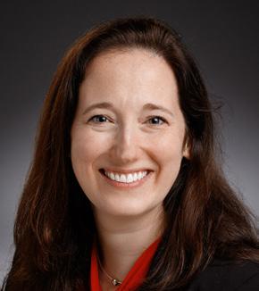
Gwen Lomberk, PhD
Former Chief, Division of Research, Department of Surgery
Adjunct Professor
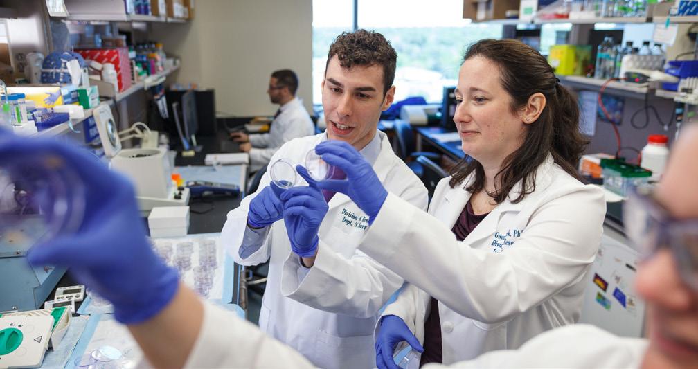
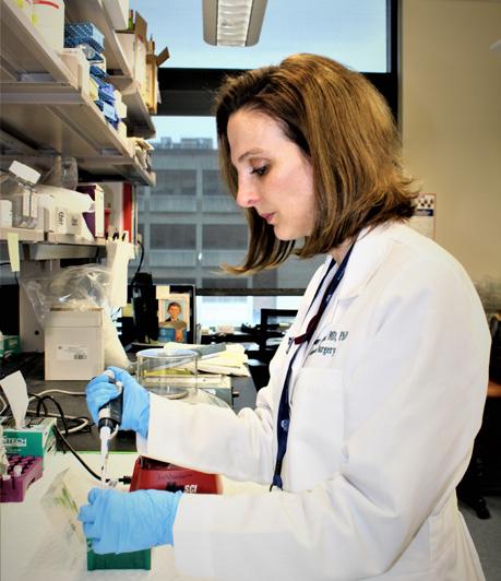
As I reflect on my nine years serving as Chief of the Division of Research within the Department of Surgery, I am extremely grateful for the opportunity to work alongside talented staff, surgeons, scientists, and trainees to build and support a research enterprise that advances our department’s academic mission. It has been a privilege to work with an extraordinary group whose dedication, creativity, and professionalism define the success of the Division. What we have built together is the product of sustained effort, a common vision, and a collective commitment to advancing the research mission.
During this period, the Division of Research was established as a centralized, reliable infrastructure to support investigators across the full lifecycle of research, bringing together previously distributed research support into a coordinated, cohesive framework. Early on, we focused on building robust pre-award services, formalized as Surgery Pre-Award Support Services (SurPASS), to ensure that faculty received consistent, accurate, and timely support during proposal development and submission. This included standardizing proposal review workflows, strengthening grant budget development, and providing hands-on administrative guidance to investigators. The Division plans to continue expanding its role into post-award support, with increasing emphasis on award management and compliance oversight to meet evolving sponsor and institutional requirements. These foundational systems have been critical for operational efficiency and for establishing the Division as a dependable partner in investigators’ scholarly work.
As the research environment became increasingly complex, the Division came to function as a strategic partner for investigators navigating regulatory, administrative, and logistical challenges through a range of initiatives. Among these efforts, clinical research support services were strengthened to assist with study coordination, protocol development, and implementation, while formal biostatistical integration agreements ensured investigators had access to specialized analytical support early in the research process. In parallel, the Division established research communication as another core function, creating a departmental research newsletter to highlight scholarly activity, share funding and policy updates, and promote connectivity across the Department of Surgery. Together, these efforts were designed to streamline research processes, improve methodological rigor, and foster a more informed and engaged research community, allowing faculty to focus their efforts on
scientific innovation, dissemination, and impact.
Equally important to infrastructure development was our emphasis on mentorship as a central element of faculty and trainee development. The Division supported mentorship by fostering meaningful relationships between faculty, trainees, and experienced, research-engaged colleagues. This work was advanced in part through the creation of CARDS, our Committee for Accelerating Research Discovery in Surgery, which brought together research-active faculty from across the clinical divisions to provide guidance on research initiatives and contribute to a department-wide vision for scholarly growth. Through CARDS, faculty played a key part in shaping and supporting a stronger research resident program, including developing a formal proposal process, reviewing submissions, maintaining regular check-ins with research residents, participating in research roundtables, and contributing to curriculum development. This collaborative, faculty-driven approach allowed mentorship to remain practical, responsive, and closely integrated with clinical practice and departmental research priorities, while also laying important groundwork for more formalized structures in the future. This approach acknowledged that research skills are best cultivated through sustained engagement, iterative feedback, and exposure to experienced perspectives, preparing investigators to navigate an increasingly competitive and shifting funding landscape.
Since 2017, the Department of Surgery has experienced measurable growth in research activity and output, reflecting the efforts of its faculty and trainees and supported by the establishment and maturation of the Division of Research. Competitive grant submissions increased by more than 60 percent from FY17 to FY25, with awards rising by nearly 80 percent over the same period, while annual publications grew by over 40 percent, reflecting both expanded scholarly productivity and broader faculty engagement.
Beyond infrastructure and mentorship, we worked intentionally to cultivate a research culture grounded in collegiality, transparency, and collective responsibility. The Division of Research has served not only as an administrative unit, but as a trusted partner and advocate for faculty seeking guidance, problem-solving, and broader institutional alignment. Strong working relationships with departments, compliance offices, clinical leadership, and senior administration have been essential to ensuring that research policies are
implemented consistently while remaining responsive to investigator needs. This collaborative approach has helped foster a culture in which research is viewed as a core pillar of the Department rather than a peripheral or isolated activity.
The challenges associated with NIH funding limitations have shaped much of the research landscape over the past decade. Reduced paylines, increased competition, and funding gaps have affected investigators at all career stages and across disciplines, requiring resilience, adaptability, and careful long-term planning. At the same time, the evolving funding environment has underscored the importance of articulating clear scientific impact, aligning research programs with unmet clinical and biological needs, and cultivating diversified sources of support. While NIH remains a cornerstone of the biomedical research enterprise, complementary partnerships with foundations, philanthropy, and external collaborators have become increasingly important in sustaining momentum and advancing high-promise ideas during periods of uncertainty. Within this context, institutional support mechanisms have become increasingly vital to sustaining research continuity and retaining talented faculty. I am particularly grateful for the work of the We Care Committee, whose support has been a tremendous asset to our Surgery research community. In addition to assisting investigators during periods of funding uncertainty, We Care has played an important role in helping promising new projects get off the ground by providing timely, targeted support that allows investigators to generate early data and advance promising projects. These efforts have helped preserve high-potential research programs, support innovation, and bridge critical transitions. Beyond its direct impact, the work of We Care has reinforced a culture of shared responsibility and mutual support within the Department of Surgery that extends beyond any single funding cycle.
In parallel with these efforts, we placed deliberate emphasis on sustainability and continuity. Our goal was not only to address immediate operational needs, but to create structures capable of enduring changes in leadership, funding climates, and institutional priorities. This included investing in experienced research administration staff, clarifying roles and workflows, and strengthening cross-unit partnerships that promote consistency and institutional memory. These efforts have allowed the Division to remain stable during periods of transition while continuing to respond to changing needs. Just as importantly, they have ensured that investigators experience consistency in support, guidance, and expectations. This
focus on durability and alignment has been central to maintaining confidence in the research enterprise and positioning the Division and Department for long-term research success.
What stands out most to me is not any single initiative, but what we built together. Through sustained effort and commitment, the Division of Research has become an organization that provides steady, proactive support while remaining responsive to new ideas and opportunities. We have put in place systems that are scalable, relationships that are durable, and governance structures that support continuity even as leadership transitions. As a result, the Division is well-positioned to continue supporting investigators in an ever-changing research environment while remaining flexible and responsive to emerging scientific opportunities and institutional priorities.
This reflection is a bittersweet one, as it marks my final update in this role. While it is difficult to step away from work that has been so meaningful, I do so with optimism and enthusiasm for the future of the Division. I am beyond grateful to the Division of Research administrative team, including Krissa, Maria, Kelly, Lynn, and Annalee, whose dedication, professionalism, and steady commitment have been essential to everything we have built. Their expertise and day-to-day support have sustained the Division and supported investigators and trainees across the Department of Surgery. From the very beginning, Krissa has been my partner in building the Division, and her leadership, sound judgment, and extensive clinical research expertise helped shape the Division at every stage while bolstering the work of the entire team.
I am especially thankful to Dr. Evans for believing in the value of this work and for giving me the opportunity to help build something that did not previously exist. His trust and unwavering support created the space to establish and grow the Division of Research into what it is today. I am confident in the Division’s future under the leadership of Dr. Kindel, whose insight, energy, and commitment to research excellence will guide the next phase of growth. The foundation is strong, the talent is exceptional, and the opportunities ahead are significant. I leave this role deeply appreciative of the colleagues and partners who shared in this effort and proud of what we built together, knowing that these shared accomplishments will endure beyond any individual tenure. I am excited to see how the Division of Research continues to grow and evolve in the years ahead. It has truly been a privilege to serve.
Associate Professor of Surgery, General Surgery Residency Program
Director, Division of Minimally Invasive and Gastrointestinal Surgery
The MCW General Surgery Residency Program has been leading the way for decades. In the last few years, there have been several innovations in surgical education that as a program, we have embraced and incorporated into our recruitment efforts and residency training.
In regard to residency recruitment, we have been conducting our interviews virtually since 2020. In this most recent recruitment season, we used a holistic review platform called Thalamus, that was offered to all residency programs from the Association of American Medical Colleges (AAMC). This platform allowed us to streamline our application reviews, ensuring we were reviewing all components of an application systemically, including scores, rotation grades, letters of recommendation, leadership experiences, etc. During the interview process, faculty used the online platform to score applicants using structured questions and a standardized rubric. We continue to receive over 1,000 applications yearly for our residency program, with all states across the country represented, and we interview 80 applicants.

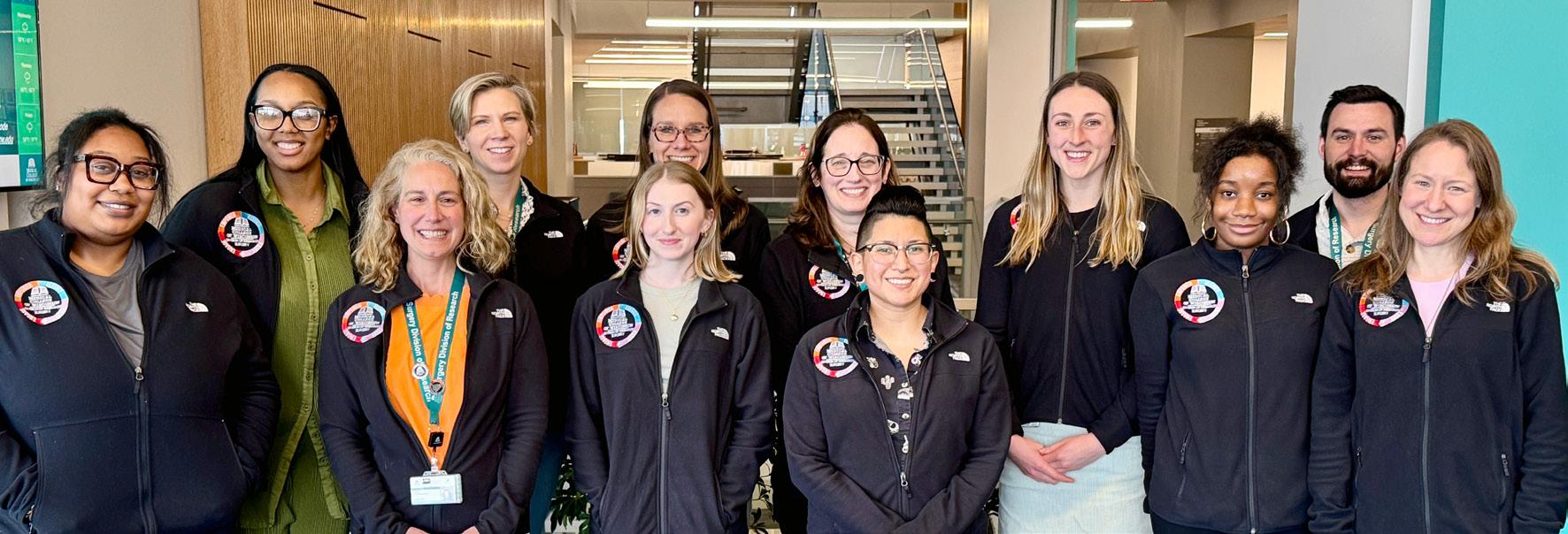
Figure 1. General Surgery Residents at the 3rd Annual Resident Retreat, with a focus on Leadership Skill Development
The training of our residency program is grounded in the pillars of our academic mission: education, mentorship and research. The clinical education the residents receive is robust, with experiences at our main quaternary referral hospital, Froedtert Hospital, Children’s Hospital, the Clement J Zablocki VA Medical Center, and three community hospital sites. Outside of their busy clinical education, we have a strong curriculum for the residents led by our Associate Program Director of Curriculum, Dr Thomas Carver. Residents have weekly reading topics and quizzes that are discussed at didactic curriculum sessions and simulation labs focused on robotic, laparoscopic, endoscopic and open surgical techniques.
Mentorship for our residents is continuous, occurring throughout their training. First-year residents are paired with a faculty member for professional mentorship, and this evolves throughout their training to include more career mentors as they decide on specialty. In addition to professional mentorship with individual faculty members, as a program we provide mentorship and support of crucial skills necessary for their professional development. This academic year, in October 2025, we had our third annual resident retreat, with a workshop focused on leadership skill development (Figure 1).
Research efforts for our residents are supported by our Associate Program Director of Professional Development, Dr Ally Istl. We have 10-12 research residents every academic year, with clinical and basic science research experiences in various subspecialties. Our residents have been incredibly productive in their research efforts, and in the last academic year, collectively had 107 publications and 131 national, regional or local scientific presentations.
Innovation and leading the way in surgical education has remained at the forefront our residency program. The American Board of Surgery initiated the use of competency-based assessments, Entrustable Professional Activities (EPAs) in general surgery residency training in July 2023. Since that time, we have had great engagement of the residents and faculty members with over 3,500 assessments completed. Given our great engagement in this initiative, we were recently enrolled in a Consortium for Assessment in Surgical Education, as one of 14 residency programs across the country.

Figure 2. Distribution of examiners' home institutions
Regionally, our efforts to lead the way in surgical education have been championed by our Associate Program Director of Assessment and Evaluation, Dr Jacqueline Blank. Last academic year we held the first Statewide Wisconsin mock orals examination. This was conducted virtually in April 2025. There were 4 residency programs participating, MCW, University of Wisconsin Madison, Gunderson and Marshfield. Across those four programs, 58 senior residents participated and were examined by 41 faculty members (Figure 2).
The MCW General Surgery residency program continues to recruit and train residents to be the best clinical surgeons with support from our amazing clinical faculty, curriculum team, faculty and research mentors. We continue to embrace and innovate, ensuring that we stay on the cutting edge of surgical education.
For more information on this topic, please email Dr. Rana Higgins at rhiggins@mcw.edu
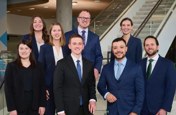
Pictured (left to right): Drs. Elise Biesboer, Monica Seadler, Amanda Witte, Brexton Turner, Taylor Jaraczewski, Santiago Rolon, Melissa Drezdzon, and Benjamin Seadler
References for Exploring the utility of magnesium alloy-based bioabsorbable stents in the treatment of atherosclerotic disease, page 8
1. Tsao, C. W.; Aday, A. W.; Almarzooq, Z. I.; Alonso, A.; Beaton, A. Z.; Bittencourt, M. S.; Boehme, A. K.; Buxton, A. E.; Carson, A. P.; Commodore-Mensah, Y. Heart disease and stroke statistics—2022 update: a report from the American Heart Association. Circulation 2022, 145 (8), e153-e639.
2. Schwartz, R. S.; Holmes Jr, D. R.; Topol, E. J. The restenosis paradigm revisited: an alternative proposal for cellular mechanisms. Journal of the American College of Cardiology 1992, 20 (5), 1284-1293.
3. Hoffmann, R.; Mintz, G. S.; Dussaillant, G. R.; Popma, J. J.; Pichard, A. D.; Satler, L. F.; Kent, K. M.; Griffin, J.; Leon, M. B. Patterns and mechanisms of in-stent restenosis: a serial intravascular ultrasound study. Circulation 1996, 94 (6), 1247-1254.
4. Ormiston, J. A.; Serruys, P. W. Bioabsorbable coronary stents. Circulation: Cardiovascular Interventions 2009, 2 (3), 255-260.
5. Fang, X.; Wang, K.; Han, D.; He, X.; Wei, J.; Zhao, L.; Imam, M. U.; Ping, Z.; Li, Y.; Xu, Y. Dietary magnesium intake and the risk of cardiovascular disease, type 2 diabetes, and all-cause mortality: a dose–response meta-analysis of prospective cohort studies. BMC medicine 2016, 14, 1-13.
6. Heublein, B.; Rohde, R.; Kaese, V.; Niemeyer, M.; Hartung, W.; Haverich, A. Biocorrosion of magnesium alloys: a new principle in cardiovascular implant technology? Heart 2003, 89 (6), 651-656.
7. Maier, J. A.; Malpuech-Brugère, C.; Zimowska, W.; Rayssiguier, Y.; Mazur, A. Low magnesium promotes endothelial cell dysfunction: implications for atherosclerosis, inflammation and thrombosis. Biochimica et Biophysica Acta (BBA)-Molecular Basis of Disease 2004, 1689 (1), 13-21.
8. Fu, J.; Su, Y.; Qin, Y.-X.; Zheng, Y.; Wang, Y.; Zhu, D. Evolution of metallic cardiovascular stent materials: a comparative study among stainless steel, magnesium and zinc. Biomaterials 2020, 230, 119641.
9. Nicol, P.; Bulin, A.; Castellanos, M. I.; Stöger, M.; Obermeier, S.; Lewerich, J.; Lenz, T.; Hoppmann, P.; Baumgartner, C.; Fischer, J. Preclinical investigation of neoatherosclerosis in magnesium-based bioresorbable scaffolds versus thick-strut drug-eluting stents. EuroIntervention 2020, 16 (11), e922-e929
10. Liu, Y.; Zheng, Y.; Chen, X. H.; Yang, J. A.; Pan, H.; Chen, D.; Wang, L.; Zhang, J.; Zhu, D.; Wu, S. Fundamental theory of biodegradable metals—definition, criteria, and design. Advanced Functional Materials 2019, 29 (18), 1805402.

@MCWSurgery
@MCWSurgOnc
@MCWMIGS
@MCWTraumaACS
@MCWPancreasProgram

@MCWSurgery
@MCWEndoSurg
@MCWMIGS
@MCWPancProgram
@MCWPedSurg
@MCWTraumaACS
@MCWSurgResearch
@MCWSurgOnc
@MCWVascSurg


@MCWSurgery
@MCWGenSurg_Residents
MCW Department of Surgery

Samantha Leonard, MD, MEd
General Surgery Resident, Medical College of Wisconsin Research Fellow, Children’s Wisconsin & Midwest Pediatric Surgery Consortium
Traumatic injuries remain the number one cause of mortality in children, highlighting the importance of excellent pediatric trauma care. Trauma video review (TVR) has been utilized since the 1980s to evaluate the trauma resuscitation during the critical first hour of trauma care.1 A survey published in 2020 suggested that 30% of adult Level I and II trauma centers in the United States currently utilize TVR.2 While there is no published data, this number is likely lower at pediatric trauma centers.
Creating a TVR program requires a number of steps for successful implementation that can take several years. Having discussions with key stakeholders is typically the first step. This includes, but is not limited to, hospital administration, the legal team, information technology, as well as leaders within emergency medicine, trauma surgery, and nursing. Once all parties are in agreement to proceed, a hospital must invest in the necessary technology, including cameras, sound systems, and software. The final step is addressing the logistics, including who will access the videos, where the videos are stored, how long the videos are stored, and who will review which videos.3–5
Children’s Wisconsin has been recording trauma resuscitations for several years, reviewing the tapes for specific quality improvement objectives, generally within individual departments and only occasional ad hoc multidisciplinary review. We saw an opportunity to expand both our use of TVR and our trauma education programming by implementing a formal multidisciplinary educational TVR arm. The previous institutional trauma education was focused on multi-disciplinary trauma simulation aimed at nursing, respiratory therapy, pharmacists, and physicians in emergency medicine, trauma surgery, and intensive care. A small multidisciplinary group was created to develop an educational TVR program consisting of pediatric trauma surgeons, an adult trauma surgeon, emergency medicine physicians, and a nurse trauma program manager.
Prior to developing and implementing new educational programming, we distributed a survey to all who participate in trauma resuscitations to not only gauge their current perceptions and comfort surrounding trauma resuscitations but also perform a needs assessment for a formal TVR program. Comfort levels related to each component of a trauma resuscitation (pre-arrival preparation, emergency medical services (EMS) handoff, primary survey/vitals/ intravenous (IV) access, secondary survey, procedures, wrap-up) were also ascertained. Questions utilized a Likert scale from 1 (pre -
pared or very comfortable) to 5 (unprepared or very uncomfortable) as well as yes/no answers. Additionally, we asked participants to share their experience with, perception of, and interest in TVR. Questions focused on the following elements: quality, knowledge, personal performance, team performance, personal benefit, and institutional benefit. They utilized yes/no/unsure answers.
We received responses from 131 individuals, across 10 different roles and specialties: pediatric emergency medicine (PEM) physicians, surgeons, anesthesiologists, pediatric intensive care physicians, nurses, OR staff, social workers, chaplains, radiology technicians, and pharmacists. This correlated with a response rate of 24% (131/553).
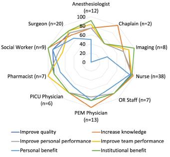
Figure 1. Radar plot of the utility of TVR for specific element improvement by role, seen as percent “yes”
Overall, 70% of respondents felt prepared to participate in a trauma resuscitation. When asked separately about blunt and penetrating trauma separately, 62% of respondents reported feeling prepared for blunt trauma and 62% reported feeling prepared for penetrating trauma. There was no significant difference between respondent role-type for overall (p=0.078), blunt (p=0.218), or penetrating (p=0.253) trauma preparedness. The percentage of providers feel-
ing very comfortable with any individual component of the resuscitation ranged from 50-63%. Fifty percent felt very comfortable with the secondary survey, and 63% felt very comfortable with the pre-arrival preparation. When broken down by role, there was a significant difference in comfort level with EMS handoff (p=0.014), primary survey (p=0.0007), secondary survey (p=0.0001), and procedures (p=0.0007). Lastly, 71% of respondents desired additional trauma education.
Forty percent of respondents noted previous video review experience. Respondents felt TVR would generally have a positive impact, ranging from 69% (provide personal benefit) to 85% (provide institutional benefit). When analyzed by role, there was no difference in any element except “provide personal benefit” (p=0.016) where the highest positive responses came from PEM physicians (84.6%), nurses (89.5%), and social workers (88.9%). Figure 1 demonstrates responses by role for each element. When asked specifically who should be present at a TVR, nurses (89%), fellows (89%), attendings (87%), respiratory therapists (76%), residents (73%), advanced practice providers (APP) (57%), and pharmacists (56%) were the most cited provider types. Less frequently named potential participants included medical students, APP students, social workers, chaplains, child life specialists, phlebotomists, radiology technicians, and other trainees/orientees. Finally, 39% of respondents felt the reviews should be held quarterly and 34% felt they should be held monthly. Respondents said they would commit to attending quarterly (40%), semi-annually (23%), or monthly (20%) most often.
This data showed that, while self-reported preparedness is similar between groups and specialties, individual comfort levels with each component of a resuscitation varied by role and component. It highlighted that all who are involved in trauma resuscitations would benefit from educational initiatives focused on discrete elements of trauma resuscitations. Additionally, this data strongly supported the implementation of a multidisciplinary TVR program at our institution with a positive perception and interest from all roles. The findings supported buy in and informed details related to participants and frequency.
With this data in hand, our committee designed and implemented a multidisciplinary educational TVR program. We built on the simulation program that already existed, alternating between simulation and TVR sessions. Both sessions utilized the same patient scenario that highlighted team lead communication and infrequently performed procedural skills. Pre- and post-surveys were distributed to participants to guide future iterative improvements.
1. Hoyt DB; S Steven R; Fridland, Peggy Hollingsworth; Mackersie, Robert C; Hansbrough, John F; Wachtel, Thomas L; Fortune, John B. Video recording trauma resuscitations: an effective teaching technique. The Journal of trauma. 1988;28(4):435-440. doi:10.1097/00005373-198804000-00003
2. Dumas RP; V Michael A; Hatchimonji, Justin S;W, Lucy; Maher, Zoe; Holena, Daniel N. Trauma video review utilization: A survey of practice in the United States. American journal of surgery. 2019;219(1):49-53. doi:10.1016/j.amjsurg.2019.08.025
3. Leonard SP, Flynn-O’Brien KT, Milia DJ, Holena DN. Trauma Video Review: A Guide To Implementation. Curr Trauma Rep. 2025;11(1):22. doi:10.1007/s40719-025-00297-z
4. Dumas RP, Cook C, Holena DN, et al. Roll the Tape: Implementing and Harnessing the Power of Trauma Video Review. Journal of Surgical Education. 2022;79(6):e248-e256. doi:10.1016/j. jsurg.2022.08.010
5. Williams KS, Pace C, Milia D, Juern J, Rubin J. Development of a Video Recording and Review Process for Trauma Resuscitation Quality and Education. West J Emerg Med. 2019;20(2):228231. doi:10.5811/westjem.2018.12.40951
For more information on this topic, please email Dr. Samantha Leonard at sleonard@mcw.edu

Momodou L. Jammeh, MD
Assistant Professor of Surgery, Division of Vascular and Endovascular Surgery

Cazza Czerniak,
PhD
Graduate Student
Cardiovascular disease remains an important cause of mortality and loss of productivity globally. While open surgical revascularization remains the gold standard for treating patients with vascular disease, patient frailty and advances in endovascular technology have led to greater use of percutaneous interventions for vascular disease1. Endovascular manipulation of vessels with balloons and/ or stents results in localized vascular injury.2 In turn, this can induce pathological remodeling, such as neointimal hyperplasia, leading to vessel occlusion or in-stent restenosis. Traditional permanent metal-based stents, often placed for acute vessel recoil at the time of intervention, can impair physiological vasomotor and endothelial function while enhancing chronic inflammatory responses.3 As such, bioabsorbable vascular scaffolds offer several theoretical advantages over traditional metal stents.4 This includes the potential to provide vessel support without perturbing normal endothelial function or blunting the acute, proliferative responses that lead to neointimal hyperplasia. Additionally, the complete dissolution of these temporary scaffolds may prevent the chronic inflammatory response to metal stents that drives delayed restenosis.
Magnesium (Mg), an essential mineral active in an array of biological processes, including vascular smooth muscle function, has been associated with improved prognosis in individuals with cardiovascular disease.5 Mg-based alloys have attracted increased interest for biomedical applications due to their favorable biode gradability profile and mechanical properties comparable to those of bone.6 While the biological environment can induce measurable changes in material performance, the metal cations (MX+) re leased during biodegradation can also significantly affect vascular pathophysiology. Mg2+ is often associated with anti-inflammatory properties and is typically intracellularly bound to ribosomes, poly nucleotides, and ATP. Mg2+ deficiency has been directly linked to endothelial dysfunction, progressive atherosclerosis, and ar terial inflammation, possibly through dysregulation of antioxidant balance and resultant oxidative stress.7 Remarkably, dietary Mg intake has been highly correlated with reduced blood lipid levels,
Roger Guillory II, PhD
Assistant Professor of Biomedical Engineering

Pardis
Taheri, PhD
Postdoctoral Fellow
reduced atherosclerosis and vascular calcification, and improved endothelial function in patients. Mg2+ is the most well-tolerated bioabsorbable metal cation in supraphysiological concentrations due to its large intracellular concentrations (10-30mM) required for functionality, the lack of concentration gradients across cell membranes, and redox inertness.
There is a growing body of preclinical evidence on improved atherosclerotic lesion characteristics after Mg-alloy stent placement and biodegradation. For instance, intravascular optimal coherence tomography of patients undergoing coronary stenting with a drug-eluting Mg-alloy stent demonstrated a reduction in lipid streaks and stabilization of the fibrous cap after resorption.9 Similarly, preclinical studies using a polymer-coated Mg-alloy stent also demonstrated reduced rates of neo-atherosclerosis compared with traditional permanent metal-based stents. In the Guillory lab, in vivo experiments aimed at elucidating the bioactivity of Mg implants have demonstrated that these implants produce supraphysiological concentrations of Mg within the atherosclerotic neointima and the vessel wall. As shown in Figure 1, mass spectrometric analysis (A) of tissue samples following Mg wire implantation demonstrated high deposits in the neointima and adventitia (B).
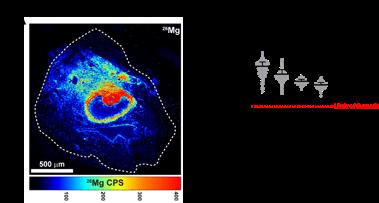
Figure 1. Quantification of Magnesium (Mg) alloy-related and endogenous elements. Red line is the limit of detection per element (A); Mapping of metallic elements Mg, Y, (B) Dot plot showing raw data extracted from (Mg, Y, respectively) each mass spectrum (pixel) and plotted according to the regions. Region sample sizes (# of pixels) are n=364 (corrosion product), n=882 (neointima), n=448 (Adventitia close), n=612 (Adventitia far). A Brown-Forsythe test for variance was run, followed by a Welch ANOVA with post hoc Dunnett multiple-comparisons test. ****P<0.0001
In addition, recent in vivo studies involving Mg wires implanted in the hypercholesterolemic, atherosclerotic-mimicking microenvironment of apolipoprotein E (ApoE) knockout mice have provided some insight into the bioactivity of Mg-alloy implants. Compared with Platinum wire implants, Mg alloy wires exhibit a significant reduction in neointimal volume on histological assessments of samples collected 30 days after implantation (Figure 2A and 2B). On closer examination with Oil red O staining, an inverse correlation between neointimal burden and Mg concentration was observed (Figure 2C). Immunological staining and viability assessments of bone marrow-derived macrophages demonstrated markedly reduced lipid uptake in the presence of Mg (Figure 2D). Taken together, these observations are consistent with the hypothesis that Mg may mitigate the pathological activity of inflammatory cells in lipid-rich environments.
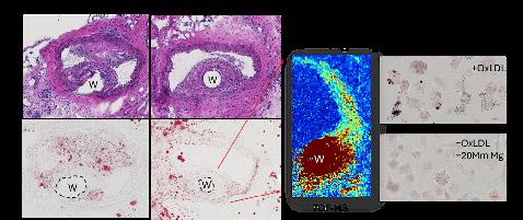
Figure 2. Mg implant behavior in APOE-/- mice for 30 days (A) H&E staining of cryosections from APOE-/- implanted with Pt and Mg (B) Oil Red O staining of the cryosections with red circle marking wire location (C) LA-ICP-TOF-MS of one section with Mg wire implanted and its location denoted as “W” (D) Oil Red O staining of bone marrow derived macrophages cultured in the presence of OxLDL and Mg
Our current efforts are focused on developing Mg-alloy-based stents with the potential to be translated from the lab to clinical use. At the most basic level, we are experimenting with ways to minimize the strut size of the Mg alloy wire used for stent manufacture while maximizing radial force through braid angle and annealing temperature. In addition, early and rapid bioabsorption, referred to as corrosion, can affect the durability and efficacy of bioabsorbable implants. Recent in vitro testing in the Guillory lab, involving Mg alloy wires implanted in the abdominal aorta of hyperlipidemic ApoE knockout mice, demonstrated increased, dose-dependent bioabsorption rates in the presence of lipoproteins. A common means of tuning bioabsorbable Mg alloys to modulate corrosion rate is through sur-
face modifications.10 The two strategies of interest for surface modification are polymer coating and anodization. The former creates a physical barrier in the form of a polymer biofilm, while the latter forms a more stable oxide layer to reduce acute degradation and promote endothelialization. Finally, the impact of our selected Mg alloy stents on the lesion progression and restenosis will be characterized through endovascular implantation in an ApoE knockout rat model of hyperlipidemia and atherosclerosis. The larger size of these animals relative to mice facilitates intravascular stent deployment via tail-artery access rather than direct abdominal aortic implantation via laparotomy, mimicking current clinical practice.
In summary, Mg alloy–based bioabsorbable stents hold promise as viable alternatives to permanent metal-based stents in the treatment of cardiovascular diseases. Ongoing efforts to optimize stent design and corrosion kinetics through alloy selection and surface modification are critical steps toward achieving predictable performance and clinical durability. As these engineering advances are integrated with rigorous in vivo evaluation in clinically relevant models, Mg-alloy bioabsorbable stents may offer a transformative alternative to permanent metallic implants, with the potential to restore vascular function while reducing long-term complications associated with permanent metal implantation.
For more information on this topic, please email Dr. Momodou L. Jammeh at mjammeh@mcw.edu
Ezra Teitelbaum, MD, MEd
Associate Professor of Surgery, Division of Minimally Invasive and Gastrointestinal Surgery
Over the past 50 years there has been a steady evolution towards the use of minimally invasive techniques for the treatment of surgical disease. This has resulted in tremendous improvements in patient outcomes, with decreased pain, convalescence, and complications. Two of the main drivers of this change have been the parallel development of laparoscopic (and now robotic) surgery and flexible endoscopy. Both minimally invasive surgery and flexible endoscopy are now core components of general surgery, and often are used in a complementary and concurrent manner. This article will highlight some of the current ways in which flexible endoscopy is being utilized to augment the treatment of surgical disease.
Technologic advances, such as fiberoptics and digital video, allowed for the development of flexible endoscopy as a diagnostic modality in the 1970s and 80s.1 This revolutionized the diagnosis of gastrointestinal disease, allowing for surgical intervention at earlier disease stages (e.g., gastric and colon cancer) and the avoidance of surgery altogether with more prompt and accurate initiation of medical therapy (e.g., peptic ulcer disease). Diagnostic flexible endoscopy has continued to evolve over the past 50 years, with the introduction of digital high-resolution cameras, advanced imaging modalities (e.g., Narrow Band Imaging, chromo-endoscopy, endomicroscopy), and endoscopic ultrasound (EUS).
Since the inception of flexible endoscopy, general surgeons have played an important role in its technological advancement, clinical application, and related outcomes research. Currently, diagnostic colonoscopy and upper endoscopy are two of the most common procedures performed by general surgeons in the United States.2 This is especially true in more rural communities without as ready access to gastroenterology care.
Training in flexible endoscopy during general surgery residency presents an important challenge, given the breadth of surgical procedures that surgical trainees must achieve competency in. For example, a recent study showed that surgery residents performed an average of 67 colonoscopies and 35 upper endoscopies over the course of training.3 This is compared with GI fellows who are required to perform a minimum of 130 upper endoscopies and 140 colonoscopies during training and often graduate with multiples more. As a result, general surgeons cannot rely on raw numbers alone to gain competency in flexible endoscopy, and we must be
intentional in the way we train residents in these procedures. One important solution for ensuring high-quality endoscopy training has been the development of the Fundamentals of Endoscopic Surgery (FES) high-stakes exam and associated curriculum, and passing FES is now a requirement for residency graduation. Additionally, the use of simulator-based training has become an essential augmentation to clinical experience during training.
As diagnostic endoscopy has become ubiquitous in the care of GI conditions, the therapeutic capabilities of flexible endoscopy have rapidly expanded. Initial procedures such endoscopic polypectomy, control of GI bleeding, and feeding access with percutaneous endoscopic gastrostomy (PEG) (first performed by a surgeon, Dr. Jeffrey Ponksy!), have led to ever more complex interventions. One such area is the advent of so-called “third-space” endoscopy, which takes placed within the submucosal potential space of the bowel wall (intraluminal and intraperitoneal are the 1st and 2nd spaces).
Third-space endoscopy was born out of the concept of Natural Orifice Transluminal Endoscopic Surgery (NOTES). The idea of NOTES was to use an endoscope inserted through a “natural orifice” to perform intraperitoneal surgery via an opening in the viscera. While such early experimental procedures such as transgastric and transvaginal cholecystectomy never became routine, the concepts and techniques of NOTES were translated to a number of third-space procedures that have since become standard-of-care.
The initial third-space procedure to gain traction was peroral endoscopic myotomy (POEM). Used to treated achalasia and other esophageal motility disorders, POEM creates a controlled myotomy across the lower esophageal sphincter (LES). During POEM, a standard flexible gastroscope is used and a 1 cm incision is made in the mucosa of the distal esophagus. The endoscope is advanced through the mucosotomy and a tunnel is created through the submucosal space of the esophagus and onto the stomach. Finally a myotomy is performed to divide the LES and the mucosotomy is closed with clips.
Since it was first performed in 2008 (by another surgeon, Dr. Haru Inoue!), POEM has become the most common procedure for treatment of achalasia, with randomized trials demonstrating superior efficacy to pneumatic dilation and superior safety compared with laparoscopic Heller myotomy.4,5 This third-space myotomy concept
has since been adapted to treated gastroparesis (G-POEM or POP) and Zenker’s diverticulum (Z-POEM).
Management of Perforations and Surgical Complications
Since its early days, flexible endoscopy has been used intraoperatively in order to evaluate anatomy, mucosal ischemia, and test for anastomotic leak. Increasingly, endoscopy has come to replace surgery as a first-line approach to treating perforations, leaks, and other complications postoperatively.
The introduction of self-expanding metal stents (SEMS) was a major advance in the treatment of naturally occurring and iatrogenic perforations. Diseases such as Boerhaave’s Syndrome and anastomotic leak after esophagectomy, that once often required open surgical drainage via thoracotomy and esophageal diversion with esophagostomy can now be treated with SEMS purely endoscopically. Multiple endoscopic clips (both through- and over-the-scope) and endoscopic suturing devices have also been developed for defect closure. While SEMS, clips, and endoscopic suturing allow for the control of acute perforations, they still require drainage of infection on the outside of the leak via percutaneous or surgically placed drains.
A newer concept in the endoscopic treatment of perforations is the concept of “internal drainage”. The idea is to drain infection back into the bowel lumen and allow for the leak abscess cavity to granulate inward, ending in a small, swallow diverticulum. Two primary modalities have been used for internal drainage: endoscopic vacuum assisted closure (EVAC) and internal drain placement.6 For EVAC, the leak cavity is debrided and irrigated endoscopically and then a VAC sponge is affixed to a nasogastric tube and brought into the leak cavity. This requires repeat endoscopies to exchange the EVAC sponge, performed once or twice weekly. Internal drain placement, typically using double pigtail biliary stents, is often used for smaller leak defects without the presence of necrotic debris. Both strategies can often obviate the need for external drain placement.
There is no doubt that flexible endoscopy has become, and will remain, an essential component of general surgery. It is imperative that we continue to train general surgery residents and GI surgery subspecialty fellows to perform high-quality endoscopy, so they can effectively apply it as a tool to enhance diagnostic accuracy and lessen the morbidity of interventions. In the future, advances in instrumentation and potentially the introduction of endoscopic robotics will continue to push the boundaries of what can be accomplished via natural orifices and further blur the boundaries between what is considered “endoscopy” and “surgery."
1. Ponsky JL, Strong AT. A History of Flexible Gastrointestinal Endoscopy. Surg Clin North Am. 2020 Dec;100(6):971-992.
2. Decker MR et al. Specialization and the current practices of general surgeons. J Am Coll Surg. 2014 Jan;218(1):8-15.
3. Sohail AH et al. Can my surgeon scope? Trends in endoscopy training volume and experience among general surgery residents in the United States: a nationwide analysis. Surg Endosc. 2024 Mar;38(3):1491-1498.
4. Ponds FA. Effect of Peroral Endoscopic Myotomy vs Pneumatic Dilation on Symptom Severity and Treatment Outcomes Among Treatment-Naive Patients With Achalasia: A Randomized Clinical Trial. JAMA. 2019 Jul 9;322(2):134-144.
5. Werner YB, Endoscopic or Surgical Myotomy in Patients with Idiopathic Achalasia. N Engl J Med. 2019 Dec 5;381(23):22192229.
6. Ward MA, Factors that promote successful endoscopic management of laparoscopic sleeve gastrectomy leaks. Surg Endosc. 2021 Aug;35(8):4638-4643.
For more information on this topic, please email Dr. Ezra Teitelbaum at eteitelbaum@mcw.edu

Anna Tatakis, MD
General Surgery Resident Sarah Cottrell-Cumber, DO
Surgical Critital Care Fellow
Introduced in 1980, the percutaneous gastrostomy tube (PEG) has become the standard approach for providing durable enteral access in patients requiring long-term nutritional support.1 Patients who require durable enteral access represent a highly heterogeneous population, encompassing a wide range of diagnoses, illness severities, and trajectories of recovery. While gastrostomy tube placement can be clearly beneficial for some conditions, determining whether and when to place a PEG is often clinically and ethically complex. Despite the widespread use of PEG tubes and known complications, there is variable and limited data regarding the true necessity and appropriate timing of PEG placement.2,3 Complicating this decision further are nonclinical influences, including family preferences, provider comfort, and systemic constraints such as long-term care facility restrictions against temporary feeding access, such as nasogastric or nasojejunal feeding tube.
The Acute Care Surgery team at Froedtert Hospital (FH) noted these non-clinical factors surrounding PEG placement and performed a retrospective review of PEG patients and their outcomes.4 It was hypothesized that PEG placement carries significant morbidity and that a proportion of procedures would prove unnecessary as patients regain nutritional independence prior to discharge or progress to end of life. The study evaluated 233 patients who had a PEG placed in 2023-2024, and evaluated return to oral intake at discharge, characterized the proportion of procedures performed for facility disposition, and assessed the associated complication rate.
Our study findings demonstrated that most patients undergoing PEG placement had resumed oral intake at the time of discharge (n=139, 59.7%), and of those, 18.7% (n=26) had returned to nutritional independence. The median time to return to oral intake was 11 days (IQR 3-30), and the median time to hospital discharge from PEG placement was 12 days (IQR 6-22). The overall number of patients who had any complication was 24.5%, nearly half of which required a radiologic, surgical, or endoscopic procedure to manage the complication.
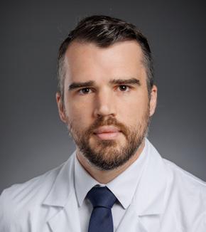
Patrick Murphy, MD,MPH, MSc
Associate Professor of Surgery, Division of Trauma and Acute Care Surgery
Ultimately, one in five patients were not meaningfully utilizing their PEG as they transitioned to hospice care, experienced in-hospital mortality, or were nutritionally independent at the time of discharge. These findings suggest that a significant number of PEGs are placed prematurely or unnecessarily, particularly given 37% are placed for facilitating hospital discharge. Patients who underwent PEG placement for disposition purposes had a significantly shorter time to resumption of oral intake, and a higher proportion of patients with oral intake at the time of discharge.
Our data aligns with prior literature in showing that a portion of patients will recover their ability to eat safely within days to weeks.5,6 The most recent American Heart Association, American Stroke Association, and American Society for Parenteral and Enteral Nutrition (ASPEN) guidelines recommend gastrostomy tube placement be reserved for those with anticipated persistent inability to swallow safely greater than 2-3 weeks or expected need of enteral nutrition of 4-6 weeks.7,8 Delaying PEG placement may provide sufficient time for patients to recover to the point of no longer requiring durable feeding access or allow the clinical trajectory to declare itself, particularly for patients who ultimately transition to hospice or die during their hospitalization. Allowing time for patients and families to understand prognosis and goals of care may reduce exposure to invasive procedures that ultimately have limited benefit, avoid procedure-related complications near the end of life, and better align nutritional decisions with a palliative approach that prioritizes comfort and quality of life.
A PEG consult often feels emotionally charged for the surgeon and there is a dichotomy of responses:

It is much less time consuming to simply perform the procedure compared to the emotional and time investment of the latter. Yet, the clinical and ethical challenge often has many surgeons diving down the rabbit hole of the PEG consult.
Many patients in need of a PEG cannot make that decision for themselves, and the decisions fall to their families and surrogate decision makers. One of the most helpless feelings as a surgeon is not having a reasonable alternative to offer a family when we are not recommending a PEG. To a family, not offering durable access for nutrition is often perceived as “you want my loved one to starve to death” because nutrition is a basic necessity for life. An alternative to discuss is hand feeding, where the patient is allowed to eat by mouth with assistance. It is time consuming, often a limitation for nursing staff, may not meet nutritional goals, and may be a higher aspiration risk. Yet hand feeding restores the social and pleasure aspect of eating that tube feeds can’t. Through discussions with family members, the benefit of showing love through eating by mouth often outweighs the associated risks.
Difficult questions from the family also arise during this discussion. Sample language to common questions may help ease the tension for both physicians and family members around this difficult topic. Aligning with the recommendation of the Acute Care Surgery team to delay PEG placement until immediately preceding discharge, this “cool off” period also allows families to reflect on the discussion, ask additional questions, or pivot care goals.
“Is my loved one starving to death?”
“What if they’re hungry?”
“Our religion says we must give them nutrition somehow. Aren’t we causing their death by not feeding them?”
“I’m worried that forcing them to eat or giving non-oral nutrition will not help them feel better and may make them feel worse. At this stage, most people don’t experience hunger the way that we’re used to. As the body slows down, it often rejects food.”
“Tell me more about your beliefs and concerns.”
“The [stroke, cancer, etc.] is what is causing their death.”
“Aren’t they suffering?” “I can tell how much you care about them. We can provide a lot of comfort with good mouth care and sips or swabs of liquids. This is something you can help us with if you wish.”
“I can’t watch them waste away.” “This is difficult. I can only imagine it might feel helpless, since food is often how we show our love and concern for people when they are sick.”
The PEG is an emotionally charged topic for surgeons, patients and their families. While not technically challenging, the discussion around a PEG is a complex, high-stakes decision that requires navigating deep ethical, emotional, and clinical dilemmas, often for frail or cognitively impaired patients. Our Acute Care Surgery team discovered that 20% of patients were not meaningfully utilizing their PEG as they transitioned to hospice care, experienced in-hospital mortality, or were nutritionally independent at the time of discharge. Surgeons must weigh prognosis and potential for recovery against the risks and long-term implications of tube dependence. Time is
often the most helpful variable when deciding the necessity and timing of a PEG tube. Despite the relative simplicity of the procedure, the PEG consult will remain a challenge to surgeons. We recommend delaying PEG placement until discharge to allow surgeons and families to reflect on the significance of a feeding tube and the patient’s condition to change, stabilize, or improve.
For more information on this topic, please email Dr. Anna Tatakis at atatakis@mcw.edu or Dr. Patrick Murphy at pmurphy@mcw.edu
1. Hanners Gutierrez J, Klein K, Bimali M, Sanders J. Rethinking tube feeding in palliative care: Impact on pneumonia, depression, and mortality in patients with dysphagia and lifelimiting illness. PLoS One. 2025 Oct 7;20(10):e0333895. doi: 10.1371/journal.pone.0333895. PMID: 41056338; PMCID: PMC12503241.
2. Welbank T, Kurien M. To PEG or not to PEG that is the question. Proc Nutr Soc. 2021 Feb;80(1):1-8. doi: 10.1017/ S002966512000703X. Epub 2020 May 22. PMID: 32441238.
3. Plonk, W. M. (2005). To PEG or Not to PEG. Practical Gastroenterology .
4. Tatakis A, Wilson D, Holland H, Patin B, Perlin S, Biesboer E, LaGraize N, Somberg L, Carver T, de Moya M, Murphy P. Reassessing the timing of percutaneous gastrostomy tube placement: Too many too soon. J Trauma Acute Care Surg. 2026 Jan 21. doi: 10.1097/TA.0000000000004882. Epub ahead of print. PMID: 41632481.
5. George BP, Kelly AG, Schneider EB, Holloway RG. Current practices in feeding tube placement for US acute ischemic stroke inpatients. Neurology. 2014 Sep 2;83(10):874–82.
6. Mandaville A, Ray A, Robertson H, Foster C, Jesser C. A Retrospective Review of Swallow Dysfunction in Patients with Severe Traumatic Brain Injury. Dysphagia. 2014 Jun;29(3):310–8.
7. Powers WJ, Rabinstein AA, Ackerson T, Adeoye OM, Bambakidis NC, Becker K, et al. Guidelines for the Early Management of Patients With Acute Ischemic Stroke: 2019 Update to the 2018 Guidelines for the Early Management of Acute Ischemic Stroke: A Guideline for Healthcare Professionals From the American Heart Association/American Stroke Association. Stroke. 2019 Dec;50(12):e344–418.
8. Boullata JI, Carrera AL, Harvey L, Escuro AA, Hudson L, Mays A, et al. ASPEN Safe Practices for Enteral Nutrition Therapy. J Parenter Enter Nutr. 2017;41(1):0148607116673053.
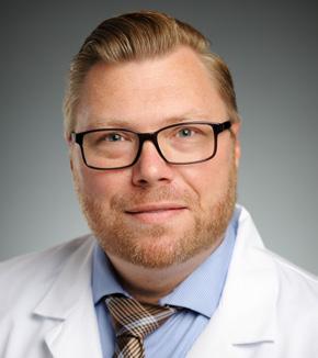
H. Adam Ubert, MD
Assistant Professor of Surgery, Division of Cardiothoracic Surgery
Extracorporeal membrane oxygenation (ECMO), along with mechanical circulatory support (MCS), represents some of medicine’s most technologically sophisticated life-support interventions. In its basic form, ECMO involves placement of large bore cannulas to circulate blood from the body, through an external membrane that removes carbon dioxide and adds oxygen before returning this highly oxygenated blood to the body. This device is deployed during cardiac and/or pulmonary failure and provides end organ support during periods of critical illness that would have otherwise proved fatal. As technology has improved, this ability to support the failing organs over a prolonged period has created ethical and moral dilemmas regarding patient selection, informed consent, resource allocation, and most notably, end-of-life decision making when prolonged care is deemed futile.
ECMO has been increasingly utilized for temporary support for cardiac or pulmonary failure. According to ELSO, the international consortium for extracorporeal life support, the use of ECMO has increased exponentially over the past decade. Despite the increased knowledge, utilization, and improvement in technology, overall survival remains at 54%.1 When ECMO is initiated emergently, it is often placed as a bridge to a decision, as the prognosis may be uncertain. Ideally, ECMO is placed as a bridge to recovery, but it can also be used as bridge to transplant or durable mechanical device. If it is determined the patient will not recover organ function, or is not a transplant or durable device candidate, this bridge can be considered a “bridge to nowhere,” creating an ethical dilemma regarding futility of ongoing medical care. In this scenario, the patient may improve sufficiently but could not survive outside of the acute care setting without the use of ECMO. In a non-decisional patient, it is incumbent on the health care team to engage the patient’s surrogate decision makers regarding treatment goals. Difficult conversations arise when the patient is mentally competent, interactive, and able to express their wishes. These wishes may not align with other family members’ desires, or the care teams concerns about futility with ongoing treatment. Adding to the difficulties, prolonged treatment with ECMO is resource intensive, technically challenging, associated with high cost, and often impeded by complications including bloodstream infections, coagulopathic bleeding, and neurologic injury.2 When a patient wishes to continue prolonged care, despite the life sustaining treatment being deemed futile, ethical concerns arise.
Ethical principles are evident throughout all health care decisions. When making difficult decisions, balancing the patient’s respect for autonomy, non-maleficence, beneficence, and justice with integrity from the medical team is necessary.
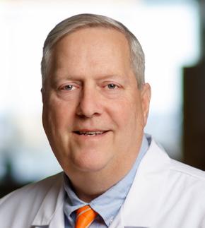
Lucian Durham, III, MD, PhD
Associate Professor of Surgery, Division of Cardiothoracic Surgery
Respect for autonomy Fundamental right of a patient with decisionmaking capacity to make independent, informed decisions about their own medical care, free from coercion from providers
Non-maleficence (avoid futility)
“Doing no harm,” requiring providers to avoid intentional harm, negligence, or providing ineffective treatment
Beneficence Act in the patient’s best interest, tailoring care to the patient’s specific needs, values, and definitions of well-being
Justice Duty to treat the patient fairly
Integrity The medical team's integrity is maintained by refusing to provide treatments that are no longer medically indicated or, in their judgment, only causing harm.
Take for example, a 52 y/o male who presents to the hospital with acute decompensation secondary to worsened respiratory failure despite maximal mechanical ventilatory support. A multidisciplinary decision is made to initiate veno-venous ECMO for lung support as a bridge to recovery. As the patient improves neurologically, the patient exhibits signs of pulmonary fibrosis and is unable to be liberated from ECMO support. At this point, transplant appears to be the only viable option for long-term survival. A transplant workup is performed and due to multiple co-morbidities and/or social factors, the patient is refused for transplant at multiple centers. With all options exhausted, continued ECMO support is deemed futile and goals of care discussions are held with the patient. The patient expresses he is content with life in the hospital and is happy to interact with family when they visit. Because of this, he refuses to consider withdrawal of care as an option. What are the appropriate options for this patient?
As recommended by the American Thoracic Society (ATS) and the American College of Critical Care Medicine (ACCM), shared decision making should be the default approach to making major decisions in the ICU.4 Based on ethical principles, the care team should maintain the patient’s respect for autonomy in deciding his medical care. Similarly, the team should try to respect the patient’s beneficence and justice, but does that interfere with the integrity of the medical team? All these questions provide a complicated scenario that requires multidisciplinary discussion including potential
guidance from the Palliative care and Ethics teams to determine the correct course of action.
As a result of these issues, multiple medical societies, including the ATS, ACCM, and the AMA have supported a process-based approach to unilateral decision making to withdraw interventions that are deemed inappropriate. This process-based approach often requires broad agreement, within and outside the health care profession, that ongoing treatment is inappropriate. If this has been fulfilled, then it may be ethical to unilaterally withdraw ECMO support.5-7 On the contrary, some ethicists have argued that withdrawal of life sustaining treatment against the patient’s wishes would cause undue emotional suffering, constituting cruelty, and therefore should not be considered.8 It is argued, “No matter what the initial intended purpose was, ECMO as a treatment option only makes sense if it aligns with the patient’s goals. If the goal of living a life that the patient finds meaningful or even minimally acceptable can still be met with ECMO support, then it is not futile to provide it.”9 Unfortunately, the idea of unilateral withdrawal of care in a fully competent patient remains controversial. Regardless, the health care team should continue to provide emotional support while ongoing decisions are made. Often, the use of Palliative care teams can help provide insight into the end-of-life process and help determine the patient’s long-term goals. Patients facing this difficult decision need to trust that their health care teams continue to provide compassionate care while maintaining the principles of medical ethics. As such, it is imperative that a multidisciplinary approach is utilized on a case-by-case basis and routine communication with the patient and family is necessary to provide optimal care, even in cases deemed futile.
1. (ELSO Registry International Summary, 2024)
2. Ventetuolo CE, Muratore CS. Extracorporeal life support in critically ill adults. Am J Respir Crit Care Med. 2014;190(5):497-508.
3. Mueller PS. Ethical and legal concerns associated with withdrawing mechanical circulatory support: A U.S. perspective. Front Cardiovasc Med. 2022 Jul 26;9:897955. doi: 10.3389/fcvm.2022.897955.
4. Kon AA, Davidson JE, Morrison W, Danis M, White DB; American College of Critical Care Medicine; American Thoracic Society. Shared Decision Making in ICUs: An American College of Critical Care Medicine and American Thoracic Society Policy Statement. Crit Care Med. 2016 Jan;44(1):188-201. doi: 10.1097/CCM.0000000000001396.
5. American Thoracic Society. Withholding and withdrawing life-sustaining therapy. Ann Intern Med. 1991 Sep 15;115(6):478-85. doi: 10.7326/0003-4819-115-6-478.
6. Consensus statement of the Society of Critical Care Medicine's Ethics Committee regarding futile and other possibly inadvisable treatments. Crit Care Med. 1997; 25:887-891
7. Medical futility in end-of-life care: report of the Council on Ethical and Judicial Affairs. JAMA. 1999; 281:937-941
For more information on this topic, please email Dr. Ubert at aubert@mcw.edu or Dr. Durham at ldurham@.mcw.edu
ADULT PATIENTS
All Non-cancer Requests Referrals: 855-314-0951
Transfers/Consultations: 877-804-4700
Clinical Cancer Center Referrals: 866-680-0505
Transfers/Consultations: 877-804-4700
PEDIATRIC PATIENTS
Referrals/Transfers/Consultations: 414-266-2460
For more information, please visit mcw.edu/surgery.
8. Abrams DC, Prager K, Blinderman CD, Burkart KM, Brodie D. Ethical dilemmas encountered with the use of extracorporeal membrane oxygenation in adults. Chest. 2014;145(4):876–882.
9. Childress, AM. COUNTERPOINT: Is It Ethically Justifiable to Withdraw Extracorporeal Membrane Oxygenation Against the Wishes of a Patient With Decision-Making Capacity When Cure Is Not Possible? No. CHEST, Volume 166, Issue 6, 1283 – 1285.
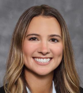
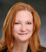
Meghan Conroy, MD, MS General Surgery Resident
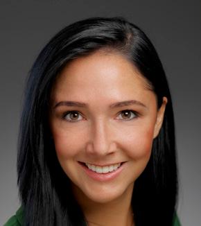
Kim Lamansky President and CEO of Kathy's House
Financial toxicity (FT) refers to the measurable financial burden and personal distress experienced by patients because of the costs associated with cancer care.1 This burden arises from a combination of medical, non-medical, and indirect expenses. Medical costs include out-of-pocket (OOP) expenses related to medications, hospitalizations, and therapies such as surgery and radiation. Non-medical expenses such as transportation, lodging, food, and childcare required to facilitate treatment further compound this burden. In addition, indirect costs including loss of employment or reduced income during treatment can significantly affect financial stability. FT has been shown to adversely impact patients’ psychological well-being, social functioning, compliance with medical care, and oncologic outcomes.2 This article aims to outline the drivers and downstream effects of FT and to highlight actionable steps healthcare providers can take to address this often under-recognized consequence of cancer care.
The causes of FT are multifactorial, and patient and cancer characteristics all play a role.2 Patients diagnosed with advanced or recurrent cancer tend to experience more FT due to the burden of multimodal treatment approaches involving surgery3, radiation4, and systemic therapy, as well as the challenges of seeking treatment at tertiary referral centers.5 Surgery and associated complications can lead to FT via delayed return to work and reliance on long term disability.6 Radiation therapy incurs high non-medical costs due to the required daily travel,7 and systemic therapy can lead to FT based on the cost of medications and travel to infusion centers. Patients with rare cancers are more susceptible to FT as they require specialized treatment and access to clinical trials at tertiary referral centers, which may require more travel and logistical considerations.8, 9
Patients with low income and members of historically marginalized racial or ethnic groups experience higher rates of FT, mirroring other concerning trends in our healthcare system.10 Uninsured patients experience higher rates of FT, although those with insurance are
Ally Istl, MD, MPH Assistant Professor of Surgery, Division of Surgical Oncology
certainly not immune.5, 11 High-deductible insurance plans have become more prevalent, leading to patients paying more OOP for clinic visits, treatment, and surveillance, often at specialized centers where co-payments and the cost of images and lab tests are greater.12 At Froedtert Hospital and the Medical College of Wisconsin (MCW), some patients must change their payer or insurance policy to a plan with a higher premium simply to participate in life-saving clinical trials.13, 14
FT affects outcomes through several mechanisms. Patients experiencing financial distress are less likely to undergo routine cancer screening and present with more advanced disease. Once diagnosed, they are more likely to experience delays in care and less likely to receive guideline-concordant treatment.15, 16 Patients reporting FT demonstrate lower adherence to prescribed therapies and are more likely to forgo healthcare appointments, including cancer treatments and mental health services.17 Prescription medications with higher OOP costs are more likely to be discontinued, a concern that is particularly pronounced for patients on second or third line systemic therapies, which are inconsistently covered by insurance.18-20 Additionally, FT is independently associated with anxiety and depression in cancer patients.21
Financial toxicity translates into worse survival outcomes. A nationally representative study demonstrated that cancer survivors reporting financial hardship had a significantly higher risk of mortality even when controlling for sociodemographic factors and insurance status.22 Cancer patients who filed for bankruptcy experienced nearly an 80% increased risk of mortality compared with matched controls.23 Adverse financial events including collections, chargeoffs, and foreclosures have also been associated with increased mortality risk.24 Based on the pervasive and severe consequences of FT in our cancer patient population, strategies to address the root causes of FT are urgently needed.
There are opportunities to address FT at the provider, institutional, health system, and policy levels. While systemic reform is essential, clinicians can address this problem in their day-to-day practice. First, we must develop a basic understanding of treatment-related costs and have transparent conversations with patients about what to expect. Patients often face the greatest OOP costs during the perioperative period, which may come as a surprise without advance counseling.25 Awareness of medication coverage by their insurer can also make a difference. Some intravenous formulations may have lower or no copays compared with oral alternatives. Suggesting an affordable formulation may prevent patients from discontinuing treatment altogether.26 Providers should also be familiar with lower-cost pharmacy options including direct-to-consumer programs that can help reduce financial strain.27
Another important strategy is minimizing low-value care. Avoiding unnecessary laboratory tests and imaging, reducing redundant clinic visits, and utilizing telehealth when appropriate can substantially decrease patient burden. This is particularly relevant for patients who travel long distances for care, as many do to receive care at MCW. Early referral to support services such as financial counseling, social work, or patient assistance programs is critical when financial concerns are identified. Finally, discussions about goals of care are not only good clinical practice but can prevent costly interventions that may not align with a patient’s priorities. Surgeons should be transparent about expected outcomes, recovery time, potential complications, and the likelihood of symptom relief, especially when considering palliative operations.28
Finally, institutions and departments play a key role in supporting patients through structural solutions. Screening for FT can help identify vulnerable patients, while tracking financial distress as a quality metric like patient satisfaction or surgical site infections can improve accountability at the health system level.29 Locally, MCW patients may benefit from resources such our financial services and assistance program, though eligibility requirements can be restrictive and require significant financial documentation. Another invaluable resource is Kathy’s House, which provides affordable housing for patients and families receiving medical care in Milwaukee. With most guests reporting improved access to care, reduced anxiety, and enhanced protection against severe financial hardship, Kathy’s House highlights how community-based support can meaningfully mitigate FT. Addressing financial toxicity ultimately requires systemic action, but small, intentional changes in everyday practice can have a profound impact on patients’ ability to access and complete cancer care.
For more information on this topic, please email Dr. Conroy at mconroy@mcw.edu or Dr. Ally Istl at aistl@mcw.edu
References continued on page 19
1. Witte J, Mehlis K, Surmann B, et al. Methods for measuring financial toxicity after cancer diagnosis and treatment: a systematic review and its implications. Ann Oncol. 2019;30(7):1061-1070. doi:10.1093/ annonc/mdz140
2. Lentz R, Benson III AB, Kircher S. Financial toxicity in cancer care: Prevalence, causes, consequences, and reduction strategies. Journal of Surgical Oncology. 2019;120(1):85-92. doi:10.1002/jso.25374
3. Brooks GA, Li L, Uno H, Hassett MJ, Landon BE, Schrag D. Acute hospital care is the chief driver of regional spending variation in Medicare patients with advanced cancer. Health Aff (Millwood). 2014;33(10):17931800. doi:10.1377/hlthaff.2014.0280
4. Palmer JD, Patel TT, Eldredge-Hindy H, et al. Patients Undergoing Radiation Therapy Are at Risk of Financial Toxicity: A Patient-based Prospective Survey Study. Int J Radiat Oncol Biol Phys. 2018;101(2):299-305. doi:10.1016/j.ijrobp.2018.03.014
5. Rose L, Rajasekar G, Nambiar A, et al. Estimated Out-of-Pocket Costs for Patients With Common Cancers and Private Insurance. JAMA Netw Open. 2025;8(7):e2521575. doi:10.1001/jamanetworkopen.2025.21575
6. Regenbogen SE, Veenstra CM, Hawley ST, et al. The personal financial burden of complications after colorectal cancer surgery. Cancer. 2014;120(19):3074-3081. doi:10.1002/cncr.28812
7. Kalbasi A, Kamrava M, Chu FI, et al. A Phase II Trial of 5-Day Neoadjuvant Radiotherapy for Patients with High-Risk Primary Soft Tissue Sarcoma. Clin Cancer Res. 2020;26(8):1829-1836. doi:10.1158/1078-0432. CCR-19-3524
8. Heus E de, Duijts SFA, Zwan JM van der, et al. The gap between rare and common cancers still exists: Results from a population-based study in the Netherlands. European Journal of Cancer. 2022;167:103-111. doi:10.1016/j.ejca.2022.03.001
9. de Heus E, Engelen V, Dingemans I, et al. Differences in health care experiences between rare cancer and common cancer patients: results from a national cross-sectional survey. Orphanet J Rare Dis. 2021;16(1):249. doi:10.1186/s13023-021-01886-2
10. Gordon LG, Merollini KMD, Lowe A, Chan RJ. A Systematic Review of Financial Toxicity Among Cancer Survivors: We Can’t Pay the Co-Pay. Patient. 2017;10(3):295-309. doi:10.1007/s40271-016-0204-x
11. Yabroff KR, Doran JF, Zhao J, et al. Cancer diagnosis and treatment in working-age adults: Implications for employment, health insurance coverage, and financial hardship in the United States. CA Cancer J Clin. 2024;74(4):341-358. doi:10.3322/caac.21837
12. Shih YCT, Xu Y, Bradley C, Giordano SH, Yao J, Yabroff KR. Costs Around the First Year of Diagnosis for 4 Common Cancers Among the Privately Insured. JNCI: Journal of the National Cancer Institute. 2022;114(10):1392-1399. doi:10.1093/jnci/djac141
13. Kaehny S. Through the Looking Glass: The Lived Experience of a Rare Tumor Diagnosis. Panel Presentation presented at: Musculoskeletal Oncology and Sarcoma Symposium; January 2026. https://moss. cme-congresses.com/

Ugwuji N. Maduekwe, MD, MMSc. MPH
Associate
Dean and Deputy Director, Advancing a Healthier Wisconsin Endowment; Associate Professor of Surgery, Division of Surgical Oncology
Imagine being able to answer a question like this: Are patients from rural Wisconsin communities less likely to receive timely surgical intervention for appendicitis than their urban counterparts—and if so, why? Or consider a different angle: Do patients from neighborhoods facing greater socioeconomic challenges experience more complications after elective surgery, even when we account for clinical factors? These are exactly the kinds of questions that keep surgical researchers up at night. Until recently, answering them required cobbling together data from multiple sources, navigating complex data use agreements, and hoping the pieces fit together. That has changed.
Before describing what is new, a bit of context about who we are. The Advancing a Healthier Wisconsin Endowment (AHW) is a statewide health philanthropy housed at MCW, established through a generous gift from Blue Cross & Blue Shield United of Wisconsin. Our mission is to improve the health of the people of Wisconsin by funding research, education, and community partnerships that address the state's most pressing health challenges. Over the years, AHW has supported hundreds of investigators and community organizations working on everything from chronic disease prevention to healthcare workforce development to improving care for underserved populations.
Beyond grantmaking, AHW invests in programming and capacity-building initiatives that give Wisconsin researchers tools they would not otherwise have access to. One of our newest efforts is a partnership with the Wisconsin Health Information Organization (WHIO) that makes statewide healthcare claims data available to MCW faculty.
Through this partnership, MCW researchers now have access to claims data covering millions of Wisconsin residents across multiple years. This is not just another database—it represents a new capability for understanding how care is delivered across our state, who is being reached, and where gaps may exist.
For surgical faculty, this could support a range of research questions. Want to study variation in surgical outcomes across Wisconsin hospitals? The data includes standardized cost information that assigns the same dollar figure to each procedure regardless of payer or site of care, enabling comparisons that were previously difficult. Curious whether neighborhood-level socioeconomic factors are associated with complications after elective procedures?
Enhancement files allow you to link clinical data to validated measures of community characteristics. Interested in understanding referral patterns or how patients move through the healthcare system before and after surgery? The claims data captures encounters across providers and settings.
A trauma surgeon could examine whether time-to-intervention varies by geography and insurance status. A surgical oncologist could study regional differences in access to cancer surgery and follow patients across their treatment journey. A health services researcher could investigate whether quality improvement interventions are reaching the communities that need them most. These are population-level questions that matter for improving surgical care in Wisconsin—and answering them requires population-level data.
As with all of our work, AHW has tried to lower the barriers to getting started. We have negotiated access, built infrastructure, and developed user guides so that faculty can focus on their research questions rather than logistics. Researchers will still need to obtain their own IRB approval, but we have worked to make the data access process as straightforward as possible.
We also want to remind colleagues that AHW offers funding to support this kind of work. Our Seed Grants provide up to $50,000 for pilot projects completed within 12 months—useful for generating preliminary data for larger federal applications. Our Momentum Grants offer up to $250,000 for 24-month projects ready to move from concept to implementation. Both prioritize work that addresses the health needs of Wisconsin residents.
We are also always looking for faculty to serve as grant reviewers. Serving as a reviewer provides valuable insight into the funding process and can strengthen your own future applications. We provide CME credit for your time, and we especially value clinical perspectives from surgical specialists.
Keep an eye out for a formal announcement of the WHIO launch in the coming months. In the meantime, the datasets are available, and we wanted to give the Department of Surgery an early introduction. The questions waiting to be explored—about access, outcomes, cost, and how care reaches different communities across Wisconsin—are exactly the kinds of questions surgical researchers are well positioned to address.
If any of this sparks an idea, we would love to hear from you. Whether you have a specific project in mind, want to explore how these data might complement your existing work, or simply want to learn more, please reach out. The best surgical research often starts with a conversation—and we are ready to have that conversation with you.

For questions about the WHIO datasets, contact Madison Van Allen at mvanallen@mcw.edu.
For information about AHW funding opportunities or to express interest in serving as a grant reviewer, visit ahwendowment.org. References for Hidden Side Effects: Confronting Financial Toxicity in Cancer Care, page 16 cont.
14. Istl A. The Patient Cost of Cancer Care. Presented at: Medical College of Wisconsin Department of Surgery Grand Rounds; April 2024.
15. Saris DH, Pena D, Haggerty AF, Taunk NK, Ko EM, Smith AJB. Insurance status and time to radiation care after pathologic diagnosis for cervical cancer patients. Gynecol Oncol Rep. 2023;47:101177. doi:10.1016/j.gore.2023.101177
16. Awan S, Saini G, Gogineni K, et al. Associations between health insurance status, neighborhood deprivation, and treatment delays in women with breast cancer living in Georgia. Cancer Med. 2023;12(16):17331-17339. doi:10.1002/cam4.6341
17. Smith GL, Banegas MP, Acquati C, et al. Navigating financial toxicity in patients with cancer: A multidisciplinary management approach. CA Cancer J Clin. 2022;72(5):437-453. doi:10.3322/caac.21730
18. Hwang C, Agulnik M, Schulte B. Prices and Trends in FDA-Approved Medications for Sarcomas. Cancers (Basel). 2024;16(8):1545. doi:10.3390/cancers16081545
19. Streeter SB, Schwartzberg L, Husain N, Johnsrud M. Patient and plan characteristics affecting abandonment of oral oncolytic prescriptions. J Oncol Pract. 2011;7(3 Suppl):46s-51s. doi:10.1200/ JOP.2011.000316
20. Rugge M, Buja A, Tropea S, et al. Direct Costs of Care for Adults with Soft Tissue Sarcomas: A Population-Based Study. Cancers (Basel). 2022;14(13):3109. doi:10.3390/cancers14133109
21. Arastu A, Patel A, Mohile SG, et al. Assessment of Financial Toxicity Among Older Adults With Advanced Cancer. JAMA Netw Open. 2020;3(12):e2025810. doi:10.1001/jamanetworkopen.2020.25810
22. Yabroff KR, Han X, Song W, et al. Association of Medical Financial Hardship and Mortality Among Cancer Survivors in the United States. J Natl Cancer Inst. 2022;114(6):863-870. doi:10.1093/jnci/djac044
23. Ramsey SD, Bansal A, Fedorenko CR, et al. Financial Insolvency as a Risk Factor for Early Mortality Among Patients With Cancer. JCO. 2016;34(9):980-986. doi:10.1200/JCO.2015.64.6620
24. Khan H, Li L, Yu K, et al. Risk of early mortality in patients with cancer experiencing adverse financial events. JCO. 2023;41(16_suppl):6503-6503. doi:10.1200/JCO.2023.41.16_suppl.6503
25. Paro A, Hyer JM, Shaikh CF, Pawlik TM. Financial Impact of Out-of-Pocket Costs Among Patients Undergoing Resection for Colorectal Carcinoma. Ann Surg Oncol. 2022;29(9):5387-5397. doi:10.1245/s10434-02211755-2
26. Hunter WG, Zhang CZ, Hesson A, et al. What Strategies Do Physicians and Patients Discuss to Reduce Out-of-Pocket Costs? Med Decis Making. 2016;36(7):900-910. doi:10.1177/0272989X15626384
27. Kouzy R, El Alam MB, Corrigan KL, Lalani HS, Ludmir EB. Patient-Level Savings on Generic Drugs Through the Mark Cuban Cost Plus Drug Company. JAMA Health Forum. 2024;5(6):e241468. doi:10.1001/ jamahealthforum.2024.1468
28. Thalji SZ, Tsai S, Gamblin TC, et al. Outcomes of palliative-intent surgery in retroperitoneal sarcoma-Results from the US Sarcoma Collaborative. J Surg Oncol. 2020;121(7):1140-1147. doi:10.1002/jso.25890
29. 29. Blinder VS, Deal AM, Ginos B, et al. Financial Toxicity Monitoring in a Randomized Controlled Trial of Patient-Reported Outcomes During Cancer Treatment (Alliance AFT-39). J Clin Oncol. 2023;41(29):46524663. doi:10.1200/JCO.22.02834
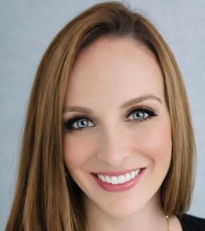
Chief, Division of Research
Assistant Professor of Surgery, Division of Minimally
Invasive and Gastrointestinal Surgery
The Department of Surgery hosted the 2025 Fall Research Symposium. This is an annual event led by the Division of Research to showcase research efforts from medical students, residents, fellows, graduate students, and post-doctorate learners housed with the Department of Surgery. This year the department had 25 presentations, covering a breath of topics from minimally invasive surgery to graduate medical education (Table 1). Symposium judges awarded two presenters the distinction as symposium winners, Lauren Cohen, BS mentored by Dr. Chandler Cortina, and Dr. Jennifer Schuh mentored by Dr. Amy Wagner. These awards were bestowed based on the highest judge’s scores following criteria including scientific content, oral presentation skills, and audience question management.
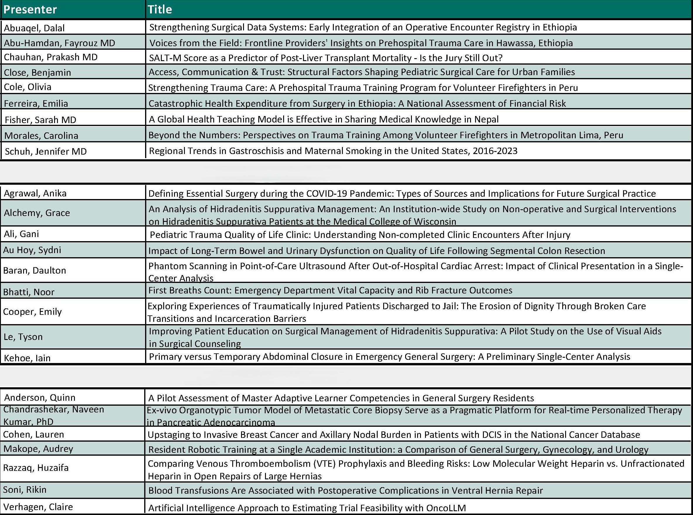
Table 1. 2025 Fall Research Symposium Presenters and Abstract
Title
Ms. Cohen’s abstract was entitled “Upstaging to invasive breast cancer and axillary nodal burden in patients with DCIS in the National Cancer Database.” The authors describe that ductal carci-
noma in situ (DCIS) is a non-obligate precursor to invasive breast cancer (iBC). The standard of care for DCIS consists of surgery, adjuvant radiation for those who undergo lumpectomy, and risk-reducing endocrine therapy. Surgical axillary nodal staging can be considered for patients who are at high risk for upstaging to iBC and those undergoing mastectomy; however, surgical axillary nodal staging is associated with increased morbidity. Furthermore, recent trials have supported the omission of surgical nodal staging in select patients with early stage iBC based on tumor receptor subtype and normal axillary imaging. There is a paucity of data on contemporary rates of DCIS upstaging on surgical pathology and nodal burden in those who undergo surgical axillary nodal staging; therefore, they aimed to identify modern national rates of DCIS upstaging to invasive disease and axillary nodal burden.
The authors performed a retrospective cohort study using data from the National Cancer Database from 2018–2022. Patients age ≥18 years, female sex, and diagnosed with clinical DCIS (cTis N0 M0) who received upfront surgery were included. Rates of upstaging were defined as any invasive disease seen on breast or axillary surgical pathology (pTNM status). Surgical axillary nodal staging was coded as no axillary surgery, sentinel lymph node biopsy (SLNB), or axillary lymph node dissection (ALND). Pathologic N stage was used to assess axillary nodal burden in those who received SLNB or ALND. The authors found that a total of 144,628 patients met inclusion criteria. The median patient age was 61 years (IQR 51–69), most had no comorbidities (83.1%) with private insurance (54.9%). 68.4% of patients were NH-White, 13.8% were NH-Black, and 7.6% were Hispanic. The majority had estrogen receptor positive (83.8%) and progesterone receptor positive (64.9%) disease, and about half received adjuvant risk-reducing endocrine therapy
(51.5%) and radiation therapy (51.9%). Most patients received lumpectomy (70.8%) and 29.2% underwent mastectomy. 16.4% of the cohort upstaged to iBC on final surgical pathology. The authors concluded that most patients with DCIS (56.8%) did not have surgical axillary nodal staging, 38.3% had SLNB, and 2.8% had ALND. Of those who underwent surgical axillary nodal staging (n=59,457), 97.5% were found to be pN0, 2.4% were found to be pN1, and only 0.1% were pN2.
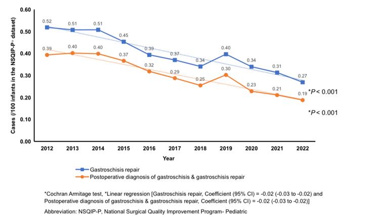
Figure 1. Decreasing trend of gastroschisis cases in the NSQIP-P, years 2012-2022. NSQIP-P, National Surgical Quality Improvement ProgramPediatric. As published in [1].
Since only 16.4% of patients with DCIS upstaged to iBC on surgical pathology and the upstaging with nodal involvement was considerably low, findings support the consideration of omitting surgical axillary nodal staging for most patients with DCIS. The study also underscores the necessity of prospective trials to assess the long-term outcomes of this approach.
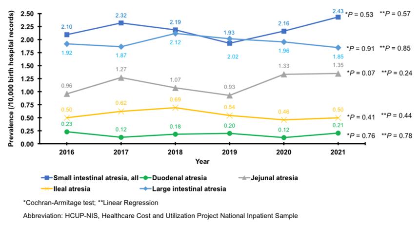
Figure 2. Trend analysis of small and large intestinal atresia prevalence. HCUP-NIS, years 2016-2021. HCUP-NIS, Healthcare Cost and Utilization Project- National Inpatient Sample. As published in [1]
Dr. Schuh’s abstract describes that after decades of rising prevalence, gastroschisis rates have declined since 2014 (Figure 1) [1]. Other gastrointestinal atresia rates attributable to in utero vascular accidents have not changed in prevalence suggesting a different pathophysiologic origin of gastroparesis (Figure 2) [1]. It is unclear if the decrease in gastroschisis trends varies by region. While the exact cause of gastroschi-
sis remains unknown, several genetic and environmental risk factors, including maternal smoking, have been implicated. The authors aimed to examine regional trends in gastroschisis and maternal smoking. They conducted a national population-based cross sectional study using birth certificate data from 2016 – 2023, analyzing gastroschisis prevalence and maternal smoking across four census regions: Northeast, Midwest, South, and West. Linear regression assessed trends, and population attributable risk estimated the proportion of cases linked to smoking. The authors found that among 92 million live births, gastroschisis prevalence declined nationally and across all regions, with the highest prevalence in the Midwest (which declined from 3.3 to 2.10 per 10,000). Tobacco use during pregnancy decreased in all regions (p<0.001), but only the Midwest (p=0.010) and South (p=0.045) showed declines among mothers of affected infants. Population attributable risk for smoking declined in the Northeast, Midwest, and South, but was not significant. The authors concluded that gastroschisis rates and smoking-related risk are decreasing in each region of the United States. Continued region-specific health efforts, especially in the West, are needed to sustain and improve progress.
For more information about the Department of Surgery Division of Research and all the amazing work being performed by our faculty and learners, please visit www. mcw.edu/departments/surgery/research.
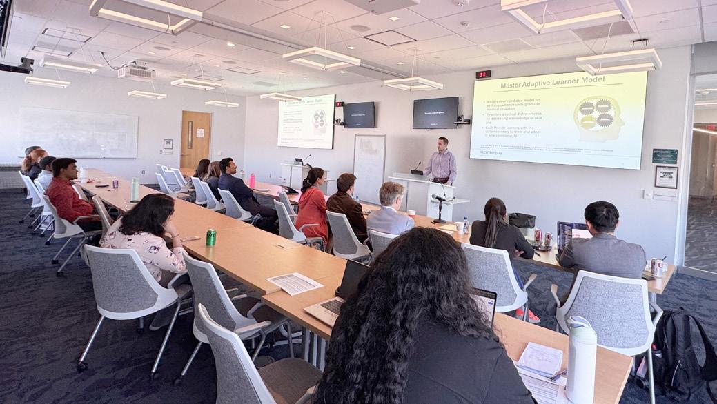
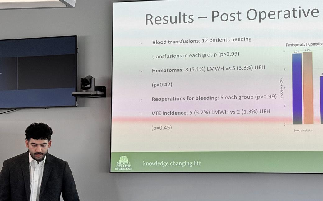
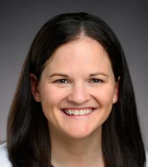
Catherine McManus, MD, MS, FACS, joined the Department of Surgery faculty in August 2025 as an Associate Professor of Surgery. Dr. McManus is a board-certified endocrine surgeon with advanced training from Columbia University and a Master’s degree in biostatistics. She specializes in thyroid, parathyroid, and adrenal surgery, with national leadership in endocrine surgery education, innovation in minimally invasive and ablative techniques, and a robust portfolio of funded research and peer-reviewed scholarship.
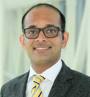
Narendra Battula, MD joined the Department of Surgery faculty in December 2025 as an Associate Professor of Surgery. Dr. Battula brings exceptional expertise in liver and multi-organ transplantation, with training at leading United Kingdom centers, including King’s College Hospital London and the renowned Liver Unit at University Hospitals Birmingham, as well as fellowship training at Ochsner Health in New Orleans. Dr. Battula is certified in robotic surgery using the da Vinci® system and has advanced expertise in hepatobiliary and transplant surgery. His clinical practice includes adult and pediatric liver transplantation, living donor surgery, and complex multi-organ transplants, including liver–kidney, heart–liver, and liver–lung. His clinical interests include transplant oncology and innovative organ preservation techniques, including normothermic machine perfusion. In addition to his clinical work, Dr. Battula serves as Medical Director of the Transplant Inpatient Unit and as Patient Safety and Quality Officer for the Transplant Service Line, supporting high-quality, coordinated transplant care.
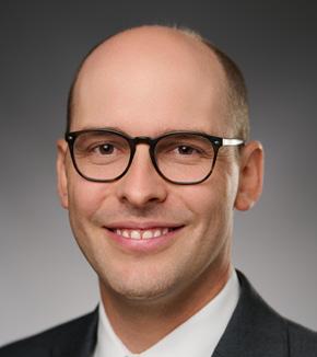
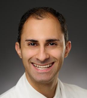
Matthew Harris, MD, joined the Department of Surgery faculty in October 2025 as an Assistant Professor of Surgery. Dr. Harris also serves as Medical Director for Organ and Tissue Donation at Versiti. He completed general surgery residency at Yale–New Haven Hospital followed by fellowship training in abdominal transplant and hepatopancreatobiliary surgery at Northwestern University. Dr. Harris’s clinical and research interests focus on liver and kidney transplantation, donor organ optimization, and normothermic machine perfusion technologies, with an active portfolio of funded research and peer-reviewed scholarship aimed at expanding the utilization of donor organs and improving transplant outcomes.
Siavash Raigani, MD, joined the Department of Surgery faculty in August 2025 as an Assistant Professor of Surgery. Dr. Raigani earned his medical degree from Case Western Reserve University, completed his general surgery residency at Massachusetts General Hospital, and went on to complete a fellowship in abdominal transplant surgery at Duke University. Dr. Raigani brings a strong academic interest in ex vivo machine perfusion of donor organs, along with valuable research experience, to the Medical College of Wisconsin. His clinical practice focuses on liver, kidney, and pancreas transplantation, and he also performs peritoneal dialysis surgery for patients with renal failure.
Jeffrey Anderson, MD, FACS, joined the Department of Surgery faculty in September 2025 as an Assistant Professor of Surgery. Dr. Anderson is board-certified in General Surgery and Surgical Critical Care, with fellowship training in burn, trauma, and critical care at the University of Washington and Harborview Medical Center. Dr. Anderson’s clinical and academic interests include trauma resuscitation, burn care, surgical critical care, wound healing, and quality and safety in acute care surgery. He has held multiple leadership roles in surgical intensive care and burn programs, serves on national trauma and critical care committees, and is an active investigator with sustained grant funding and numerous peer-reviewed publications.
Erin Buchanan, MD, joined the Department of Surgery faulty in September 2025 as an Assistant Professor of Surgery. Dr. Buchanan completed her general surgery residency at the Medical College of Wisconsin and fellowship training in vascular and endovascular surgery at the University of Virginia Health. Dr. Buchanan’s clinical and academic interests include vascular surgery education, endovascular and open peripheral revascularization, and complex aortic pathology. She is an active researcher and author with peer-reviewed publications, national presentations, and contributing chapters in leading vascular surgery textbooks.
Bariatric & Minimally Invasive Gastrointestinal Surgery
Amir Ghaferi, MD, MSc, MBA
Matthew Goldblatt, MD
Jon Gould, MD, MBA
Rana Higgins, MD
Andrew Kastenmeier, MD
Tammy Kindel, MD, PhD
Kathleen Lak, MD
Philip Redlich, MD, PhD
Wen Hui Tan, MD
Ezra Teitelbaum, MD, MEd
Cardiac Surgery
Ahmed Ali, MD
G. Hossein Almassi, MD
Lucian Durham III, MD, PhD
Robert Jaquiss, MD
Takushi Kohmoto, MD, PhD, MBA
James Mace, Jr., MD
Jorge Mascaro Carvajal, MD
Michael Mitchell, MD*
Paul Pearson, MD, PhD
Stefano Schena, MD, PhD
H. Adam Ubert, MD
Colorectal Surgery
Jed Calata, MD
Katherine Hu, MD
Kirk Ludwig, MD
Carrie Peterson, MD, MS*
Timothy Ridolfi, MD, MS
Community Surgery
Marina Affi Koprowski, MD
Marc de Moya, MD
Kaizad Machhi, MD
Allegra Saving, MD
Eric Soneson, MD
Mark Timm, MD
Pediatric General & Thoracic Surgery
John Aiken, MD*
Christina Bence, MD
Casey Calkins, MD*
Brian Craig, MD
John Densmore, MD*
Katherine Flynn-O’Brien, MD, MPH
David Gourlay, MD*
Tammy Kindel, MD, PhD
Christopher Laird, MD
Dave Lal, MD, MPH*
Caroline Maloney, MD, PhD
Jose Salazar Osuna, MD, PhD*
Jack Schneider, MD*
Amy Wagner, MD*
Research Faculty
Mohammed Aldakkak, MD
John Baker, PhD
Young-In Chi, PhD
Christian Kastrup, PhD
Gwen Lomberk, PhD
Nikki Lytle, PhD
Angela Mathison, PhD
Aoy Tomita Mitchell, PhD
M. Muska Nataliansyah, MD, PhD, MPH
Kirkwood Pritchard, Jr., PhD
Raul Urrutia, MD
Surgical Oncology – Breast Surgery
Adrienne Cobb, MD, MS*
Chandler Cortina, MD, MS*
Amanda Kong, MD, MS*
Caitlin Patten, MD*
Jasmine Walker, MD, MPH
Tina Yen, MD, MS
Surgical Oncology – Endocrine Surgery
Douglas Evans, MD*
Catherine McManus, MD, MS
Tracy Wang, MD, MPH*
Tina Yen, MD, MS
Surgical Oncology – Hepatobiliary & Pancreas Surgery
Kathleen Christians, MD
Callisia Clarke, MD, MS
Douglas Evans, MD*
Karen Kersting, PhD, LCP
Y. David Seo, MD
Surgical Oncology – Regional Therapies
Callisia Clarke, MD, MS
Alexandra Istl, MD, MPH
Anai Kothari, MD, MS
Ugwuji Maduekwe, MD, MMSc, MPH
Thoracic Surgery
Mario Gasparri, MD
Mallory Hunt, MD, MBE, MSTR
Paul Linsky, MD
Transplant Surgery
Emre Arpali, MD, PhD
Narendra Battula, MD
Matthew Cooper, MD
Ty Dunn, MD, MS
Matthew Harris, MD
Kondragunta Rajendra Prasad, MD, MS
Jenessa Price, PhD
Siavash Raigani, MD
Badi Rawashdeh, MD
Stephanie Zanowski, PhD
Trauma/ACS
Jeffrey Anderson, MD
Marshall Beckman, MD, MA*
Jacqueline Blank, MD*
Kelly Boyle, MD
Nathan Carlson, MD
Thomas Carver, MD*
Joshua Dilday, DO*
Marc de Moya, MD*
Terri deRoon-Cassini, PhD, MS
Christopher Dodgion, MD, MSPH, MBA*
Anuoluwapo Elegbede, MD, MS*
Timothy Geier, PhD, LP
Allyson Hynes, MD MSCE
Katie Iverson, MD, MPH*
Jeremy Levin, MD*
Christina Megal, DNP, APNP
David Milia, MD*
Rachel Morris, MD*
Patrick Murphy, MD, MSc, MPH*
Todd Neideen, MD*
Jacob Peschman, MD, MSPE*
Andrew Schramm, PhD
Libby Schroeder, MD, MS*
Lewis Somberg, MD, MSS*
Sydney Timmer-Murillo, PhD
Colleen Trevino, MSN, FNP, PhD
Sarah Vanderlinden, DMSc, PA-C
Vascular & Endovascular Surgery
Shahriar Alizadegan, MD*
Kellie Brown, MD, FACS*
Erin Buchanan, MD
Mitchell Dyer, MD, MSc
Joseph Hart, MD, MHL
Momodou L. Jammeh, MD
Dean Klinger, MD
Nathan Kugler, MD*
Brian Lewis, MD
Mona Li, MD*
Michael Malinowski, MD, MEHP, FACS*
Peter Rossi, MD*
Abby Rothstein, MD*
Affiliated Institution Program Directors
Gary Sweet, MD (Wausau)
Nicholas Meyer, MD (Columbia St. Marys)
Chief Advance Practice Providers
Courtney Johnson, PA-C, Ambulatory Chief APP
Maria Wellenstein, APNP, Inpatient Chief APP
Chief Surgical Residents (20252026)
Elise Biesboer, MD
Administrative Chief Resident
Melissa Drezdzon, MD
Administrative Chief Resident
Taylor Jaraczewski, MD, MS
Santiago Rolon, MD
Ben Seadler, MD
Monica Seadler, MD
Brexton Turner, MD
Amanda Witte, MD
*Participates in Community Surgery/Off-Campus Locations.
Learn more at mcw.edu/ surgery or follow us on social media.


Leading the Way is published biannually by The Department of Surgery at MCW. Leading the Way is written for physicians for medical education purposes only. It does not provide a complete overview of the topics covered and should not replace the independent judgment of a physician in clinical practice. The opinions expressed by our faculty are those of the individual authors and may not reflect the opinions of the Medical College of Wisconsin. ©2024 Editors: Katie Iverson, MD, MPH and Maria McQuestion (with assistance from Dr. Evans and the Department faculty)