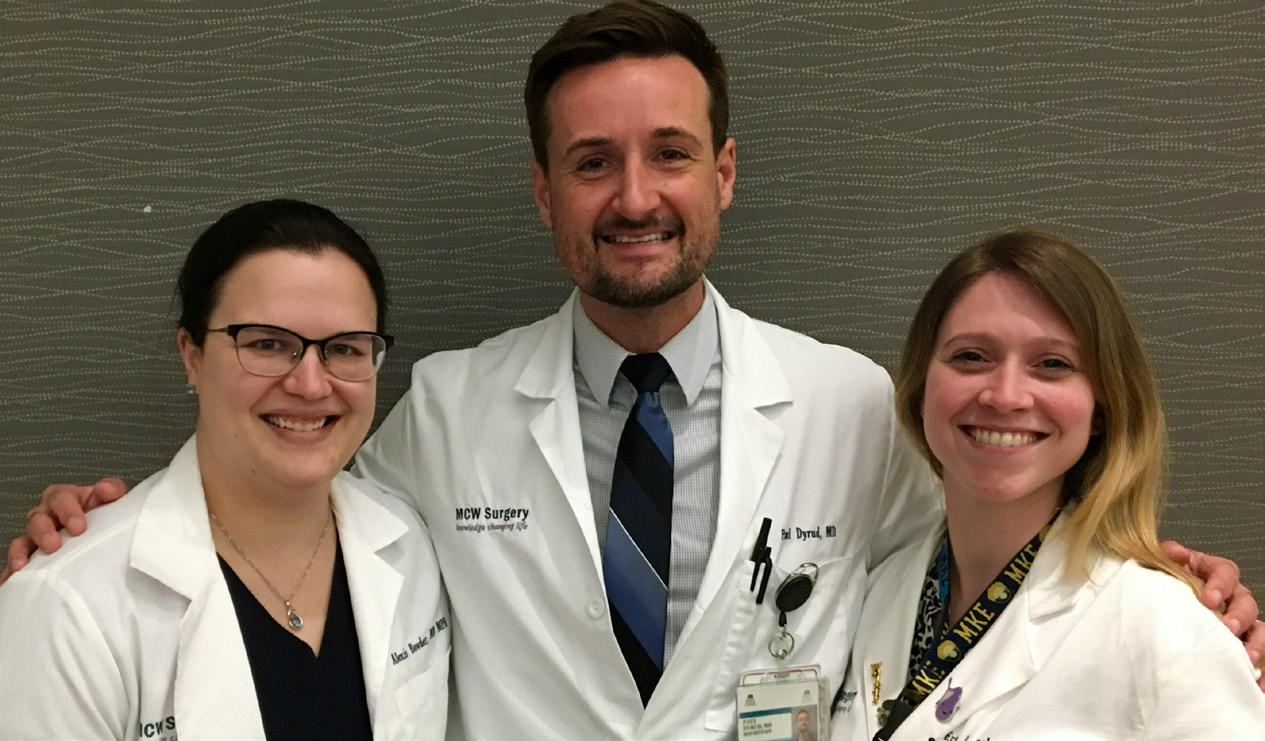
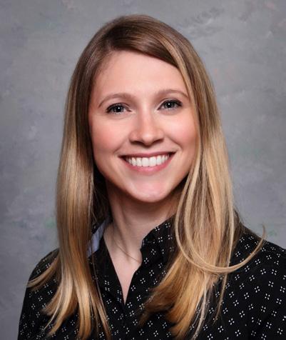
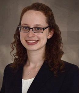
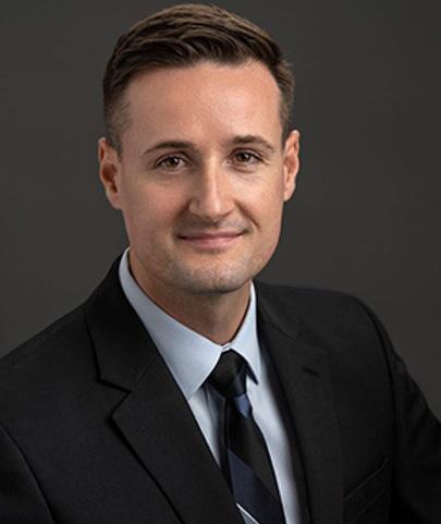
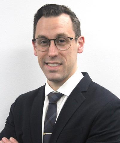
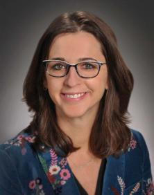
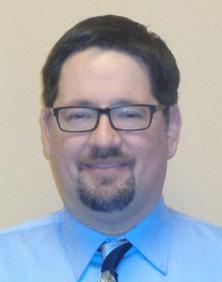
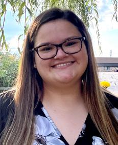
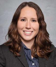
“Leading the Way” has grown just as much as our department; the number of featured articles in this edition is a prime example. While the articles focus on different areas within surgery and medicine, they all have one thing in common – they reflect the extraordinary amount of time that our faculty, residents and fellows have their minds on some aspect of innovation and discovery. The outpouring of contributions to “Leading the Way” represents the tip of the department’s iceberg of academic productivity. Such academic achievement creates an atmosphere of always asking why – the most important part of how we practice medicine and work together. Questioning everything from clinical guidelines and pathways to all aspects of patient management and importantly, to department and institutional policies, means that we are thinking all the time. Creating a culture which accepts, and expects, us to question the status quo will result in us writing the clinical guidelines and pathways, embracing the time and resources needed to succeed in research and academics, and developing a culture of open communication in an environment which can sometimes be stressful and uncomfortable. Pictured on the cover of this issue of “Leading the Way” are our incoming Chief Residents who will continue the tradition of questioning how and why we do things – ensuring that we treat the patients of tomorrow better than we are treating the patients of today. Thank you to all our faculty and residents who contributed to this amazing issue of “Leading the Way.”
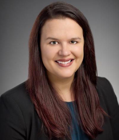
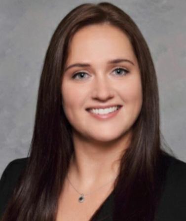
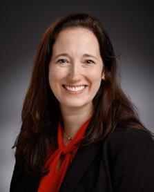
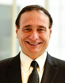
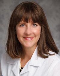
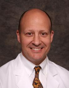
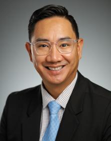
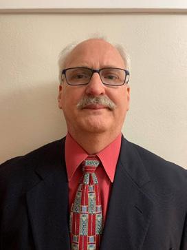
AY 2022-2023 Chief Residents (top left to bottom right): Alexis Bowder MD, Erin Buchanan MD, Paul Dyrud MD, Kyla Fredrickson MD, Ashley Krepline MD, Nathan Smith MD
(Left to right) Alexis Bowder MD, Paul Dryud MD, Erin Buchanan MD
Gastroschisis Outcomes of Delivery Study
Amy Wagner, MD Professor of Surgery; Section Chief, Division of Pediatric Surgery
What is Gastroschisis?
Gastroschisis (Fig. 1) is the most common congenital defect of the abdominal wall. During normal development, the abdominal wall remains open, and the intestines are found outside the fetus early in gestation. The intestines should return to the abdomen and the abdomen should close by 12 weeks gestation. However, in cases of gastroschisis, the intestines remain outside the abdomen, protruding through a small defect. Therefore, gastroschisis is typically diagnosed after 14 weeks gestation using prenatal ultrasound. Gastroschisis is diagnosed in about 1 out of every 2,000 births1; however, this number is increasing2. Fetuses diagnosed with gastroschisis have an increased risk of damage to or loss of the intestine, which can be caused by the caustic amniotic fluid or the defect narrowing and constricting blood flow. Gastroschisis patients also have an increased risk of fetal growth restriction and fetal demise (stillbirth). These risks may be higher in infants carried to full term; therefore, some providers choose to deliver these patients early to try and mitigate these risks. While some providers choose to deliver gastroschisis patients early, others believe delivering early comes with equal or more risk as these patients are born prematurely (late pre-term).
Infants born prematurely have an increased risk for jaundice, infections, low birth weight, need of extra respiratory support, and neurocognitive deficits. Unfortunately, there are conflicting data (based mostly on retrospective studies) to determine the optimal delivery timing in gastroschisis.3,4 The lack of rigorous scientific evidence has caused much controversy in the medical community, prompting institutions across the country to vary delivery timing to anywhere from 34 to 38 weeks gestation. Due to the lack of standard-of-care guidelines for pregnancies with fetal gastroschisis,5 Dr. Amy Wagner and colleagues designed the Gastroschisis Outcomes of Delivery Study.
Study Overview
The Gastroschisis Outcomes of Delivery (GOOD) Study is a national, multi-site, randomized controlled trial designed to evaluate delivery timing of infants diagnosed with gastroschisis and provide evidence of optimal delivery timing. The recruitment goal for the study is 800 mother-fetus pairs by 2025. Currently, 26 centers across the country are participating in the study. The trial has two intervention arms, a 35-week delivery group and an expectant management group with a goal of 38 weeks. Our
Chris Fueger, MS GOOD Study Program Manager
Maddie Rundell, BS Clinical Research Coordinator I
hypothesis is that delivery at 35 0/7 – 35 6/7 weeks gestation in stable mothers and fetuses with gastroschisis will be superior to expectant management of the pregnancy with a goal of 38 0/7 – 38 6/7 weeks gestation. The three aims of the study are (1) to compare risk of mortality and major morbidity for infants with gastroschisis between the early and late delivery arms, (2) to determine maternal, fetal, and neonatal intermediate secondary outcomes for gastroschisis cases in early and late delivery arms, and (3) to establish a national registry of clinical data and biobank comprised of gastroschisis patients.
The study design is a randomized, parallel assignment model with an intention-to-treat analysis utilizing a primary weighted composite outcome. Outcomes include stillbirth (after 20 weeks gestational age), neonatal/infant death prior to Neonatal intensive care unit (NICU) discharge, respiratory morbidity, gastrointestinal morbidity, and sepsis.
Mothers with a singleton pregnancy diagnosed with stable gastroschisis and followed by one of 26 participating centers across the country are eligible to participate. The treating physician will evaluate the patient to determine if they meet the eligibility criteria. Eligible patients are then approached to discuss participation in the study. Patients are consented into the study (until 33 6/7 weeks gestation) and are randomized between 33 0/7 and 33 6/7 weeks gestation. Each site uses its own randomization
Fig. 1: Fetal gastroschisis
Gastroschisis Outcomes of Delivery (GOOD)
table to create equal groups across all institutions and eliminate site-specific bias in the data due to the site’s standard clinical practices. After randomization, both the mother and infant are followed by study staff until initial NICU discharge of the infant.
Future Directions
Another area within gastroschisis lacking robust evidence is the etiology of the disease. We plan to create a national bank of bio-specimens (blood sample or buccal swab) collected from neonates enrolled in the GOOD study to learn more about possible genetic or epigenetic factors which cause gastroschisis and may give insight into the increasing incidence. We are collaborating with Dr. Reddi and her colleagues at the Precision Medicine Laboratory (PML) to help us investigate the cause or genetic associations in these patients. This has the potential to be the largest ever bio-bank and genetic analysis for gastroschisis patients.
In addition to further investigation with the bio-bank, we are also planning our next clinical phase of the trial which will be to investigate the intermediate outcomes of gastroschisis patients between 2 and 5 years of life. Factors such as cognitive/neurodevelopment, gastrointestinal motility, social determinants of health, and quality of life will be examined.
Endorsements & Sponsors
The GOOD Study has been endorsed by the North American Fetal Therapy Network (NAFTNet). Sponsors of the study include: We Care Fund for Medical Innovation and Research, NAFTNet, Stanek Fund, Ruth St. John and John Dunham West Foundation Inc., Melitta S. Pick and Joan M. Pick Charitable Trust, and the Eunice Kennedy Shriver National Institute of Child Health & Human Development of the National Institutes of Health Award Number R01 HD104607.
FOR ADDITIONAL INFORMATION on this topic, visit mcw. edu/surgery or contact Dr. Amy Wagner at awagner@ chw.org.
REFERENCES
1. Bhat V, Moront M, Bhandari M. Gastroschisis: A state-ofthe-art review. Children. 2020;7(12):302. doi:10.3390/ children7120302.
2. Jones AM, Isenburg J, Salemi JL, Arnold KE, Mai CT, Aggarwal D, Arias W, Carrino GE, Ferrell E, Folorunso O, Ibe B, Kirby RS, Krapfl HR, Marengo L, Mosley BS, Nance AE, Romitti PA, Spadafino J, Stock J, Honein MA. Increasing prevalence of gastroschisis — 14 States, 1995–2012. MMWR Morb Mortal Wkly Rep. 2016;65:23-6. doi: http://dx.doi.org/10.15585/ mmwr.mm6502a2.
3. Landisch RM, Yin Z, Christensen M, Szabo A, Wagner AJ. Outcomes of gastroschisis early delivery: A systematic review and meta-analysis. J Pediatr Surg. 2017;52(12):196271. Epub 2017/09/28. doi: 10.1016/j.jpedsurg.2017.08.068. PubMed PMID: 28947324.
5. Amin R, Domack A, Bartoletti J, Peterson E, Rink B, Bruggink J, Christensen M, Johnson A, Polzin W, Wagner AJ. National practice patterns for prenatal monitoring in gastroschisis: Gastroschisis outcomes of delivery (GOOD) provider survey. Fetal Diagn Ther. 2018:1-6. Epub 2018/05/24. doi: 10.1159/000487541. PubMed PMID: 29791899.
REFERENCES FROM PAGE 5, CONTINUED
Precision Medicine: An Individualized Approach to Trauma Care
8. McDaniel DO, Hamilton J, Brock M, et al. Molecular analysis of inflammatory markers in trauma patients at risk of postinjury complications. J Trauma Acute Care Surg. 2007;63(1):147-158.
9. Thompson CM, Holden TD, Gail RR, et al. Toll-like receptor 1 polymorphisms and associated outcomes in sepsis following traumatic injury, a candidate gene association study. Ann Surg. 2014;259(1):179-185.
10. Drewry AM, Ablordeppey EA, Murray ET, et al. Comparison of monocyte human leukocyte antigen-DR expression and stimulated tumor necrosis factor alpha production as outcome predictors in severe sepsis: a prospective observational study. Crit Care. 2016;20(1):334.
Welcome to our 2022-2023 General Surgery Interns
Michael Brinton, MD
Allison Couillard, MD
Sarah Fisher, MD
Yoon Jae (Alice) Lee, MD
Alexa Lisevick, MD
Gesina Thiry, MD
Shanita Thomas, MD
Kaitlyn Sonnentag, MD, Rural
Tara Mather, MD, Plastics
Aishwarya Ramamurthi, MD, Plastics
Matthew Henry, MD, Prelim IR
Michael Rabaza, MD, Prelim IR
Abdul-Rahman AbdelReheem, MD, Prelim
Ayokunle Olowofela, MD, Prelim
John Shepherd, III, MD, Prelim
Arya Anvar, MD, Urology
Kwesi Asantey, MD, Urology
Carissa Battle, MD, Urology
Precision Medicine: An Individualized Approach to Trauma Care
Background
Christina Georgeades, MD
General Surgery Resident; Pediatric Surgery
Research Fellow, Division of Pediatric Surgery
Gwen Lomberk, PhD
Joel and Arlene Lee Endowed Chair in Pancreatic Cancer Research; Chief, Division of Research; Director, Basic Science Research; Associate Professor of Surgery and Pharmacology & Toxicology; Co-Leader, Precision Oncology Initiative, Cancer Center
Trauma is the fourth leading overall cause of death in the United States, and is the leading cause of death before the age of 45.1 The National Academies of Sciences, Engineering, and Medicine has estimated that approximately 30,000 trauma-related deaths could be prevented each year with implementation of more adequate trauma care, particularly on a personalized level.2 Precision medicine, which accounts for an individual’s unique environment, lifestyle, and genetic makeup, could potentially allow providers to ascertain the optimal management strategy for each specific trauma patient, leading to more accurate and efficient care.3 The utilization of precision medicine would ideally provide real-time information after injury about an individual’s distinct physiology to determine what their likely clinical course would be. This would ultimately not only prevent mortality, but also improve clinical outcomes.
Relevant
Genomic, Epigenomic, Proteomic, and Metabolomic Factors
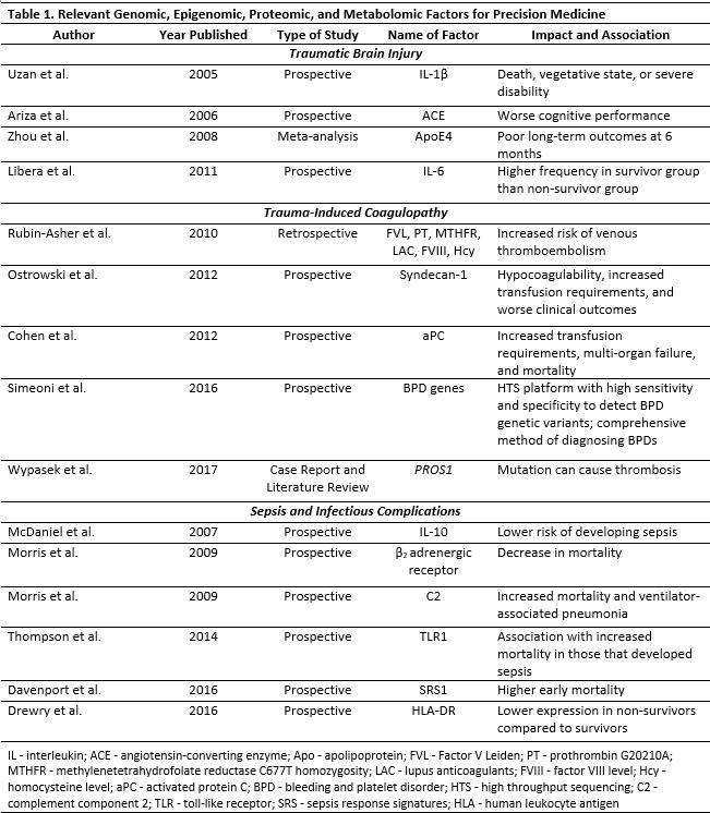
There are certain genomic, epigenomic, proteomic, and metabolomic factors that have been identified in relation to trauma, and they may serve as potentially useful clinical precision medicine assays [Table 1]. In particular, traumatic brain injury, trauma-induced coagulopathy, venous thromboembolism, and sepsis are topics with rich possibilities for implementation of precision medicine that could positively impact a patient after injury. Although certain genes have been identified that link traumatic brain injury and clinical outcomes, none have been directly evaluated for use in real-time clinical decision-making. There are at least 33 traumatic brain injury genes associated with injury response and neurocognitive reserve, particularly relating to variation in the bloodbrain barrier.4 An example of relevant proteomic factors are interleukins, which are a group of cytokines that play an important role in the inflammatory response after traumatic brain injury. Interleukins have been linked with clini-
Raul Urrutia, MD
Director, Genomic Sciences and Precision Medicine Center; Warren P. Knowles Chair of Genomics and Precision Medicine; Professor, Departments of Surgery, Biochemistry and Physiology; Associate Director, Precision Medicine, CTSI
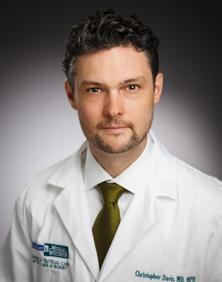
Christopher S. Davis, MD, MPH
Assistant Professor, Division of Trauma & Acute Care Surgery and the Institute for Health and Equity; Associate Program Director, General Surgery Residency Program; Community and Institutional Engagement Pillar Faculty/Well-being and Resilience, Kern Institute for the Transformation of Medical Education
cal outcomes. While some interleukins are associated with poor outcomes such as death or severe disability, others are associated with improved survival. Early assessment of these factors in the acute trauma setting could identify individuals that are more at risk for specific morbidity than others.
Trauma-induced coagulopathy, occurring with the activation of coagulation and fibrinolytic pathways after major hemorrhage, is connected with molecular markers that can indicate abnormal bleeding after traumatic injury. Certain coagulopathic factors, such as the syndecan-1 protein and activated protein C levels, are associated with hypocoagulability, higher transfusion requirements, and worse outcomes.5,6 Genetics also play a role in determining the efficacy of venous thromboembolism prophylaxis and the risk of venous thromboembolism after trauma. For example, a deficiency in a vitamin-K dependent natural anticoagulant called Protein S could lead to increased risk of venous thromboembolism occurrence.7 The deficiency is found in 1.5% - 7% of thrombophilic patients.7 Promptly identifying trauma patients at higher risk of bleeding or thrombotic events through genetic factors could mitigate the risk of adverse events.
Post-traumatic sepsis and infectious complications are associated with many molecular markers. Cytokines, chemokines, and growth factors are markers that mediate the impact of the pro- and anti-inflammatory response. For example, patients with a low-producing genotype for interleukin-10 have a lower risk of developing sepsis.8 Expression levels of factors such as toll-like receptor protein, which recognizes bacterial substrates, and human leukocyte antigen-DR, which is involved in the immune response, can also influence the outcomes of trauma patients who develop sepsis due to their involvement with an immunocompromised state.9,10 The numerous factors that affect sepsis outcomes after traumatic injury provides
a rich opportunity for potentially identifying a marker early in the hospital course that could guide management to positively impact a patient’s subsequent clinical outcome.
Future Directions
The utilization of precision medicine in the acute setting after a traumatic injury has the opportunity to guide management decisions and positively affect outcomes. There are many physiologic and individualized markers that have been identified, but the ability to apply these markers and analyze genetic differences in a real-time setting is lacking. Future research is needed to identify which factors provide the highest yield of applicable information and subsequently can lead to the development of effective and efficient assays to be used in the clinical setting.
The Division of Trauma and Acute Care Surgery is collaborating with the Genomic Sciences and Precision Medicine Center to initiate precision medicine research with the goal of ultimately identifying genetic and molecular factors to be used in clinical assays in the early period of
an acute traumatic injury. The trauma patient population deserves an individualized approach that can optimize their care and recovery.
Conclusions
The opportunity of translating specific genomic, epigenomic, proteomic, and metabolomic factors into realtime clinical adjuncts to guide management of traumatic injury is promising. The development of diagnostic methods can potentially assist with anticipating the physiologic response to trauma based on an individual’s unique genetic makeup and variation, with the ultimate goal of reducing preventable death. The advent and application of precision medicine in trauma could revolutionize patient management, which is an endeavor worth striving for.
FOR ADDITIONAL INFORMATION on this topic, visit mcw. edu/surgery or contact Dr. Christina Georgeades at cgeorgeades@mcw.edu.
Table 1: Relevant Genomic, Epigenomic, Proteomic, and Metabolic Factors for Precision Medicine; Adapted from Davis CS, Wilkinson KH, Lin E, et al. Precision medicine in trauma: a transformational frontier in patient care, education, and research [published online ahead of print, 2021 Nov 16]. Eur J Trauma Emerg Surg. 2021;1-6. doi:10.1007/s00068-021-01817-7
REFERENCES
1. Centers for Disease Control and Prevention. 10 Leading Causes of Death by Age Group, United States - 2020. WISQARS - Fatal Injury Data. Published February 20, 2020. https://wisqars.cdc.gov/fatal-leading
2. National Academies of Sciences, Medicine. A national trauma care system: integrating military and civilian trauma systems to achieve zero preventable deaths after injury. Mil Med. 2016;182(5):1563-1565.
3. Genetics Home Reference. What is precision medicine? U.S. National Library of Medicine. Published September 22, 2020. https://ghr.nlm.nih.gov/primer/ precisionmedicine/definition
4. Kurowski BG, Treble-Barna A, Pitzer AJ, et al. Applying systems biology methodology to identify genetic factors possibly associated with recovery after traumatic brain injury. J Neurotrauma. 2017;34(14):2280-2290.
5. Cohen MJ, Call M, Nelson M, et al. Critical role of activated protein C in early coagulopathy and later organ failure, infection and death in trauma patients. Ann Surg. 2012;255(2):379-385.
6. Ostrowski SR, Johansson PI. Endothelial glycocalyx degradation induces endogenous heparinization in patients with severe injury and early traumatic coagulopathy. J Trauma Acute Care Surg. 2012;73(1):60-66.
7. Makris M, Leach M, Beauchamp NJ, et al. Genetic analysis, phenotypic diagnosis, and risk of venous thrombosis in families with inherited deficiencies of protein S. Blood. 2000;95(6):1935-1941.
References continued on page 3
Lending a Hand: The Utility of Hand-Assisted
Introduction
Melissa K. Drezdzon, MD General Surgery Resident
Carrie
Y. Peterson, MD, MS
Associate Professor, Division of Colorectal Surgery
Kirk A. Ludwig, MD
Professor and Chief, Division of Colorectal Surgery; Vernon O. Underwood Professor in Colon Cancer Research
Diverticulitis is a common problem, and the incidence is steadily increasing, while the affected patients are getting younger.1 Approximately 15% of patients with diverticulitis will require surgery in their lifetime, and 20% of those cases are complicated by the presence of a fistula.2 Colovesical fistula is the most common diverticulitis-associated fistula, compromising 65% of all cases, and symptoms related to these fistulas can significantly impact a patient’s quality of life. 3 The 2020 American Society of Colon and Rectal Surgeons (ASCRS) Guidelines for the treatment of colonic diverticulitis recommends elective colectomy for patients with diverticulitis complicated by fistula.4
Diverticulitis-associated fistulas present unique operative challenges due to significant inflammation and anatomic distortion, often impairing structure identification and mobilization. Due to case difficulty, these procedures were traditionally performed via an open approach; however, the feasibility and safety of minimally-invasive approaches has been demonstrated.2,3 Nevertheless, the laparoscopic approach to these cases is technically challenging and has high conversion-to-open rates.2,5–8 The Hand-Assisted Laparoscopic Surgery (HALS) technique is a minimally invasive approach that can be used to tackle these challenging cases. HALS surgery preserves the tactile feedback, manual blunt dissection, and figure fracture techniques afforded in open surgery, while offering minimally-invasive benefits including smaller incisions, shorter length of stay, and earlier return of bowel function.3,5,6,8
The aim of our study was to review the experience with HALS colectomy used to manage diverticular disease complicated by fistula within our Division of Colorectal Surgery. Since many of these fistulas were colovesical, we also sought to evaluate our intraoperative management
Jed F. Calata, MD Assistant Professor, Division of Colorectal Surgery
Mary F. Otterson, MD, MS Professor, Division of Colorectal Surgery
Timothy J. Ridolfi, MD, MS
Associate Professor, Division of Colorectal Surgery
of the fistula tract and post-operative Foley catheter management.
Materials and Methods
Patient Selection
All patients who underwent elective resection for diverticular disease-associated fistula between January 2008 and August 2021, within the Division of Colorectal Surgery, were identified. Clinical and operative details were collected via chart review. Exclusion criteria included diagnosis of Crohn’s disease and patients who underwent emergent surgical intervention for complications of diverticulitis, including free perforation and feculent peritonitis.
Operative Technique
In the standard HALS approach within the Division of Colorectal Surgery, patients are positioned in low lithotomy and a Foley catheter is placed. Ureteral stent placement is at the discretion of the surgeon. A 6-7 cm midline, periumbilical incision is made and a GelPort (Applied Medical) is inserted. In some cases, a Pfannenstiel incision is used for the hand port. A 5-mm suprapubic port and a 5-mm right lower quadrant port are placed. The descending colon is mobilized with routine splenic flexure mobilization and left ureter identification. The fistula tract is approached with a combination of sharp dissection and finger fracture technique. A LigaSure device (Medtronic) is used to divide the mesentery and the proximal rectum is divided with an Endo-GIA stapler using a blue cartridge (Medtronic). The left colic artery is divided, and the specimen is extracorporealized and resected proximally via the hand port incision. Continuity is restored via an intracorporeal end-to-end anastomosis with an EEA circular stapler (Ethicon), followed by a pneumatic leak test. Duration of post-operative Foley catheter decompression is at the
Laparoscopic Surgery in Diverticulitis with Fistula
discretion of the operating surgeon. In cases of colovesical fistula, the catheter is removed after cystography.
Results
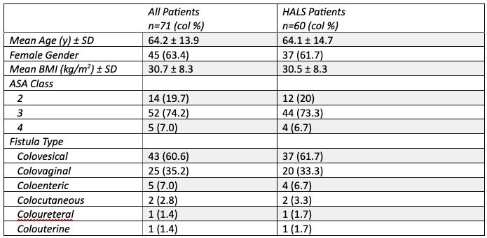
Seventy patients met study criteria. Sixty patients were treated with the HALS approach, seven by open approach, two by laparoscopic approach, and two with robotic-assisted laparoscopy. Table 1 shows the patient characteristics of the full cohort and the HALS cohort.
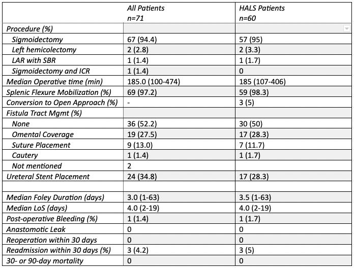
Of the 60 patients that underwent a HALS approach, colovesical fistula was the most frequent diagnosis (61.7%). Table 2 shows the intraoperative and postoperative data for the full and HALS cohorts. The fistula tract was inspected and left without intervention in 30 (50%) of the HALS procedures. Omental coverage of the tract remnant occurred in 17 (28.3%) cases and sutures were placed in 7 (11.7%) cases. Ureteral stents were used to aid in left ureter identification in 17 (28.3%) cases. Median post-operative Foley catheter duration was 3.5 days (1-63) after HALS resection and median length of stay was 4 days (2-19). Three (5%) of the HALS patients were readmitted within 30 days. There were no mortalities within 90 days among all included patients. Conversion from HALS to an open approach occurred in three cases (5%).
Discussion
Our experience using HALS for treating diverticulitis with fistula is notable. Our conversion rate with HALS (5%) is quite low compared to other studies, reporting conversion rates for laparoscopic surgery between 18-50%, with the largest study (Martinolich, et al.) citing a conversion rate of 34.9%.2,7,9 Few studies have evaluated outcomes of HALS approach for diverticulitis-associated fistula, and available studies are small and retrospective in nature, with reported conversion rates between 0-25%.3,6,10
In addition to low conversion rates, our study demonstrates that the HALS approach is associated with a short length of stay, achieving one of the major goals of minimally invasive surgery. Our median length of stay for all patients who underwent resection was 4 days, with prior studies reporting lengths of stay from 4.6-7 days.2,3,6 In addition, we found a low rate of intra- and post-operative complications.
In terms of our secondary aims, there were no cases of urine leak from the fistula tract among studied patients and the median Foley duration was 3.5 days for HALS patients. The longest catheter duration was 63 days, in a patient who presented with a rectourethral fistula. Few patients required a Foley catheter at discharge and need for a catheter was not a barrier to discharge.
Conclusion
Diverticulitis-associated fistulas present unique technical challenges to surgeons and can make minimally-invasive surgery difficult. The HALS technique is used for most
diverticulitis-associated fistula cases performed within the Division of Colorectal Surgery at MCW. This study represents the largest series of these cases reported in the literature. Our outcomes show that these cases are not only feasible and safe, they are associated with a low conversion rate, a low complication rate and a short hospital length of stay. We feel that the HALS approach is definitely a sound alternative to standard laparoscopic or open surgery for complex diverticulitis cases.
Table 1: Patient characteristics. HALS (Hand-assisted laparoscopic surgery), Standard Deviation (SD), Body mass index (BMI), America Society of Anesthesiologists physical status classification (ASA)
Table 2: Intraoperative and post-operative characteristics. HALS (Hand-assisted laparoscopic surgery), Low anterior resection (LAR), Small bowel resection (SBR), Ileocecal resection (ICR), Length of stay (LoS)
FOR ADDITIONAL INFORMATION on this topic, visit mcw.edu/surgery or contact Dr. Melissa Drezdzon at mdrezdzon@mcw.edu.
REFERENCES ON PAGE 13
Why Quality Improvement is Important in Residency
Katherine Scheidler, MD General Surgery Resident, Quality Improvement Research Resident
Dr. Atul Gawande, well-known surgeon, public health leader, and writer, wrote “Ingenuity is often misunderstood. It is not a matter of superior intelligence but of character. It demands more than anything a willingness to recognize failure, to not paper over the cracks, and to change. It arises from deliberate, even obsessive, reflection on failure and a constant searching for new solutions.”1 Shortly after starting medical school I received my first primer in safety and quality improvement as a gift from my brother— the book Better: A Surgeon’s Notes on Performance by Atul Gawande. In his books, he highlights real incidents and stories and gives us in the surgical community opportunities to reflect on our own patients, cases, and hospitals. Where are the opportunities for improvement in myself and my institution? How can we promote better outcomes for our patients? Was there a way I could have done that better? As a resident, I know I answer “yes” to that last question every day. In surgical residency, we are continually learning nuances to diagnoses, surgical and non-operative management strategies, effective communication – not to mention surgical technique. Therefore, safety and quality improvement must be an integral part of resident education to ensure that we are asking these important questions throughout our careers. Quality improvement and patient safety are often discussed goals to develop practices and systems to provide the best possible care for our patients. In 2000, the Institute of Medicine authored the book, To Err is Human: Building a Safer Health Care System, positing that as many as 98,000 patients die yearly due to a preventable medical error.4 As its title notes, we are human. The medical professionals themselves do not shoulder the blame for these errors, but instead a health care system that lacks supports in place to help prevent these errors. The aviation community has provided many examples of safety and quality improvement practices. My brother is a helicopter pilot in the Navy and since I began residency, we frequently discuss how safety practices in aviation have been adapted in surgical practice – like creating checklists and addressing latent risk factors and human error.3 Since the book was published, some practices have been implemented and some systems issues addressed, yet there remains room for improvement that should focus on “patient safety, care effectiveness, patient-centeredness, timeliness, care efficiency, and equity.”4 These six characteristics should be at the core of our initiatives.
As residents, we are in a unique position to both learn from quality improvement interventions, as well as ask the questions that prompt studies. Quality and safety presentations such as “Morbidity & Mortality” conferences or our Department of Surgery “Quality Minute” are learning opportunities for the presenter and the listener alike. Junior residents have an opportunity to hear senior residents teach about the natural course of some conditions and complications; while the presenters are encouraged to pursue new literature. Though these opportunities for learning are accessible and invaluable, I have found that we have far more chances to learn from quality improvement and safety initiatives than are simply on our schedules. The American College of Surgeons developed a program call the NSQIP Quality in Training Initiative (QITI) to give residents access to our own outcomes data – similar to that the hospital receives. Comparing our own outcomes to the institutional opportunities for improvement allows residents to take personal observations on the wards and in the operating room and bring our perspective to the table. As the QITI curriculum mentions, residents are often the “front-line workers” and therefore have a unique angle on problems that are not evident on the institutional level. 5 Additionally, in the vast domain of “quality improvement” each resident has their own interests and focal points from which to draw ideas and investigation: from health care disparities to global health and surgical subspecialties to post-operative care. In Atul Gawande’s introduction to Complications: A Surgeon’s Notes on an Imperfect Science he wrote, “A resident has a distinctive vantage on medicine. You are an insider, seeing everything and a part of everything; yet at the same time you see it anew.”2
I think that if we ask ourselves “why focus on quality improvement and safety in residency?” all of our responses would be “for our patients.” Beyond educational opportunities and the future endeavors we are all doubtless planning, we are all focused on the good of the patient in front of us. We have all cared for the patient who is discouraged over another complication or who has recently been readmitted more often than they have been home. We all hope and strive for good outcomes for the patients
Figure 1: ACS NSQIP Logo
Figure 2: Quality in Training Initiative Logo
REFERENCES FROM PAGE 21
with which we develop bonds over daily 5AM discussions. I cannot count the number of times I tell my patients on discharge day, “Take care. We will see you again – let’s just make sure it’s in clinic!” With patients’ well-being in the forefront, quality improvement in residency can be as simple as asking why. When we hear something that piques our interest at conference or on rounds, be curious. Is there a way we can make the system work better for our patients? Is there a way we can do better for our patients?
FOR ADDITIONAL INFORMATION on this topic, visit mcw.edu/surgery or contact Dr. Katherine Scheidler at kscheidler@mcw.edu.
REFERENCES
1. Gawande A. Better: A Surgeon’s Notes on Performance. London: Profile Books; 2010.
2. Gawande A. Complications: A Surgeon’s Notes on an Imperfect Science. London England: Profile Books; 2010.
3. Kapur N, Parand A, Soukup T, Reader T, Sevdalis N. Aviation and healthcare: a comparative review with implications for patient safety. JRSM Open. 2015 Dec 2;7(1):2054270415616548. doi: 10.1177/2054270415616548. PMID: 26770817; PMCID: PMC4710114.
4. Kohn LT, Corrigan JM, Donaldson MS, editors. To Err is Human: Building a Safer Health System. Washington, DC: National Academies Press; 2000.
5. Practical QI: THE BASICS OF QUALITY IMPROVEMENT EDUCATION The Quality In-Training Initiative: An ACS NSQIP Collaborative [Internet]. American College of Surgeons; [cited 2022Mar16]. Available from: https://qiti.acsnsqip. org/ACS_NSQIP_2017_QITI_Curriculum.pdf
Developing an Engaging and Effective Didactic Curriculum for General Surgery Residents
1. Joyner BD. An historical review of graduate medical education and a protocol of Accreditation Council for Graduate Medical Education compliance. The Journal of Urology. Jul 2004;172(1):34-39.
2. Weigelt JA, Simpson DE, Anderson RC, Brasel KJ, Redlich PN. A PGY1 curriculum--meeting a need for changing times. Current Surgery. Nov-Dec 2006;63(6):410-417.
3. Webb TP, Weigelt JA, Redlich PN, Anderson RC, Brasel KJ, Simpson D. Protected block curriculum enhances learning during general surgery residency training. Arch Surg. Feb 2009;144(2):160-166.
4. Parikh JA, McGory ML, Ko CY, Hines OJ, Tillou A, Hiatt JR. A structured conference program improves competencybased surgical education. American Journal of Surgery. Aug 2008;196(2):273-279.
5. de Virgilio C, Stabile BE. Weekly reading assignments and examinations result in sustained improvement in American Board of Surgery In-Training Examination (ABSITE) scores. The American Surgeon. Oct 2005;71(10):830-832.
6. Winer LK, Cortez AR, Kassam AF, et al. The Impact of a Comprehensive Resident Curriculum and Required Participation in “This Week in SCORE” on General Surgery ABSITE Performance and Well-Being. Journal of Surgical Education. Nov-Dec 2019;76(6):e102-e109.
7. Chick RC, Adams AM, Peace KM, et al. Using the Flipped Classroom Model in Surgical Education: Efficacy and Trainee Perception. Journal of Surgical Education. Jun 28 2021.
8. Zhu J, Li Y, Cox MF, London J, Hahn J, Ahn B. Validation of a Survey for Graduate Teaching Assistants: Translating Theory to Practice. Journal of Engineering Education. 2013;102(3):426-443.
9. McAvoy P, Hunt T, Culbertson MJ, McCleary KS, DeMeuse RJ, Hess DE. Measuring student discussion engagement in the college classroom: a scale validation study. Studies in Higher Education. 2021:1-15.
Congratulations and a huge thank you to our departing Chief Residents (and their post-residency plans) — we will miss you!
Christina Bence, MD
Pediatric Surgery Fellowship
Emory University/Children’s Hospital of Atlanta
Atlanta, Georgia
Kelly Boyle, MD
Trauma /Acute Care Surgery and Surgical Critical Care Fellowship
University of Southern California – Los Angeles Medical Center
Los Angeles, California
Bonnie Chow, MD, MA
Abdominal Transplant Surgery Fellowship
Stanford University Stanford, California
Emma Gibson, MD
General Surgery
Aurora West Allis Memorial Hospital West Allis, Wisconsin
Andrew Goelz, MD
General Surgery
St. Joseph’s Ascension Hospital Milwaukee, Wisconsin
Katherine Hu, MD
Colon and Rectal Surgery Fellowship
Cleveland Clinic Foundation Cleveland, Ohio
Zoe Lake, MD
Surgical Critical Care Fellowship
University of Kansas Kansas City, Kansas
Matthew Madion, Jr., MD
Minimally Invasive Surgery Fellowship
University of Tennessee Knoxville, Tennessee
Erin Strong, MD, MBA, MPH
Hospice and Palliative Medicine Fellowship
Northwestern University Chicago, Illinois
Food be thy Medicine: Microbiome as a Therapeutic Target
Jed F. Calata, MD Assistant Professor, Division of Colorectal Surgery
From the colloquial wisdom of ‘you are what you eat’ to Hippocrates supposedly coining the phrase ‘let thy food be thy medicine and thy medicine be thy food’, health and diet have long been associated together and is a central element to many traditional forms of medicine. And like many other examples in history, as we have come to better understand human physiology and disease, we have grown to appreciate traditional wisdom in modern scientific context. This, in particular, has been profound in our understanding of one of the most difficult and vexing diseases we treat in colorectal surgery, inflammatory bowel disease (IBD).
Although historically regarded as a disease of westernized nations, the global spread of IBD has gone beyond borders as we are seeing increasing incidence worldwide.1,2 The growing incidence of ulcerative colitis seen in newly industrialized countries in the far east nearly exactly mirrors the growing incidence of ulcerative colitis
seen in the western world after the industrial revolution.1 While we do know that genetics play a role in the development of IBD, it is unlikely to be the sole driver behind this change, implicating the urbanization of diet, lifestyle, and environments as important factors. Alteration of the intestinal microbiome is one of the proposed mechanisms that may be enacting these changes.
Intestinal microbiome, the complex community of bacteria and organisms that reside in our gut, has an important role in IBD pathogenesis. Numerous studies have shown that IBD patients have broad changes in their intestinal microbiota profile compared to individuals without IBD.3 The urbanization of our diet to include processed foods, animal proteins, animal fats, emulsifiers, and artificial sweeteners and is low in vegetables, fiber, and complex carbohydrates leads to a lack of microbiome diversity. In turn, this causes an opportunistic increase in mucosa associated inflammation-promoting bacteria (Figure 1).4
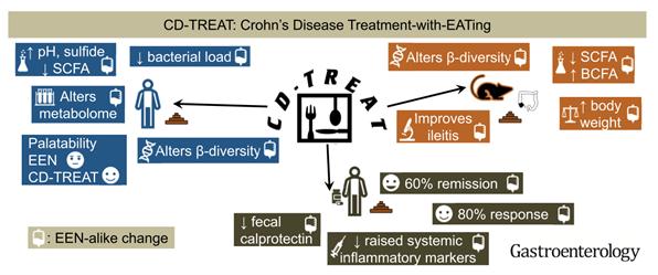
This has led to new and exciting research in the development of food-based treatment of IBD. In fact, therapeutic dietary treatment of Crohn’s disease, named ‘exclusive enteral nutrition’ (EEN), is being utilized in pediatric patients and is already considered as first line therapy in Europe and Australia since 2014.5 EEN is a polymeric or elemental enteral feed diet given as a patient’s sole dietary source, either orally or via nasogastric tube, over a 6-8 week period. EEN has been shown to be equally effective as steroids in inducing remission and mucosal healing in pediatric patients with acute Crohn’s flares; however, EEN is a very restrictive diet with difficult compliance and limited prolonged use which has affected adoption of the therapy in the United States as well as by adult patients.5,6 To increase acceptance and overcome these barriers, a group from Scotland published a very interesting study utilizing an ‘ordinary’ food diet based on a similar nutritional profile as EEN called the CD-TREAT diet. When tested in healthy adults, they were able to produce similar microbiome shifts as seen in EEN diet patients. They further performed a pilot study treating 5 children with active Crohn’s disease with 8 weeks of the CDTREAT diet and were able to achieve clinical remission in 3 of the 5 patients, with 1 additional patient showing marked decrease in fecal calprotectin de-
Figure 2: Crohn’s Disease Treatment with EATing (Reprinted with permission) 6
Figure 1: Effects of urbanization of diet
and New Frontier for Research
spite having some persistent mild symptoms (Figure 2).6 These dietary therapeutic treatments highlight that what we eat can influence the health of our microbiome, and in turn, the health of our gut.
Could it really be this simple? Was your grandmother on to something much deeper when she told you to eat your peas and carrots? Is Western medicine finally catching up to the wisdom of traditional remedies? The microbiome and, in particular, the dietary influence on the microbiome are a growing target for IBD therapy and research with some very promising early results. What I find even more compelling is the possibility that microbiome induced inflammation may be playing a role in the outcomes of non-IBD surgical patients and is a research target that may help us continue to improve operative outcomes in the future.
FOR ADDITIONAL INFORMATION on this topic, visit mcw.edu/ surgery or contact Dr. Jed Calata at jcalata@mcw.edu.
REFERENCES
1. Ng SC, Kaplan GG. Understanding and Preventing the Global Increase of Inflammatory Bowel disease. Gastroenterology. 2017 Jun;152(8):2084
2. Ng SC, Shi HY, Hamidi N, et al. “Worldwide incidence and prevalence of inflammatory bowel disease in the 21st century: a systematic review of population-based studies” Lancet. 2017; 390(10114): 2769-2778.
3. De Cruz P, Prideaux L, Wagner J, et al. Characterization of the gastrointestinal microbiota in health and inflammatory disease. Inflamm Bowel Dis. 2012; 18:372-390
4. Zuo T, Kamm MA, Colombel JF, et al. Urbanization and the gut microbiota in health and inflammatory bowel disease. Nat Reve Gastroentrol Hepatol. 2018 Jul; 15(7):440-452
6. Svolos V, Hansen R, Nichols B, et al. Treatment of Active Crohn’s Disease with an Ordinary Food-based diet that replicates Exclusive Enteral Nutrition. Gastroentrology. 2019; 156:1354-1367
Advanced GI MIS & Bariatric Surgery Fellowship
Wen Hui Tan, MD
Faculty
Division of Minimally Invasive & Gastrointestinal Surgery
Medical College of Wisconsin
Complex General Surgical Oncology Fellowship
Erin P. Ward, MD
Post-Fellowship Plans Pending
Endocrine Surgery Fellowship
Marisa Bartz-Kurycki, MD
Post-Fellowship Plans Pending
Pediatric Surgery Fellowship
Ali Mejaddam, MD
Pediatric Surgery
Loma Linda Children’s Hospital
Loma Linda, California
Surgical Critical Care Fellowship
Kathryn Haberman, MD
Acute Care Surgery Fellowship
Medical College of Wisconsin
Milwaukee, Wisconsin
Surgical Critical Care Fellowship
Nolan Rudder, MD
Faculty
Gundersen Lutheran Medical Center
La Crosse, Wisconsin
Surgical Critical Care Fellowship
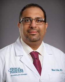
Motaz A. Selim, MD
Assistant Professor of Surgery
Transplant Division
Medical College of Wisconsin
Surgical Critical Care Fellowship, Pediatric Track
Amanda Witte, MD
Pediatric Surgery Research Fellowship
Children’s Wisconsin
Milwaukee, Wisconsin
Trauma/Acute Care Surgery Research Fellowship
Juan Jose Figueroa, MD
General Surgery Resident
University of Texas Medical Branch
Galveston, Texas
Trauma/Acute Care Surgery Research Fellowship
Courtney Pokrzywa, MD
Surgery Resident
New York Presbyterian Hospital
Columbia University Medical Center
Vascular Surgery Fellowship
Simon Fraser, DO
Vascular Surgery
OhioHealth Riverside Methodist Hospital
Columbus, OH
Trauma-Informed Care in Surgical Practice: Awareness of Possible
Sydney Timmer-Murillo, PhD, MS
Post-doctoral Fellow in Trauma and Health
Psychology, Division of Trauma & Acute Care Surgery
Terri A. deRoon-Cassini, PhD, MS
Associate Professor, Division of Trauma & Acute Care Surgery, Psychiatry & Behavioral Medicine, Institute for Health and Equity; Director, MCW Comprehensive Injury Center
A patient recently required consultation from a surgical specialist after developing chronic pain in both wrists following severe injuries. The specialist subsequently recommended surgical intervention to alleviate chronic pain and improve overall functionality. The goal of the patient, an avid athlete, was to be more physically active again and engage in daily activities with diminished pain. Despite her goals, the patient hesitated for several months prior to scheduling the procedure due to fear around potential complications, being more dependent on others in recovery, and how possible complications might make her functioning even worse. To feel more comfortable moving forward with the procedure, she requested a followup appointment with the surgical specialist to explain her anxiety and hesitation. The surgeon subsequently met with the patient, and in an attempt to alleviate her anxiety, he said, “there’s no reason to be worried about surgery,” and then quickly moved on. In the end, the patient never scheduled the recommended surgery.
Decades of psychological research have unequivocally demonstrated that traumatic experiences (e.g., child abuse, domestic violence) have lasting, often deleterious consequences across the lifetime.1 For some individuals, these consequences come in the form of hypervigilance and hyperarousal. The resulting fear and anxiety can cause individuals to avoid much needed medical care.2 Those who do engage in healthcare may do so with considerable distress. Trauma-informed care (TIC) is an approach to healthcare that appreciates the impact a patient’s history of psychological trauma may have on their experience of healthcare. Providers educated about TIC can improve healthcare delivery to increase patient engagement and decrease re-traumatization during medical care. This approach creates a safe, empowering, and healing environment for the patient.
The seminal work of Vincent Felitti, MD, and the Centers for Disease Control originally produced findings documenting the relationship between trauma exposure in childhood and health comorbidities in adulthood.3 The core of TIC in healthcare includes two principles: universal trauma precautions and trauma-specific provision of
Tim Geier, PhD, MS
Assistant Professor, Division of Trauma & Acute Care Surgery, Psychiatry & Behavioral Medicine
Andrew Schramm, PhD
Assistant Professor, Division of Trauma & Acute Care Surgery
care.2,4 Regardless of whether a provider knows of a patient’s trauma history, TIC is best practice. Provider behaviors that are sensitive to the issues of patients with a history of trauma will be beneficial to all patients, as they promote patients’ comfort and communication.2 TIC can include soliciting permission to engage in care (i.e., to foster safe environment), consistently announcing oneself prior to entering a room (i.e., to reduce startle response), and providing ample time for patient questions (i.e., to improve patient collaboration and a greater sense of control). These small adjustments are designed to reduce potential triggers and to promote trust, which can improve the patient’s experience. In addition to such universal precautions, TIC includes the assessment of trauma history, promoting providers’ understanding of the impact of psychological trauma on healthcare engagement, and encouraging provider self-reflection on their own responses to patients whose behavior is influenced by their history of psychological trauma. While this transformative approach can influence patient experience of healthcare, its benefits extend beyond the patient, as provider burnout has been shown to decrease in healthcare systems that have adopted TIC.5
The need for trauma informed care is evidenced by research reporting just over 90% of people are exposed to at least one psychologically traumatizing event in their lifetime. On average, people living in the United States (US) experience at least four traumas, including physical and sexual assault, domestic violence, gun violence, and motor vehicle crashes. Trauma occurs throughout the lifespan, and in childhood, trauma is often referred to as adverse childhood experiences (ACEs), including childhood abuse and neglect, substance abuse or domestic violence occurring in the household, and chronic stressors such as racism or experiences with discrimination.3 There are clear consequences of psychological trauma, including the development of posttraumatic stress disorder (PTSD), which has a prevalence rate in the US of just over 8%. Less obvious is the insidious impact of trauma, whether during childhood or in adulthood, that can lead to chronic health problems and maladaptive coping behaviors. The toxic,
Psychological Trauma History Should Inform Patient Care
chronic stress of trauma is linked with medical comorbidities in a dose-response relationship, whereby the more traumatic events in ones’ life, the more medical comorbidities in adulthood.1 Specifically, a person who has experienced four or more traumatic events, particularly during childhood, is 2.4 times more likely to have a stroke, 1.9 times more likely to be diagnosed with cancer, 12 times more likely to attempt suicide, and 7 times more likely to misuse alcohol.6
The outcomes from this case example could have been different. Unknown to her providers, the patient had a significant trauma history that directly impacted her ability to trust in providers and healthcare systems. Although she recognized the impact of trauma in her life, she also needed her provider to understand her experience to build her comfort and confidence in their plan of care. Outcomes could have improved had the provider: 1) assessed and acknowledged her history and 2) validated her current concerns in treatment. Similarly, giving the patient space for discussion, the provider could have involved the patient in treatment planning to foster their collaboration and promote her sense of safety and control. TIC is not about adding substantial time to the patient visits, but rather tailoring our work to validate patient experiences and provide care with patients’ specific needs in mind. As humans, we are each a collection of unique past experiences, and there is not a “one size fits all” way to engage with and treat every patient. Knowing this, we have both the responsibility and honor to make important shifts in our behaviors and acquire knowledge that can foster significant changes for the patient experience.
FOR ADDITIONAL INFORMATION on this topic, visit mcw. edu/surgery or contact Dr. Terri A. deRoon-Cassini at tcassini@mcw.edu.
REFERENCES
1. Quinn, M. (2021). Epidemiologic Linkages Between Childhood Trauma, Health, and Health Care. Global Mental Health Ethics, 171-190. Springer, Cham.
2. Reeves, E. (2015) A Synthesis of the Literature on TraumaInformed Care, Issues in Mental Health Nursing, 36:9, 698709, DOI: 10.3109/01612840.2015.1025319
3. Felitti, V. J., Anda, R. F., Nordenberg, D., & Williamson, D. F. (1998). Adverse childhood experiences and health outcomes in adults: The Ace study. Journal of Family and Consumer Sciences, 90(3), 31.
4. Raja, S, Hasnain, M, Hoersch, M, Gove-Yin, S, Rajagopalan, C. (2015). Trauma Informed Care. Medicine, Family & Community Health, 38(3), 216-226 doi: 10.1097/ FCH.0000000000000071
5. Kuehn, B. M. (2020). Trauma-informed care may ease patient fear, Clinician Burnout. JAMA, 323(7), 595-597.
Lending a Hand: The Utility of Hand-Assisted Laparoscopic Surgery in Diverticulitis with Fistula
1. Hanna MH, Kaiser AM. Update on the management of sigmoid diverticulitis. World journal of gastroenterology. 2021;27(9):760-781. doi:10.3748/wjg.v27.i9.760
2. Martinolich J, Croasdale DR, Bhakta AS, et al. Laparoscopic Surgery for Diverticular Fistulas: Outcomes of 111 Consecutive Cases at a Single Institution. Journal of Gastrointestinal Surgery. 2019;23(5):1015-1021. doi:10.1007/s11605-0183950-3
3. Spector R, Bard V, Zmora O, Avital S, Wasserberg N. Hand-assisted Laparoscopic Colectomy for Colovesical Fistula Associated With Diverticular Disease. Surgical Laparoscopy Endoscopy & Percutaneous Techniques. 2014;24(3). https://journals.lww.com/surgical-laparoscopy/ Fulltext/2014/06000/Hand_Assisted_Laparoscopic_ Colectomy_for.12.aspx
4. Hall J, Hardiman K, Lee S, et al. The American Society of Colon and Rectal Surgeons Clinical Practice Guidelines for the Treatment of Left-Sided Colonic Diverticulitis. Diseases of the Colon & Rectum. 2020;63(6):728-747. doi:10.1097/ DCR.0000000000001679
5. Aalbers AGJ, Biere SSAY, van Berge Henegouwen MI, Bemelman WA. Hand-assisted or laparoscopic-assisted approach in colorectal surgery: a systematic review and meta-analysis. Surgical endoscopy. 2008;22(8):1769-1780. doi:10.1007/s00464-008-9857-4
6. Lee SW, Yoo J, Dujovny N, Sonoda T, Milsom JW. Laparoscopicvs. Hand-Assisted Laparoscopic Sigmoidectomy for Diverticulitis. Diseases of the Colon & Rectum. 2006;49(4). https://journals.lww.com/dcrjournal/Fulltext/2006/49040/ Laparoscopicvs__Hand_Assisted_Laparoscopic.5.aspx
7. Cirocchi R, Cochetti G, Randolph J, et al. Laparoscopic treatment of colovesical fistulas due to complicated colonic diverticular disease: a systematic review. Techniques in Coloproctology. 2014;18(10):873-885. doi:10.1007/s10151014-1157-5
8. Targarona EM, Gracia E, Garriga J, et al. Prospective randomized trial comparing conventional laparoscopic colectomy with hand-assisted laparoscopic colectomy. Surgical Endoscopy And Other Interventional Techniques. 2002;16(2):234-239. doi:10.1007/s00464-001-8168-9
9. Klarenbeek BR, Veenhof AA, Bergamaschi R, et al. Laparoscopic sigmoid resection for diverticulitis decreases major morbidity rates: a randomized control trial: short-term results of the Sigma Trial. Annals of surgery. 2009;249(1):3944. doi:10.1097/SLA.0b013e31818e416a
10. Bartus CM, Lipof T, Sarwar SCM, et al. Colovesical Fistula: Not a Contraindication to Elective Laparoscopic Colectomy. Diseases of the Colon & Rectum. 2005;48(2):233-236. doi:10.1007/s10350-004-0849-8
The Balancing Act: Liver Transplantation for
Motaz Selim, MD
Assistant Professor, Division of Transplant Surgery
The list of indications for liver transplantation has only been growing since the first successful transplantation by Starzl in 1963. Expansion of that list is hindered mainly by the short supply of donated organs. The United States (US) Congress passed the National Organ Transplantation Act (NOTA) in 1984 to address organ shortage and set standards for organ allocation. Input from ethicists, clinicians, and the general public has been incorporated in developing the allocation rules. The consensus was to use the concept of “equity” as the prioritization parameter for organ allocation, effectively prioritizing the “sickest first”. That said, proper stewardship of organs mandates considering the concept of “efficiency” as well. Poor posttransplant outcomes due to the disease process (e.g., advanced or aggressive cancers) or patient factors (e.g. too sick or noncompliant) should be weighed carefully against benefits before transplant is considered. As evidence of survival benefit accumulates, novel indications for liver transplantation are considered (e.g., cholangiocarcinoma, metastatic colon cancer, etc.)
Liver transplantation for alcoholic liver disease has always been one of the topics that stir debate. The social stigma of alcohol use and addiction is one aspect, the other is the difficulty in predicting patients’ ability to maintain sobriety. The six months sobriety rule has been traditionally used by transplant centers and third-party payors as a prerequisite for listing, hoping for clinical improvement with abstinence. It was also used as a predictor of longterm sobriety, yet research failed to show strong correlation with post-transplant abstinence.1 The six months rule is not mandated by the United Network for Organ Sharing (UNOS) for listing for liver transplantation. The literature indicates that alcohol use should be viewed as a mental health disorder rather than a choice. Comprehensive psychosocial evaluation would provide a more reliable prediction of post-transplant abstinence.1
The percentage of liver transplants performed for alcoholic liver disease has risen from 16% of all liver transplants in the United States in 2010 to 35% in 2020.2 This is more relevant to our institution, considering that Wisconsin is one of the states with the highest rates of binge drinking, with an 18% increase in alcohol-attributable deaths in 2020.3
Of the two presentations of alcoholic liver disease, chronic alcoholic cirrhosis is more common. Cirrhotic patients will usually have enough time to undergo evaluation and show sobriety and commitment to the transplant
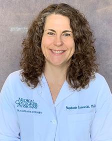
Stephanie C. Zanowski, PhD
Assistant Professor, Division of Transplant Surgery and Psychiatry; Director of Psychology Services, Medical College Physicians; Director, Transplantation Mental Health
Jenessa S. Price,
PhD
Assistant Professor, Surgery & Psychiatry, Division of Transplant Surgery; Director, MCW Health Psychology Practicum Program
process. In contrast, acute alcoholic hepatitis (AAH) patients, typically younger and previously healthy, present with acute inflammation of the liver and life-threatening acute liver failure in the setting of binge drinking or excessive alcohol intake. 40% of those patients will not respond to steroid treatment and will be left with no other medical treatment options. Without liver transplantation, they face a grim prognosis of 75% 6-month mortality.4 With not much time left and as sick as they are, traditional transplant selection protocols based on the six months sobriety rule have failed many of those patients. Ethicists argue that a standard evaluation process tailored for this unique situation should be in place to avoid systematically excluding those patients.5
The first reports of early liver transplantation for AAH date back to the 1990s, but it was not until 2011 that a prospective multicenter trial was published.6 More centers in the US and across the world began offering transplants to those patients and published their experience.7 The survival benefit of transplantation in those patients is undeniable with a 77-100% six months survival versus 25% without transplantation. The rates of recidivism were 11-28%, similar to reported rates in cirrhotic patients who have been transplanted after demonstrating six months sobriety.8 Clinical guidelines from US and Europe advocate for early liver transplantation for qualifying AAH patients who fail steroid therapy.9,10 The selection criteria in the published studies are not standardized and the best way for evaluating the risk of recidivism is still debatable.
We proposed that a comprehensive mental health program involved in pre-transplant assessment and posttransplant support can a) predict with acceptable accuracy which patients have the highest chances of maintaining sobriety post-transplant and b) provide ongoing access to mental health and Alcohol and Other Drug Abuse (AODA) resources to support post-transplant patients and help maintain abstinence. Comprehensive evaluation for patients who met the medical criteria included calculating the Stanford Integrated Psychosocial Assessment for Transplantation (SIPAT) score and interviewing the patients and their identified support system by transplant psy-
Acute Alcoholic Hepatitis
chologists. The goal was to identify risk and protective factors that would impact post-transplant outcomes. Potential transplant candidates were provided an individualized treatment regimen implemented during their hospitalization while awaiting transplantation. Patients were required to engage in AODA treatment as medically able and commit to completing treatment post-transplant as a condition of listing. Patients were closely monitored for overall compliance (medical therapies, nutrition, and physical therapy), and their participation in AODA treatment and psychotherapy. Looking back at our experience with this model, comprehensive evaluation for liver transplantation was applied to 83 patients who failed medical therapy for AAH between April 2015 and April 2020. Of the evaluated patients, 65% were eventually listed (54% of them were transplanted while 35% died while waiting for an organ) and 35% were declined. The demographic and clinical parameters were similar in both cohorts. The listed cohort had significantly better SIPAT scores (median 40 versus 57; p<0.01), better availability of social support (100% versus 76%; p<0.01) and less prevalence of active smokers (30% versus 66%; p<0.01). Both cohorts demonstrated similar severity and duration of alcohol use. One-year survival of patients who were not transplanted was a dismal 23% compared to 92.5% in transplanted patients (p<0.01) [Figure 1]. The rate of recidivism was 3%, the lowest among the published studies.
REFERENCES
1. Rice, J.P. and M.R. Lucey. Should length of sobriety be a major determinant in liver transplant selection? Curr Opin Organ Transplant, 2013. 18(3): p. 259-64.
2. Kwong, A.J., et al., OPTN/SRTR 2020 Annual Data Report: Liver. Am J Transplant, 2022. 22 Suppl 2: p. 204-309.
3. Wisconsin Department of Health Services. DHS Interactive Dashboards: Alcohol Death Module. [cited 2022 3/9/2022]; Available from: https://www.dhs.wisconsin.gov/aoda/ deaths-county.htm.
4. Louvet, A., et al., The Lille model: a new tool for therapeutic strategy in patients with severe alcoholic hepatitis treated with steroids. Hepatology, 2007. 45(6): p. 1348-54.
5. Donckier, V., et al., Ethical considerations regarding early liver transplantation in patients with severe alcoholic hepatitis not responding to medical therapy. J Hepatol, 2014. 60(4): p. 866-71.
6. Mathurin, P., et al., Early liver transplantation for severe alcoholic hepatitis. N Engl J Med, 2011. 365(19): p. 1790-800.
7. Marot, A., et al., Liver transplantation for alcoholic hepatitis: A systematic review with meta-analysis. PLoS One, 2018. 13(1): p. e0190823.
This data suggests that an integrated transplant mental health program can facilitate access to lifesaving liver transplantation and improve outcomes among the high acuity patients who failed medical treatment for AAH. This can be achieved by providing a thoughtful multidisciplinary approach that balances individual risks and protective factors associated with abstinence and overall post-transplant success.
FOR ADDITIONAL INFORMATION on this topic, visit mcw. edu/surgery or contact Dr. Motaz Selim at mselim@ mcw.edu.
8. Weeks, S.R., et al., Liver Transplantation for Severe Alcoholic Hepatitis, Updated Lessons from the World’s Largest Series. J Am Coll Surg, 2018. 226(4): p. 549-557.
9. Singal, A.K., et al., ACG Clinical Guideline: Alcoholic Liver Disease. Am J Gastroenterol, 2018. 113(2): p. 175-194.
10. European Association for the Study of the Liver. Electronic address, e.e.e. and L. European Association for the Study of the, EASL Clinical Practice Guidelines: Management of alcohol-related liver disease. J Hepatol, 2018. 69(1): p. 154181.
Figure 1. Patient survival without transplant versus survival after transplant (relative to admission date)
State-of-the-Art Simulation to Improve ECMO
Background
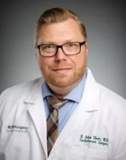
H. Adam Ubert, MD
Assistant Professor, Division of Cardiothoracic Surgery
In a time where technology seems to be advancing at a feverish pace, the use of simulation in medical education is not a new concept. The era of “modern” simulators can be traced back to the early 1700’s where various teams focused on birthing simulators to teach techniques of delivery to midwives and surgeons.1,2 As technology improved, so did simulation. Today, more advanced simulators are being used for all aspects of medicine, including refining surgical techniques and concepts in critical care. There have been multiple studies that highlight the role of simulation-based training to improve rapid assessment, establish efficient medical care, and reduce complications.3 One clinical scenario where simulation is emerging is in patients requiring extracorporeal membrane oxygenation (ECMO) for respiratory and cardiac failure. Patients requiring ECMO remain one of the highest acuity patients and as such, complications and emergencies can have a direct negative influence on patient outcomes. To minimize these events, simulation-based training can provide standardized opportunities for care members to recognize early issues before they lead to patient harm.4 Similarly, simulated ECMO scenarios provide opportunities to recognize and correct common physiologic derangements in a low-stress environment.5
The Froedtert & the Medical College of Wisconsin ECMO Program and Simulation Center
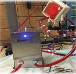
In 2018, the ECMO program at Froedtert & the Medical College of Wisconsin became a member of the Extracorporeal Life Support Organization (ELSO). Since that time, our ECMO program has grown into one of the largest and most successful programs in the country in terms of volume, outcomes, and lack of complications. By 2021, the program was designated as a Gold Center of Excellence. Certainly, integral to this success has been the establishment of the ECMO simulation program, which was started in 2019 in the Froedtert & the Medical College of Wisconsin Simulation Center. Our current ECMO simulator uses an EigenFlow simulator (Curtis Life Research) along with the SimMan 3G (Laerdal Medical). By using the EigenFlow system, it is possible to simulate thrombus in cannula, line obstruction, air emboli, and manipulate vital signs and changes in pulmonary and cardiac function. The state-ofthe-art ECMO simulator allows the creation of complex scenarios based on real world events. Through multi-disciplinary development, we have created a wide variety of
Lucian A. Durham III, MD, PhD
Associate Professor, Division of Cardiothoracic Surgery; Director of Mechanical Circulatory Support/ECMO
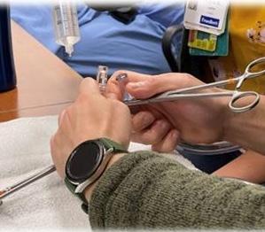
EigenFlow – Curtis Life Research Deairing the circuit
simulations that encompasses both common scenarios, such as changing an oxygenator and Harlequin syndrome, as well as relatively uncommon, potentially catastrophic events such as accidental decannulation and air entrapment. Throughout each year, there are two sessions, each with two to three simulations, which allow for a total of four to six newly created simulated patient scenarios per year. These simulated scenarios allow our intensivists, surgeons, ECMO nurses, and perfusionists the opportunity to recognize potential complications and obtain handson experience for remedying these issues. To say that these simulations have helped improve care at the bedside would be an understatement. It is not uncommon for members of the care team to diagnose a patient care issue, such as the sound the circuit makes when there is air entrainment, and quickly recognize the problem because they have previously heard that sound during simulation training.
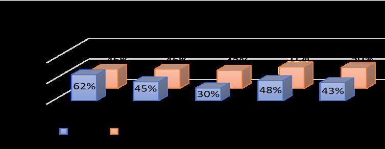
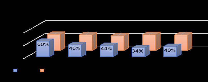
Our ECMO program has become a large referral center that provides exceptional care for the highest acuity patients. ELSO routinely benchmarks the member programs and maintains a database pertaining to mortality and complications. The F&MCW ECMO program has consistently measured below the national average for mortality (Figure 1) and complications (Figure 2). Not surprisingly, a drastic decline in mortality and complications was seen after implementing ECMO simulations in 2019, a testament to the improved care provided through simulationbased learning. The COVID-19 pandemic created a new patient population with difficult to treat pathophysiology, but from the knowledge gained and experience obtained, we have been able to create new simulation models that have helped improve our overall outcomes for the use of ECMO in COVID and non-COVID patients.
Care and Training
While simulation for ECMO may be the most complex as it requires multiple simulators running simultaneously, the Sim Center provides a large skills lab, allowing for hands-on training through multiple specialties. The ability to mimic health care environments, such as an operating room, the intensive care unit, and trauma bay, allows for continued education through adult-based learning. The wide variety of simulation offered at our Sim Center have been utilized in Obstetrical training, sexual assault nurse examination simulations, critical care scenarios, difficult airway, and other clinical areas that augment our basic medical education. Similarly, the Sim Center is used in conjunction with our nurse residencies and nurse competencies to provide inter-professional simulation.
It has been shown that the use of simulation to acquire knowledge about ECMO and recognize early complications is superior to the educational methods using interactive mobile learning and even experiential, non-formal training.6 Through state-of-the-art ECMO simulation, along with the exceptional multidisciplinary care provided by the team of nurses, coordinators, advanced practice providers, perfusionists, pharmacists, physical therapists, intensivists, and surgeons, our team has created a wellrespected and nationally known ECMO program that successfully cares for the highest acuity patients.
For more information about our Sim Center please go
to www.froedtert.com/sim-center or https://Froedtert. wistia.com/medias/xybmqzgycb to watch a brief video.
FOR ADDITIONAL INFORMATION on this topic, visit mcw. edu/surgery or contact Dr. H. Adam Ubert at aubert@ mcw.edu.
REFERENCES
1. GH, B. (1991). Development of simulators in medical education. Gesnerus, 48 Pt 1:7-28.
2. Jones F, P.-N. C. (2015). Simulation in medical education: brief history and methodology. The principles and Practice of Clinical Research, 1:46-54.
3. Armenia S, e. a. (2018). The role of High-fidelity team based simulation in acute care settings: A systematic review. Surgery Journal, 136-151.
4. Brum R, R. R. (2015). Simulation training for extracorporeal membrane oxygenation. Ann Card Anaesth, 18: 185-90.
5. S, S. (2017). Simulation-based training for handling extracorporeal membrane oxygenation emergencies. Journal of Thoracic Disease, 3649-3651.
6. Gannon W, e. a. (2021). Simulation versus interactive mobile learning for teaching extracorporeal membrane oxygenation to clinicians: A randomized trial. Critical Care Medicine, doi: 10.1097/CCM.0000000000005376.
Figure 1: ECMO mortality comparing FMLH vs ELSO registered database
Figure 2: ECMO complications comparing FMLH vs ELSO registered database
Figure 3: ECMO simulation in progress
Trauma Video Review: Advancing the Goals of
Daniel N. Holena, MD
Associate Professor and Director of Research, Division of Trauma and Acute Care Surgery
Imagine an untelevised sporting event played in an empty stadium, except for those participating in the game. There are no fans, no cameras, no sportscasters. The home team is a heavy favorite, with a roster loaded with talent and a string of dominating wins. At the end of this unwitnessed game, the score is announced: the home team has lost by an overwhelming margin. Outside the stadium in the darkening sky, the team captain is interviewed by the press. The fans are upset. What happened in there they ask, and what can we do to make sure this does not happen again?
“Well,” says the captain “It’s true that we lost, but there were some problems with our equipment, and the trainers weren’t really as helpful as they could have been, and of course we got some really bad calls from the referees. Led by my shining example of athleticism, we as a team played to the best of our outstanding ability, as we always do, so there wasn’t really anything we could have done better.”
Nodding and murmuring, the fans and reporters appear satisfied with the response, except for one who speaks up.
“What you say is most certainly true,” she says, “but isn’t there some more objective information you can provide about what happened in there?”
“Of course!” says the team captain cheerfully. “Later today I’ll write a note that contains my recollection of the game. Well, a game anyway. It’s actually a templated note, but I’ll be sure to update the relevant details!”
In the real world, of course, major athletic events are not just experienced by the team members but are often videorecorded. Reviewing footage of these games is a key element of performance improvement, so coaches and players can understand where the team is performing well and where improvement is needed. So important is this practice that players in the National Football League may review up to 4 hours of game tape per day and even veteran players report that this endeavor helps them to improve their game. If such a practice is in widespread use to help professional sports teams play at the top of their game, then it is perhaps counterintuitive that this method of performance improvement is vanishingly rare in medicine, where so much more is on the line.
There is, however, one area in healthcare where audiovisual review has taken root: the initial evaluation and management of injured patients. In fact, the audiovisual review of trauma resuscitations, or Trauma Video Review
David J. Milia, MD
Associate Professor and Trauma Medical Director, Division of Trauma and Acute Care Surgery
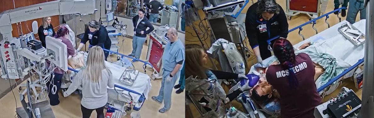
(TVR, Figure 1), has been a practice at high-performing centers for over three decades.1 This is perhaps unsurprising given the strong alignment between the benefits of TVR and the clinical care, educational, and research missions of the academic trauma center.2
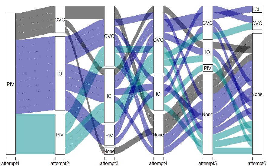
As a principle in quality improvement, it is difficult to demonstrate improvement in that which is not measured. For example, experienced trauma providers know that intravascular access in patients in hemorrhagic shock can be extremely difficult; once-plump veins are collapsed, resisting the efforts of the needle to find them. How then to best get access to allow for volume resuscitation? One could review the medical record to see what methods were most often employed, how long they took, and what their success rates were, but even under the best circumstances such information is recorded only sporadically. In contrast, TVR can provide granular data not available in the medical record (Figure 2). Using such data from a TVR quality improvement program, one study found that only 43% of attempts at peripheral intravenous access were successful in patients in extremis. Similarly, only 44% of central venous access attempts were successful with the added drawback of taking a median of ~3 minutes per attempt. In contrast, intraosseus attempts were successful in 92% of cases and only took ~30 seconds per attempt.3 Using TVR to measure the timing and success rates of these access attempts informed a change in practice at the study site for patients in extremis arriving without access. Early attempts at central venous catheter insertion were all but abandoned and insertion of two intraosseous access devices quickly became the first line of therapy.
When it comes to surgical education, there is an old aphorism that the worst thing about every other night
Figure 1. Typical view from a TVR platform.
the Academic Trauma Center
Figure 2. Data from TVR: an alluvial plot of intravascular access attempts over time during trauma resuscitation. PIV = Peripheral IV; IO = Intraosseous device; CVC = Central Venous Catheter.
call is missing half the cases. This need not be true when there is a TVR program in place, as learners may still find educational benefit, even from cases where they were not physically present. This education can come in the form of personalized feedback provided to learners in individual review sessions but can also take the form of a multidisciplinary conference in which challenging and interesting cases are reviewed in a group setting. Indeed, survey data from participants of one such TVR educational conference demonstrates that attendees find the experience to be of high educational value.4
From a research perspective, a critical barrier to study of processes of care during trauma resuscitation is data collection. For instance, high impact low frequency procedures such as resuscitative thoracotomy occur only sporadically, even at busy trauma centers. Studying such procedures using prospective data collection would require continuously present and available research personnel, a resource beyond the means of most trauma centers. Audiovisual review systems can eliminate this barrier, while at the same time providing data that is subject to far less missingness and measurement bias than prospective or retrospectively collected data. Such efforts have led to insights into both the technical and non-technical aspects of resuscitative thoracotomy.5,6
Although the benefits of TVR programs are manifold, only about one-third of U.S. Level 1 and 2 trauma centers have such programs. When surveyed, centers cited staff concerns, perceived medicolegal risk, and financial constraints as barriers to the institution of TVR programs. The division of trauma at the Medical College of Wisconsin was an early adopter of TVR technology and first instituted a program in the 1990s. This was taken offline sec-
ondary to regulatory concerns, but fortunately was able to be reinstated in 2015.7 Since that time, the trauma video review program at the Medical College of Wisconsin has continued to execute the clinical care, education, and research aims of the academic trauma center.
FOR ADDITIONAL INFORMATION on this topic, visit mcw.edu/ surgery or contact Dr. Daniel Holena at dholena@mcw.edu.
REFERENCES
1. Hoyt DB, Shackford SR, Fridland PH, Mackersie RC, Hansbrough JF, Wachtel TL, Fortune JB. Video recording trauma resuscitations: an effective teaching technique. The Journal of trauma. Apr 1988;28(4):435-40.doi:10.1097/00005373198804000-00003
2. Vella MA, Dumas RP, Holena DN. Supporting the Educational, Research, and Clinical Care Goals of the Academic Trauma Center: Video Review for Trauma Resuscitation. JAMA Surg. Mar 1 2019;154(3):257-258. doi:10.1001/ jamasurg.2018.5077
3. Chreiman KM, Dumas RP, Seamon MJ, Kim PK, Reilly PM, Kaplan LJ, Christie JD, Holena DN. The intraosseous have it: A prospective observational study of vascular access success rates in patients in extremis using video review. Conference Paper. Journal of Trauma and Acute Care Surgery. 2018;84(4):558-562. doi:10.1097/TA.0000000000001795
4. Davis L, Johnson L, Allen SR, Kim PK, Sims CA, Pascual JL, Holena DN. Practitioner perceptions of trauma video review. Article. Journal of trauma nursing : the official journal of the Society of Trauma Nurses. 2013;20(3):150-154. doi:10.1097/ JTN.0b013e3182a172b6
5. Dumas RP, Chreiman KM, Seamon MJ, Cannon JW, Reilly PM, Christie JD, Holena DN. Benchmarking emergency department thoracotomy: Using trauma video review to generate procedural norms. Article. Injury. 2018;49(9):16871692. doi:10.1016/j.injury.2018.05.010
6. Dumas RP, Vella MA, Chreiman KC, Smith BP, Subramanian M, Maher Z, Seamon MJ, Holena DN. Team Assessment and Decision Making Is Associated With Outcomes: A Trauma Video Review Analysis. Article. Journal of Surgical Research. 2020;246:544-549. doi:10.1016/j.jss.2019.09.033
7. Williams KS, Pace C, Milia D, Juern J, Rubin J. Development of a Video Recording and Review Process for Trauma Resuscitation Quality and Education. West J Emerg Med. Mar 2019;20(2):228-231. doi:10.5811/westjem.2018.12.40951
Developing an Engaging and Effective Didactic Curriculum for
Jacob Peschman, MD Assistant Professor, Division of Trauma & Acute Care Surgery; General Surgery Residency Associate Program Director, PGY 2-5 Curriculum Co-Director
Our Curriculum History
Despite a nearly 100-year history of formalized graduate medical education in the United States, the Accreditation Council for Graduate Medical Education (ACGME) did not require formal resident instruction and assessment of competency until 1998.1 At the Medical College of Wisconsin, our response to this ACGME requirement included the development of a Protected Block Curriculum (PBC) for Post Graduate Year (PGY) 1 residents.2 At the time, this was a novel idea, as protected education time had not been typical in residency training. The leaders of the PBC initiative tried to balance the principle that residents were learners, not just a workforce, with the acknowledgement that not removing them from clinical responsibilities would lead to patient care disruptions. Ultimately, the group decided that dedicating one week for education every two months would allow us to provide protected time, while trying to limit the impact on clinical care.2 The initial iteration of this plan included a four-day PGY1 PBC, a fourday PGY2 PBC, and a monthly PGY3-5 curriculum. While some changes were made over the past five years (primarily that the PGY2 and PGY3s were grouped together), the original block format remained intact for almost 10 years. A follow-up study of our experience was published in 2009, which demonstrated the effectiveness of our model. When compared to the historic control, average ABSITE scores in the PGY1/2 PBC residents increased from 60.3% to 71.2% (p=0.004),3 respectively. During the same period, other programs instituted protected education time, typically as a once a week curriculum consisting of several hours. The results from this model were mixed overall. While surveys of residents’ satisfaction were positive (98% scored the program positively and there was a 37% improvement in how well residents felt prepared for their ABSITE), the actual ABSITE performance results varied by institution.4,5 No subsequent data on the efficacy of a weekly protected time model is available nor has anyone transi-
Thomas Carver, MD
Associate Professor, Division of Trauma and Acute Care Surgery, PGY 2-5 Curriculum CoDirector
Nicole Dixon, BBA
Education Coordinator III, Division of Education
tioned from a PBC to a weekly model to directly compare the outcomes of each system.
Our New Model
A downside to the PBC model was that residents would lose a large amount of dedicated education time if they missed one session. Due to this concern, there had been some preliminary discussion about changing the curriculum block to have fewer days on a more frequent basis. Shortly after this, the entire structure of education delivery was upended by the COVID-19 pandemic. During the initial peak of cases in 2020, a weekly virtual education curriculum was adopted to meet social distancing rules and to accommodate the changes in how our resident complement was allocated. By the end of the 20202021 academic year, the change to weekly sessions had been well-received and the residents requested that we continue the weekly curriculum. This proposal was supported by the Division of Education and after months of discussion, the PBC was exchanged for a weekly PGY2-5 Wednesday morning curriculum.
While this weekly structure is the resident preferred model, it has created several challenges in areas that require ongoing assessment. First, this represents the first time that almost 70% of the resident workforce is unavailable at the same time, adding additional workload to faculty and Advanced Practice Providers (APPs). Second, the weekly education sessions now include a more heterogenous group of learners as PGY2s participate in the same session as PGY5s. This raises the concern of how to appropriately engage varying level residents: not too basic for seniors or too advanced for juniors. Additionally, we must ensure that the education itself prepares them for their surgical careers, performance on the yearly ABSITE, and board certification examinations. Lastly, with the weekly structure, residents are no longer as protected from their clinical responsibilities, running the risk that residents may not adequately prepare for the session or as actively participate in discussions. This concern about inadequate preparation was one of the reasons why the PBC had been chosen in the first place.
To address the content of our curriculum, we elected
Figure 1: Dr. Nathan Smith (PGY4) practices with Dr. Marc de Moya on a resuscitative thoracotomy trainer during a skills session.
General Surgery Residents
to adopt the standardized curriculum developed by the Surgical Council on Resident Education (SCORE). This Week in SCORE (TWIS) is utilized by over 97% of surgical training programs and utilizes assigned readings on weekly topics, patient care scenarios, and customizable quizzes that allow residents to complete the modules at their own pace.6 After implementation of mandatory quiz curriculums, other programs have reported improved ABSITE scores and improved resident well-being.6 Winer, et al showed that by providing a structured but self-directed format, residents feel less depersonalized with an improved culture of education at their institutions.6 Our TWIS sessions are led by faculty who are usually subject matter experts for the specific topic. Facilitators do not have a format that they are required to follow but have been asked to utilize Socratic questioning and open-ended questions to facilitate a discussion of the topic. Basic concepts (anatomy, physiology), surgical techniques, and clinical decision making (diagnostic work up, management of common complications) are often presented in a mock oral boards format, which is based on an evidence-based practice called the “flipped classroom model.” This educational approach is structured around the learner studying the basic concepts independently and using dedicated class time for a deeper facilitated discussion of the topic, as opposed to receiving a content-based lecture. A recent study showed that this method was preferred by 90% of residents to traditional lecture-based didactics.7
In addition, we provide two additional hours once per month for each PGY class to provide both technical and non-technical skills training. Technical sessions have included pig and cadaver labs, laparoscopic common bile duct exploration, resuscitative thoracotomy trainers, and endovascular surgical training. Non-technical sessions have included faculty panels on leadership, career planning and managing difficult patient outcomes.
Our Outcomes
Ensuring that this curriculum was as effective and engaging as our prior model was one of our primary goals. While not a perfect measure of the effectiveness of the curriculum, our 2022 ABSITE scores would suggest we are doing reasonably well. ABSITE scores from 2020 compared to 2022 (the ABSITE was optional in 2021) are essentially unchanged (Table 1). While our PGY2 scores have slightly decreased, they continue to outperform the national average and we have seen improvements in the PGY3 and 4 scores when compared to the national average (Table 2).
To address trainee engagement, a 14-item post-session survey was designed in consultation with the University of Wisconsin’s Discussion Project program. Surprisingly, there is no standardized tool to assess engagement/effectiveness in graduate medical education. To fill this gap, we
Clinical Training Year.
took items from validated engagement/effectiveness surveys used in other graduate teaching settings and adapted them to assess our curriculum.8,9 We have found that the majority of residents “Agree” or “Strongly Agree” that they participated in that week’s discussion, were asked questions appropriate for their training level by the facilitator, and felt the session was beneficial to their overall education. PGY5 and PGY2 residents have equally found the sessions engaging to their level of training. In addition, these surveys allow us to provide individual results to faculty volunteers for their own professional development in education and for their academic promotion portfolios.
The new curriculum model appears to be meeting the needs of our residents, and as we move forward, it will continually be assessed to make sure it is effective and engaging, providing a valuable addition to resident education. The entire department’s dedication to education is reflected in the success of this new program and could not have been accomplished without the support of our Faculty and APPs.
FOR ADDITIONAL INFORMATION on this topic, visit mcw.edu/surgery or contact Dr. Jacob Peschman at jpeschma@mcw.edu.
REFERENCES ON PAGE 9
Table 1: ABSITE average percentage of correct answers by Clinical Training Year.
Table 2: ABSITE Observed (Program Average) to Expected (National Average) percentage of correct answers by
Providing Surgical Care Globally – We Should Do Our Part
Reproduced from February 2022 F&MCW Better Together – Periop Pulse newsletter
Dean Klinger, MD Professor, Division of Vascular and Endovascular Surgery
Of the 7 billion people in the world, it is estimated that about 5 billion do not have access to emergency life-saving operations. The World Health Organization recognizes the disparity in the delivery of surgical care in the world and is calling on organizations to help rectify this problem. The disparity looks insurmountable, but that should not deter us from trying to make a difference.
When I am at the scrub sink, I am frequently engaged in conversations about my global health trips to Nepal. When I first started going to Nepal, I asked myself “Why am I here?” and “How is my being here making a difference?” I stepped back and listened to the health care providers and asked numerous questions. I came to discover that my primary role was going to be in the role of teaching and doing collaborative projects.
wife team that were trained as paramedics. The training to become a paramedic is basically two years beyond high school. I taught wound care, basic surgical techniques and performed some minor operative procedures.
Where I have concentrated most of my efforts, and believe I have had the most impact, is at Kathmandu University Hospital in Dhulikhel. It is here that I have given lectures and am working to help develop the school’s curriculum in teaching vascular disease. I spend time with the surgeons in their clinics, operating theaters and share ideas on operative techniques. We have initiated an annual International Vascular Symposium and have started some collaborative research projects. In the future, with the assistance of Drs. Malinowski, Beckman, and Mitchell, we hope to expand collaborations in the areas of angiography, vascular surgery, trauma and critical care.
On my first trip, there was a pediatrician that was doing all the surgery at a remote hospital. He was very bright and skilled, but wanted instruction in doing intestinal anastomosis. At the local market we came upon some goat intestines, and in the evenings had surgical technique teaching sessions. On my second trip, I worked at a remote clinic in the foothills of the Himalayan Mountains. The health care there was provided by a husband-and-
As I look forward, there needs to be a succession plan and my hope is that our efforts continue. Eighty percent (80%) of senior medical students applying to a general surgery residency are looking for a program that includes a Global Health Rotation. Within our general surgery residency, we have 8 general surgery residents who are taking the Graduate Medical Education Global Health Scholars academic enrichment program. This two-year training program is designed to help train physicians to become actively involved in the development of global health initiatives. Additionally, a new two-year Global Health Fellowship has been established in the Department of Surgery to allow for the development of a major focus in global health as a career choice. We have one resident that will be starting the fellowship this summer with plans to accept one individual into the program yearly. To date, we have had 6 surgical residents do away electives and another 7 have wished to go abroad but have not been able to travel due to the pandemic and travel restrictions.
As you can see, there is considerable interest in Global Health. This is a credit to the young surgeons we are training and to the Department of Surgery for being supportive
Patients waiting to be seen at Mercy Clinic in Chepuwa in the foothills of the Himalayan Mountains
Kathmandu University Dhulikhel Hospital
to Help Make It Happen
of these efforts. For more information on the Department of Surgery’s Global Surgery programs, please visit the Global Surgical Programs web site.
Drs. Alexis Bowder (resident), Dean Klinger and Elizabeth Traudt (resident), 2019
To learn how you can support these efforts by donating to The Global Health Endowment Fund, please visit https://mcwsupport.mcw.edu/surgeryglobalhealth or by scanning the QR code.
FOR ADDITIONAL INFORMATION on this topic, visit mcw.edu/surgery or contact Dr. Dean Klinger at dklinger@mcw.edu.
Past Lectureships & Grand Rounds
Missed our past events?
Visit our webpage to view our library of Grand Rounds and Lectureship presentations at www.mcw.edu/departments/ surgery/news/grand-rounds.
Named Lectureships
The Department of Surgery sponsors eight annual lectures, each honoring the lectureship’s namesake for their important contributions to surgical education, research, and patient care. Join us for these upcoming 2022 events.
June 1, 2022
21st Annual Marvin Glicklich Lectureship – Alan Flake, MD, Ruth M. & Tristram C. Colket, Jr. Endowed Chair in Pediatric Surgery, Children’s Hospital of Philadelphia
June 8, 2022
61st Annual Carl W. Eberbach Memorial Lectureship – Carla Pugh, MD, PhD, Professor of Surgery, Stanford University School of Medicine; President, Society of Black Academic Surgeons
June 15, 2022
41st Annual Milton A. Lunda Trauma Lectureship – Karen Brasel, MD, MPH, Professor of Surgery, Trauma, Critical Care & Acute Care Surgery, Oregon Health & Science University School of Medicine
August 10, 2022
35th Annual C. Morrison Schroeder Lectureship – Yue-Yung Hu, MD, MPH, Assistant Professor of Pediatric Surgery, Ann & Robert H. Lurie Children’s Hospital; Associate Program Director, General Surgery, Feinburg School of Medicine, Northwestern University
November 2, 2022
7th Annual Gale L. Mendeloff Lectureship –Adnan Alseidi, MD, Ed.M, Professor of Surgery, Division of Surgical Oncology; Vice Chair for Education, Department of Surgery, University of California San Francisco
For additional information, please contact surgeryevents@mcw.edu
To refer a patient or request a transfer/consultation, please use the references below:
One of the characteristics that distinguishes elite academic medical institutions is the presence of long-established and growing donor-supported endowments and current-use funds that provide financial resources for scholarly activities such as research and education.
Although not always considered by the public in evaluating a university or college’s reputation, access to philanthropic funds plays a critical role in recruiting top-tier faculty to an institution.
These funds are also a measure of the trust philanthropists have in the mission and leadership of an institution to drive new knowledge while also leaving a legacy for the benefactors and their families.
Philanthropy bridges the gaps in healthcare by providing clinicians and researchers with essential funds to develop new innovations, investigate new discoveries, and provide educational training and growth opportunities for the next generation of healthcare providers.
Through the We Care Fund within the Department of Surgery, MCW accelerates the development of advanced, life-saving solutions by providing funding opportunities for faculty to trailblaze and pioneer new medicine. With more than $2.1 million raised in the last 12 years, the We Care Fund has supported 21 grants that have investigat-
“Having the ability to offer a named faculty position or access to a fund supporting research activities as part of an overall recruiting package is of immense importance.”
— Dr. Joseph Kerschner, MD, Provost and Executive Vice President of the Medical College of Wisconsin
ed an array of new ideas by faculty of all stages of their careers. Many grant recipients have received additional funding from world-renowned institutions, such as the National Institutes of Health, for their innovative research. Join us today as we continue to raise philanthropic support for the We Care Fund. Your generosity will provide patients and families with hope through advanced treatments for cancer, organ failure, complications from trauma, and cardiovascular diseases.
If you would like to donate, please visit us online using the QR code. If you would like to make a gift with cash or a check, please print the Giving Form at https://www.mcw. edu/giving/ways-to-give/donatenow and make your check payable to the Medical College of Wisconsin. Please be sure to indicate that you would like your gift directed to the We Care Fund. Please send your gift to the address listed below:
Jason Kraiss Associate Vice President
MCW Office of Institutional Advancement P.O. Box 26509 Milwaukee, WI 53226
Franklin H. Martin, MD, FACS: From Rural Boyhood to Distinguished Surgeon
Gordon L. Telford, MD, Professor Emeritus, is the author of a new biography of Franklin H. Martin, MD, founder of the American College of Surgeons.
“Franklin H. Martin, MD, FACS: From rural boyhood to distinguished surgeon” details Dr. Martin’s childhood in rural Wisconsin and his accomplished medical career. The book provides insight into how Dr. Martin’s illustrious work ethic, solidified during his early years working a variety of jobs, propelled Dr. Martin to clinical and academic success in medicine.
Dr. Martin is celebrated as the
ACS Clinical Congress
October
16-20
|San Diego, CA
founder of both the Clinical Congress of the Surgeons of North America (1910) and the American College of Surgeons (1913).
About the Author
Dr. Telford joined the Medical College of Wisconsin surgical faculty in 1982 as an Assistant Professor in the Division of General Surgery. He became Professor in 1994 and continued his work with the Division through his retirement in 2017. During his time at MCW, Dr. Telford served in several leadership roles including as Division Chief of General Surgery, and Chief of Surgical Services from 2006-2017 at Zablocki VA Medical Center. His biography of Dr. Martin is available as a free download for ACS members at https://www.facs.org/ franklinhmartin, or for purchase on Amazon.
Plan to join us on Monday, October 17, 2022 at the MCW Department of Surgery / Marquette Medical Alumni Association reception during the American College of Surgeons Clinical Congress in San Diego, California. The reception will be held from 6:00 p.m. to 8:00 p.m. at The University Club atop Symphony Towers, 750 B Street in the Laureate Ballroom on the 34th Floor.
Medical College of Wisconsin
New YouTube Series
Meet the new faces of the Department of Surgery! Watch our Faculty Focus series now on the Medical College of Wisconsin YouTube channel. Scan the QR code to watch the series and don’t forget to subscribe!
Leading the Way New Faculty
Division of Surgical Oncology
Adrienne Cobb, MD, MS, will join the Department of Surgery faculty in September as Assistant Professor of Surgery from the University of Texas M. D. Anderson Cancer Center where she recently completed her breast surgical oncology fellowship. She received her medical degree from Indiana University School of Medicine and completed her residency in general surgery at Loyola. Dr. Cobb also completed her Master’s degree in Clinical Research Methods and Epidemiology at the Loyola University Stritch School of Medicine.
John W. Haeberlin, MD
John W. Haeberlin, MD, will join the Department of Surgery faculty in September as Assistant Professor of Surgery. Dr. Haeberlin has provided surgical care to patients in communities throughout Illinois and Wisconsin, most recently in Washington County and Grafton. He was the Trauma Director at Swedish American Hospital in Rockford, Illinois and also served in the United States Army in
Division of Vascular and Endovascular Surgery
Mitchell R. Dyer, MD, MSc, will join the Department of Surgery faculty in September as an Assistant Professor of Surgery and Radiology from the University of Pittsburgh, where he is completing a Vascular Surgery fellowship. He received his medical degree from the University of Pittsburgh and remained there for his general surgery and vascular surgery training. He received a Master of Science degree from the University of Pittsburgh Institute of Clinical Research Education. Dr. Dyer will provide care
Dr. Cobb brings with her an experienced background in breast cancer outcomes and disparities research which she will continue here at MCW. Dr. Cobb will provide clinical care of patients on the breast surgery service at Drexel Town Square Cancer Center and Froedtert Hospital.
Division of Trauma and Acute Care Surgery
various surgical positions, including as Chief of General Surgery and Cancer Committee Chair at Moncrief Army Hospital in Fort Jackson, South Carolina. He received his medical degree from the University of Wisconsin School of Medicine and Public Health and completed general surgery residency at William Beaumont Army Medical Center in El Paso, Texas. Dr. Haeberlin will provide care to patients on the trauma and acute care surgery service at Froedtert Menomonee Falls Hospital.
to patients at Froedtert Hospital and his basic science laboratory will be located in the Versiti Blood Research Institute, in collaboration function with Dr. Christian Kastrup. Dr. Dyer was raised in Green Bay so we’re excited to welcome him back to Wisconsin – and we assume he is still a Packers fan!
Adrienne Cobb, MD, MS
Mitchell R. Dyer, MD, MSc
THE MEDICAL COLLEGE OF WISCONSIN DEPARTMENT OF SURGERY
FACULTY BY SPECIALTY
Bariatric & Minimally Invasive Gastrointestinal Surgery
Matthew I. Goldblatt, MD
Jon C. Gould, MD, MBA
Rana M. Higgins, MD
Andrew S. Kastenmeier, MD
Tammy L. Kindel, MD, PhD
Kathleen Lak, MD
Philip N. Redlich, MD, PhD
Cardiac Surgery
G. Hossein Almassi, MD
Nilto C. De Oliveira, MD
Lucian A. Durham III, MD, PhD
Viktor Hrasksa, MD, PhD
David L. Joyce, MD, MBA
Lyle D. Joyce, MD, PhD
Takushi Kohmoto, MD, PhD, MBA*
R. Eric Lilly, MD*
James E. Mace, MD
Michael E. Mitchell, MD*
Paul J. Pearson, MD, PhD
Elyan Ruiz Solano, MD
Stefano Schena, MD, PhD
H. Adam Ubert, MD
Ronald K. Woods, MD, PhD*
Colorectal Surgery
Jed F. Calata, MD
Kirk A. Ludwig, MD
Mary F. Otterson, MD, MS
Carrie Y. Peterson, MD, MS*
Timothy J. Ridolfi, MD, MS
Community Surgery
T. Clark Gamblin, MD, MS, MBA
Kaizad Machhi, MD
Kevin V. Moss, MD
Eric A. Soneson, MD
Mark A. Timm, MD
Pediatric General & Thoracic Surgery
John J. Aiken, MD*
Casey M. Calkins, MD*
Brian T. Craig, MD
John C. Densmore, MD*
Katherine T. Flynn-O’Brien, MD, MPH
David M. Gourlay, MD*
Tammy L. Kindel, MD, PhD
Dave R. Lal, MD, MPH*
Keith T. Oldham, MD*
Pediatric General & Thoracic Surgery, continued
Jose H. Salazar Osuna, MD, PhD
Thomas T. Sato, MD*
Jack G. Schneider, MD*
Kyle Van Arendonk, MD, PhD
Amy J. Wagner, MD*
Research Faculty
Mohammed Aldakkak, MD
John E. Baker, PhD
Young-In Chi, PhD
Mats Hidestrand, PhD
Christian J. Kastrup, PhD
Gwen Lomberk, PhD
Angela J. Mathison, PhD
Aoy T. Mitchell, PhD
Mochamad Muska Nataliansyah, MD, MPH, PhD
Kirkwood Pritchard, Jr., PhD
Raul A. Urrutia, MD
Surgical Oncology –Breast Surgery
Adrienne N. Cobb, MD (9/22)
Chandler S. Cortina, MD*
Amanda L. Kong, MD, MS*
Caitlin R. Patten, MD*
Tina W.F. Yen, MD, MS
Surgical Oncology –Endocrine Surgery
Sophie Dream, MD*
Douglas B. Evans, MD*
Jennifer L. Rabaglia, MD, MSc
Tracy S. Wang, MD, MPH*
Tina W.F. Yen, MD, MS
Surgical Oncology –Hepatobiliary & Pancreas
Surgery
Kathleen K. Christians, MD
Callisia N. Clarke, MD, MS
Douglas B. Evans, MD*
T. Clark Gamblin, MD, MS, MBA
Karen E. Kersting, PhD, LCP
Susan Tsai, MD, MHS
Surgical Oncology –Regional Therapies
Callisia N. Clarke, MD, MS
T. Clark Gamblin, MD, MS, MBA
Surgical Oncology –Regional Therapies, continued
Anai N. Kothari, MD, MS
Ugwuji N. Maduekwe, MD, MMSc, MPH
Thoracic Surgery
Mario G. Gasparri, MD*
David W. Johnstone, MD*
Paul L. Linsky, MD*
Transplant Surgery
Francisco A. Durazo, MD
Calvin M. Eriksen, MD
Johnny C. Hong, MD
Joohyun Kim, MD, PhD
Priyal Patel, MD
Terra R. Pearson, MD
Jenessa S. Price, PhD
Motaz A. Selim, MBBCh, MSC, MD
Stephanie Zanowski, PhD
Michael A. Zimmerman, MD
Trauma/ACS
Marshall A. Beckman, MD, MA*
Nathan A. Carlson, MD
Thomas Carver, MD
Christopher S. Davis, MD, MPH
Marc A. de Moya, MD
Terri A. deRoon-Cassini, PhD
Christopher Dodgion, MD, MSPH, MBA
Anuoluwapo F. Elegbede, MsC, MD
Timothy J. Geier, PhD, LP
John Haeberlin, MD (9/22)
Daniel N. Holena, MD, MCSE
Katie Iverson, MD, MPH
Christina Megal, DNP, APNP, FNP-C, CWON-AP, CFCN
David J. Milia, MD*
Rachel S. Morris, MD
Patrick B. Murphy, MD, MSc, MPH
Todd A. Neideen, MD
Jacob R. Peschman, MD
Andrew T. Schramm, PhD
Libby Schroeder, MD
Lewis B. Somberg, MD, MSS*
Colleen Trevino, MSN, FNP, PhD
Travis P. Webb, MD, MHPE
Vascular & Endovascular Surgery
Shahriar Alizadegan, MD*
Kellie R. Brown, MD*
Mitchell R. Dyer, MD, MSc (9/22)
Joseph P. Hart, MD, RVT, RVPI
Christopher P. Johnson, MD
Dean E. Klinger, MD
Nathan W. Kugler, MD*
Brian D. Lewis, MD
Mona S. Li, MD*
Michael J. Malinowski, MD*
Neel Mansukhani, MD
Peter J. Rossi, MD*
Abby Rothstein, MD*
Allan M. Roza, MD
Affiliated Institution Program Directors
Gary T. Sweet Jr., MD Aspirus Wausau Hospital
James Rydlewicz, MD AuroraGrafton
Nicholas Meyer, MD Columbia
St. Mary’s Hospital
Joseph C. Battista, MD St. Joseph’s Hospital
Justin Dux, MD, Waukesha Memorial Hospital
Chief Advance Practice Providers
Stephen W. Robischon, PA-C Ambulatory Chief
Cynthia L. Schulzetenberg, PA-C Inpatient Chief
Chief Surgical Residents (2022-2023)
Alexis Bowder, MD
Erin Buchanan, MD
Paul Dyrud, MD
Kyla Fredrickson, MD
Ashley Krepline, MD
Nathan Smith, MD
Administrative Chief Residents (2022-2023)
Erin Buchanan, MD
Paul Dyrud, MD
Department of Surgery
8701 Watertown Plank Road
Milwaukee, WI 53226
Department of Surgery
Dedicated to Clinical Care, Research, and Education
• Cardiothoracic Surgery
• Colorectal Surgery
• Community Surgery
• Congenital Heart Surgery
• Surgical Education
• Minimally Invasive & Gastrointestinal Surgery
• Pediatric Surgery
• Research
• Surgical Oncology
• Transplant Surgery
• Trauma/ACS
• Vascular & Endovascular Surgery
Leading the Way is published three times yearly by The Medical College of Wisconsin
Leading the Way is written for physicians for medical education purposes only. It does not provide a complete overview of the topics covered and should not replace the independent judgment of a physician about the appropriateness or risks of a procedure for a given patient.