The Future of Discovery at VCU Health
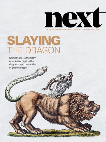
SLAYING THE DRAGON Chimeritope Technology offers new hope in the diagnosis and prevention of Lyme disease
Winter 2018–2019
Welcome to NEXT Dear Friends, Welcome to our second issue of NEXT, a magazine that we started last year to share the incredible innovations in patient care, research and education being discovered every day on the MCV Campus at VCU Health. The exciting advances made by our world-class healthcare providers improve and save lives right here in our community, and the impact is felt around the world. In this issue, the research and innovation that we present include a broad range of patient care, from prevention and diagnosis to treatment and recovery. Beginning with our cover story, we explore Dr. Richard Marconi’s research to prevent and diagnose Lyme disease by developing a vaccine for dogs, a more accurate diagnostic test for humans, and the science that will lead to a human vaccine in the not-too-distant future. We investigate new wearable technology that lets people know when they are experiencing a cardiac arrest and automatically alerts emergency medical personnel. We also look at a new method of determining what medications trauma patients are taking in the event they are unconscious or disoriented when they arrive at the emergency department. Looking at treatment and recovery, we start at VCU Massey Cancer Center, which became the first location in Virginia to offer CAR T-cell therapy this summer. This new treatment involves genetically modifying the immune system to attack cancer cells. We investigate state-of-the-art care that uses deep brain stimulation to help people with Parkinson’s disease and other movement disorders regain control of their motor skills. We also explore a fresh look at recovery to address the ongoing opioid crisis through a study that guides opioid overdose survivors directly from the emergency department into long-term treatment facilities. This level of research and development requires significant funding to bring ideas from the bench to the bedside. VCU Health is fortunate to garner public and private research awards, and philanthropic funding is playing an increasingly important role, especially in moving early stage research projects forward. In fiscal year 2018, the MCV Campus health science schools and college received $155.4 million in new research awards, raising the total MCV Campus sponsored research amount to $621 million. We hope you enjoy learning about the innovative work being accomplished at VCU Health and feel inspired to join our community and be a part of the next great discovery.
Margaret Ann Bollmeier
Harry R. Thalhimer
PRESIDENT, MCV FOUNDATION
BOARD CHAIR, MCV FOUNDATION
WINTER 2018–2019
03
Personalized Medicine at Its Finest Oncologists are mobilizing a patient’s immune system to better target and fight cancer.
06
Temperature and Time Technology may soon alert 911 dispatchers of cardiac arrest. A pioneering post-arrest team is ready to treat survivors.
22
A Better Way to Identify Medications Trauma patients in the emergency department who can’t identify their medications now have a voice.
26
COVER STORY
Chimeritope Technology:
Paul Fisher, M.Ph., Ph.D., discusses his research
12
An Avant-Garde Approach to Preventing and Diagnosing Lyme Disease Researchers have developed a canine vaccine and human diagnostic test in the battle against
Faculty Spotlight and what motivates him to fight cancer.
28
Lyme disease.
Second Opportunities for Lasting Change Researchers are working across Virginia to reduce opioid overdose recidivism and death.
About the Cover Named after the fire-breathing hybrid monster in Greek
32
Electrifying the Brain
mythology, modern-day
Deep brain stimulation effectively treats movement
chimeritope technology fights
disorders, and doors are opening for it to help
diseases by combining DNA
those with dementia.
from different organisms. Read more about the history of the mythological Chimera on page 17.
M C V F O U N DAT I O N
1
Personalized Medicine at Its Finest CAR T-cell Therapy Offers Hope to Cancer Patients When Other Treatments Fail CAR T-CELL THERAPY OFFERS HOPE TO CANCER PATIENTS WHEN OTHER TREATMENTS FAIL
By Alex Henley
C
hemotherapy, radiation and surgery have been the hallmarks of cancer treatment over the last several decades, and targeted drug therapies have been added to the arsenal in recent years. But what happens when these methods don’t result in a cure? In the most severe cases, patients have been left with limited options, such as hoping for a new clinical trial or entering hospice care to manage their symptoms. Immunotherapy is offering fresh hope to patients through a new approach — adoptive cell transfer — which mobilizes the patient’s own immune system to attack tumors. CAR T-cell therapy is a type of adoptive cell transfer that has successfully passed clinical trials and is now offered at select academic health centers and hospitals to treat patients with aggressive types of B-cell lymphoma, cancer of the lymphatic system, and B-cell acute lymphoblastic leukemia, a type of blood cancer. CAR is an abbreviation for chimeric antigen receptors (this exciting medical advance is explained later in this article). Additional clinical trials studying the benefits of CAR T-cells in other cancers are also underway. This summer, VCU Massey Cancer Center announced it was the first in Virginia to offer an FDA-approved CAR T-cell therapy as an official treatment center for YESCARTA® by Kite Pharma. This therapy treats adult patients with non-Hodgkin’s lymphoma, also known as recurrent or refractory B-cell lymphoma, who have already tried at least two types of other treatments without success. Massey also announced that it will soon offer KYMRIAH® by Novartis, another FDA-approved CAR T-cell therapy, in partnership with the Children’s Hospital of Richmond at VCU for children and young adults with recurrent or refractory B-cell acute lymphoblastic leukemia. “I’ve been doing bone marrow transplant for 30 years and I view this as ‘Transplant 3.0’ — the next evolution in using
cells of the immune system to treat cancer when standard therapies don’t get the job done,” said John M. McCarty, M.D., a professor in the VCU School of Medicine, director of VCU Massey Cancer Center’s Immunotherapies and Transplantation Program and the G. Watson James Endowed Professor of Hematology. Dr. McCarty explained how often the immune system may allow a cancer to develop when part of its role is to eliminate it. “T-cells are a type of white blood cell and they are the sentries of the immune system. For a cancer to develop, usually one of three things happens: either the T-cells don’t recognize the cancer cell type as foreign, there aren’t enough T-cells, or the T-cells are exhausted by the cancer itself. Traditionally, in Transplant 2.0, we’ve resolved a lack of sufficient T-cells or T-cell exhaustion through allogeneic transplant, which involves implanting someone else’s immune system into a patient to look for the cancer and eliminate it. The problem with this therapy is it’s not specific only to the type of cancer and there can be significant side effects, such as graft-versus-host disease. “CAR T-cell therapy is a more comprehensive solution because it addresses all three scenarios. It engineers the patient’s own immune cells to recognize and target tumor cells, it expands the patient’s quantity of T-cells, and it reactivates and reenergizes the patient’s T-cells to kill the targeted cells.” CAR T-cell therapy involves a multistage process. First, the patient’s blood lymphocytes are collected through an outpatient process called leukapheresis that typically lasts three to four hours. The patient’s lymphocytes are then sent to an FDA-approved manufacturing facility where their T-cells are isolated, and the requested chimeric antigen receptor (CAR) genetic constructs are added to reprogram them into CAR T-cells. This process can take from
A stimulated genetically reprogrammed CAR T-cell performs targeted killing of a cancer cell.
M C V F O U N DAT I O N
3
“Previously, we haven’t had a lot of additional treatment options for patients with B-cell leukemias and lymphomas who didn’t respond to chemotherapy or had their cancer return. CAR T-cell therapy is giving those patients a second chance. ... It’s a very effective therapy that is yielding positive, long-term results.” Dr. John M. McCarty
two-and-a-half to three weeks, during which time the patient may receive some chemotherapy with their local oncologist to keep their disease in check. Once the CAR T-cells are received at Massey’s Cellular Therapeutics Laboratory, the patient is given a low-intensity chemotherapy designed primarily to make room in their immune system for the new CAR T-cells to operate freely. The CAR T-cells are administered to the patient through an IV infusion, and the patient’s augmented immune system is then armed for battle. The chimeric antigen receptors that are now an upgraded part of these warrior cells reside on the surface of the T-cells and allow these new super-powered T-cells to hunt and attach to a certain antigen, or protein, called CD19, on the lymphoma or ALL cells. CD19 is one of the most prolific biomarkers for B-cell lymphomas and leukemias because it is present in all B-cells and is seen in especially high quantities in B-cell-derived cancer cells. CAR T-cells are chimeric because the binding capabilities of the antigen receptor are synthetically married to the pathogen-killing abilities of an individual’s T-cell — targeted precision medicine at its finest. “Ideally you would want the target, in this case CD19, to be present on just the cancer cells and nowhere else,” Dr. McCarty told us. “This therapy gets rid of the lymphoma, but it may also attack some healthy lymph node tissue. In those situations, we help patients replace their B-cells through IV immunoglobulin therapy, but this is one reason why this therapy is intended for patients who haven’t responded to other therapies. Soon, studies here at VCU Massey will explore how this new therapy will stack up against more traditional high-dose transplantation strategies, such as in non-Hodgkin’s lymphoma, for example. “Previously, we haven’t had a lot of additional treatment options for patients with B-cell leukemias and lymphomas who didn’t respond to chemotherapy or had their cancer return. CAR T-cell therapy is giving those patients a second chance.
4
N E X T / W I NT E R 2 018 – 2 019
The clinical trials are showing complete responses in 82% of young ALL patients, with over 55% of lymphoma patients surviving past the one-year mark in preliminary data. We expect to hear even more exciting news in the coming months as more patients are treated and followed long term after these treatments and clinical trials. It’s a very effective therapy that is yielding positive, long-term results to those previously with fewer effective options. It’s also more conducive to the patient — one round of treatment with local monitoring for a month is certainly preferable to multiple rounds of chemotherapy or even a month-long in-hospital stay for a transplant. These CAR T-cells remain in the body and have the ability to perform constant surveillance and attack future cancer cells.” While these results are compelling, CAR T-cell therapy is not without risks or potential side effects, and one of the most serious is cytokine release syndrome, also called a “cytokine storm.” The purpose of the CAR T-cell therapy is to wake up the immune system and direct it to go into attack mode to kill cancer cells. This activation and further expansion of the CAR T-cell army is a necessary and important part of making these cells effective combatants against a patient’s disease. As part of this response, cytokines are released as cell-signaling proteins of the immune system. This is an inflammatory response, and if it expands rapidly or dramatically and isn’t monitored and modulated, the patient can experience side effects, such as fever, fatigue, difficulty breathing, a drop in blood pressure or neurological symptoms. “These side effects are manageable and reversible, but it takes a small city to take care of these patients to anticipate, monitor and manage untoward complications and side effects,” said Dr. McCarty. “We have trained over 500 people across VCU Health to be part of the comprehensive integrated healthcare team to take care of CAR T-cell patients no matter how they may enter into our healthcare
Leadership of the VCU Massey Cancer Center CAR T-cell therapy team includes (left to right) Gary Simmons, D.O.; Harold Chung, M.D.; Christina Wiedl, D.O.; William Clark, M.D.; and John McCarty, M.D. (not pictured, Amir Toor, M.D., and Judy Davis, R.N.). Photo: Kevin Schindler
system clinic, emergency room, after-hours clinic, or while inpatient. We have a whole system of technology and protocols to flag and escalate these patients to the appropriate team to address their needs for rapid and appropriate interventions. A cytokine storm can be a serious condition that requires an immediate response. Patients receiving CAR T-cell therapy must stay in the Richmond area for one week after treatment and remain within a 30-minute drive of Massey for one month so that we can monitor them. They’re instructed to come to our emergency department if they have any adverse symptoms, even something as minor as a headache or a low-grade fever.” It was this level of investment in infrastructure and expert coordination across departments at VCU Health that allowed Massey Cancer Center to be selected as one of only 61 providers to offer YESCARTA® and one of only 69 to offer KYMRIAH® nationwide. “We want to make sure the patients who come here are given the best treatment for them,” said Dr. McCarty. “There is an extensive consultation and screening process for CAR T-cell therapy participants, and we approach this with the same careful process we have in place for evaluating transplantation candidates for cellular therapy. We have extensive experience in adapting our resources to any new regulatory infrastructure behind new treatments and we’re used to dealing with seriously ill patients who require multiple, clinically complex therapies.”
Massey Cancer Center has begun treating its first patient with CAR T-cell therapy and has several more patients going through the consultation and screening process. “Our initial treatment responses have been far more than promising and satisfying to our team, and especially for those involved.” Dr. McCarty told us. “Future potential uses for this technology are immense and could include targeting other hematologic cancers, as well as solid tumors, such as neuroblastoma, some sarcomas, and HPV-related cancers. “The beauty of this technology is it has the promise and potential to truly marry precision with personalized medicine — that’s the future of cancer treatment. Massey Cancer Center has the tools to support high-quality research from seed money with the first investigator to carrying out complex clinical trials and care. This allows us to bring cutting-edge therapy to the people of the Mid-Atlantic region and will allow us to continue to add therapies that will make Massey a medical destination for patients to receive world-class care.” If you would like to be a part of the next great cancer breakthrough and support the excellent patient care at Massey, please go to www.MCVFoundation.org, click the Give Now button, and search for the “Bone Marrow Transplant General Fund.”
M C V F O U N DAT I O N
5
Temperature Time
&
How VCU Health is Counteracting the Deadly Outcomes of Cardiac Arrest By Eric Peters
More than 350,000 people in the U.S. experience an out-of-hospital cardiac arrest every year, and only 10% of those individuals survive.1 Not to be confused with a heart attack — which involves blocked arteries, a lack of blood flow to the heart muscle and often an accompanying tightness or pressure in the chest — a cardiac arrest occurs when the pumping system of the heart suddenly and unexpectedly fails. This is often due to an electrical or serious rhythm disturbance that stops the heart from pumping blood to the brain and other vital organs. Within 10 to 20 seconds of suffering a cardiac arrest, a person is totally unconscious and collapses with no pulse. If victims are resuscitated in time, they most often remain in a coma and face a host of additional challenges as their bodies recover from such a catastrophic event, and it is in these vital times — from resuscitation to post-cardiac arrest care — that VCU Health has been a national leader in treatment and research for more than a decade. This leadership began in 2004 when VCU Health established the Advanced Resuscitation, Cooling Therapeutics and Intensive Care (ARCTIC) program, making it one of the first two hospitals in the U.S. to offer its patients the specialized life-saving continuum of care. Holding this position as an early adopter of the process means VCU Health developed and shaped many of the best practices and techniques that are used today in hospitals around the world when administering the therapy.
ADVANCED RESUSCITATION The ARCTIC program, led by Mary Ann Peberdy, M.D., the C. Kenneth Wright Professor of Cardiology at VCU School of Medicine, is a continuum of care that begins with resuscitation in the field that is strengthened and supported through coordination with regional community members, highly trained EMS professionals and partner hospitals. Dr. Peberdy and her husband, Joseph P. Ornato, M.D., professor and chairman of VCU Health’s Department of Emergency Medicine, led a landmark international study2 published in 2004 that showed when a lay person performed CPR and used an automated external defibrillator, it doubled the chances of out-of-hospital cardiac arrest survival compared to CPR alone. Leveraging the study — in which Dr. Ornato was the overall steering committee chair and Dr. Peberdy was the VCU/Richmond principal investigator — the American Heart Association lobbied Congress to pass the Cardiac Arrest Survival Act of 2006, which required all federal buildings, airlines and airports in the country to install automated external defibrillators in public places and train staff to use them. COOLING THERAPEUTICS Survivors of cardiac arrest usually remain in a coma after being resuscitated because of the lack of blood flow to the brain that occurred during the cardiac arrest. Throughout this vital time following resuscitation, a patient’s body
M C V F O U N DAT I O N
7
undergoes a massive inflammatory response that contributes to ongoing multi-organ injury. To prevent and reverse this damage to the brain and other vital organs, ARCTIC patients undergo cooling therapeutics administered through a catheter placed in the femoral vein. This process chills the body temperature to 92 degrees for 24 hours, then gradually returns the body temperature to normal. Multiple beneficial mechanisms are at play as this cooling procedure is administered. Like applying ice to a swollen ankle, it blunts the inflammatory process and swelling of the brain, but, in addition, it directly reduces the cellular effects of the ischemia reperfusion injury, which is tissue damage caused when blood supply returns to tissue after a period of that tissue lacking oxygen. The cooling procedure also lowers the patient’s metabolic rate so the injured organs do not require as much energy. INTENSIVE CARE Next, in the intensive care portion of the ARCTIC process, physicians and caregivers continuously monitor brain waves and focus heavily on ventilation and hemodynamic and metabolic parameters to improve outcomes. Patients are in a coma due to the brain injury caused by the cardiac arrest, and they are at a high risk for having seizures due to the brain injury. The only way to identify these seizures and treat them if they occur is to continuously monitor the brain activity with an electroencephalogram (EEG). Maintaining
adequate brain blood flow and protecting the brain from the effects of reperfusion injury are very important at this critical time, and these factors can be altered by changes in ventilation, oxygenation and blood pressure. Additionally, the VCU Health ARCTIC program was the first in the country to perform detailed neurocognitive testing in survivors during this intensive care portion of treatment to identify subtle signs of brain injury and begin early rehabilitation. All of these therapies proved to be so beneficial after being implemented at VCU Health that in 2008 the health system began partnering with EMS systems and hospitals in the region to identify additional patients who might be eligible for advanced post-cardiac arrest therapies. “Many smaller hospitals simply don’t have the volume of patients or the multidisciplinary expertise available 24 hours a day, seven days a week, to be able to provide this therapy,” Dr. Peberdy said. “And we felt that all patients in the region should be able to get this life-saving therapy if they have a cardiac arrest.” As a result of this regional cooperation and leadership, VCU Health is now one of the largest post-arrest centers in the country, treating approximately 90 patients per year. “To put that in perspective, many other hospitals our size treat maybe only five to 10 patients a year,” Dr. Peberdy said. WHEN NO ONE IS THERE TO HELP Survivors who get the opportunity to benefit from programs like ARCTIC at VCU Health often experience their cardiac arrests at places like the gym, surrounded by trained employees and others who can help, but that isn’t the case with many who suffer cardiac arrest. Of the more than 350,000 victims each year, Dr. Ornato said half collapse while no one is around to help or call for assistance, and when it comes to cardiac arrest, there couldn’t be a worse scenario than being alone. “Most of the time, all that is needed to reset the electrical system of the heart and get it started again is a shock from an automated external defibrillator or other device,” said Dr. Ornato. “But here’s the problem: every minute that goes by when a person’s heart stops, the odds that a shock will restart the heart drop 10%. So, if you shock in less than
Joseph P. Ornato, M.D., professor and chairman of the Department of Emergency Medicine, and Mary Ann Peberdy, M.D., C. Kenneth Wright Professor of Cardiology and medical director of the ARCTIC program, have worked together in the cardiac resuscitation field for decades. At VCU Health, they have created one of the world’s most renowned cardiac resuscitation programs. Photo: VCU University Relations
8
N E X T / W I NT E R 2 018 – 2 019
THE PERSONAL TOUCH
From Routine to Life-changing How One Couple is Saying ‘Thank You’ After a Cardiac Arrest
On an April morning in 2017, Bob and Margaret Hershberger woke up, ran a few errands and went to their local gym in Williamsburg for a workout. As they went about a usual day, they had no idea that within hours Bob would be in a coma and flown to VCU Medical Center in Richmond. The day’s shift from routine to life-changing began soon after Bob and Margaret arrived at the gym. “I was back at some of the weight machines and heard this loud thump, and everybody was looking around to see what was going on,” Margaret said. “I looked up to where Bob was on the bike, and he wasn’t there anymore.” Bob, a 70-year-old U.S. Air Force veteran and retired executive vice-president of the Williamsburg Chamber and Tourism Alliance, had suffered a cardiac arrest and fallen off his exercise bike. Gym staff administered CPR, and when emergency responders arrived, they used defibrillators three times to restart his heart and then took him to a hospital in Williamsburg to be stabilized. After Bob suffered his cardiac arrest and was resuscitated, his chances of survival and recovery greatly improved when he was airlifted to VCU Health’s MCV Campus and placed under the care of Dr. Peberdy, medical director of the ARCTIC post-cardiac arrest program. Bob arrived in Richmond at VCU Health in a coma, had his body temperature lowered to 92 degrees to protect his organs, received detailed neurocognitive testing as he recovered, and then underwent double bypass surgery before leaving the hospital. Within six months, Bob was back to exercising regularly. Inspired by his return to good health and the care he received, Bob announced in September 2017 that he and Margaret would make a gift to VCU Health through the MCV Foundation. “Normally, the chances of surviving an out-of-hospital cardiac arrest such as this are less
Bob Hershberger visits with Mary Ann Peberdy, M.D. Dr. Peberdy is medical director of the ARCTIC program at VCU Health and oversaw Bob’s treatment after he suffered a cardiac arrest in 2017. Photo: Courtesy of Bob Hershberger
than 5%, but the cooling procedure changes the odds,” Bob said at the MCV Foundation Discovery Series event where he announced his gift. “Tonight, I’m here as a testimony of Dr. Peberdy’s clinical research and care, as well as the medical assistance I received from the many dedicated professional caregivers who treated me throughout the entire process.” Now, just over one year later, Bob and Margaret have announced a second gift — one that will expand the scope of those who can benefit from and survive thanks to the ARCTIC program. The gift will support a clinical study to test the wearable technology described on page 10. “It’s philanthropic support from people like the Hershbergers that allows novel concepts to move forward to ultimately get larger funding from the National Institutes of Health,” said Dr. Peberdy. “We needed a commercial-level prototype for this device, and no funding source is really available to give you the amount of money to initially prove the concept, so this study would have been dead in the water if it hadn’t been for the support of the Hershbergers.”
M C V F O U N DAT I O N
9
a minute, almost everyone is going to survive. By five minutes you’re down to a 50/50 chance, and by 10 minutes you’re down to less than a 10% chance. That’s the dilemma, time is so critical and unforgiving in cardiac arrest.” This dilemma is why Dr. Ornato has partnered with faculty and students at the VCU School of Engineering to develop a way to ensure people don’t have to be alone when they experience a cardiac arrest. He began brainstorming and speaking on the topic in the 1990s, but technology didn’t catch up with his idea until now. In 2017, a team composed of faculty and students from the Weil Institute of Emergency and Critical Care Research at VCU, VCU Health’s Departments of Emergency Medicine and Cardiology, and VCU’s Department of Biomedical Engineering developed a device that can be worn on the wrist and can detect cardiac arrest instantly, triggering the victim’s cellphone to dial 911 and activate its speaker and video functions so dispatchers can talk to the victim. If there is no response, cardiac arrest is assumed and emergency responders are dispatched to the GPS location, which is accurate to within 20 feet. The GPS information also includes data about altitude to better direct emergency personnel to specific floors in high-rise buildings. If there is no response from the victim and a bystander is present, the dispatcher can prompt the bystander to perform CPR until emergency personnel arrive, resuscitate and, in Central Virginia, send patients to VCU Health’s ARCTIC program. The wearable device Dr. Ornato’s team has developed works by measuring pulse in the same way as current wearable smart devices, but with a much more robust array of sensors that enable it to more accurately measure and continuously monitor blood flow. It also includes more effective signal processing that improves accuracy and transmission, which will be a key component of reducing false alarms. Another critical component of the device, as well as the response system that will be in place around it, is the unique perspective of those who created it. Dr. Ornato is the medical director for Richmond Ambulance Authority and operational medical director for the Henrico County Division of Fire. He has involved 911 dispatchers, first responders and administrators in designing the overall response system. He also is an editor of the journal Resuscitation and writes national and international guidelines on resuscitation. Now that the multidisciplinary team has developed an effective device, the crucial step of getting it onto wrists in the public involves proving and fine-tuning its sensitivity
10
N E X T / W I NT E R 2 018 – 2 019
and specificity, and that’s where a gift from Bob and Margaret Hershberger is making a very big impact (see their story on page 9). Their support is enabling VCU Health cardiologists Kenneth Ellenbogen, M.D., and Santosh Padala, M.D., to conduct a clinical study that will test the device on 50 participants who are undergoing internal defibrillator implantation procedures at the VCU Medical Center that initiate either brief cardiac arrest or abnormal heartbeat rhythms as part of the procedure. By simply putting the wrist device on participants during these procedures, researchers will have a unique opportunity in a controlled, but real-life, situation to prove the device’s effectiveness as it gathers vital data and communicates with cellular devices and emergency services. Once the clinical study has shown the device is reliable and effective, Dr. Ornato sees its path to success as one that blends with existing devices like smart watches that are already being worn for other reasons. This is because less than 1% of patients who have a sudden, unexpected cardiac arrest know they have a problem before their heart suddenly stops, so just giving the device to high-risk individuals wouldn’t reach enough of the population. “If we can prove the concept with studies like the one we’re conducting — prove the safety and the value and the accuracy — our dream is to license this technology to companies that already make wrist devices,” Dr. Ornato said. “That way, people would be purchasing something that they were already going to purchase, not really expecting it to someday save them. Our technology can be invisible in the background, and then, God forbid, if their heart stops beating one day, it will have been the critical purchase of their life.” If you would like to support lifesaving cardiac resuscitation research to help those who may suffer cardiac arrest in the future, please visit www.MCVFoundation.org and contact any of our development officers, or go directly to the Give Now link on our website and select the “Weil Institute Current Fund” from the dropdown menu.
1 According to the American Heart Association: www.ahajournals.org/doi/10.1161/ CIR.0000000000000558. 2 Hallstrom, A.P.; Ornato, J.P.; Weisfeldt M.; Travers, A.; Christenson, J.; McBurnie, M.A.; Zalenski, R.; Becker, L,B,; Schron, E.B.; Proschan, M.; New England Journal of Medicine, August 2004, PMID: 15306665.
Building the World’s Premiere Resuscitation Program Right Here in Richmond In 2016, the Weil Institute of Emergency and Critical Care Research, which is widely regarded as the premier basic science cardiopulmonary resuscitation research laboratory in the world, was searching for an academic health center into which they could integrate. When the Weil Institute’s leaders selected VCU Health, it was a clear choice. The Weil Institute’s board voted unanimously to relocate to the academic health center in Richmond largely because of VCU Health’s position as a leader in cardiac resuscitation and post-cardiac arrest care. Now that the expertise in research available within the Weil Institute has combined with the expertise and leadership in care at VCU Health, Richmond is home to one of the strongest comprehensive basic science, clinical and translational cardiac resuscitation programs in the world. What this means is that the most promising basic science research outcomes now quickly and easily translate from the laboratory to the best care providers at the bedside and in the field. The Weil Institute, founded in 1991, is named after the late Max Harry Weil, M.D., Ph.D., who is considered the father of critical care medicine. Wanchun Tang, M.D., who was trained by Dr. Weil, became the institute’s director after Dr. Weil passed away in 2011. Dr. Tang joined VCU Health as professor of emergency medicine in 2016 and continues to work as the Weil Institute’s director. At the Weil Institute, staff perform research on a broad array of emergency medicine and critical care topics.
Recent research has focused on improving outcomes of CPR, circulatory shock, life-threatening heart failure, acute lung failure and overwhelming infections that produce septic shock. The Weil Institute also provides advanced training for physicians, scientists and engineers, as well as sponsors professional and community-based conferences. Most importantly, this research, education and outreach fosters collaboration that is critical to improving human health. Dr. Peberdy, who, along with Dr. Ornato, is a co-deputy director of the institute, said, “Because of the Weil Institute, we now work with investigators who have very successful independent labs exploring topics pertinent to resuscitation, but not directly related. If you look at traumatic brain injury, for example, and you look at the brain injury that occurs after cardiac arrest, there is something that can be learned and translated from one lab to another. It allows us to be at the forefront of looking at very novel therapeutics and treatment for cardiac arrest.” ABOVE: Drs. Ornato and Peberdy celebrate the opening of the Weil Institute of Emergency and Critical Care Research at VCU with Wanchun Tang, M.D. (left to right), the institute’s director. Drs. Ornato and Peberdy are co-deputy directors of the institute, which is named after its founder, the late Max Harry Weil, M.D., Ph.D. (pictured in center), who is considered the father of critical care medicine. Photo: Julia Rendleman, VCU University Marketing
M C V F O U N DAT I O N
11
CHIMERITOPE TECHNOLOGY An Avant-Garde Approach to Preventing and Diagnosing Lyme Disease By Alex Henley
M C V F O U N DAT I O N
13
“As a kid growing up in Brooklyn, we didn’t have a lot of ticks,” quipped Richard T. Marconi, Ph.D., a Lyme disease expert and professor at VCU School of Medicine’s Department of Microbiology and Immunology. His path to studying Lyme disease and other spirochetal infections began during his postdoctoral training at the Roche Institute of Molecular Biology in Nutley, New Jersey. “In the mid-1980s, every time you turned on the news there was something about this new mysterious illness that was showing up in the population and came to be known as Lyme disease. It was causing debilitating symptoms in both human and veterinary populations, and I thought this was an area where I could make an important contribution.” At that time there were just a few major labs in the U.S. studying Lyme disease, and Dr. Marconi went to one of those — Rocky Mountain Laboratories, National Institutes of Health in Hamilton, Montana, where it just so happened a gentleman named Dr. Willy Burgdorfer had made a groundbreaking discovery. BORRELIA BURGDORFERI — A FORMIDABLE FOE Lyme disease was named after Lyme and Old Lyme, two towns in Connecticut where there was a cluster of cases in the 1970s involving children and adults who presented with atypical arthritic symptoms. In 1982, Willy Burgdorfer, Ph.D., discovered that the disease was caused by a unique type of bacteria called a spirochete, and it was named Borrelia burgdorferi in his honor. Bacteria are often identified by their shape, and spirochetes are spiral. It is this spiral structure that allows them to move quickly in a corkscrew-like motion, burrowing deeply into the tissue of their host. This deep drilling into the tissue, as well as their ability to change shape and lie dormant, make spirochetes a formidable foe, able to hide from the host’s immune system. “Lyme disease is a significant and growing human and veterinary health concern,” Dr. Marconi explained. “For Lyme disease to persist in nature, the bacteria must pass back and forth between mammals and Ixodes ticks. The ticks that transmit Lyme disease in North America are commonly referred to as the black-legged or deer tick. In some patients the infection initially presents with the development of a bull’s eye rash, called an erythema migrans, which typically
14
N E X T / W I NT E R 2 018 – 2 019
Richard T. Marconi, Ph.D., professor at VCU School of Medicine’s Department of Microbiology and Immunology, has developed a Lyme disease vaccine for dogs with the longest duration of immunity claim currently available. Photo: Karl Steinbrenner
develops at the site of the tick bite. The rash expands over time before finally resolving. After this initial stage of infection the spirochetes spread throughout the body within a matter of days. The spirochetes can infect tissue, organs and cross the blood-brain barrier, gaining access to the central nervous system.” Because of this rapid movement throughout the body, patients can present with such a variety of symptoms that obtaining a clear diagnosis is often challenging. Early stage Lyme disease is characterized by fever, chills, headache, fatigue, muscle and joint aches, and swollen lymph nodes — symptoms that could easily be taken for any number of illnesses that general practitioners are more commonly on the lookout for. Another complication in diagnosing Lyme disease is the low accuracy of most diagnostic tests and inter-lab variation in interpreting test results. “Spirochetes are amazing creatures and they’re remarkably diverse,” Dr. Marconi told us. “Due to the existence of many different genetic ‘versions’ of this bacteria, development of a diagnostic test that can detect infection with diverse strains, or a vaccine that is broadly protective, was a significant challenge.” If not diagnosed and treated early, some patients may develop what is commonly called Chronic Lyme Disease
and what researchers call Post-Treatment Lyme Disease Syndrome. Whether PTLDS represents an active infection or is a consequence of the original infection is the subject of considerable research and debate. What the research and medical community do know is that the symptoms associated with long-term infection can be debilitating and may include severe fatigue; headache and neck stiffness; body rashes; arthritis; facial palsy; tendon, muscle, joint and bone pain; heart palpitations; dizziness and shortness of breath; inflammation of the brain and spinal cord; nerve pain; numbness and tingling in the hands or feet; and neurological symptoms, including memory loss, cognitive impairment, depression, anxiety, irritability, insomnia, psychosis and others.1 In addition, the spirochete bacteria can be difficult to eradicate with a standard round of antibiotics because they spread so widely throughout the body, and their deep burrowing allows them to hide in organs and tissue. Because the actual number of Lyme disease cases is vastly underreported, the Centers for Disease Control and Prevention conducted two studies using data from laboratory tests and insurance claims to develop a realistic estimate of the actual public health burden. Based on this research, the CDC estimates there are 300,000 people diagnosed with Lyme disease in the U.S. each year; however, because the data is several years old, many experts posit the number of new infections is much higher, possibly double. Lyme disease is now the most common vector-borne illness in the U.S.2 Fortunately, Dr. Marconi’s groundbreaking work is positioned to change that.
A HIGH-TECH APPROACH TO PREVENTION Faced with a disease that spreads rapidly throughout the body, exhibits symptoms that mimic other illnesses, is hard to diagnose and even harder to treat due to its dynamic form and genetic variability, researchers at the Marconi lab have focused their efforts on prevention and improved diagnostics. “What has consistently bothered me is the notion that the best we can do in terms of prevention after so many years of study is to tuck your pants into your socks, perform ‘tick checks’ and apply sprays containing permethrin,” said Dr. Marconi. “With the increasing incidence of Lyme disease, I couldn’t accept that this was the best we could do — I thought there’s got to be a better way.” Dr. Marconi set up his lab at VCU School of Medicine in 1994, and after many years of basic science research, he and his team began working on the development of a Lyme disease vaccine in 2007. There have been attempts by other labs at developing a vaccine in the past. LYMErix™ was a vaccine distributed by GlaxoSmithKline from 1998–2002; however, it was taken off the market due to dwindling sales as some recipients reported side effects that resembled Lyme disease, including arthritis and autoimmune reactions. While the CDC reviewed the claims and found no evidence that the vaccine was causing these symptoms, the damage was done. Public perception and lack of commercial success dampened the enthusiasm of pharmaceutical companies, which partly explains why we don’t have a vaccine available for use in humans at the present time.
Another complication in diagnosing Lyme disease is the low accuracy of most diagnostic tests and inter-lab variation in interpreting test results. “Spirochetes are amazing creatures and they’re remarkably diverse,” Dr. Marconi told us.
M C V F O U N DAT I O N
15
Chimeritope Technology – Canine Vaccine
OspC Chimeric Protein
OspA Protein
Vanguard® crLyme Canine Vaccine
Due to the high variability of OspC proteins, Dr. Marconi and his team selected the seven strains most commonly seen in canine Lyme disease. The colored portions represent the epitope that triggers an immune response. DNA from all seven epitopes was combined to create one OspC chimeritope (chimeric epitope). The OspC chimeritope was then combined with the OspA protein to formulate a vaccine that offers the broadest protection on the market. Illustration: Tom Edwards
Vaccines work by introducing proteins from a diseasecausing agent that have been identified to stimulate the immune system via an antibody response. These foreign proteins are called antigens, but it is the immune system’s antibody response that ultimately kills the pathogen. One of the most important aspects of developing an effective vaccine is identifying the optimal antigens to use in it. The unique features of the spirochete bacteria make this process extremely problematic. The spirochetes are stealthlike and have the ability to produce a large number of antigens, referred to as outer surface proteins, that they can switch to protect themselves and adapt to different environments. Dr. Marconi explained that these outer surface proteins are similar to wearing different coats in different environments. The midgut of an unfed tick is a very harsh environment, so the bacteria wear one type of coat called OspA (outer surface protein A). When the tick feeds on a host and the bacteria get exposed to blood, this changes the pH and temperature of their environment. The spirochetes stop producing OspA and switch to another coat called OspC (outer surface protein C), which allows them to attach to and infect a mammalian host. Where the LYMErix™ vaccine failed — it had a modest level of efficacy around 72% — was that it focused only on OspA, targeting the bacteria while it was still inside the tick.
16
N E X T / W I NT E R 2 018 – 2 019
To achieve this level of protection, the patient has to maintain very high levels of antibodies that require three vaccine doses the first year and additional doses after that. The OspA approach is a transmission-blocking technique, so while this is ideal in theory, these types of vaccines are only effective when the spirochetes are inside the tick. Once the bacteria enter the mammal, there is no OspA production, so this type of vaccine is ineffective at generating antibodies for an immune response at that point. OSPC — A COAT OF MANY COLORS Simplicity is another reason some researchers chose to focus on the OspA protein in earlier vaccine development efforts. There is only one type of OspA protein found in North American Borrelia burgdorferi. In contrast, there are approximately 30 different variants of OspC worldwide, and 11 of these have been found in North America and Europe. Dr. Marconi and his team pressed forward at a time when there wasn’t much enthusiasm for a human vaccine, but, like all visionaries, his solution would be a game changer for both the prevention and detection of Lyme disease. He and his team found support to develop a vaccine for the canine market. Canine Lyme disease has grown to the point of one in 16 dogs testing positive, according to the Companion
The Slaying of the Chimera in Greek Mythology Animal Parasite Council. Further, the council estimates that only 30% of test results are reported, meaning the total number of infected dogs is closer to 800,000. The team began its journey toward developing a canine Lyme disease vaccine by studying the properties of OspC proteins produced by strains of the bacteria that infect dogs, and again observed a robust variance in anti-OspC antibodies that were reacting to the diverse versions of OspC. They next sought to identify the segments of OspC that stimulate the production of antibodies. Known as an epitope, this is the part of an antigen that is recognized and targeted by antibodies in an immune response. His lab identified two major OspC epitopes, but also found the amino acid sequence of these epitopes was highly variable across all the different OspC proteins that exist in nature. To overcome this challenge, Dr. Marconi’s team came up with a next-generation process he calls chimeritope technology. The team isolated DNA segments from the epitopes of seven different OspC protein strains and then used gene synthesis to recombine the DNA and create one chimeric OspC epitope. Chimeritope is formed from the words chimeric and epitope. Named after the fire-breathing hybrid monster in Greek mythology, a chimera is created by joining together the parts of different organisms. The chimeric OspC epitope was then combined with OspA to form a vaccine that can target the diverse strains of the Lyme disease bacteria and offers the broadest protection on the market. “The epitopes that we included in the chimeritope were carefully selected so that the protein would trigger production of antibodies that can recognize OspC variants associated with infections in canines,” said Dr. Marconi.3 At last — chimeritope technology was able to target the monstrous diversity of Borrelia burgdorferi. In partnership with Zoetis, the largest global animal health company, extensive safety and efficacy studies were conducted. The U.S. Department of Agriculture reviewed the data and approved the use of the vaccine in canines. The efficacy trials showed that dogs that received the vaccine did not develop symptoms of Lyme disease after exposure to infected ticks.3 After nearly 10 years of research, the canine vaccine was launched in the U.S. in January 2016 under the name Vanguard® crLyme. Vanguard® crLyme is also approved for use in Canada, and in August of 2017, Zoetis announced that the USDA granted approval for a 15-month duration
Modern-day chimeritope technology fights diseases by combining DNA from different organisms and is named after the Chimera, a fire-breathing monster found in Greek mythology. Made up of three different animals to form one terrifying beast, she wreaked havoc on the people of ancient Lycia and was described by multiple authors, the earliest reference found in Homer’s The Iliad. “She was of divine stock, not of men, in the fore part a lion, in the hinder a serpent, and in the midst a goat, breathing forth in terrible wise the might of blazing fire.”1 According to legend, King Iobates in Lycia was asked to kill the Greek hero Bellerophon, but he did not want to anger the gods by killing Bellerophon himself, so he sent him into battle against the unconquerable Chimera. Though this challenge represented an almost certain death, Bellerophon was joined by his winged horse Pegasus. Bellerophon mounted Pegasus, who flew high into the sky, allowing Bellerophon to avoid the fiery darts of the Chimera and kill her by shooting from above. According to Vergil’s Aeneid, the Chimera is one of the creatures guarding the gates of Hades.2 The myth of the Chimera lives on in popular culture today. The unique creatures that are the heroes and villains of many modern-day books and movies have chimeric origins and qualities. Fortunately, as two articles in this magazine illustrate, chimeric technology has moved beyond fantasy into cuttingedge science that is helping solve real medical problems. In these stories, the chimera is the hero. You can read more about how chimeric technology is being used to target cancer cells in our CAR T-cell article on page 2.
1. Homer, The Iliad, translation by A.T. Murray, Ph.D., in two volumes, (Cambridge, Mass., Harvard University Press; London, William Heinemann, Ltd., 1924), 6.180, retrieved from Perseus Digital Library at www.perseus. tufts.edu/hopper/. 2. Vergil, Aeneid, (trans. Fairclough), 6.287, retrieved from www.theoi.com/ Ther/Khimaira.html.
Image Source: Wellcome Library, London
M C V F O U N DAT I O N
17
of immunity claim, the longest of any canine Lyme disease vaccine on the market. “One of the most important aspects of efficacy of an annual vaccine is that it truly lasts the full 12 months or more. In the past, breaks have been associated with Ixodes tick exposure as circulating OspA titers have waned prior to the end of the year with other Lyme vaccines currently on the market,” said Richard E. Goldstein, D.V.M., DACVIM, DECVIM-CA, chief medical officer at the Animal Medical Center in New York City. “Now that Vanguard® crLyme has a proven 15-month duration of immunity, that is no longer a concern. Veterinarians and pet owners can be confident that Vanguard® crLyme vaccination will provide excellent protection to help prevent Lyme disease in their dogs, and that protection is in place for more than a year.” USING CHIMERITOPE T ECHNOLOGY FOR DIAGNOSTIC T ESTING Lyme disease is known as “the great imitator” because it includes such a wide range of symptoms that it can resemble anything from juvenile arthritis to fibromyalgia, chronic fatigue syndrome, lupus, multiple sclerosis or any number of other diseases. This makes a diagnosis based on clinical symptoms difficult and crystallizes the importance of a definitive test. Historically, diagnostic tests that included OspC as a diagnostic antigen have not accounted for the diversity of the protein, causing a false negative result as much as 56% of the time,4 and leaving thousands to suffer without a clear diagnosis. Dr. Marconi and his team applied the same chimeritope technology used in the canine vaccine to develop a human Lyme disease diagnostic test. While there are differences in the variants of OspC that are produced in humans versus canines, the underlying technology is the same. Vaccines work because the bacteria’s epitope stimulates an immune response via the production of antibodies. In a diagnostic test, these same epitopes can be used to detect the very antibodies that they induce. Antibodies bind to the epitope as part of the immune response to identify and kill the pathogen. Dr. Marconi’s team used a standard ELISA-based approach, which stands for enzyme-linked immunosorbent assay. Using the ELISA method, researchers take the diagnostic proteins, which are referred to as capture antigens, and use them to coat the surface of small wells
18
N E X T / W I NT E R 2 018 – 2 019
that are present on ELISA plates. They then add the patient’s blood serum, allowing the capture antigen time to bind with antibodies that might be present, and then wash the plates to clear unrelated materials. If the capture antigen has bound to an antibody, those bound molecules won’t wash away and provide evidence that the patient has either an active or previous infection. The standard model of testing hasn’t worked with Lyme disease assays that use a natural version of OspC because there are so many variants of the protein. There would need to be multiple assays run to test for a reaction between each different OspC epitope and antibody. The Lyme disease tests on the market that test for OspC typically only screen for one type. Even if pharmaceutical companies were to develop tests for every type of OspC, which they haven’t, that would be an expensive and inefficient process, especially with a fast-moving disease where time is of the essence. Dr. Marconi’s solution uses the ELISA method, but instead of an epitope from a singular OspC protein, he uses his chimeritope technology. The chimeritope joins together epitopes from the various OspC proteins and will bind with any of the different OspC antibodies that could exist in a patient’s blood serum. One definitive test for a variety of antibodies — that’s what makes chimeritope technology a game changer. VCU Innovation Gateway and Dr. Marconi’s lab have partnered with a company called Global Lyme Diagnostics to bring this test to market. It was launched in June 2017 as the “GLD Test” and is now available as a screening and diagnostic tool for patients and physicians. WHAT’S NEXT? “With the canine vaccine and human diagnostic test in hand, the trifecta would be a human vaccine,” said Dr. Marconi. “I’m working on that now with a really talented group of people, and we hope to advance to clinical trials as rapidly as possible. The success of the Vanguard® crLyme vaccine has been a tremendous boost and shows that the key components of the science are essentially complete. Our next step is to establish the right partnerships so that we can garner the funds and support to move into human clinical trials of the human vaccine. “Another likely outcome would be vaccines that protect against other tick-borne pathogens — babesiosis, ehrlichiosis, Rocky Mountain spotted fever and
“With the canine vaccine and human diagnostic test in hand, the trifecta would be a human vaccine. I’m working on that now with a really talented group of people.” Richard T . Marconi A fluorescent image shows the Lyme disease spirochetes adhering to particulate matter while growing in the laboratory. Photo: Courtesy of Dr. Marconi
anaplasmosis to name a few. We’re currently working on a prototype vaccine for anaplasmosis, and we’re also looking at developing a vaccine for the equine market.” While seemingly unrelated, Dr. Marconi and his team are performing research on periodontal disease. Another class of spirochete bacteria called Treponema denticola has been identified as one of the causes of periodontal disease, which can damage the gums and supporting structures in the mouth. Researchers have also linked an overgrowth of bacteria in the mouth to other problems elsewhere in the body, including heart disease. “We are working to develop vaccines or therapeutic antibodies that offer clinicians less invasive approaches for preventing and treating periodontal disease,” Dr. Marconi told us. Chimeritope technology is also being used in vaccines for other disease categories. For patients with concerns about the adjuvants in vaccines or receiving too many vaccines at one time, this technology could allow multiple disease epitopes to be incorporated into fewer vaccines. “It’s really a major undertaking to secure funding and conduct the testing that is required to obtain FDA approval and bring a human vaccine to the market,” Dr. Marconi told us. “Resources are critical to driving this forward. The public helped bring Lyme disease to the forefront and encouraged the funding and appropriate support. We wouldn’t be where we are today without the public backing. “I’ve worked with incredibly dedicated people in my lab for many years — you can’t complete this type of work
on your own. We’re grateful for the support that we’ve received from so many different sources, including the National Institutes of Health, the Global Lyme Alliance, and the Steven & Alexandra Cohen Foundation. That support is essential to advance development and establish corporate partnerships.” If you would like to contribute to Dr. Marconi’s research to end Lyme disease and other related spirochete infections, please contact Brian Thomas, vice president and chief development officer at the MCV Foundation, at 804-828-0067 or Brian.Thomas@MCVFoundation.org.
1. According to the Centers for Disease Control and Prevention www.cdc.gov/lyme/ signs_symptoms/index.html and the International Lyme and Associated Diseases Educational Foundation iladef.org/education/lyme-disease-faq/. 2. According to the Centers for Disease Control and Prevention www.cdc.gov/lyme/ faq/index.html, www.cdc.gov/lyme/stats/index.html. 3. There are multiple studies leading up to the creation of a vaccine. One such study was published in The Veterinary Journal, November 2013, PMC3872846. A summary of the development of the canine vaccine is available at www.zoetisus.com/ products/dogs/vanguard-crlyme/. 4. Stricker R.B. and Johnson L., The BMJ (British Medical Journal), November 2007, PMC2078675.
M C V F O U N DAT I O N
19
THE PERSONAL TOUCH
THE RELIEF OF A DIAGNOSIS The average Lyme disease patient see five doctors over two years to get a diagnosis1 — Craig Suro is one of them. In June 2015, Craig set up a trail camera in the woods for an afternoon of archery hunting underneath the Huguenot Bridge on the James River. Two days later he found two ticks on his waistline and removed them. Two weeks later he felt off — something was wrong but he wasn’t sure what. He had a cold that wasn’t getting any better and the pain in his head and neck felt like whiplash. A month later his symptoms progressed to cognitive impairment that resulted in not being able to make decisions, exaggerated emotions and not having any confidence. He went to his general practitioner, who ran a full panel of bloodwork — everything came back normal. Because of the tick bites, Craig’s doctor ran one of the CDC-recommended Western blot tests for Lyme disease and it came back negative. He put Craig on doxycycline for two weeks as a prophylactic measure and assured him that would kill any Lyme bacteria, just in case the test wasn’t picking up the infection. “By the end of August, I was unable to function as a human being,” Craig told us. “I could not leave the house. I was diagnosed as having a midlife crisis by my general practitioner, and he said nothing was physically wrong with me.” This is the story of countless Lyme disease patients. One day everything is fine, and then suddenly life is drastically altered. Overcome with a range of physical and mental symptoms, these patients are often unable to complete everyday activities they used to perform with ease. And the worst part is, many of them are told by the medical professionals they turn to for help that nothing is medically wrong with them. Craig and his family took a trip to the beach later that summer to see if that would help him relax. “I couldn’t drive the car, have a conversation or even go down to the beach without major anxiety,” Craig said. “I made a living by taking business risks, and now here I am, unable to turn left or right.” After finally receiving the right diagnosis and treatment for Lyme disease, Craig Suro and his daughter are back to enjoying healthy, full lives. Running is one of their favorite activities, and here they celebrate completing a local turkey trot race together last Thanksgiving. Photo: Courtesy of the Suro family
20
N E X T / W I NT E R 2 018 – 2 019
Craig suffered a major medical episode on the beach trip. “I felt lightheaded and fell to the floor. I was sweating profusely, drooling and was able to call out and crawl on the floor toward my wife, who called an ambulance. Thirty minutes later I was in the ambulance and my vitals were back to normal. The EMTs said they had never seen anything like this.” Craig had an MRI in September which showed everything as normal. He asked his doctor to run the Lyme disease tests again when he had another medical episode in September, and the results came back negative. After a third breakdown in October, Craig went to an endocrinologist who was able to run tests that showed his sympathetic nervous system was through the roof while his parasympathetic nervous system measured normal. The doctor told Craig his blood levels were showing something was wrong, but he couldn’t determine what it was. Craig even went to a psychologist and a psychiatrist in case it was “in his head” as his general practitioner had suggested. Both mental health experts told him they thought there was an underlying physiological problem that was causing the psychological symptoms. They sent him back to his medical doctor. “At the six-month mark it became just unbearable,” Craig said. Fortunately, at a lunch to catch up with an old friend he got just the break he needed. “In the restaurant, I’m hyperventilating and everything is too bright, but by this point I’m used to it and just try to fend it off. It’s hard when you look fine on the outside — everyone just assumes you’re nuts. “I’m an analytical guy and the only delta over the last several months was those two ticks. When I mentioned this to my friend he said, ‘If you think there’s a chance it’s Lyme disease, you’ve got to talk to Dr. Marconi. He was my professor and he’s right here in Richmond.’” Since this was before the Global Lyme Diagnostics test was on the market, Dr. Marconi looked at Craig’s blood under a microscope. “We were able to visualize the spirochetes in the blood sample, which is a bit unusual,” Dr. Marconi explained. “This meant there was a high bacterial load and Craig was most likely suffering from Lyme disease.”
“When you hear the results, you literally stop what you’re doing and just bawl,” Craig said. “Not having a diagnosis is such a torture. I don’t think Dr. Marconi has any idea what he did for me. It was such a relief. I thought, ‘I’ve got hope now and I know what to do.’” Dr. Marconi is a researcher and doesn’t treat patients, so Craig found a Lyme-literate doctor and began a long course of antibiotic treatment. Due to the nature of the spirochete, if antibiotics are used as a treatment, much longer courses are often required to completely rid the body of the bacteria. The two-week treatment Craig’s general practitioner gave him near the onset of his symptoms wasn’t long enough to kill off the infection. After six months, Craig’s blood tests came back clear of the spirochete bacteria. He also found out the Lyme disease had caused encephalitis, which is swelling in the brain, that didn’t show up on his earlier MRI. As the brain is one of the slowest parts of the body to heal, it took a year-and-a-half for him to make a full recovery. But the story doesn’t end here. In May 2017, Craig’s daughter leaned into him and told him she was so very tired and had a headache. They tracked her symptoms, which occurred often enough to warrant concern. Craig and his wife were able to quickly get their daughter to their Lyme-literate doctor. By this time the Global Lyme Diagnostic test was on the market, and it came back positive for Lyme disease. Craig’s daughter immediately began a course of antibiotics for four weeks, and by the 22nd day she sprang back to her normal self again. “I often wonder, did I go through everything I went through to save my daughter?” Craig said. “I’ve helped two of my friends get a diagnosis and they’ve helped their friends.” As for Dr. Marconi, Craig says, “I owe him dearly. He literally, quite literally, saved my life. People need to understand that Lyme disease is a huge, real, legitimate problem. Rich gave me the hard data I needed to prove I had an illness and find a doctor who could help me.”
1. According to the International Lyme and Associated Diseases Educational Foundation.
M C V F O U N DAT I O N
21
A Better Way By Alex Henley
VCU Health Trauma Surgeon and VCU School of Pharmacy Translational Scientist Collaborate to Identify Patient Medications in the ER Medication reconciliation is the term used when doctors and nurses try to determine all the medications — including frequency and dosage — that a patient is currently taking. While this may seem like a straightforward discussion in the relaxed setting of a scheduled appointment with a family doctor, what happens in a trauma setting when the patient is unconscious or disoriented and unable to answer? “When a patient comes in after a major trauma like a car crash, I need to know what medical problems the person has and what medications they are taking that might affect my decision to operate on them emergently,” explained trauma surgeon Sudha Jayaraman, M.D., associate professor of surgery, VCU School of Medicine, and co-director, Program for Global Surgery at VCU Health. “The process that we have right now is haphazard and time-intensive, and it’s like that across the country. First, we ask the patient, but maybe the person has a bad head injury or is in shock and can’t remember. Next, we try to find family members, but they may not be available immediately or they may only
22
N E X T / W I NT E R 2 018 – 2 019
be able to describe the color of the pills and not remember the names of the medications. We then try to locate the patient’s primary care physician or pharmacist, but they can’t tell us if that person is actually taking their prescribed medications. “It seems like a simple question, but it’s actually quite complicated. We need to know what medicines patients have in their systems or what they are supposed to be taking. We need to know if they are on blood-thinning medicines or if they took their spouse’s pain medicine because their knee was hurting that day or if they skipped their heart medicine because it upset their stomach that morning. Some patients carry a list of medications in their wallet, which is helpful, but I still would have no idea if the person took the medication the morning of their car crash. These are questions we can’t answer right now — and that’s at both smaller hospitals and major medical centers. Across the U.S., our current medication reconciliation process is inaccurate 25–80% of the time.” One scientific paper reviewed 22 studies involving 3,755 hospital patients and found that the current method
VCU Health trauma surgeon Sudha Jayaraman, M.D., and VCU School of Pharmacy assistant professor Dayanjan “Shanaka” Wijesinghe, Ph.D., collaborate on using mass spectrometry as a new method of analyzing what medications are in a patient’s blood. Photo: Karl Steinbrenner
to measure mass, specifically mass/charge ratios. This process is described further below. Scientists build a library of the various retention times and mass/charge ratios of molecules, which they use to compare the molecule in question to, in order to identify it.
of medication reconciliation by asking the patient, family members, family doctor or pharmacist contained errors in up to 67% of the cases. Further, the investigators estimate that 11–59% of these errors were clinically important.1 The current process is also time and labor intensive for doctors and nurses — they end up spending time on the phone trying to track down information rather than making decisions and caring for their patient. In an environment that demands both speed and accuracy, Dr. Jayaraman knew she had to figure out a better process. “I was meeting with a new colleague and discussing how more people are on blood-thinning medications, but one of the challenges is that there is currently no reliable test to identify some of these medications. He jumped in and said, ‘We have a technology that could be applied to that!’” explained Dr. Jayaraman. That colleague was none other than VCU School of Pharmacy assistant professor Dayanjan “Shanaka” Wijesinghe, Ph.D., director of the Laboratory of Pharmacometabolomics & Companion Diagnostics. “In metabolomics, we study the underlying biochemical abnormalities that lead to diseases,” explained Dr. Wijesinghe. “One of the first things we do is identify all the drugs in a patient’s system and remove that data from our analysis — it’s a data cleanup method. This technology has been used in research for a while. We started talking and thought maybe we could apply it in a clinical setting with trauma patients.”
STEP 1: Ultra Performance Liquid Chromatography Because there are many molecules that can have the same mass, researchers begin by measuring retention times. In the specific chromatographic technique used at VCU Health, the molecules are forced at very high pressure through a column packed with silica particles containing chains of hydrocarbons that are 18 carbons long. As the molecules pass through this forest of hydrocarbons, based on their chemical properties, they interact to different degrees with the hydrocarbon chains, thereby varying the time it takes different molecules to move through the columns. The time it takes for a molecule to exit this column is called its retention time (the time the molecule is retained in the column) and is used to identify specific classes of molecules. With this approach, even if two molecules have the same mass, they will have different retention times. These separated molecules are then sent to the HRMS for mass measurement.
MASS SPECTROMETRY — HOW IT WORKS The technology Dr. Wijesinghe referenced is called Ultra Performance Liquid Chromatography coupled to High Resolution Mass Spectrometry — abbreviated as UPLC-HRMS or even shorter as LC-MS. It takes place inside two machines coupled together with the first called a chromatographic separation device (a UPLC in this case) and the second machine called a mass spectrometer (an HRMS in this case). In metabolomics, researchers begin by taking approximately 200 microliters of plasma and preprocessing it to remove large molecules, such as DNA and proteins. They then use the chromatographic separation device to separate and measure speed, specifically retention time, and the mass spectrometer
STEP 2: High Resolution Mass Spectrometry The type of mass spectrometer used in VCU Health’s analysis is referred to as a Quadrupole Time of Flight. This type of mass spectrometer has three main stages. In the first stage, molecules are converted into gas ions and pass through a quadrupole mass analyzer. The quadrupole creates an electromagnetic field via four parallel rods that impose a specified radio frequency (RF) and direct current (DC) voltage across the rods onto the stream of ions passing through. This is done because researchers know that at a set RF/DC voltage level, only ions with a specific mass/charge ratio will pass through the entire length of the quadrupole. This allows them to filter out the molecules they’re not interested in and expedite the process by only allowing ions with a specific mass/charge ratio to pass through to the next stage.
Even with chromatographic separation, some molecules that are very similar will have the same mass and retention times. In such instances, researchers need to break them apart and measure the mass/charge ratios of their smaller fragments to identify them. The second stage is used in this scenario. The mass spectrometer accelerates the ions inside
M C V F O U N DAT I O N
23
the aptly named collision cell and crashes them into an inert gas, typically nitrogen. Because of their chemical structure, the ions break apart in predictable ways and generate fragments with predictable mass/charge ratios. Researchers maintain a database of the mass/charge ratios of both intact ions and their fragments, which they can use for comparison and identification. “The way I explain it to my students is if somebody provided five different types of cars and said I’m not going to tell you what these vehicles are, but I’m going to tell you how much they weigh, can you figure out which cars they are?” said Dr. Wijesinghe. “We know an SUV weighs more than a compact car, so we can figure that out without much trouble. However, a Toyota Camry and Honda Accord might weigh the same, so you need to start taking the car apart piece by piece. You might find out that they have different steering wheels with different weights, so you would use the weights of the component parts to identify the car. That’s what we do with mass spectrometry.” The third stage aids in confirming the identification of the molecule by measuring mass/charge ratio at a very high level of accuracy through a time of flight mass analyzer. Here the ions are pulsed through a tube maintained in a vacuum. Based on their mass/charge ratio, ions will move through this tube
at different, but predictable, speeds, and the time it takes to reach the end of the tube is measured (i.e., time of flight). The retention times from chromatographic separation coupled to this three-fold process of measuring the mass/ charge ratios of the intact ion and fragmented ions allows researchers to accurately identify the molecules and corresponding medications that are present in a patient’s blood plasma. MOVING FROM THE LAB TO A CLINICAL SETTING In 2014, Drs. Jayaraman and Wijesinghe put together a proposal for a clinical investigation to determine the feasibility of using LC-MS testing in a trauma setting. As Dr. Jayaraman was seeing increasing numbers of patients on blood thinners, they began by testing LC-MS as a means of identifying three different blood thinners — apixaban, dabigatran and rivaroxaban. First, they established and validated the retention time and mass/charge metrics that would be entered in the database and used as the standard for measurement for each of the three medications. Next, they enrolled 356 patients age 65 and above who were admitted to VCU Medical Center’s Level I trauma center from 2015–2016. They obtained consent from the patients to run LC-MS tests on blood samples that were already drawn for other testing purposes and would have otherwise been discarded. They wanted to identify any of the three blood-thinning medications and compared these results with what the patients reported on their medical records. The results of the clinical investigation were incredibly accurate, with an overall sensitivity and specificity of 94.87% and 99.11%, respectively. This means the LC-MS technology was able to correctly detect a patient was on blood-thinning medications with 94.87% accuracy and also able to correctly identify the exact medication with 99.11% accuracy. “These results are very exciting and show that LC-MS would be a valuable test to aid in medication reconciliation, especially with trauma patients. We weren’t expecting to see
Dr. Jayaraman and Kimberly Baldwin, R.N., read through a medication list and discuss a patient’s case in the surgical/trauma ICU at VCU Medical Center. Photo: Kevin Schindler
24
N E X T / W I NT E R 2 018 – 2 019
100% accuracy because we know the current medication reconciliation process is flawed and the medical records we were comparing the tests to weren’t 100% accurate. For example, a patient may have reported being on a medication, but had a fall and was taken to the hospital before taking their prescription that morning, so the test would have been correct in not identifying the medication in their system, but would have shown up as a false negative in the results because it didn’t match what the patient reported in their medical record,” said Dr. Wijesinghe. An equally important outcome of this investigation is the dramatic increase in speed to obtain accurate medication information. The current process of interviewing the patient, family, primary care doctor and pharmacist can take up to two days to obtain enough information to make a safe clinical decision. The goal of the LC-MS test being developed is to provide this information in 30 minutes or less.2 WHAT’S NEXT? “There are 1,500 FDA-approved medications in the U.S. and the list is growing every day,” said Dr. Jayaraman. “In the emergency setting, there are about 200 –300 medications that are really important to identify. We’d like to use LC-MS testing to establish a library of identification metrics for these top 200–300 medications. We plan to create an overall test panel as well as panels for different groups of patients. For example, the testing needed on a patient with a heart attack is different than that needed for a patient who has just been in a car crash.” Drs. Jayaraman and Wijesinghe are closing in on that goal thanks to the funding they have received through grants from VCU’s C. Kenneth and Dianne Wright Center for Clinical and Translational Research (read more about this center on page 31), VCU President’s Quest for Commercialization Fund through VCU Innovation Gateway, and a Commonwealth Research Commercialization Award from the state. This funding will allow them to establish a library of metrics for 150 medications, which is a highly technical and time intensive process that requires obtaining analytical-grade medications, performing the LC-MS testing and reproducing the results in an extensive validation process. They have a patent pending and are working with VCU Innovation Gateway to advance this technology and make it more widely available.“There are so many great opportunities
Dr. Wijesinghe examines a patient’s plasma sample extract before analyzing it with the chromatographic separation device and mass spectrometer on the MCV Campus at VCU Health. Photo: Kevin Schindler
within VCU Health for innovation. It’s a very collaborative institution that fosters outside-the-box thinking. The support we’ve gotten and continue to get is critical to the success of this idea and other ideas. That’s the benefit of being at an academic health center — we’re at the cutting edge because this institution pulls together people with diverse strengths and interests, who wouldn’t normally cross each other’s path, to approach a problem in different ways. Where else would I run into a researcher with a mass spectrometer? “I see patients on a daily basis where I wish I had this test. It would be so helpful in our clinical decision-making and make the care we provide safer. I can’t think of a physician who wouldn’t want this test,” said Dr. Jayaraman. If you would like to contribute to this work, please visit www.MCVFoundation.org and contact any of our development officers, or go directly to the Give Now link on our website, select “Pharmacy Current Fund” from the dropdown menu and note this research project on the form.
1. Kwan, J.L.; Lo, L.; Sampson, M.; Shojania, K.G.; Annals of Internal Medicine, March 2013, PMID: 23460096. 2 Drs. Jayaraman and Wijesinghe have published an article, “Metabolomics and Precision Medicine in Trauma: The State of the Field,” in the July 2018 issue of Shock, a medical journal. The digital version can be accessed at journals.lww.com/ shockjournal/Fulltext/2018/07000/Metabolomics_and_Precision_Medicine_in_ Trauma__.2.aspx. They expect to publish two more articles in Shock in the coming months.
M C V F O U N DAT I O N
25
FACULTY SPOTLIGHT
Innovation By Eric Peters
PAUL B. FISHER, M.PH., PH.D., FNAI Professor and Chair, Department of Human & Molecular Genetics, VCU School of Medicine Director, VCU Institute of Molecular Medicine (VIMM) Thelma Newmeyer Corman Endowed Chair in Oncology Research and member of the Cancer Molecular Genetics research program, VCU Massey Cancer Center
This year, Paul B. Fisher, M.Ph., Ph.D., FNAI, and his colleagues from VCU Health and other institutions uncovered a potential “Achilles heel” of glioblastoma multiforme (GBM), the most common and deadly form of brain cancer. Their study1 identified a protective mechanism that can potentially be used to develop new and more effective GBM treatments. Since Dr. Fisher joined VCU Health in 2008, his research lab has identified and created novel genetic elements that diagnose cancer, selectively kill cancer cells, stimulate the immune system and work alongside cancer therapies, such as radiation and chemotherapy, to maximize the effectiveness of treatment. Dr. Fisher, an inventor on 55 issued U.S. patents and author or co-author on approximately 600 peer-reviewed publications and reviews, was recognized for his innovation in December 2017 when he was elected to the National Academy of Inventors. The academy wrote in its announcement that “election to NAI Fellow status is the highest professional accolade bestowed to academic inventors who have demonstrated a prolific spirit of innovation in creating or facilitating outstanding inventions that have made a tangible impact on quality of life, economic development and welfare of society.”
Paul B. Fisher, M.Ph., Ph.D., FNAI, utilizes novel genetic elements identified and created in his lab to diagnose cancer, kill cancer cells, stimulate the immune system and work alongside cancer therapies to maximize effectiveness of treatment. He was named “Virginia Scientist of the Year” in 2014, was elected to the National Academy of Inventors in 2017, and was selected as a Fellow of the National Foundation for Cancer Research (NFCR) in 2018. Photo: Courtesy of VCU Massey Cancer Center
26
N E X T / W I NT E R 2 018 – 2 019
“I am a true believer in team science and have been fortunate to have outstanding collaborators at the VCU School of Medicine. ... These interactions have allowed us to capitalize on our own discoveries.” Paul Fisher, M.Ph., Ph.D., FNAI
In addition, Dr. Fisher was named “Virginia Scientist of the Year” in 2014, and in 2018 he was selected as a Fellow of the National Foundation for Cancer Research (NFCR). We spoke with him recently and asked a few questions about his research and his time at VCU Health. WHAT IS THE FOCUS OF Y OUR RESEARCH? Our research focuses on defining the molecular and biochemical basis of cancer development and progression to metastasis, which is the spread from a primary tumor to other sites in the body. With this information, we believe it will be possible to develop improved diagnostic, preventive and therapeutic approaches for cancer, with the ultimate aim of early detection, preventing and curing this devastating spectrum of diseases. Our laboratory was the first to clone MDA-7/IL-24 and MDA-9/Syntenin. MDA-7/IL-24 is a tumor suppressor gene, which selectively induces cell death in diverse cancer cells. MDA-9/Syntenin, which is elevated in a majority of cancers, is a cellular protein that interacts with other proteins to play a pivotal role in cancer invasion and metastasis. We have shown using genetic and pharmacological approaches that blocking MDA-9/Syntenin expression and its interactions with other proteins disrupts pathways mediating cancer cell invasion and metastasis. This can dramatically suppress tumor cell penetration into normal tissue and the metastatic spread of multiple cancers, including those in the brain, breast, prostate, pancreas and liver. WHY IS VCU HEALTH A GOOD PLACE FOR Y OU TO CONDUCT YOUR RESEARCH? I am a true believer in team science and have been fortunate to have outstanding collaborators at the VCU School of Medicine, VCU Massey Cancer Center and externally. These interactions have allowed us to capitalize on our own discoveries and combine them with the facilities and expertise
of other outstanding investigators, which has significantly enhanced the outcomes of our research programs. The VCU Institute of Molecular Medicine (VIMM) provides a conduit for investigators with overlapping research interests in cancer to collaborate and develop projects in which the sum is greater than its parts. With continued future support from the School of Medicine and Massey, we hope to progress our research projects from the laboratory into the clinic. WHAT IS IT ABOUT Y OUR RESEARCH THAT MOTIVATES OR EXCITES YOU? The ability to comprehend complex processes in nature has always intrigued me. Moreover, the capacity to develop and test hypotheses to better understand physiologically important processes and how they can go awry, resulting in cancer, has always intrigued me. I strongly believe that by understanding the nuances of cancer development and progression, inevitable inroads into improved therapy will become evident. I am highly motivated by a desire to have our research translate into new ways of diagnosing, treating and preventing cancer. Although newer technologies are logarithmically improving our scientific understanding of cancer, this disease remains a major clinical problem that diminishes quality of life and has touched us all, either personally, or through family members, friends or colleagues. If you or a family member has been touched by cancer and you would like to support research like Dr. Fisher’s, please visit www.MCVFoundation.org, click Give Now at the top, and find “Massey Cancer Center” in the dropdown menu.
1 Talukdar, S.; Pradhan, A. K.; Bhoopathi, P.; Shen, X.; August, L.A.; Windle, J.J.; Sarkar, D.; Furnari, F.B.; Cavenee, W.K.; Das, S.K.; Emdad, L.; and Fisher, P.B.; Proceedings of the National Academy of Sciences, May 2018, PMID: 29760085.
M C V F O U N DAT I O N
27
SECOND OPPORTUNITIES FOR LASTING CHANGE Ensuring Opioid Overdose Survivors Get the Long-Term Care They Need By Eric Peters
Those who survive an opioid overdose usually do so because they’re found in varying states of consciousness by family members, friends, caregivers or first responders before their breathing stops completely. These survivors gain an additional opportunity at life, but oftentimes, because of the nature of their illness, they can’t use their new opportunities for change and recovery. F. Gerard “Gerry” Moeller, M.D., director of the VCU C. Kenneth and Dianne Wright Center for Clinical and Translational Research, began contemplating these missed opportunities one day in 2017 after hearing from colleagues in the VCU Health Emergency Department. “They came to me and said they were seeing overdose patients time and time again, and they felt like they just weren’t accomplishing anything,” Dr. Moeller said. “They were reviving the patients, but then the survivors weren’t getting into long-term treatment.” Dr. Moeller, who is internationally known for his translational research on impulsivity and addictions, is keenly aware of the importance long-term treatment plays in pulling people out of a deadly spiral like the one his colleagues described to him, and he wanted to help. Before exploring solutions, Dr. Moeller needed to better understand the problem. Utilizing the expertise available on the Wright Center’s biomedical informatics team, he found some alarming preliminary numbers. The data showed that opioid overdose visits to the VCU Health Emergency Department went from approximately 270 in 2015 to more than 650 in 2017, and from all of those visits, as many as one in five patients experienced a repeat overdose or died within 12 months of their initial overdose. Dr. Moeller knew these numbers demanded action toward finding the reason survivors weren’t getting the help they needed, and, most importantly, toward identifying a new approach to helping the survivors avail the opportunities they’d been given for a new life. WHY ARE SURVIVORS NOT GETTING HELP? The current protocol in treating overdose victims involves administering naloxone, a medication that eliminates opioid intoxication and reverses an overdose. Naloxone has been very effective in saving lives, thanks in large part to advances that enable family members, caregivers and first responders to keep the medication on hand and administer intranasally or via a single injection as soon as they encounter a victim. Naloxone is an opioid receptor antagonist, which means it stops the effects of whatever opioid has been misused by binding to and blocking opioid receptors in the brain.
After an overdose victim arrives at the emergency department, often having already received naloxone, he or she is stabilized, monitored for some time and then referred to a long-term outpatient facility where appropriate follow-up treatment, usually for addiction, can begin. Dr. Moeller believes this referral is the critical moment in the treatment paradigm that can and should be changed. That’s because naloxone is very effective at saving lives, but it also causes acute opioid withdrawal, leading to nausea, vomiting, sweating, diarrhea, chills, cravings, impulsivity and poor decision-making. “One of the behavioral definitions of impulsivity is the lack of ability to delay your gratification,” Dr. Moeller said. “So, if you’re in withdrawal, you have all these symptoms and you wish you were dead. You realize that when you walk out the door of the hospital you can get something that will make you feel better — it’s heroin, or it’s oxycodone, or it’s a pill. Even though you know you just almost died from an overdose, the threat of that happening again is in the future.” Because of this impulsivity, which is often amplified because of life-saving medication, many patients never go to clinics when they’re referred for long-term treatment, opting instead to seek an immediate fix. WHAT CAN BE CHANGED TO BETTER ENCOURAGE LONG-TERM CARE? Instead of referring overdose survivors, who are likely experiencing acute withdrawal, to long-term care after they leave the emergency department, Dr. Moeller is testing the effectiveness of initiating long-term treatment before the survivors ever leave the hospital. The goal here is to counteract withdrawal symptoms and reduce impulsivity. The Phase IV clinical trial, called the Virginia Overdose Treatment Initiative (VOTIVE), began enrolling patients in August 2018. For those who agree to participate, Dr. Moeller’s team, working inside the emergency department, makes contact and provides a medication called buprenorphine that reverses the withdrawal symptoms and initiates long-term treatment. Patients are then given a referral within 72 hours to the outpatient clinic, where they continue medication and counseling for addictions. Approved to treat chronic opioid addiction, buprenorphine is a treatment that many patients don’t ever begin taking
M C V F O U N DAT I O N
29
Nearly one in five opioid overdose survivors who are treated at VCU Medical Center either experience another overdose or die within 12 months of their original visit. F. Gerard “Gerry” Moeller, M.D., director of the VCU Wright Center for Clinical and Translational Research, is leading a statewide project to help opioid overdose survivors pull themselves out of this deadly cycle.
VCU’s Leadership in Addiction Research VCU is home to some of today’s most well-recognized leaders in the field of addiction studies, and its historic, decades-spanning relationships with the National Institutes of Health (NIH) and National Institute on Drug Abuse (NIDA) continue to distinguish VCU’s leadership in addiction research. • In FY 2017, VCU received 227 awards from NIH, totaling $77.3 million, including 39 research grants from NIDA. • VCU is the highest-ranked institution in Virginia for addiction-focused research funding from NIH, and ninth in the country in funding from NIDA and the National Institute on Alcohol Abuse and Alcoholism. • VCU Health researchers have a long history of leadership in basic research on the science of addiction, including giants in the field like Robert Balster, Ph.D., Billy Martin, Ph.D., and William Dewey, Ph.D., among others. These VCU Health scientists worked in the addiction research space for many years, before it became front-page news.
30
N E X T / W I NT E R 2 018 – 2 019
F. Gerard "Gerry" Moeller, M.D., director of the VCU C. Kenneth and Dianne Wright Center for Clinical and Translational Research, is the principal investigator on a clinical trial that is initiating long-term care for opioid overdose survivors inside emergency departments. Photo: Kevin Schindler
after their overdose because they choose to forego outpatient treatment where it’s administered. Buprenorphine binds to and activates the same receptors in the brain that opioids activate, which reduces withdrawal symptoms, but it produces fewer intoxicating effects. In addition, buprenorphine reduces the effects of opioids that might be taken around the same time, making those opioids less desirable and even less likely to cause overdose. The type of buprenorphine this trial is testing is administered in a once-monthly injectable formulation. Indivior Inc., which is headquartered in Richmond, has been developing medications to treat opioid use disorder for more than 20 years and is providing the formulation known as SUBLOCADE™ for use in the trial. The new injectable formulation all but eliminates the possibility of it being diverted or sold outside of the clinic for misuse. In addition to administering buprenorphine as early as possible before survivors leave the hospital, the long-term care component of VOTIVE is vitally important to the recovery of survivors. In Richmond, referrals to long-term outpatient care guide participants to the VCU Health MOTIVATE Clinic, where social workers, nurses and physicians monitor patients’ progress weekly, provide behavioral counseling sessions in individual and group settings, and administer buprenorphine monthly. Improving the likelihood that overdose victims reach this long-term component of care is essential, Dr. Moeller said. “Addiction really is a chronic medical disorder. Like diabetes and hypertension, a one-time treatment is not going to solve the problem, so patients need chronic medication and behavioral treatments like group therapy to help them with lifestyle changes.” Dr. Moeller will serve as principal investigator on the trial and will work with Robert Lipsky, Ph.D., director of translational research in the Department of Neurosciences
at Inova Fairfax Hospital, and Warren Bickel, Ph.D., professor of psychiatry and director of the Addiction Recovery Research Center at Virginia Tech Carilion Research Institute. The trial is funded in part by a $500,000 Virginia Catalyst grant from the Virginia Biosciences Health Research Corporation. The fact that this is a Phase IV clinical trial means it will study how well a treatment that has already been approved for sale works over a long period of time, in specific circumstances and with a specific population. The team will enroll more than 100 participants across the state and follow each for a year, monitoring their progress and recidivism. If the results show what Dr. Moeller expects, which is a significant drop in repeat overdose and death
rates compared to previous data, this trial will establish a new paradigm for treatment of patients after opioid overdose that can be utilized nationally to help survivors get the help they need before it’s too late. If you would like to support addiction research at VCU Health, please visit www.MCVFoundation.org, click Give Now at the top, choose Search for other funds, and search for “Addiction Psychiatry Research Fund.”
The Wright Center:
Research Infrastructure for a University This past May, VCU’s C. Kenneth and Dianne Wright Center for Clinical and Translational Research received the largest National Institutes of Health grant in the university’s history. The $21.5 million award will support the Wright Center in its mission to advance university and community research from basic laboratory science to treatments that improve human health. The grant would not have been possible without the generosity of Ken Wright, who gave $16 million in 2015 to support the center by establishing six distinguished chairs in clinical and translational research and a physician-scientist scholars program, all of which are named for Ken and his late wife Dianne. The Wright Center serves researchers from the life, physical, computer and social sciences, as well as engineering and economics, and serves as an academic bridge for researchers from their home departments to resources across the university and the community. Through its programs, the center provides advanced imaging services, clinical research services, education, community-engaged research support and biomedical informatics. The fastest growing of these programs is biomedical informatics, a service that utilizes the massive amount of information from de-identified electronic medical histories of patients contained in the electronic health record. It combines that information with other data to find answers that lead to preventions or new treatments for diseases. This core’s growth is thanks to Mr. Wright, who provided another gift this year of $5 million to create
a new 6,000-square-foot space where more than a dozen specialists will Ken Wright, one of VCU’s most generous and impactful serve the university’s and benefactors, has been community’s research needs. especially kind to the MCV Campus. In 2015, he “The goals we have for gave $16 million to support the biomedical informatics and name the C. Kenneth program are to improve our and Dianne Wright Center for Clinical and Translational ability to mine the electronic Research, and this year he health record and combine gave $5 million more to support the center’s it with other information biomedical informatics like genomic information program. Photo: Kevin Schindler and brain imaging,” said F. Gerard “Gerry” Moeller, M.D., director of the Wright Center and the first of the center’s distinguished chairs. One example is scanning for mild traumatic brain injury, which doesn't have a strong signal if a radiologist looks at an MRI alone. But, combining the data from that MRI with electronic health record data could be very beneficial. The biomedical informatics team would use machine learning to ask what previously unnoticed small signatures appear every time in millions of data points, and they could specify by age, gender and other factors. “This year’s grant and Mr. Wright’s most recent gift are going to dramatically enhance our biomedical informatics capabilities,” Dr. Moeller said. “We’re expanding into those areas where there are really massive amounts of data so we can look at diseases in ways we haven’t been able to do before.”
M C V F O U N DAT I O N
31
Electrifying
THE BRAIN
By Eric Peters
Deep Brain Stimulation is Treating Movement Disorders and Moving Closer to Treating Dementia Related to Parkinson’s Disease Kathryn Holloway, M.D., a professor in the Department of Neurosurgery at VCU School of Medicine and neurosurgical director of the Hunter Holmes McGuire VA Medical Center Parkinson’s Disease Research, Education and Clinical Centers, specializes in what has been called the most complicated object in the known universe — the human brain. She sounds like an electrician when she talks about the circuits, electrical signals and switches that she works with daily, but it’s clear she is not working with a typical circuit board. For years, her electrical focus in the brain has been an “off switch,” which, despite what the term might suggest, actually restores in her patients the ability to move more normally and helps them do everything from drink a cup of coffee to walk. Through a procedure called deep brain stimulation, she accesses this “switch” to treat those who suffer from any of several movement disorders, including Parkinson’s disease, a chronic and progressive neurological disorder that causes tremors, slow movement, stiffness and impaired balance. These symptoms are caused by deterioration in a part of the brain called the basal ganglia, which is a group of nuclei, or clusters of neurons, near the center of the brain. Basal ganglia deterioration makes the “brakes” of a person’s motor system overactive, and by “turning off” some of the overactive braking areas, motor function can be improved. Dr. Holloway is a pioneer and leader in deep brain stimulation, which works by implanting neurostimulators and two electrodes in the shape of thin wires that send electrical impulses to specific targets in the brain. She formed a team in the late 1990s dedicated to understanding and perfecting what was a new procedure at the time, and now she uses it to treat Parkinson’s disease, essential tremor and other neurological disorders. While participating in the defining clinical trials in deep brain stimulation, Dr. Holloway partnered with a medical equipment development firm to create a new deep brain stimulation surgical tool called
NexFrame,™ which she continues to refine to improve patient comfort, increase accuracy and reduce surgery time. “Deep brain stimulation allows patients to return to activities that they had to give up due to Parkinson’s, which can be as basic as walking or feeding themselves, or as complex as riding a bicycle or painting,” Dr. Holloway said. “The most important thing we provide is more years of enjoyable living.” WITH SUCCESS COMES NEW CHALLENGES: TARGETING DEMENTIA As Dr. Holloway’s team continues to improve deep brain stimulation and maximize the years of enjoyable living for those who are fighting Parkinson’s disease, their success is leading to questions and challenges that they are determined to meet. One of the major challenges is dementia, which is one of the later-stage symptoms of Parkinson’s disease. “Dementia hasn’t garnered much attention in the past because patients would often be so debilitated that it was sort of just a footnote in their decline,” Dr. Holloway said. “Now that we have people functioning so well through deep brain stimulation that they’re up and walking longer, we’re really seeing the dementia much more clearly.” Dementia in Parkinson’s disease patients is caused by deterioration in the nucleus basalis of Meynert, an area of the brain that essentially writes short-term memories down and distributes them throughout the rest of the brain for long-term storage. Because this is the area of deterioration, and especially because of its important cognitive role, Dr. Holloway’s team is focusing on the nucleus basalis of Meynert as they prepare to address dementia through deep brain stimulation. Before Dr. Holloway’s team begins treating dementia through deep brain stimulation, they’re conducting three preclinical studies to ensure they can make the best possible
M C V F O U N DAT I O N
33
Rachel Van Aken, an intraoperative monitoring technologist on Dr. Kathryn Holloway’s research team, adjusts a cap with 256 electrodes that the team uses in its research. The data it gathers will guide brain surgeons in using deep brain stimulation to treat cognitive challenges, such as dementia, in patients battling Parkinson’s disease. Photo: Kevin Schindler
impact. Dr. Holloway’s post-doctoral trainee, Deepak Kumbhare, Ph.D., is critical to these efforts, and he had help this summer from an enthusiastic group of medical students, including Evan Hughes, Adel Azghadi and Zi Huang. These projects, all of which focus on the nucleus basalis of Meynert area within the brain, are described below. STUDY 1: Creating a Guide to Target the Nucleus Basalis of Meynert One study the team is conducting will help determine whether they’re stimulating the correct location within the brain during deep brain stimulation. This is a challenge they haven’t previously faced because patients who undergo deep brain stimulation to improve motor functions remain awake during the procedure and can demonstrate in real time whether motor activities (grasping a cup, writing, etc.) change in response to stimulation. Changes in cognitive function aren’t as easy to identify in real time. To better understand where stimulation should be targeted, the team is using quantitative dense array electroencephelogram (QEEG), which utilizes a cap with 256 electrodes to gather data and analyze it using machine learning algorithms. Participants all have existing deep brain stimulation implants, and based on the QEEG readings, as their neurostimulators are activated,
34
N E X T / W I NT E R 2 018 – 2 019
Dr. Holloway’s team is working to identify signatures of various parts of the brain that they can use as guides when they begin to target dementia in Parkinson’s disease patients. STUDY 2: Restoring Cells in the Nucleus Basalis of Meynert Through Stimulation Another question Dr. Holloway’s team is investigating is whether stimulation can have a regenerative effect on parts of the brain. They hypothesize that using deep brain stimulation to directly target the nucleus basalis of Meynert, which plays a role in memory storage, could induce restorative changes in the brain to protect against or repair ongoing damage. “Electrical stimulation has been shown to cause new brain cells to be created, so when we stimulate this dying or deteriorating area of the brain, we may actually rescue the cells from impending death,” Dr. Holloway said. “We have good reason to suspect this is the case, but no one has ever looked into this question.” STUDY 3: Mimicking Brainwaves with Deep Brain Stimulation The team’s third project is examining how deep brain stimulation signals can integrate with the brain’s various
“Electrical stimulation has been shown to cause new brain cells to be created, so when we stimulate this dying or deteriorating area of the brain, we may actually rescue the cells from impending death.” Kathryn Holloway, M.D., pioneer in deep brain stimulation
Dr. Kathryn Holloway addresses the audience at an MCV Foundation Discovery Series event in 2017. She and her team conduct research in deep brain stimulation to treat Parkinson’s disease, essential tremor and other neurological disorders at VCU Health and the Hunter Holmes McGuire VA Medical Center (see story on page 36). Photo: Skip Rowland
oscillatory patterns, or brainwaves. The form of these brainwaves vary depending on their source and their role. Their sources are driven by either mechanisms within individual brain cells or by interactions between different brain cells, and their role is to facilitate circuit coordination, information transfer or motor output. Since the nucleus basalis of Meynert has connections throughout the brain related to memory transfer, identifying and utilizing the appropriate brainwaves that relate to information transfer and interaction between brain cells could be quite beneficial to someone who is
struggling with memory loss. To investigate this opportunity, the team is examining the use of short electrical bursts that mimic innate nucleus basalis of Meynert behavior and brainwaves instead of using continuous electrical signals as they’ve done in the past with motor-related deep brain stimulation. As these initial studies contribute to a growing overall knowledge of the universe’s most complicated object, teams like Dr. Holloway’s are ready to change the way we all live, grow, thrive and, hopefully, remember. If you’re interested in learning about ways to support this research to help guide surgeons in using deep brain stimulation to treat dementia related to Parkinson’s and other diseases, please visit www.MCVFoundation.org/Give. If you would like to give directly to a Parkinson’s disease fund, click on the Give Now link at the top of our website, choose Search for other funds, and search for “Parkinson’s.”
Dr. Holloway’s team includes (left to right) VCU School of Medicine students Zi Huang, Adel Azghadi and Evan Hughes; intraoperative monitoring technologist Rachel Van Aken; and postdoctoral trainee Deepak Kumbhare, Ph.D. The team is conducting research into techniques and possibilities of how deep brain stimulation can treat dementia. Photo: Kevin Schindler
M C V F O U N DAT I O N
35
THE PERSONAL TOUCH
A Partnership to Serve Heroes and the Community At the end of World War II, more than 100,000 of our nation’s heroes returned home in need of healthcare. To meet the demand, the Veterans Affairs medical centers, known as the Veterans Administration at the time, began partnering with medical schools throughout the country to care for veterans and to train young clinicians. Seventy years later, VCU Health and the Hunter Holmes McGuire VA Medical Center, which was founded in Richmond in 1942, maintain an exceptionally strong partnership that fosters world-class care for our heroes and community, promotes the research which makes that care possible, and provides the education to ensure healthcare continues to advance and evolve. Services at the Hunter Holmes McGuire VA Medical Center are often provided by physicians and other care providers who have dual appointments at that facility and at VCU Health, and those services are available to more than 200,000 veterans who travel from 52 cities and counties covering 22,515 miles of central and southern Virginia and parts of northern North Carolina. MCV Foundation trustee Charles Bryan Jr., Ph.D., — someone who personally knows the care, research and education at the Hunter Holmes McGuire VA Medical Center — introduced Dr. Holloway as his doctor at a foundation event last year. “Thirteen years ago, I was diagnosed with Parkinson’s, and last year I had the privilege of having my brain operated on by one of my real heroines, Kathryn Holloway,” he said. “Before my deep brain stimulation, I was taking more than 20 pills a day with only limited results and my episodes of reduced mobility were coming more frequently. After surgery, the episodes came less frequently and I was able to reduce my pill intake to no more than eight per day. It truly has improved my life.” Deep brain stimulation began at VCU Medical Center in 1997 shortly after it was approved by the Food and Drug Administration, and two years later Dr. Holloway expanded the program to begin performing the procedure at Hunter
36
N E X T / W I NT E R 2 018 – 2 019
Holmes McGuire VA Medical Center. To date, between the two institutions, Holloway’s team has performed close to 600 deep brain stimulation procedures and the partnership has led to new technology development MCV Foundation trustee and and landmark research.1, 2, 3 veteran Charles Bryan Jr., Ph.D., “Everything we learn attends an MCV Foundation from treating patients event after introducing Dr. Kathryn Holloway as a at VCU Health can be speaker. He calls Dr. Holloway translated to the VA and his heroine and says that after she performed his deep brain vice versa,” Dr. Holloway stimulation procedure at the said. “This accelerates McGuire VA Medical Center in the discovery process and 2016, his Parkinson’s disease symptoms were greatly innovations in care. The reduced and his life improved. big data generated from Photo: Kevin Schindler our combined university and VA studies have changed the practice of medicine. It allows us to make the most of the research resources at each facility instead of duplicating them at each institution.” In addition to treating Parkinson’s disease and dementia, Dr. Holloway’s team foresees the partnership and the resources it provides addressing depression, obesity and epilepsy using deep brain stimulation. “The combined research and care at the Hunter Holmes McGuire VA Medical Center and VCU Health makes a larger impact on human health than either could alone,” Dr. Holloway said.
1 Weaver, F.M., et al., Journal of the American Medical Association, Jan. 2009, PMID: 19126811. 2 Follett, K.A., et al., New England Journal of Medicine, June 2010, PMID: 20519680. 3 Weaver, F.M., et al., Neurology, July 2012, PMID: 22722632.
NEXT is published by the MCV Foundation to share the latest breakthroughs occurring at VCU Health and the positive impact these exciting innovations have on our patients. MANAGING EDITOR:
Alex Henley ASSOCIATE EDITOR:
Eric Peters GRAPHIC DESIGN:
Big River Advertising SCIENTIFIC ADVISORY PANEL:
Joseph T. DiPiro, Pharm.D. Alpha A. “Berry” Fowler III, M.D. Steven R. Grossman, M.D., Ph.D. F. Gerard Moeller, M.D. Jerome F. Strauss III, M.D., Ph.D. Wanchun Tang, M.D. The Medical College of Virginia Foundation was established in 1949 to inspire and steward philanthropic resources for our MCV Campus Partners at VCU Health. The MCV Foundation manages more than $500 million in assets to ensure VCU Health remains at the forefront of excellence and innovation in patient care, research and education as one of the top academic health centers on the East Coast. Through our 1,500 funds, we provide scholarships, professorships, research and program funds to support the life-saving work occurring at VCU Health every day. Our MCV Campus partners include: VCU College of Health Professions, VCU School of Dentistry, VCU School of Medicine, VCU School of Nursing, VCU School of Pharmacy, VCU Massey Cancer Center and VCU Medical Center. To learn more, visit : www.mcvfoundation.org Please address comments or subscription requests to: Alex Henley MCV Foundation 1228 East Broad Street Box 980234 Richmond, VA 23298 alex.henley@vcuhealth.org
In our next issue, we will interview David Cifu, M.D., who serves as principal investigator on VCU’s largest federal grant and oversees a national consortium of universities and hospitals that are studying traumatic brain injuries in military personnel.
NON-PROFIT ORGANIZATION US POSTAGE PAID PERMIT NO. 1182 RICHMOND, VA Box 980234 Richmond, Virginia 23298-0234
A P U B L I C AT I O N O F M C V F O U N DAT I O N