Why Detailed Surface Examination Is Key to Ocular Health
The surface of the eye serves as the first line of defense against environmental irritants, pathogens, and physical injury It includes critical structures such as the cornea, conjunctiva, tear film, and eyelids Even minor abnormalities in these areas can lead to discomfort, reduced vision quality, and long-term complications if left undetected.
A detailed surface examination is therefore a fundamental component of comprehensive eye care. It allows clinicians to identify subtle signs of disease, monitor ongoing conditions, and provide early interventions that protect visual function In modern ophthalmology, precise evaluation techniques have significantly improved the ability to assess ocular surface health accurately and efficiently.
Understanding the Ocular Surface
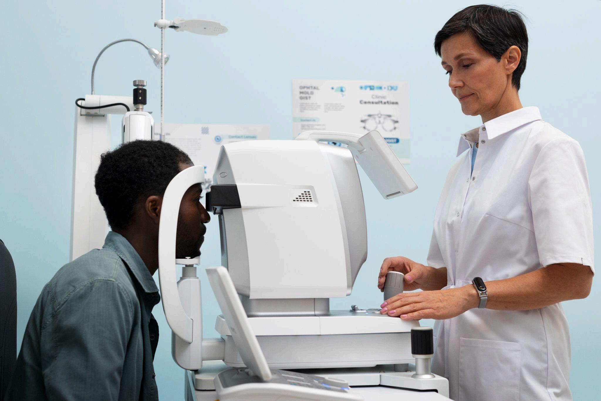
The ocular surface is a delicate and highly specialized system The cornea, responsible for most of the eye’s focusing power, must remain transparent and smooth for optimal vision The
conjunctiva protects and lubricates the eye, while the tear film maintains hydration and provides antimicrobial defense.
Any disruption in this system whether due to dryness, infection, inflammation, or trauma can compromise both comfort and visual clarity. Patients may initially experience mild irritation or blurred vision, but underlying issues can progress rapidly without proper diagnosis
A detailed examination ensures that these structures are evaluated thoroughly, allowing clinicians to detect even micro-level irregularities before they escalate into more serious conditions
Early Detection of Corneal Disorders
The cornea plays a crucial role in refracting light onto the retina Because it is transparent, even small defects can significantly affect visual performance. Conditions such as corneal abrasions, keratitis, dystrophies, and early degenerative changes often begin with subtle surface alterations
Through magnified inspection and focused illumination, clinicians can identify tiny epithelial defects, infiltrates, or irregular tear film distribution Early detection enables prompt treatment, preventing complications such as scarring or vision loss.
Routine surface evaluation also helps monitor contact lens wearers, who are at increased risk of corneal irritation and infection. By identifying early warning signs, practitioners can recommend appropriate adjustments or preventive measures
Identifying Tear Film and Dry Eye Issues
Dry eye disease is one of the most common ocular complaints worldwide It occurs when the tear film becomes unstable or insufficient, leading to discomfort and fluctuating vision However, diagnosing dry eye requires more than simply noting symptoms.
A detailed surface assessment allows clinicians to evaluate tear film breakup time, debris presence, and meibomian gland function. These findings help determine whether dryness is evaporative, aqueous-deficient, or mixed in nature
Accurate diagnosis ensures targeted treatment strategies, such as artificial tears, anti-inflammatory therapy, or gland management procedures. Without proper surface examination, dry eye conditions may be misdiagnosed or inadequately managed
Detecting Conjunctival and Lid Abnormalities
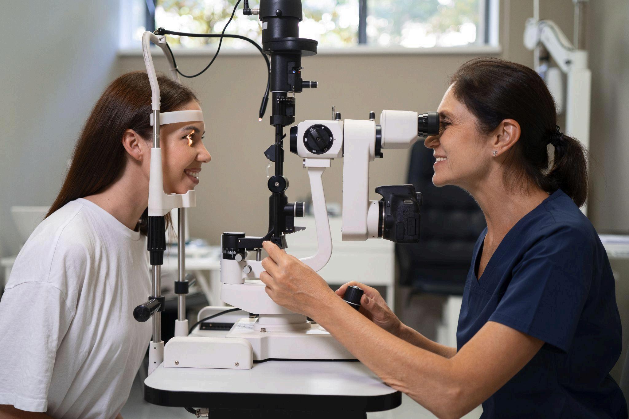
The conjunctiva and eyelids contribute significantly to ocular protection and lubrication. Inflammation, allergic reactions, or infections often manifest as redness, swelling, or discharge However, some conditions may present with subtle vascular changes or minor structural abnormalities.
A thorough examination helps identify early conjunctivitis, pinguecula, pterygium, and lid margin disorders such as blepharitis. Detecting these issues early prevents progression and reduces patient discomfort
Moreover, evaluating eyelid alignment and blinking patterns can reveal functional abnormalities that impact tear distribution and overall ocular health