Why a Camera Fundus Is Critical for
Identifying Early Vision Loss Signs
Vision loss rarely occurs suddenly without warning In most cases, the early signs develop gradually within the retina and optic nerve, long before a patient notices any change in eyesight. These subtle changes often go undetected during routine vision checks, making early diagnosis a challenge. This is where advanced retinal imaging becomes essential.
Modern ophthalmology depends on precise imaging tools to uncover hidden abnormalities inside the eye By providing a detailed view of the retina, these technologies help clinicians identify early warning signs and take preventive action before irreversible damage occurs.
Understanding Early Signs of Vision Loss
Early vision loss can be caused by a variety of conditions, including diabetic retinopathy, glaucoma, macular degeneration, and retinal vascular disorders These conditions typically begin with minor structural or vascular changes in the retina
For example, small microaneurysms, tiny hemorrhages, or deposits such as drusen may appear in the early stages of disease These signs are often invisible to the patient and may not cause immediate symptoms. However, they serve as critical indicators of underlying pathology.
Specialists are trained to recognize these subtle changes, which often appear long before noticeable vision impairment develops.
Role of Retinal Imaging in Early Detection
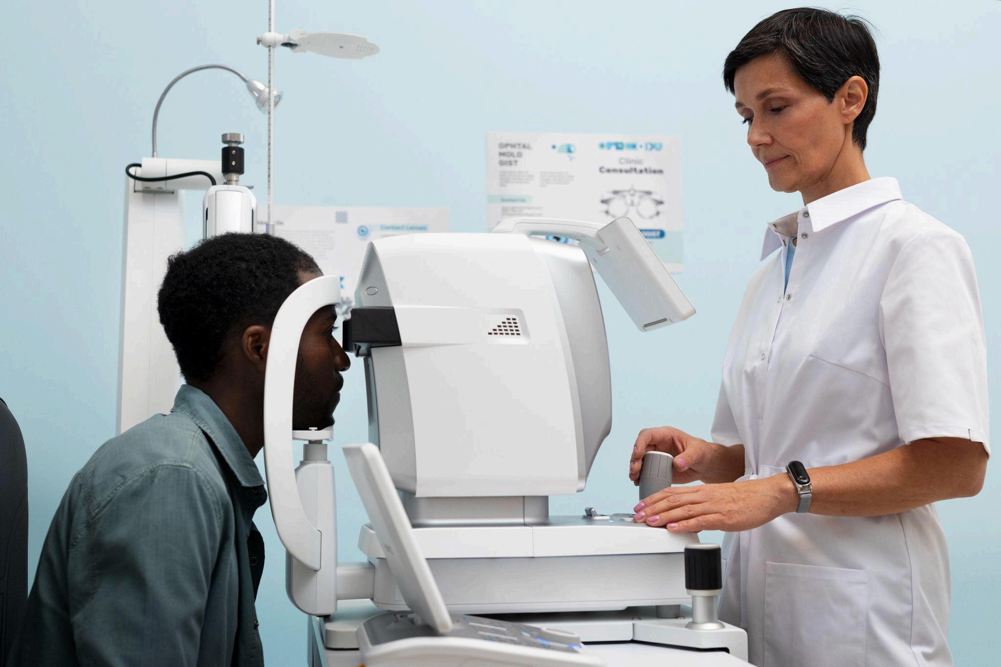
Retinal imaging allows clinicians to capture high-resolution images of the eye’s internal structures, including the retina, macula, optic disc, and blood vessels. These images provide valuable insights into the health of the eye and help detect abnormalities at an early stage
Fundus photography is one of the most widely used imaging techniques for this purpose. It documents the condition of the retina and creates a baseline for future comparisons This enables clinicians to track changes over time and identify the progression of disease
Additionally, retinal imaging is non-invasive and quick, making it suitable for routine screenings and long-term monitoring
Importance of Advanced Imaging in Detecting Early Vision Loss
A camera fundus is a crucial diagnostic tool that captures detailed images of the retina, enabling clinicians to identify early signs of vision loss with high accuracy. These images provide a comprehensive view of the eye’s internal structures, allowing for precise evaluation
Through this imaging technique, specialists can detect:
● Microvascular abnormalities in retinal blood vessels
● Early optic nerve damage
● Subtle macular changes
● Small hemorrhages or lesions
● Deposits indicating degenerative conditions
These findings are often the earliest indicators of serious eye diseases Detecting them at this stage allows for timely intervention and better management of the condition.
Early Detection and Its Clinical Importance
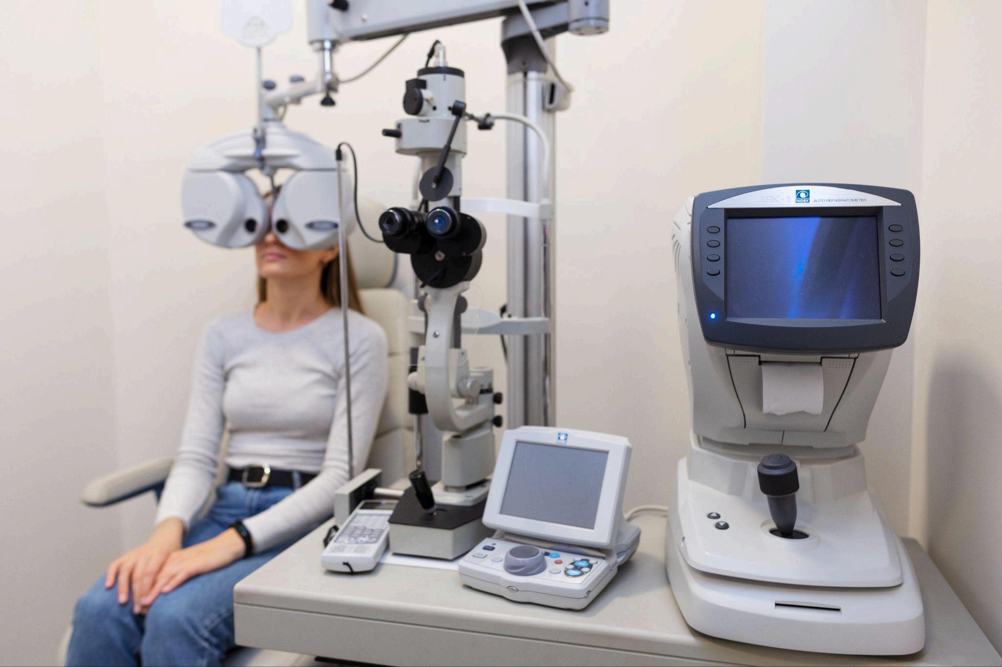
Early detection is the most effective way to prevent vision loss Many eye diseases can be managed successfully if identified in their initial stages. However, once significant damage occurs, the effects are often irreversible
Retinal imaging enables clinicians to detect abnormalities before symptoms appear This proactive approach allows for early treatment, which can slow or even halt disease progression.
For example, early detection of diabetic retinopathy can prevent severe complications, while identifying optic nerve changes early can help manage glaucoma effectively. Fundus imaging plays a vital role in detecting these conditions and guiding treatment decisions