Serum and Urinary Hormones During Pregnancy and the Peri- and Postpartum Period in an Asian Elephant (Elephas maximus)
Janine L. Brown and John Lehnhardt
Smithsonian Institution, National Zoological Park, Conservation and Research Center, Front Royal, Virginia (J.L.B.) and Department of Mammals, Washington, DC (J.L.)
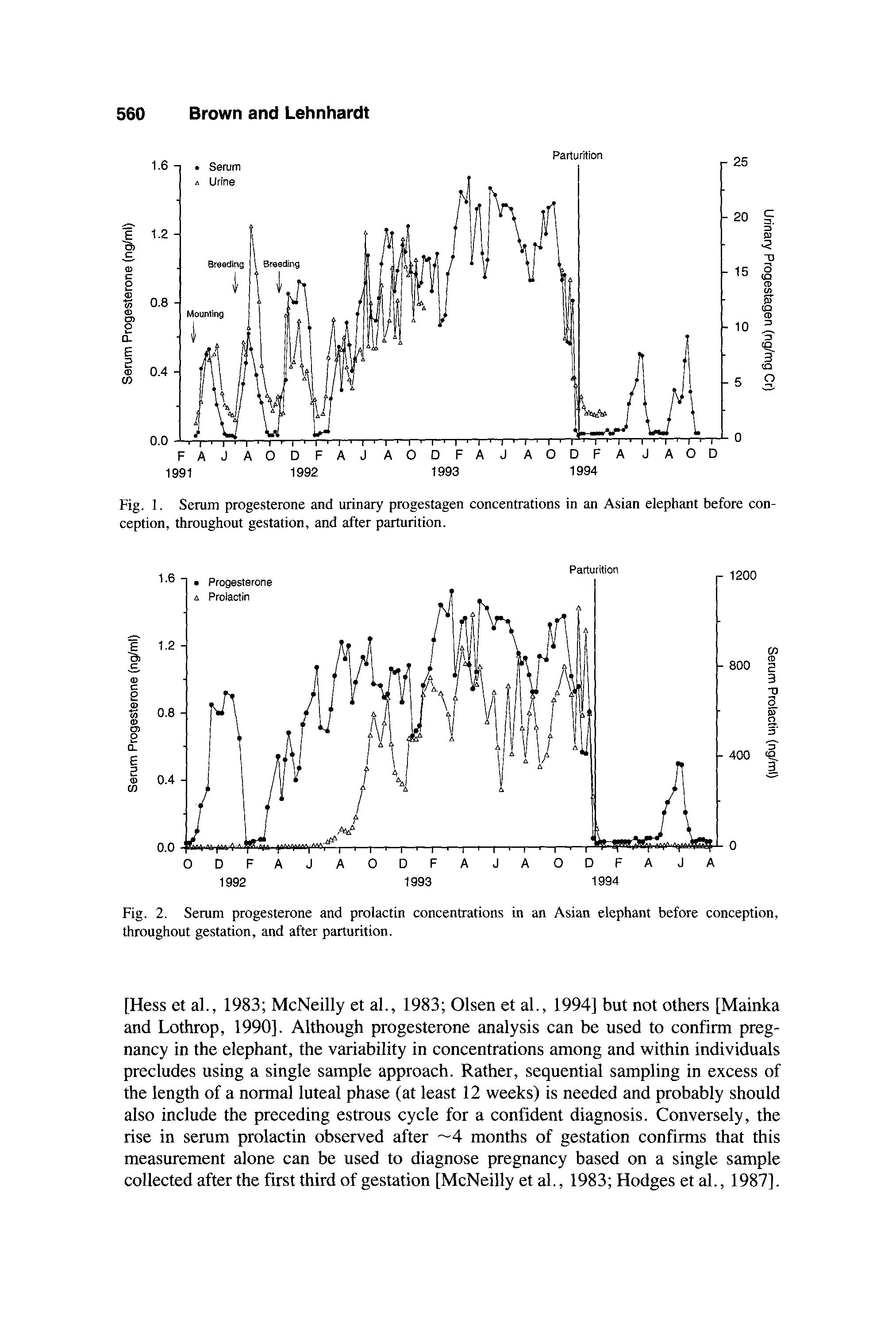
Blood and urine samples were collected weekly from an Asian elephant (Elephas maximus) for 10 months before conception, throughout pregnancy, and for 10 months after parturition. Additional daily samples were collected for 41 days before through 10 days after parturition to define endocrine events during the peripartum period. During gestation, serum progesterone concentrations increased gradually and, after -13 weeks, were higher (P < 0.05) than those observed during the nonpregnant luteal phase. Concentrations peaked at -12 months of gestation, gradually declined during the last month, and then decreased sharply to nondetectable levels 2 days before parturition. A 12 week lactational anestrus was observed before cyclicity resumed. The urinary profile of progestagen excretion paralleled that of circulating progesterone (r = 0.79; P < 0.05); however, radioimmunoassay of HPLC-separated fractions of urinary eluates indicated that this immunoactivity was not associated with native progesterone. After remaining basal through the first 16 weeks of gestation, serum prolactin concentrations increased to 100-fold about midterm and remained elevated until after parturition. Neither serum nor urinary cortisol concentrations were altered during pregnancy, but both increased markedly the day after parturition and remained elevated above prepartum levels for several weeks thereafter. These data indicate that analysis of serum prolactin can confirm pregnancy in the Asian elephant after -4 months of gestation and that daily monitoring of serum or urinary progestagens is useful for predicting parturition. 0 1995 Wiley-Liss, Inc.*
Key words: parturition, progesterone, cortisol, prolactin
Received for publication January 23, 1995; revision accepted April 21, 1995.
Address reprint requests to Janine L. Brown, Conservation and Research Center, 1500 Remount Road, Front Royal, VA 22630.
0 1995 Wiley-Liss, Inc. *This article is a US Government work and, as such, is in the public domain in the United States of America.
556 Brown and Lehnhardt
INTRODUCTION
The Asian elephant (Elephas maximus) has endured a relationship with man for more than 4,000 years when it was first captured for domestication. However, while once occupying vast areas of Asia, its distribution over the Indian subcontinent and Southeast Asia is now restricted largely to forested areas that have not yet been overtaken by human settlements [Olivier, 1978; Sukumar, 19891. Today, Asian elephants are used extensively in the timber industries of Myanmar, Thailand, Nepal, and India as well as for ceremonial and religious purposes and ecotourism. Because of poor breeding success, however, these domestic stocks are generally replenished by capturing wild elephants, which ultimately contributes to their declining numbers [Sukumar, 19891. In North America, fewer than 20% of the Asian elephants have produced offspring, due in large part to the physical, economical, and logistical problems associated with transporting elephants between institutions for breeding and the danger and difficulty of maintaining bulls in captivity [Balke et al., 19881. In the future, acquisition of replacement elephants from wild populations likely will be restricted, thus emphasizing the need to improve natural breeding as well as develop assisted reproduction techniques to maintain stable, genetically healthy populations. Developing controlled breeding programs for elephants depends on a thorough knowledge of reproductive physiology, including data on reproductive cycles, pregnancy, and parturition. While an extensive database now exists on hormonal patterns during the estrous cycle in Asian elephants [Hess et a]., 1983; Hodges et al., 1987; Plotka et al., 1988; Mainka and Lothrop, 1990; Brown et al., 1991; Taya et al., 1991; Niemuller et a]., 1993; Olsen et al., 19941, data on endocrine changes during pregnancy are limited and based on fewer animals [Hess et al., 1983; Hodges et al., 1987; Mainka and Lothrop, 1990; Olsen et al., 19941. Using endocrine assessments to diagnose pregnancy and predict parturition could greatly facilitate the captive management of this species. In the present study, we evaluated the secretory dynamics of progesterone, prolactin, and cortisol throughout gestation and the postpartum period in an Asian elephant. In addition, daily blood and urine samples were analyzed before and after parturition to provide a more complete characterization of the endocrine events surrounding the peripartum period.
MATERIALS AND METHODS
Animal Husbandry and Sample Collection
The female used in this study was orphaned at several months of age in 1976 and hand-raised at the Pinnewela Elephant Orphanage in Sri Lanka. She was acquired by the National Zoological Park (NZP) in 1977. At study onset, she was housed in a herd comprised of one African and two other Asian females and had access to an indoor/outdoor enclosure at all times. Animals were fed a pelleted supplement and fresh fruits and vegetables (bananas, apples, carrots) three times daily, with mixed grass hay and water available at all times. All elephants were trained and maintained in a free-contact management system with extensive keeper interaction.
On 13 March 1991, the female was transported to the Burnet Park Zoo (Syracuse, NY), which maintains one adult Asian bull (20 years of age) and three Asian cows. A male calf was born into the group in July 1991- Three days after arrival, the cow was introduced to the bull, who showed immediate interest and attempted several
Pregnancy Hormones in an Asian Elephant 557 mountings. Subsequently, the bull was given daily access to all cows but was housed separately at night. Breeding activity between the bull and NZP female was observed on 11 July and 2 November 1991. An unobserved conceptive mating occurred during the third week of February 1991, and, after 9 months of gestation, the cow was transported back to the NZP, where she immediately rejoined the herd. At 2148 h on 14 December 1993 she gave birth to a 120 kg female calf -3 h following amnionic membrane rupture. Observable discomfort due to onset of heavy contractions occurred - 1.5 h before birth. Although the fetal membranes were passed along with the calf at birth, the placenta was retained and milk production was reduced (as evidenced by poor initial growth performance by the calf). The cow was subsequently treated multiple times with oxytocin (20 IU i.m. twice on day 1; 2-4 injections of 40-240 IU/day i.v. or i.m. on days 7-18 postpartum) and once with estradiol cypionate (10 mg i.m. on day 16 postpartum) before the bulk of placental tissue was expelled 25 days postpartum.
Blood samples (10 ml) were collected from a vein on the caudal aspect of the downside ear using a 22 gauge vacutainer needle and tube while the cow was in lateral recumbency. Samples were collected weekly from the time of arrival at Burnet Park, throughout gestation, and for 10 months after parturition. Additional blood samples were collected daily for 41 days prior to parturition through 9 days postpartum (3 November-23 December 1993). Weekly urine samples (-5 ml) were collected by midstream catch until 41 days before parturition when collection frequency was increased to daily. After parturition, urine samples were obtained daily for 7 days and then weekly for -2.5 months. Unfortunately, urine samples collected between 8 November 1992 and 14 November 1993 were inadvertently destroyed before analysis. In general, all blood and urine samples were collected in the morning between 0800 and 1200 h. Blood samples were centrifuged (500g) within 2 h of collection and all sera and urine stored at -20°C until analyzed.
Radioimmunoassays
Progesterone in serum and unextracted urine was measured using a solid-phase I radioimmunoassay (RIA) kit (Coat-A-Count; Diagnostic Products Corporation, Los Angeles, CA). The antisera crossreacts 100% with progesterone, 43.8% with 5a-pregnane-3P-ol-2O-one, 31.1% with SP-pregnane-3a-ol-20-one, 23.3% with 5P-pregnane-3p-ol-20-one, 16% with 5a-pregnane-3a-ol-20-one, 3.5% with 5ppregnane-3P,20a-diol, 2% with 20a-dihydroprogesterone, 1.3% with SP-pregnane3,20-dione, and < 1% with 5P-pregnane-3a, 17a-dio1, 5a-pregnane-3a,20au-diol, 5a-pregnane-3P,20a-diol717a-hydroxyprogesterone and 5p-pregnane-3a 17, 20atrio1 [Wasser et al., 1994; kit literature]. Serial dilutions of serum and urine pools were parallel to the standard curve. Addition of 0.10, 0.50, 2.0, 10.0, 20.0, and 40 .O ng/ml progesterone resulted in a net recovery of 0.09, 0.38, 2.13, 10.9, 24.7, and 43.0 ng/ml (y = 1.09~+ 0.26; r = 0.99) from serum and 0.11, 0.45, 2.08, 10.5, 19.6, and 41.2 ngiml (y = 1.02~- 0.06; r = 0.99) from urine. Assay sensitivity was 0.03 ng/ml, and the intra- and interassay coefficients of variation were <lo%.
Serum prolactin was measured by heterologous RIA using an anti-human prolactin antisera (NIDDK-anti-hPRL-3) and ovine prolactin (NIDDK-oPRL-1-2) label and standards. The assay was incubated at room temperature for 2 days in a total
558 Brown and Lehnhardt
volume of 500 (11. Standards (100 pl; 0.039-20 @tube) and/or sample (10-100 (11) were brought up to 300 p1 with phosphate buffer (0.01M PO,, 0.5% BSA, 2 mM EDTA, 0.9% NaC1, 0.01% thimerosal, pH 7.4) and incubated with antibody (1: 25,000 in 100 11.1) for 3 h followed by the addition of '251-prolactin (20,000 cpm in 100 11.1). On day 2, antibody-bound complexes were precipitated by incubation for 1 h with sheep anti-rabbit gamma globulin (1:200 in 1 ml containing 5% polyethylene glycol) and centrifugation for 30 min at 2,500g. The antibody generally bound 2025% of the I2'I-prolactin with 3% nonspecific binding. Serial dilutions of elephant serum pools were parallel to the standard curve. Upon addition of 0.16, 0.31, 0.63, 1.25,2.5, and 5 .O ng ovine prolactin, 0.17,0.29,0.66, 1.17,2.68, and 4.97 ng were recovered after subtraction of endogenous hormone (y = 1.OOx - 0.01; r = 0.99).
Assay sensitivity was 0.05 ng/tube, and the intra- and interassay coefficients of variation were <10%.
Cortisol in serum and urinary extracts was measured using a solid-phase "'1 RIA kit (Diagnostic Products Corporation) [Brown et al., this issue]. For extraction of cortisol, urine (0.5 ml) was combined with 1 ml dichloromethane in a 12 X 75 mm polypropylene tube, capped tightly, shaken gently for 10 min, and centrifuged (1,500g) for 5 min. The lower dichloromethane fraction was removed by aspiration using a Pasteur pipet and placed into a glass 12 X 75 mm tube. Six hundred microliters were then transferred to another glass 12 X 75 mm tube using a positive displacement pipet and dried under air. Samples were reconstituted in 150 p1 phosphate buffer (0.01 M PO,, 0.14 M NaCl, 0.01% merthiolate, pH 7.4), vortexed for 1 min, and stored frozen (-20°C) until analyzed. Extraction efficiency of 3H-cortisol (2,500 dpm) added to pooled urine samples was >90%. Serial dilutions of elephant serum and urinary extract pools were parallel to the standard curve. Addition of 10, 50, 100, and 200 ng/ml cortisol resulted in a recovery of 10.2,48.7, 103.1, and 203.6 ng/ml (y = 1.02~- 0.66; r = 0.99) from serum and 9.8, 51.6, 97.3, and 199.4 ng/ml (y = 0.99~+ 0.15; r = 0.99) from urine, after subtraction of endogenous hormone. Assay sensitivity was 2.5 ng/ml, and the intra- and interassay coefficients of variation were <lo%.
Urinary cortisol and progesterone concentrations were indexed by creatinine (Cr) concentrations (samples ranged from 0.15-0.87 mg Cr/ml) to account for fluctuations in fluid intake [Taussky, 19541.
High-Performance Liquid Chromatography
Reverse-phase high-performance liquid chromatography (HPLC) (Microsorb C-18 Column; Rainen Inc., Woburn, MA) was used to determine the nature of progestagen immunoactivity in unextracted elephant urine samples. Before HPLC, samples were filtered through a C-18 matrix column (Spice Cartridge; Rainen, Inc.) and eluted with 5 ml of 80% methanol to remove contaminants. Progestagen metabolites were separated using a gradient of 20-32% over 15 min, increasing to 50% over 50 min and then to 100% over 55 min. One milliliter fractions were collected over a 120 min period (1 ml/min flow rate). The elution profile of 3H-progesterone was determined in a separate run. HPLC fractions of urine eluates were taken to dryness and reconstituted in phosphate buffer, and progestagen immunoactivity was quantified by RIA.
Pregnancy Hormones in an Asian Elephant 559
Statistics
The nonluteal phase of the estrous cycle was defined as the period when serum progesterone concentrations were <0.05 ng/d for more than 2 consecutive weeks. Conversely, the luteal phase consisted of that portion of the cycle where progesterone concentrations were continuously elevated above 0.05 ng/ml. During gestation, serum progesterone and prolactin concentrations were averaged at 12 week intervals and compared to nonpregnant luteal phase concentrations using Dunnett’s procedure. Weekly concentrations of prolactin and cortisol also were considered significantly elevated above baseline when values exceeded the prepartum mean plus three standard deviations.
RESULTS
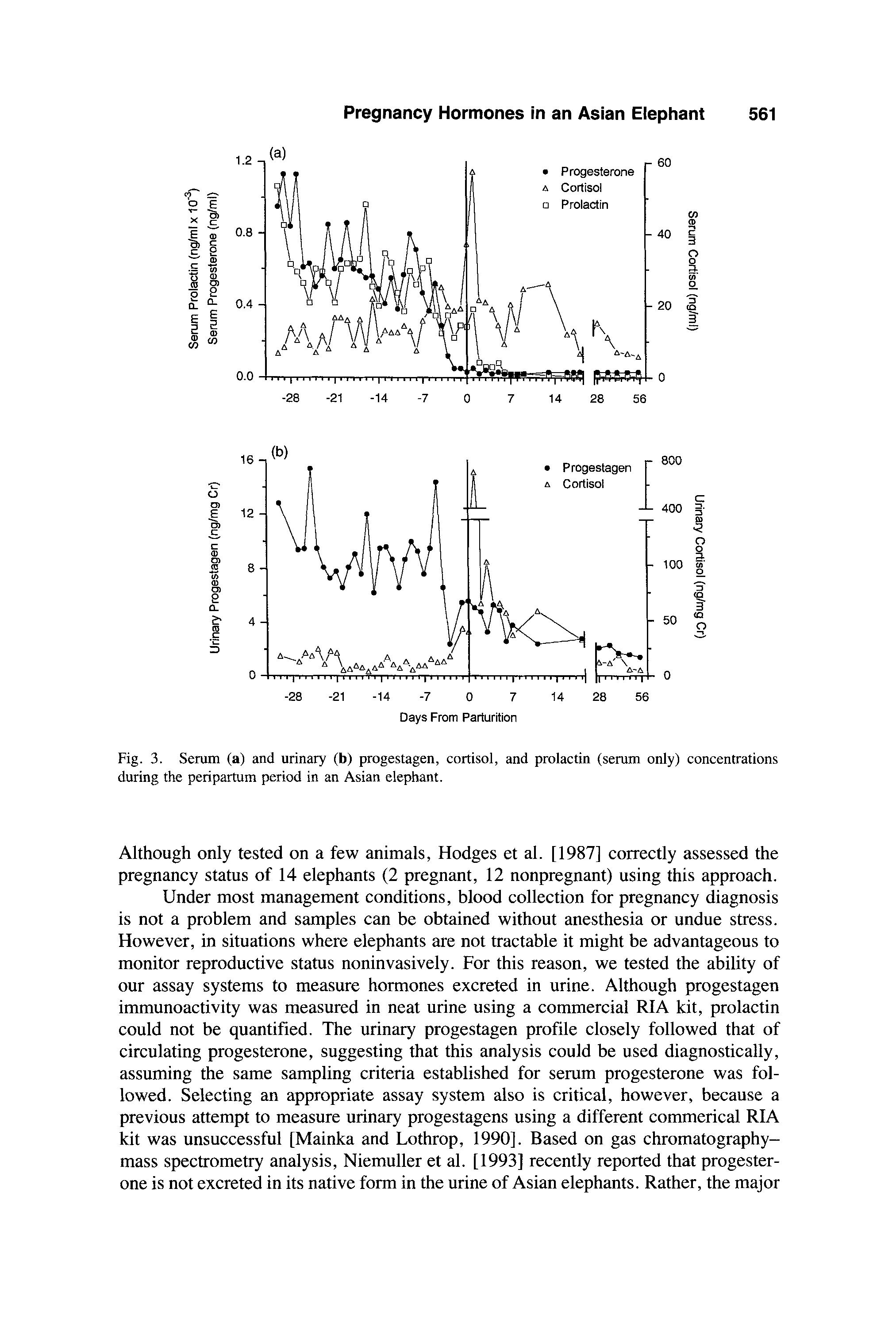
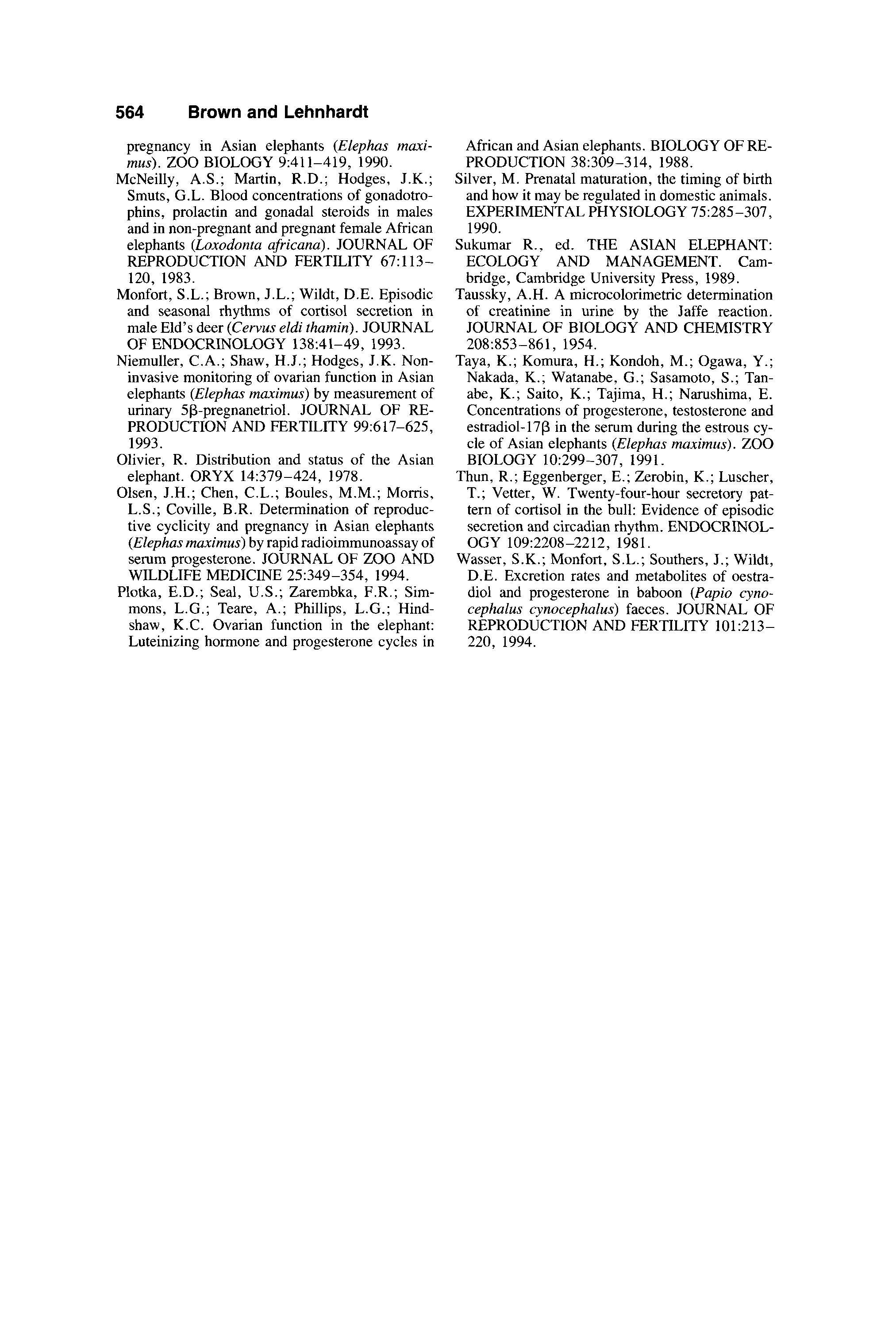
Based on serum progesterone profiles, three estrous cycles were monitored before conception and two after parturition with a mean cycle length of 16.0 t 0.5 weeks and a luteal phase length of 11.3 +_ 0.7 weeks (Fig. 1). Breeding activity was observed during the late follicular phase immediately preceding the luteal phase rise in progesterone. Serum progesterone concentrations increased gradually throughout gestation, peaked after -12 months, and then started to decline -1 month before parturition (Figs. 1,2). Serum progesterone concentrations decreased precipitously to 20% of peak concentrations 4 days before, 10% 2 days before, and to nondetectable levels 2 days before parturition (Fig. 3). After birth, a 12 week lactational anestrus was observed before estrous cyclicity resumed (Fig. 1). When evaluated at 12 week intervals, mean serum progesterone concentrations were higher (P <0.05) after 13 weeks of gestation than those observed during the nonpregnant luteal phase. The pattern of urinary progestagen excretion parelleled that of circulating progesterone and concentrations were correlated when matched on a weekly basis (r = 0.79; P <0.05) (Fig. 1). Radioimmunoassay of HPLC-separated fractions of urinary eluates determined that the majority (>95%) of this immunoactivity was detected in fractions 12-17 and was not associated with 3H-progesterone which eluted in fractions 60-68.
Serum prolactin secretion was stable throughout the estrous cycle, averaging 6.1 & 0.8 ng/ml. Concentrations remained at baseline throughout the first 16 weeks of gestation, then increased markedly, and peaked before midterm (Fig. 2). In contrast to progesterone, serum prolactin concentrations were still slightly elevated at parturition and remained 2.5- to tenfold higher than baseline throughout the first week before declining at -10 days postpartum (Fig. 3).
Basal cortisol concentrations in serum and urine during the 10 months before conception were stable, averaging 13.8 k 0.9 ng/ml and 12.5 * 1.1 ng/ml Cr, respectively, and neither was affected by pregnancy. However, serum and urinary cortisol concentrations both increased approximately threefold over baseline prior to delivery and five- and sixtyfold the day after parturition, respectively (Fig. 3). Serum and urinary cortisol concentrations fluctuated thereafter but generally remained elevated above prepartum levels for several weeks after birth (Fig. 3).
DISCUSSION
Serum progesterone concentrations during gestation were significantly greater than luteal phase levels after the first several months, a finding noted in some studies
560 Brown and Lehnhardt
Fig. 1. Serum progesterone and urinary progestagen concentrations in an Asian elephant before conception, throughout gestation, and after parturition.
Fig. 2. throughout gestation, and after parturition. Serum progesterone and prolactin concentrations in an Asian elephant before conception, [Hess et al., 1983; McNeilly et al., 1983; Olsen et al., 19941 but not others [Mainka and Lothrop, 19901. Although progesterone analysis can be used to confirm pregnancy in the elephant, the variability in concentrations among and within individuals precludes using a single sample approach. Rather, sequential sampling in excess of the length of a normal luteal phase (at least 12 weeks) is needed and probably should also include the preceding estrous cycle for a confident diagnosis. Conversely, the rise in serum prolactin observed after -4 months of gestation confirms that this measurement alone can be used to diagnose pregnancy based on a single sample collected after the first third of gestation [McNeilly et al., 1983; Hodges et al., 19871.
Pregnancy Hormones in an Asian Elephant 561
Fig. 3. during the penparturn period in an Asian elephant.
Serum (a) and urinary (b) progestagen, cortisol, and prolactin (serum only) concentrations
Although only tested on a few animals, Hodges et al. [1987] correctly assessed the pregnancy status of 14 elephants (2 pregnant, 12 nonpregnant) using this approach, Under most management conditions, blood collection for pregnancy diagnosis is not a problem and samples can be obtained without anesthesia or undue stress. However, in situations where elephants are not tractable it might be advantageous to monitor reproductive status noninvasively. For this reason, we tested the ability of our assay systems to measure hormones excreted in urine. Although progestagen immunoactivity was measured in neat urine using a commercial RIA kit, prolactin could not be quantified. The urinary progestagen profile closely followed that of circulating progesterone, suggesting that this analysis could be used diagnostically, assuming the same sampling criteria established for serum progesterone was followed. Selecting an appropriate assay system also is critical, however, because a previous attempt to measure urinary progestagens using a different commerical RIA kit was unsuccessful [Mainka and Lothrop, 19901. Based on gas chromatographymass spectrometry analysis, Niemuller et al. [ 19931 recently reported that progesterone is not excreted in its native form in the urine of Asian elephants. Rather, the major
562 Brown and Lehnhardt
progesterone metabolites consist of a and p pregnanetriols. Using HPLC, we confirmed that the immunoactivity in elephant urine was not progesterone; therefore, the antibody supplied in the Diagnostic Products kit apparently crossreacts with one or more of the progesterone metabolites.
The 12 week postpartum period noted in this individual before resumption of estrous cyclicity was short compared to the average 45-46 week lactational anestrus reported by Olsen et al. [1994] for six Asian females. Problems associated with a retained placenta, including reduced milk production, may have contributed to this finding. In other mammalian species, lactation inhibits reproduction, and weaning can reduce the postpartum anestrous period [Jainudeen and Hafez, 19801. In Asian elephants, a female whose calf died 12 h after birth resumed cycling within 10.5 weeks, whereas another female whose calf was bottle fed began cycling 8.5 weeks postpartum [Olsen et al., 19941.
Another objective of this study was to determine precisely if a prepartum decrease in progesterone signaled impending parturition. In this cow, serum progesterone concentrations declined to nondetectable levels 2 days before parturition. Thus, although management of pregnant elephants generally includes 24 h observations during the last few weeks of gestation, knowing that parturition will occur within a few days of progesterone decline assures that conditions are optimized for delivery. Urinary progestagens also decreased before parturition, although the response was not as dramatic. Nevertheless, in situations where daily blood sampling is not possible, noninvasive monitoring of excreted progestagens could provide a practical alternative.
In many mammalian species, the timing of parturition is believed to be initiated by the fetus through maturation of the hypothalamic-pituitary-adrenal axis and a corresponding increase in circulating cortisol [Silver, 1990; Ginther, 19921. Generally, increased cortisol secretion by the fetal adrenals occurs during the last few days of gestation and can be detected in the maternal circulation. To our knowledge, no reports describing cortisol patterns during pregnancy or parturition are available for the elephant. In this study, cortisol secretion was generally stable throughout gestation. On the morning of parturition, however, a threefold increase in serum cortisol was observed, followed by a fivefold increase the next morning. The response in urine was even greater, with a threefold increase observed just before parturition, followed by a sixtyfold increase the day after parturition. The magnitude difference in postpartum response between blood and urine likely is due to the dynamics of cortisol in circulation. Cortisol is secreted in a pulsatile fashion in many species [Fulkerson and Tang, 1979; Thun et al., 1981; Brown et al., 1993; Monfort et al., 19931, and accurate assessment of circulating activity can only be made by collecting multiple blood samples over time. Alternatively, analyzing cortisol excreted in urine provides a measure of adrenal status over a period of hours rather than minutes. We recently demonstrated in Asian and African elephants that urinary cortisol increases within 30 min of a rise in serum after adrenocorticotropin injection [Brown et al., this issue], thereby providing an approximately real-time evaluation. The moderate increase in cortisol observed the morning of parturition suggests that this steroid might be involved in the initiation of parturition. However, it is also possible that it reflected an increase in adrenal activity (i.e., stress), as the cow appeared nervous prior to delivery. The large surge in cortisol activity after parturition probably was due to increased stress, as she became extremely agitated and aggressively attacked her calf
Pregnancy Hormones in an Asian Elephant 563 within minutes of delivery. Although she calmed down shortly thereafter and became very attentive of her offspring, she and the other elephants in the herd remained noticeably uneasy for several weeks after the birth. In addition, the cow was subjected to numerous medical procedures in an attempt to expel a retained placenta which eventually occurred 25 days postpartum. During this time, serum and urinary cortisol were both elevated above baseline. Taken together, the results of this and our previous [Brown et al., this issue] study suggest that cortisol analysis is capable of assessing acute and chronic changes in adrenal activity in the elephant. The evaluation of cortisol in urine appears to be a better indicator of this activity, probably because the pooling of urine over time dampens minor fluctuations in cortisol secretion and improves the ability to distinguish between normal pulsatile dynamics and genuine physiological responses to stressful events.
CONCLUSIONS
1. Increased serum prolactin secretion can be used to confirm pregnancy in the
2. Daily monitoring of serum or urinary progesterone is useful for predicting
3. Elevated cortisol concentrations in serum and urine after birth suggest adrenal Asian elephant after -4 months of gestation. parturition. activity was increased during the postpartum period in this cow.
ACKNOWLEDGMENTS
The authors thank Dr. Carolee Wallace at the Burnet Park Zoo and the elephant keepers at the NZP (Marie Galloway, Liz Hile, Cindy Sparks, and Debbie Flynn) for providing excellent animal care and collecting samples. This project was supported by the Friends of the National Zoo and the NZP Mammology Department Lenox Elephant Sculpture Fund.
REFERENCES
Balke, J.M.E.; Barker, I.K.; Hackenberger, M.K.; McManamon, R.; Boever, W.J. Reproductive anatomy of three nulliparous female Asian elephants: The development of artificial insemination techniques. ZOO BIOLOGY 7:99113, 1988.
Brown, J.L.; Citino, S.B.; Bush, M.; Lehnhardt, J.; Phillips, L.G. Cyclic patterns of luteinizing hormone, follicle-stimulating hormone, inhibin, and progesterone secretion in the Asian elephant (Elephas maximus). JOURNAL OF ZOO AND
OGY 81:135-141, 1979.
dian rhythms in the plasma concentration of cortisol in sheep. JOURNAL OF ENDOCRINOLGinther, J.O., ed. REPRODUCTIVE BIOLOGY
PECTS, 2nd ed. Cross Plains, Wisconsin, Equiservices, 1992.
Hess, D.L.; Schmidt, A.M.; Schmidt, M.J. Reproductive cycle of the Asian elephant (Elephas maximus) in captivity. BIOLOGY OF REPRO-
Brown, J.L.; Schoenemann, H.M.; Chakraborty, P.K. Basal and pulsatile cortisol secretion in sheep after surgical manipulation. JOURNAL OF ANIMAL SCIENCE 71 (Suppl. 1):234, 1993.
Brown, J.L.; Wemmer, C.M. ; Lehnhardt, J. Urinary cortisol analysis for monitoring adrenal activity in elephants. ZOO BIOLOGY, (this issue). Fulkerson, W.J.; Tang, B.Y. Ultradian and circaWILDLIFE MEDICINE 22:49-57, 1991.
OF THE MARE, BASIC AND APPLIED ASDUCTION 28:767-773, 1983. YEARBOOK 26:285-289, 1987.
Hodges, J.K.; McNeilly, A.S.; Hess, D.L. Circulating hormones during pregnancy in the Asian and African elephants. INTERNATIONAL ZOO Jainudeen, M.R.; Hafez, E.S.E. Reproductive failure in females. Pp. 449-470 in REPRODUCTION IN FARM ANIMALS, 4th ed. E.S.E. Hafez, ed. Philadelphia, Lea and Febiger, 1980. Mainka, S.A.; Lothrop, C.D. Reproductive and hormonal changes during the estrous cycle and
564 Brown and Lehnhardt pregnancy in Asian elephants (Elephas maximus). ZOO BIOLOGY 9:411-419, 1990.
African and Asian elephants. BIOLOGY OF REPRODUCTION 38:309-314, 1988.
Silver, M. Prenatal maturation, the timing of birth and how it may be regulated in domestic animals. EXPERIMENTAL PHYSIOLOGY 75:285-307, 1990.
REPRODUCTION AND FERTILITY 67: 113-
McNeilly, A.S.; Martin, R.D.; Hodges, J.K.; Smuts, G.L. Blood concentrations of gonadotrophins, prolactin and gonadal steroids in males and in non-pregnant and pregnant female African elephants (Loxodontu africana). JOURNAL OF 120, 1983.
Monfort, S.L.; Brown, J.L.; Wildt, D.E. Episodic and seasonal rhythms of cortisol secretion in male Eld’s deer (Cervus eldi thamin). JOURNAL
OF ENDOCRINOLOGY 138:41-49, 1993.
Sukumar R., ed. THE ASIAN ELEPHANT: ECOLOGY AND MANAGEMENT. Cambridge, Cambridge University Press, 1989.
Taussky, A.H. A microcolorimetric determination of creatinine in urine by the Jaffe reaction. JOURNAL OF BIOLOGY AND CHEMISTRY 208:853-861, 1954.
Niemuller, C.A.; Shaw, H.J.; Hodges, J.K. Noninvasive monitoring of ovarian function in Asian elephants (Elephas maximus) by measurement of urinary 5P-pregnanetriol. JOURNAL OF RE1993.
PRODUCTION AND FERTILITY 99:617-625,
Olivier, R. Distribution and status of the Asian elephant. ORYX 14:379-424, 1978.
Olsen, J.H.; Chen, C.L.; Boules, M.M.; Morris, L.S.; Coville, B.R. Determination of reproductive cyclicity and pregnancy in Asian elephants (Elephas maximus) by rapid radioimmunoassay of serum progesterone. JOURNAL OF ZOO AND
WILDLIFE MEDICINE 251349-354, 1994.
Plotka, E.D.; Seal, U.S.; Zarembka, F.R.; Simmons, L.G.; Teare, A,; Phillips, L.G.; Hindshaw, K.C. Ovarian function in the elephant: Luteinizing hormone and progesterone cycles in
Taya, K.; Komura, H.; Kondoh, M.; Ogawa, Y.; Nakada, K.; Watanabe, G.; Sasamoto, S.; Tanabe, K.; Saito, K.; Tajima, H.; Narushima, E. Concentrations of progesterone, testosterone and estradiol-17P in the serum during the estrous cycle of Asian elephants (Elephas muximus). ZOO
BIOLOGY 10:299-307, 1991.
Thun, R.; Eggenberger, E.; Zerobin, K.; Luscher, T.; Vetter, W. Twenty-four-hour secretory pattern of cortisol in the bull: Evidence of episodic secretion and circadian rhythm. ENDOCRINOLOGY 109:2208-2212, 1981.
Wasser, S.K.; Monfort, S.L.; Southers, J.; Wildt, D.E. Excretion rates and metabolites of oestradiol and progesterone in baboon (Pupio cynocephalus cynocephulus) faeces. JOURNAL OF REPRODUCTION AND FERTILITY 101:2 13220, 1994.