16 minute read
ARTIFICIAL INTELLIGENCE IN GENOMIC MEDICINE AND DIAGNOSTICS
from DC QUANTUM
by MahaNawaz
Artificial Intelligence (AI) is a rapidly growing field in computer science that is slowly increasing its influence over diagnostics and genomic medicine. Especially: Machine Learning (ML) and Deep Learning (DL), which are:
Offering new approaches to streamline key problems in Genomic Medicine.
Advertisement
PHG Foundation
Although some ML algorithms have already been written to analyze key problems in Genomic Medicine (GM) and Genomic Diagnostics (GD), and even been used for basic testing for many years now, a surge in High-Performance Computing and Biomedical Datasets (BD) have meant that AI’s relevance in Diagnostics has been slowly growing. This article will analyze the role of AI in GD, and cover the different types of AI that can be applied in this field while looking at some case studies.
The study of Genomics
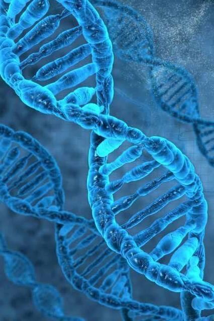
Genomics is the study of the entire genetic material of an organism [1]. Genomic medicine makes use of an individual’s genomic information to guide their clinical care and deliver more personalized strategies for diagnostic or therapeutic decision making.
The growing research in Genomic Medicine has started to allow for more personalized treatment, better palliative care, and even lead to more accurate diagnostics. Some areas of key interest in Genomics are the analysis of Phenotype Data to better aid congenital conditions, and modeling the effects of certain genes on biological processes [2].
Artificial Intelligence
Since Genomics requires a lot of data analysis, there is now a growing need for quick and accurate computational intelligence that can tackle the analysis of these large multidimensional datasets.
In this case, multidimensional refers to datasets that have many different attributes. Like how a facial feature is the result of thousands of smaller measurements like the genes in a cell.
This is why the field of Artificial intelligence is quickly gaining traction in Genomic Medicine.
Applications of Artificial intelligence in Genomics
Most aspects of GM and Diagnostics have been brushed by AI in some form. These programs can be classified as ‘narrow’ as they cover only a certain aspect of GM / GD, however, with increased development in these ‘narrow’ algorithms, they can prove an indispensable tool for physicians.
The fields of AI that are looked at most for GM are Computer Vision and Phenotype Analysis Algorithms, and AI is also being implemented in Variant Calling, which is a method used to identify ‘variants’ in a patient’s genome compared to the average person. These methods can allow for the early identification of diseases like Cancer and other Rare Genetic Diseases (RGD).
Phenotyping
In a clinical setting, phenotyping is the process of examining and reporting a patient’s physical features. This can be things like the diameter of a patient’s pupil, or the curvature of their spine, however, facial features tend to be used more often. The information gathered from phenotyping can then be surveyed by ML algorithms to create more refined BDs. Phenotype data consists mostly of genomic factors with the environment playing a moderate factor. This makes it hard for standard forms of phenotyping to separate the genomic factors from environmental factors [3].
Many Deep Learning algorithms are surfacing that support image recognition for rare disease phenotyping and they are showing great promise.
Computer Vision
Computer vision is a generic field in AI that tackles analyzing images and video. These algorithms can — with enough training data — learn to recognize almost anything, from the silhouette of a dog to the facial features that hint at down syndrome. Tying back to phenotyping, computer vision can allow us to analyze images of physical features to form suggestions and ideas on the kinds of conditions that patients might have.
Rare genetic diseases have been found to include minor facial patterns, and most syndromes havedirect phenotypes. Computer Vision algorithms can be trained — with extensive datasets to detect these phenotypes, and effectively pre-diagnose patients, aiding towards the effort to move towards preventive medicine, rather than reactive medicine.
Face2gene
A rapidly growing technology — DeepGesalt — is slowly gaining traction in Phenotype-Genotype AI. It uses information from the phenotype and genotype data of 1000s of patients to suggest genetic syndromes that a patient might have, that would otherwise be hard and time-consuming to discern. When this algorithm was tested with two external BDs the syndrome that a patient had, appeared in the top 10 of the computer’s suggestions, 90% of the time [4].
Algorithms Like Face2Gene are Becoming More Prevalent
Again, this is no way near the accuracy that can even be potentially administered in a clinical setting, but it is programs like these that will slowly see improvement and growth in the many years to come.
DNA Sequencing
Sequencing and analyzing DNA has been an indispensable tool for medical research in diagnosing hereditary diseases and other rare conditions. Variant calling is a method used for DNA analysis and it — in simple terms — looks at a patient’s DNA and compares it to a large database to find ‘variants’ or differences in the patient’s Genome that might underlie disease.
There are already many tools and techniques established for variant calling, many new deep learning models are being developed with the goal of improving the accuracy and reliability of variant calls. One algorithm being developed — Google’s DeepVariant — has proved phenomenal, having outperformed many well-known methods of variant calling.
DeepVariant looks at variant calling from a slightly un-intuitive angle, image classification [5]. In short, it takes a DNA sequence and converts it into an image. It then compares this image to other images from different datasets to identify variants. This is effective because Deep Learning algorithms are particularly good at tackling problems that involve image recognition and classification.
Conclusion
AI has many vast and intricate aspects. Its vast types allow it to become an unforgettable part of many emerging fields in science. GM has always been quite a science-fiction type area of study. Being on the cutting edge of science has meant that new discoveries are commonplace. AI is providing a way to cope with this fast-paced field of study. Applications are wide, from DNA sequencing to PhenotypeGenotype analysis to many I have not covered in this article. It provides a way of accurately diagnosing and prediagnosing patients, that can allow for huge leaps in preventive medicine. This, however, is not without its fair share of hurdles. AI is heavily dependent on accurate BDs, some of which are extremely hard to find. And more importantly, AI is a black-box system. Even the people that write AI programs cannot fully see that AI’s thought process. This creates ethical questions…
Should a robot be able to diagnosepatients if the scientists themselves cannot understand how the algorithm came to a diagnosis? What if there are conflicting opinions between scientists and robots, who’s diagnosis holds more weight?

References
[1] Ruane J, Sonnino A, Agostini A. Bioenergy and the potential contribution of agricultural biotechnologies in developing countries. Biomass and Bioenergy [Internet]. 2010 Oct [cited 2022 Jun 26];34(10):1427–39. Available from: https://www.sciencedirect.com/topics/earth-andplanetary-sciences/genomics
[2]Frey L. Artificial Intelligence and Integrated Genotype–Phenotype Identification. Genes [Internet]. 2018 Dec 28 [cited 2022 Jun 26];10(1):18. Available from: https://www.mdpi.com/2073-4425/10/1/18/htm
[3]Li X, Guo T, Mu Q, Li X, Yu J. Genomic and environmental determinants and their interplay underlying phenotypic plasticity. Proceedings of the National Academy of Sciences [Internet]. 2018 Jun 11 [cited 2022 Jun 26];115(26):6679–84. Available from: https://www.ncbi.nlm.nih.gov/pmc/articles/PMC6042117/
[4]Gurovich Y, Hanani Y, Bar O, Nadav G, Fleischer N, Gelbman D, et al. Identifying facial phenotypes of genetic disorders using deep learning. Nature Medicine [Internet]. 2019 Jan;25(1):60–4. Available from: https://arxiv.org/pdf/1801.07637.pdf?
[5]DeepVariant Blog. DeepVariant Blog [Internet]. DeepVariant Blog. 2022 [cited 2022 Jun 26]. Available from: https://google.github.io/deepvariant/
The Evolution of Sulfonylureas as Hypoglycaemic Drugs over time, their Mechanisms and how they
Treat Symptoms of Type II Diabetes Mellitus.
Introduction
Type 2 diabetes mellitus can be a difficult disease to live with and can severely affect one’s quality of life. Diabetes mellitus is a chronic condition in which your body cannot regulate your blood glucose levels, the two main types being type 1 and type 2. These are due to either an inability to produce insulin (type 1) or when the insulin produced is ineffective (type 2). Type 2 diabetes, or non-insulin dependent diabetes mellitus, can occur as a result of lifestyle factors, such as diet and obesity. These lead to insulin resistance or the inability to produce enough insulin as necessary. Currently, there are 4.1 million people in the UK with diabetes, with 90% of these cases due to type 2 diabetes. It is estimated that 1 in 10 adults will develop type 2 diabetes by 2030 (Lacobucci, 2021)
One treatment for type 2 diabetes is the use of sulfonylureas - a group of oral drugs with hypoglycaemic effects (ability to lower blood glucose levels). Since their discovery in the 1940’s, medicinal chemists have changed the structure of these drugs, to make them more effective for clinical use. These modifications have led to more favourable properties in metabolism, potency, efficacy and safety, which have made the drugs a more effective, safe and convenient treatment for type 2 diabetes mellitus. These will be discussed later on in the article.
This article will explain the chemistry of sulfonylureas, the pharmacology behind them and how they have changed over time to make them more effective in the treatment of type 2 diabetes mellitus.
Type 2 Diabetes mellitus cause
Type 2 diabetes occurs when there is a deficiency in insulin secretion by the β-cells in the pancreas, or when cells develop a resistance to insulin action (Galicia-Garcia, et al., 2020) This is usually due to obesity and an unhealthy lifestyle, including lack of exercise, and a high fatty and sugar diet. Insulin is a peptide hormone that is secreted by β-cells in the pancreas. It is responsible for lowering blood glucose levels by stimulating the conversion of glucose in the blood into glycogen to be stored in muscle, fat, and liver cells. When there is a deficiency or resistance of insulin it leads to hyperglycaemia (high blood glucose levels), due to the reduced ability to convert glucose into glycogen. This would lead to symptoms such as vomiting, dehydration, confusion, increased thirst, and blurred vision to name a few.

Physiology behind insulin secretion and structure
To understand the pharmacology of the sulfonylurea compounds, one must first understand the physiology behind the secretion of insulin.
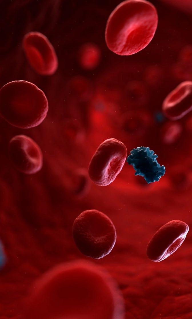
As stated above, insulin is a peptide hormone, so it is made from a polypeptide chain. Transcription of the insulin gene (found on chromosome 11) occurs and the resulting mRNA strands are translated to produce two peptide chains. These chains are held together in a quaternary structure by two disulfide bonds to form the hormone insulin (Brange & Langkjoer, 1993). Insulin secretion must be tightly controlled to maintain efficient glucose homeostasis. To do so, the secretion of insulin is regulated precisely to meet its demand. The β-cells of the pancreas contain glucose transporter 2, a carrier protein that allows facilitated diffusion of glucose molecules across a cell membrane. These transporters allow glucose to be detected and enter the β-cells. Upon cytoplasmic glucose levels rising, the pancreatic β-cells respond by increasing oxidative metabolism, leading to increased ATP in the cytoplasm (Fridlyand & Philipson, 2010). The ATP in the cytoplasm of the βcells, can bind to ATP sensitive K+ channels on the cell membrane, causing them to close. This leads to a build up of K+ ions within the cell as they are unable to leave the cell, leading to the depolarisation of the cell. The increasing positive membrane potential, leads to the opening of voltage gated Ca2+ channels, leading to an influx of Ca2+ ions. This further depolarises the cell, which triggers the release of insulin from the cell, packaged in secretory vesicles, by exocytosis (Fu, et al., 2013).
Pharmacology of sulfonylureas
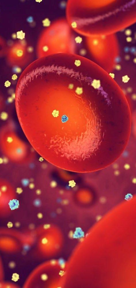
Sulfonylurea’s act inside the pancreatic β-cells On the ATP sensitive K+ channel, there are sulfonylurea receptors to which the drug binds, causing them to close. The cascade of events that follows leads to the release of insulin by the pancreatic β-cell. This mimics the activity that occurs when glucose is taken into the cell, as mentioned earlier. (Panten, et al., 1996). (possibly delete this instead as it is repeated)
This process allows more insulin to be released, to lower blood glucose levels when insufficient insulin is produced naturally. Sulfonylureas are only effective in type 2 diabetes, since insulin production is not impaired (as in type 1 diabetes), rather the release of or resistance to insulin is affected.
Common chemistry of all sulfonylureas
All the sulfonylurea drugs are characterised by their common sulfonylurea group. This functional group allows this unique group of compounds to bind to SUR on ATP sensitive K+ channels, giving it its hypoglycaemic properties. The common structure of sulfonylureas is shown in figure 1 (Fvasconcellos, 2011), with the blue R groups indicating replaceable side chains, which fluctuates between each drug development over time giving slightly different properties between the drugs. Over time, scientists have improved the drugs efficacy by changing the side compounds. Additionally, scientific research has led to development of other drugs from the same pharmacological group, but with altered side chains (again, giving them different properties) which have also improved the efficacy of the drug. These changes have altered properties of the drug such as potency, metabolism, half-life, tolerance and safety, to make the drug more effective for clinical use.
History and development of the drugs and their chemical structure
Sulfanilamide and IPTD
In 1935, a French research term discovered the active chemical in the antibiotic prontosil, known as sulfanilamide (Sorkhy & Ghemrawi, 2020). Sulfanilamide was found to be a poor antibiotic and so derivatives of it were synthesised and tested. These compounds, such as such as p-aminosulfonamide-isopropylthiodiazole (IPTD), which was used as an antibiotic for the treatment of typhoid in 1942, revealed unexpected hypoglycaemic side effects. These were discovered by French physician, Marcel Janbon (Quianzon & Cheikh, 2012) However, scientists could not identify how these side effects were caused.
In 1946, Auguste Loubatières, investigated the effect of IPTD on dogs. He administrated the drug to fully pancreatectomized and partially pancreatectomized dogs and found that the drug was ineffective in the fully pancreatectomized ones but effective in the partially pancreatectomized ones. This later lead to his conclusion that the drugs’ hypoglycaemic property was due to its ability to stimulate insulin secretion directly in the pancreatic β-cells (Loubatières-Mariani, 2007).
Carbutamide
The first sulfonylurea to be marketed as a drug for diabetes was Carbutamide. It was synthesised in East Germany by Ernst Carstens and in the early 1950’s, clinical trials for this sulfanilamide derivative Carbutamide were carried out, by Hellmuth Kleinsorge, for the treatment of urinary tract infections. However, during treatment, side effects of hypoglycaemia were also noted (Kleinsorge, 1998) –similar to those experienced by patients treated with IPTD for typhoid in 1942.
These findings were presented to Erich Haak, of the East German Ministry of Health, in 1952, which ultimately culminated in the ban of the drug. Haak later moved to West Germany where he patented the drug to be tested for antibacterial use, without disclosing the side effects of hypoglycaemia. Karl Joachim Fuchs, a doctor who was part of this drug testing, noticed symptoms of ravenous hunger and euphoria upon taking the drug himself, which were found to be due to hypoglycaemia. Following this, studies were undertaken, and a general conclusion was that Carbutamide was most effective in people over 45 years of age, who had had diabetes for less than 5–10 years and had not used insulin for more than 1–2 years (Tattersall, 2008). The use of Carbutamide was short lived as it was found to have fatal side effects in a small number of people, including toxic effects on bone marrow (National Center for Biotechnology, 2005).
The structure of Carbutamide is shown in figure 2 (Anon., 2021) It can be seen, attached to the benzene ring on the left-hand side of the sulfonylurea functional group, can be seen an amine group. Attached to a second amine group on the right side of the functional group is a four-carbon chain. As mentioned previously, it is the sulfonylurea functional group that gives rise to the drugs hypoglycaemic effects. This is the first drug to contain the sulfonylurea functional group (seen in figure 1) and the beginning of many discoveries into the treatment of non-insulin dependent diabetes mellitus.
2 Structure of Carbutamide Tolbutamide
After the discovery of the fatal side effects of Carbutamide, the next sulfonylurea drug to be synthesised was Tolbutamide; it was one of the first sulfonylureas to be marketed for controlling of type 2 diabetes, in 1956 in Germany (Quianzon & Cheikh, 2012). There were minimal changes to the chemical structure in this next development of the sulfonylureas. The amine group on the left hand side of Carbutamide was swapped for a methyl group to give Tolbutamide, shown in figure 3 (Anon., 2021), which helped reduce the toxicity of the drug. However, as a result tolbutamide was subsequently being metabolised too quickly (Monash University, 2021), which led to low levels of the (active) drug in the blood. The drugs efficacy was therefore lower than expected, resulting in it having to be administered twice a day, which was an inconvenience for patients.
Structure of Tolbutamide
Chlorpropamide
It was soon discovered that the methyl group attached to the benzene ring in Tolbutamide was the site of its metabolism (Monash University, 2021) and so it was replaced by medicinal chemists with a chlorine atom in the next drug, Chlorpropamide (see figure 4 ), (Anon., 2021). This helped reduce metabolism, giving the drug a longer half-life, so it was not cleared as quickly from the body. Indeed, a University of Michigan study found that chlorpropamide serum concentration declined from about 21 mg/100ml at 15 min to about 18 mg/100ml at 6 hours, whereas the tolbutamide serum concentration fell more rapidly from about 20 mg mg/100ml at 15 min to about 8 mg/100ml at 6 hours. Therefore, it could be seen that under experimental conditions, tolbutamide disappeared from the blood approximately 8 times faster than chlorpropamide (Knauff, et al., 1959). This would mean less frequent dosing with chlorpropamide, which would make the drug much more convenient for patients to treat type 2 diabetes. However, further research subsequently revealed that, due to the longer half-life of chlorpropamide, the hypoglycaemic effects were compounded and lasted longer than previously expected (Sola, et al., 2015) This meant that Chlorpropamide could not be administered for the safe treatment of type 2 diabetes.
4 Structure of Chlorpropamide
Glibenclamide
Glibenclamide is the first of what is known as the second-generation sulfonylureas. Introduced for use in 1984, these mainly replaced the first-generation drugs (Carbutamide, Tolbutamide, Chlorpropamide etc) in routine use to treat type 2 diabetes. Due to their increased potency and shorter half-lives, lower doses of these drugs could be administered and only had to be taken once a day (Tran, 2020). These second-generation sulfonylureas have a more hydrophobic right-hand side, which results in an increase in their hypoglycaemic potency (Skillman & Feldman, 1981) In Glibenclamide, the left-hand side of the drug changed drastically from chlorpropamide, as seen in figure 5 (Anon., 2021) This suggested to medicinal chemists, an innumerable number of possible changes that could be made to the drug, simply by changing the left and right-hand sides, resulting in better potency, safety, efficacy and convenience (Monash University, 2021). Consequently, the metabolism of the drug varied between patients, and this in addition to increased hypoglycaemia and increased incidence of Cardiovascular events (Scheen, 2021), meant that the drug is not a first choice in recommendation to treat type 2 diabetes.
Glipizide
Glipizide, figure 6 (Anon., 2021), shares the same hydrophobic structure on the right-hand side as Glibenclamide, however a few changes have been made to the lefthand group, resulting in faster metabolism. Although it has similar potency to that of Glibenclamide; however, the duration of its effects was found to be much shorter (Brogden, et al., 1979). Glipizide has the lowest elimination half-life of all the sulfonylureas, reducing the risk of the long-lasting hypoglycaemic side effects found in previous developments (Anon., 2022)
Gliclazide
Gliclazide is the most common sulfonylurea used in current medicine for the treatment of non-insulin dependent diabetes mellitus; it is part of the World Health Organisation’s most recent list of essential medicines (World Health Organisation, 2021). The chemical structure of Gliclazide can be seen in figure 7 (Anon., 2021). Fascinatingly, medicinal chemists returned to the use of a methyl group on the left-hand side of the drug, which was last seen in Tolbutamide. As mentioned before, the lefthand group on the drug, attached to the benzene ring, is responsible for the metabolism of the compound. Returning to the use of a methyl group, allows for a faster metabolism of the drug, which helped to remove the unwanted longer hypoglycaemic side effects, especially for use with elderly patients (Monash University, 2021). The right-hand group of gliclazide is comprised of two hydrophobic rings which, as mentioned previously, are responsible for its increased potency. Gliclazide has also been shown to be one of the most effective sulfonylureas. According to Harrower, three studies carried out concluded that gliclazide is a potent hypoglycaemic agent, which compares favourably with others of its type (Harrower, 1991).
disadvantages in potency and safety. The discovery of the ability to modify the left and right sides of the drug’s common structure has led to many new forms within this class, with varying properties in potency, metabolism, efficacy, and safety. The experimentation of the chemical structures over time has led to the production of more effective treatments for the disease. Currently, Glipizide and Gliclazide are the two most commonly prescribed sulfonylureas, due to their high potencies and suitable half-lives, while maintaining minimal side effects. These now provide an effective treatment in helping reduce the symptoms of type 2 diabet es and thus improving quality of life for those suffering with the disease.
References
Anon., 2021. Carbutamide. [Online]
Available at: https://www.drugfuture.com/chemdata/carbutamide.html [Accessed 27 March 2022].
Anon., 2021. Chlorpropamide. [Online]
Available at: https://www.drugfuture.com/chemdata/chlorpropamide.html [Accessed 29 March 2022].
Anon., 2021. Gliclazide. [Online]
Available at: https://www.drugfuture.com/chemdata/gliclazide.html [Accessed 30 March 2022].
Anon., 2021. Glipizide. [Online]
Available at: https://www.drugfuture.com/chemdata/glipizide.html [Accessed 29 March 2022].
Anon., 2021. Glyburide. [Online]
Available at: https://www.drugfuture.com/chemdata/glyburide.html [Accessed 29 March 2022].
Anon., 2021. Tolbutamide. [Online]
Available at: https://www.drugfuture.com/chemdata/tolbutamide.html [Accessed 29 March 2022].
Conclusion
Sulfonylureas are one of several groups of drugs used to treat type 2 diabetes. Through research and trials, they have developed significantly over time, to become one of the most prescribed medications in the effective treatment of

Type 2 Diabetes
The sulfonylureas discussed above represent significant developments in physiology and pharmacology of the group, since their initial discovery. Other sulfonylurea drugs have been synthesised and tested over the years, such as tolazamide and acetohexamide, however these are less commonly prescribed because of their
Anon., 2022. Glipizide. [Online]
Available at: https://pharmaoffer.com/api-excipientsupplier/glipizide#:~:text=About%20Glipizide&text=It%20was%20fi rst%20introduced%20in,glucose%2Dlowering%20therapy%20follow ing%20metformin.
[Accessed 29 March 2022].
Brange, J. & Langkjoer, L., 1993. Insulin structure and stability, Bagsvaerd: Novo Research Institute.
Brogden, R. N. et al., 1979. Glipizide: a review of its pharmacological properties and therapeutic use. Drugs , 18(5), pp. 329-353.
