The official magazine of the Football Medicine & Performance Association
football medicine & performance
Issue 51
In this issue Rehabilitation and Return-to-Play Following Partial Achilles Tendon Rupture in a Professional Football Player Anton O’Reilly From Risk to Regulation: How the FA Developed and Structured a Supplement Governance Framework in Elite Football Dr. Chris RosImus Breathwork in Elite Football: Translating Science into Practice Lewis Brady Risk Markers to Management: Identifying and Managing Osgood–Schlatter’s Disease in Academy Football Liam Dadd The Role of Romantic Partners in Football Injury Rehabilitation Dr. Sofie Kent The Hidden Opponent: Why Football Medicine Needs a Plan for Sexually Transmitted Infections Dr. Thom Phillips From Delayed Fracture Union to Elite Football: A Multi-Modal Approach To Meta-Tarsal Fracture Using Ortho-Biologics, Electrotherapy and Loading Measures Farzan Kamdin An Ultrasound-Based Classification for Biceps Femoris T-Junction Injuries: A Clinical Guide for Practitioners Dr. Kevin Cronin
Legal • Education • Recruitment • Wellbeing
www.fmpa.co.uk
A single injection of high molecular weight hyaluronic acid demonstrating lasting pain relief1-3
Scan the QR code to visit DUROLANE.com
References: 1. Leighton R, Åkermark C, Therrien R, et al. NASHA hyaluronic acid vs. methylprednisolone for knee osteoarthritis: a prospective, multi-centre, randomized, non-inferiority trial. Osteoarthritis Cartilage. 2014;22(1):17-25. doi:10.1016/j.joca.2013.10.009 2. Ågerup B, Berg P, Åkermark C. Non-animal stabilized hyaluronic acid: a new formulation for the treatment of osteoarthritis. BioDrugs. 2005;19(1): 23-30. doi:10.2165/00063030-200519010-00003 3. Bioventus LLC. Q-Med Molecular Weight of DUROLANE, MA-10789. Data on file, RPT-001314. June 2021. Summary of Indications for Use: Symptomatic treatment of mild to moderate knee or hip osteoarthritis. In addition, DUROLANE has been approved in the EU for the symptomatic treatment associated with mild to moderate osteoarthritis pain in the ankle, shoulder, elbow, wrist, fingers, and toes. DUROLANE is also indicated for pain following joint arthroscopy in the presence of osteoarthritis within 3 months of the procedure. Contraindications and Summary of Risks: DUROLANE should not be injected if the synovial joint is infected or severely inflamed. DUROLANE should not be injected if there is an active skin disease or infection present at or near the injection site. DUROLANE should not be injected intravascularly or extra-articularly or in the synovial tissues or capsule. DUROLANE should not be injected if the patient is known to be sensitive to hyaluronic acid-based products. DUROLANE should not be injected in patients with pre-existing chondrocalcinosis as injection may lead to an acute attack of the condition. DUROLANE should not be used in patients who have infections or skin disease at the injection site. DUROLANE has not been tested in children or pregnant or lactating women. Risks can include transient pain, swelling and/or stiffness at the injection site. Full prescribing information can be found in product labeling, or at DUROLANE.com. Bioventus, the Bioventus logo, and DUROLANE are registered trademarks of Bioventus LLC. © 2026 Bioventus LLC SMK-100133A 03/26
CEO MESSAGE The merry-go-round of staff leaving and joining clubs continues to be astonishing, yet it has become an accepted feature of the professional game. As the number of practitioners working across our sector continues to grow, so too does the movement of staff between clubs. Experienced practitioners move on, while new and often less-experienced colleagues step into their roles. For junior practitioners and recent graduates in particular, keeping pace with the latest research, developments and best practice can be challenging. With the demands of the professional game, finding the time to access and digest relevant information is not always easy. Many new practitioners may also be unfamiliar with the wealth of knowledge contained within our peer-reviewed publication, Football Medicine & Performance. It is with this in mind that we are introducing In Focus, a new mid-season edition of the magazine dedicated to a specific topic from our extensive archives. Each edition will bring together relevant articles, research and professional insight in one accessible resource, helping to ensure that valuable knowledge continues to be shared across our profession. The first edition of In Focus will focus on concussion and head injuries — an area of practice that continues to evolve and where staying informed about the latest evidence, guidance and approaches to management is essential. The publication will be disseminated to all FMPA members, as well as associations and organisations across our wider sector. Our aim is to make the knowledge and experience contained within our archives accessible to practitioners at every stage of their careers, supporting continued learning and helping to bridge the gap between established knowledge and the latest developments in football medicine and performance. Having now entered our 16th season, I am incredibly proud of how far the Association has come and, more importantly, of the role it continues to play as a support network for those working within the ever-changing environment of football. Our purpose remains as relevant today as it was when we first began, and our commitment is to continue developing and strengthening that purpose as the game around us continues to evolve. A huge part of that journey has been the continued support of the FMPA Advisory Group. Their contribution has been pivotal in helping to shape our direction, challenge our thinking and guide the Association through an increasingly complex and rapidly changing landscape. Reaching this point is a testament to everyone who has contributed to the Association over the years. I am hugely grateful to those who have supported us from the outset, to those who have joined us along the way, and to everyone who continues to give their time, expertise and commitment to the FMPA.
Eamonn S almon
Chief Executive Officer Football Medicine & Performance Association
www.fmpa.co.uk
3
Enigma Legal provides legal services and advice to managers, coaches and professionals working within the sports industry, its unique structure utilising a panel of expert barristers all with extensive experience of the sports industry, most notably their work with the League Managers Association. Enigma Legal lawyers are tried and trusted to work in a flexible, agile and responsive way, demonstrating their understanding of the unique demands of the sports industry. A creative and pragmatic approach will ensure that Enigma Legal offers real value to the FMPA Membership.
www.enigmalegal.com admin@fmpa.co.uk
FROM THE EDITORS Welcome to the second edition of the Football Medicine & Performance Association (FMPA) Journal for 2026, our 51st edition. As football medicine continues to evolve at an extraordinary pace, this issue reflects the breadth of expertise that underpins modern player care—from cutting-edge rehabilitation strategies and governance frameworks to often overlooked aspects of athlete wellbeing. We begin with an insightful case study from Anton O’Reilly, detailing the rehabilitation and return-to-play process following a partial Achilles tendon rupture in a professional football player. By combining clinical reasoning with the realities of elite sport, this article offers valuable lessons for practitioners managing complex tendon injuries. The increasing scrutiny of supplement use in elite sport makes Dr Christopher Rosimus’s contribution especially timely. His exploration of how the Football Association developed and implemented a structured supplement governance framework demonstrates the importance of robust systems that protect both player welfare and organisational integrity. Innovation in musculoskeletal care is the focus of Farzan Kamdin’s review of ortho-biologics, electrotherapy and loading strategies in managing delayed union of lower limb fractures. As practitioners seek to optimise healing while balancing the demands of elite competition, this article critically examines the evidence behind emerging treatment approaches and their role in facilitating a safe return to football. Performance extends beyond physical conditioning alone. Lewis Brady explores the growing application of breathwork in football, examining how breathing interventions may influence recovery, performance and psychological readiness. It is a fascinating reminder that seemingly simple interventions can have meaningful impacts when applied appropriately. Youth development remains central to the future of the game, and Liam Dadd provides practical guidance on identifying and managing Osgood-Schlatter disease within
Fadi Hassan Dr. Fadi Hassan Editor, FMP Magazine
academy football. His article reinforces the importance of early recognition, evidence-informed management and effective communication to support young athletes through critical stages of growth. Recovery is rarely an individual journey. Dr Sofie Kent highlights the often-overlooked role of partners in the rehabilitation process, encouraging clinicians to consider the wider support network surrounding injured players. Her perspective reminds us that successful rehabilitation depends not only on physical interventions but also on the social and emotional environment in which recovery takes place. Hamstring strain injuries represent one of the most pervasive and economically burdensome conditions in competitive football, accounting for considerable time-loss from training and competition. Dr Kevin Cronin discusses a new ultrasound paradigm for diagnosing and managing T-junction injuries of the biceps femoris which in turn helps inform load progression and rehabilitation planning in professional football. Finally, Dr Thom Phillips tackles a topic that has received remarkably little attention within football medicine: sexually transmitted infections. By arguing for a proactive and structured approach to STI education, prevention and management within football, this thought-provoking article challenges us to broaden our definition of player health and recognize an important gap in current medical planning. Collectively, the articles in this edition demonstrate that football medicine is no longer defined solely by injury diagnosis and treatment. It encompasses governance, public health, innovation, psychology, relationships and holistic athlete care. As our profession continues to develop, so too must our willingness to embrace new ideas, question established practice and collaborate across disciplines. On behalf of the editorial team, thank you to all of our authors, reviewers and contributors for sharing their expertise with the FMPA community. We hope this edition informs your practice, stimulates discussion and ultimately contributes to improving the health, wellbeing and performance of footballers at every level of the game.
Andrew Shafik Dr. Andrew Shafik Editor, FMP Magazine
www.fmpa.co.uk
Daniela Mifsud Dr. Daniela Mifsud Editor, FMP Magazine
5
ASSOCIATE EDITORS
Ian Horsley Lead Physiotherapist
Dr. Jon Power Director of Sport & Exercise Medicine
REVIEWERS
Matthew Brown Academy Sports Scientist
Mike Brown Head of Physical Performance
Dr. Avinash Chandran Director
Lisa Edwards Sports Therapist
Jake Heath Elite Sports Specialist Podiatrist
Frankie Hunter Lead Sports Scientist
Callum Innes Medical Doctor
Dr. Alessio Kenda Doctor
Dr. Danyaal Khan Academy Doctor
Dr. Dáire Rooney Doctor
Dr. Jose Padilla MD Sports Medicine Specialist
Kevin Paxton Strength & Conditioning Coach
Dr. Thom Phillips Chief Medical Officer
Dr. Manroy Sahni Medical Doctor
ABOUT
Medicine And Performance Association t/a Football Medicine & Performance Association 3 Chatburn Road, Clitheroe, BB7 2AW T: 0333 4567 897 E: info@fmpa.co.uk W: www.fmpa.co.uk FMPA_Official Officialfmpa fmpa_official LinkedIn: Football Medicine & Performance Association FMPA_Register FMPARegister fmpa_register Chief Executive Officer Eamonn Salmon eamonn.salmon@fmpa.co.uk Commercial Manager Angela Walton angela.walton@fmpa.co.uk Design Oporto Sports www.oportosports.com
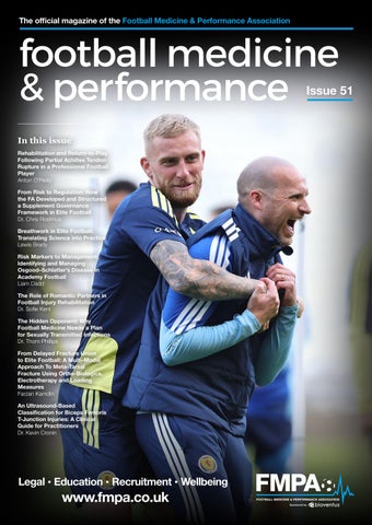
Photography Alamy, FMPA, Unsplash Cover Image Scotland’s Oliver McBurnie with sports scientist and performance coach Shayne Murphy during a training session at Barclays Hampden, Glasgow. Friday October 2, 2026. Picture: Robert Perry Credit: PA Images/Alamy
© Football Medicine & Performance Association. All rights reserved. The views and opinions of contributors expressed in Football Medicine & Performance are their own and not necessarily of the FMPA Members, FMPA employees or of the association. No part of this publication may be reproduced or transmitted in any form or by any means, or stored in a retrieval system without prior permission except as permitted under the Copyright Designs Patents Act 1988. Application for permission for use of copyright material shall be made to FMPA. For permissions contact admin@fmpa.co.uk
CONTENTS 08
Commercial News
10
Rehabilitation and Return-to-Play Following Partial Achilles Tendon Rupture in a Professional Football Player: A Case Study Anton O’Reilly
18
From Risk to Regulation: How the FA Developed and Structured a Supplement Governance Framework in Elite Football Dr. Chris RosImus
24
Breathwork in Elite Football: Translating Science into Practice Lewis Brady, Dr. Ricardo Catumbela
30
Risk Markers to Management: Identifying and Managing Osgood–Schlatter’s Disease in Academy Football Liam Dadd
36
The Role of Romantic Partners in Football Injury Rehabilitation Dr. Sofie Kent
41
The Hidden Opponent: Why Football Medicine Needs a Plan for Sexually Transmitted Infections Dr. Thom Phillips
47
From Delayed Fracture Union to Elite Football: A MultiModal Approach To Meta-Tarsal Fracture Using Ortho-Biologics, Electrotherapy & Loading Measures Farzan Kamdin
53
An Ultrasound-Based Classification for Biceps Femoris T-Junction Injuries: A Clinical Guide for Practitioners Dr. Kevin Cronin
10
18
24
30
36
41
47
53
www.fmpa.co.uk
7
commercial feature
BIOVENTUS NAMED FMPA 2026/27 LEAD SPONSOR We are delighted to announce that Bioventus UK will be the FMPA Lead Sponsor for the 2026/27 season. Following a successful partnership last season, we are excited to continue working with Bioventus and strengthen our shared commitment to supporting the healthcare and performance professionals working across professional football. Bioventus brings a strong focus on helping patients return to the activities they love, with innovative solutions including EXOGEN® and DUROLANE®. “Bioventus UK is delighted to be partnering with the FMPA once again for the 2026/2027 season. We have greatly enjoyed the opportunity to engage with the FMPA and their community of leading healthcare and performance professionals across football. As a company committed to helping patients return to the activities they love, we look forward to continuing to support FMPA members through innovative solutions such as EXOGEN and DUROLANE. We are excited for another successful year of collaboration and shared learning.” Eamonn Salmon, FMPA CEO, said: “We are pleased to welcome Bioventus as the FMPA’s Lead Sponsor for the 2026/27 season. Our partnership is built on a shared commitment to supporting the
professionals behind medicine and performance in football, while helping to connect our members with innovation, education and solutions that can positively impact player care. Bioventus UK has been a valued partner of the FMPA, and we look forward to building on that relationship and delivering an exciting programme of collaboration and shared learning throughout the season.” Together, supporting the professionals behind medicine and performance. What We Offer Bioventus is a global leader of innovations for active healing. Our mission is to partner with the healthcare community to deliver innovative medical devices and meet the evolving needs of patients, allowing them to resume and enjoy active lives. Bioventus develops and/or markets clinically proven and costeffective product offerings that are backed by clinical data. The products of Bioventus are known for their safety, innovation, and effectiveness. This includes DUROLANE, a nonsurgical injection for joint pain associated with osteoarthritis and EXOGEN, an ultrasound bone healing system that stimulates the body’s natural healing process, helping fractured bones mend. Find out more from Bioventus here www.bioventus.com
SWIMEX – AQUA THERMAE FMPA BUSINESS PARTNER SwimEx Sports Performance Pools and Accelerated recovery hot and cold deep water plunge pools. SwimEx Sports Fitness, Hydrotherapy Pools and Plunge Pools have delivered Sports Wellbeing since 1986. SwimEx’s unique paddlewheel delivers fantastic laminar flow water resistance for sports peak performance training and rehabilitation. Athletes can also commence recovery exercises much earlier due to the buoyancy of the SwimEx variable depth deep water wells alleviating their bodyweight, accelerating their return to action and helping prevent future injuries. SwimEx also offer motorised underwater treadmills for aqua running which can be included in a new pool or a drop in self propelled resistance version for existing pools. You can contact Richard Bishop MISPE direct if you would like to discuss a health check on your existing facilities or considering a future hydrotherapy project. Email richard@aquathermae.co.uk Tel: 07484 070765. www.aquathermae.co.uk
8
info@fmpa.co.uk
football medicine & performance
SPORTING EDGE UK FMPA BUSINESS CLUB Sporting Edge is the UK-based global leader in the design and provision of state-of-the-art Altitude & Environmental Facilities, trusted by the world’s most prestigious sports organisations. Key benefits for your squad: • Measurable performance gains: Our systems deliver quantifiable improvements in fitness and performance in as little as three weeks. • Accelerated return to play: Crucially, simulated altitude training allows injured players to maintain cardiovascular fitness during rehabilitation, significantly speeding up their return to match-day readiness and reducing the risk of a performance dip upon their return. • General squad fitness: Integrating altitude training into your standard protocol provides a powerful boost to the overall conditioning of your entire squad. Give your team the advantage this season! Visit www.sportingedgeuk.com or contact us today at info@sportingedgeuk.com for a free, no-obligation consultation and bespoke quote for a club-defining altitude or environmental chamber.
ONE HYPE WELLNESS FMPA BUSINESS PARTNER & RECRUITMENT SPONSOR The FMPA is pleased to welcome One Hype Wellness as an FMPA Business Partner and Recruitment Sponsor for the 2026/27 season. Their support of the FMPA reflects a shared commitment to advancing the health, wellbeing and performance of professionals working within football, while supporting the continued development of opportunities and resources for the wider FMPA community. Through their Recruitment Sponsorship, One Hype Wellness is also supporting the FMPA’s recruitment platform, helping connect talented practitioners with opportunities across the professional game. Intelligent Wellness. Personalised Performance. One Hype Wellness is redefining the approach to recovery, performance and wellbeing by bringing together advanced technology, artificial intelligence and personalised protocols. At the centre of the One Hype ecosystem is One AI, which uses individual data to help create more personalised wellness protocols and track responses over time. This technology is supported by a suite of advanced systems, including One Brain X for brain mapping and neurofeedback, One Hyperbaric for hyperbaric oxygen therapy, and One Light for whole-body red and near-infrared light therapy. For professional football, where recovery, cognitive performance and player wellbeing are increasingly important components of the high-performance environment, One Hype provides practitioners with technology designed to complement existing medical and performance strategies. More info: www.one-hype.com
CHARITY PARTNERS:
WORKING WITH:
www.fmpa.co.uk
9
feature
REHABILITATION AND RETURN-TO-PLAY FOLLOWING PARTIAL ACHILLES TENDON RUPTURE IN A PROFESSIONAL FOOTBALL PLAYER: A CASE STUDY FEATURE / ANTON O’REILLY, MSC, BSC, GSR Introduction Partial Achilles Tendon (AT) ruptures are uncommon injuries that frequently occur on a background of chronic tendinopathy. While considerable literature exists regarding the management of complete Achilles tendon ruptures and tendinopathy, there is a notable absence of consensus guidelines for rehabilitation of partial ruptures (Gatz et al., 2020). Clinicians must therefore integrate principles of tendon healing, progressive loading, and sport-specific reconditioning to guide rehabilitation.
Affiliations: Blackburn Rovers FC
football player who sustained a partial AT rupture. The case highlights key clinical considerations including imaging, tendon-specific loading strategies, plyometric progression, and sport-specific conditioning. A conceptual overview of the rehabilitation framework used in this case is presented in Figure 1 (below). Epidemiology In professional football, injuries affecting the triceps surae complex account for approximately 12–13% of all muscle injuries (Hallén & Ekstrand, 2014), with Achilles tendon pathologies representing a smaller but clinically significant proportion (Waldén et al., 2024). The majority of Achilles tendon disorders are tendinopathic
The purpose of this case study is to describe the diagnosis, rehabilitation strategy, and return-to-play progression of a professional
Diagnostics
Immobilzation
Tendon Loading
Plyometrics
Both MRI and UTC
Necessary to protect
Emphasis should be
An adequate plyometric
imaging modalities
the injury site, and
placed on making
base is necessary to be
are useful in the
maximize tissue
tendon loading as
developed prior to
management of
healing. Staged return
specific as possible to
progressing to running.
partial AT ruptures. It
to weight bearing and
the injury site.
Plyometric load should be
accessing of ranges is
overcoming stress
suggested to avoid big
shielding and
is important to identify the goals of each modality
monitored, progressions
jumps in between
avoiding excessive
progressions
mechanical strain on
Key Objective Markers Progressions through the stages of rehab should be made as objective as possible.
UTC Useful to assess quality of tendinous tissue, quantify the disorganization within the collagen matrix and assess degree of tendinopathic pathology.
Re-Imaging Helpful to assess healing status, and track progressions within tendon fibre alignment and organization throughout rehab.
Lateral
Range of Motion Use of wedges can be useful to limit dorsiflexion in early stages, and provide a gradual progression into deeper ranges of motion.
Soleus
Medial
Gastroc
Eccentric
use of various modalities can make
and continual through
should be achieved in
the later stages of rehab
order to progress to
Ground Contact Times Use objective progressions when working through SSC and GCT increases
HSR
Cumulative Loading Time Signaling for adaptation diminishes as time goes on
Structure Exposure must be tracked and staged. Volume and frequency recorded, and progressions made. Must have plan to reach end of plyometric continuum Expose Repeat
Signaling may peak at 10 minutes, anything above potentially extra mechanical strain without benefit
Progress Complete
Isometric Strength Soleus: > 2x BW Gastroc: > 2.8x BW Capacity Testing Max Single Leg Standing Raises to 60bpm: > 30 reps Jump Testing Tests to assess: Countermovement Jump Single Leg Countermovement Jump Single Leg Drop Jump 10-5 Test
Key Metrics Breaking & Concentric Force Imp - Mom (cm) RSI & RSI Mod (Test dependent) Landing Force Asymmetries Targets Very individualized. Aim for high RSI, symmetry left to right, and within 10% of patients previous best scores
Figure 1
10
objective testing. The
the reintroduction
Multidirectional Important to carry out in all planes, exposing to lateral and rotational variations, within deeper and more susceptible ranges
Contraction Type Progress through to maximize adaptation Iso
run should be based off
Key GPS metrics should
info@fmpa.co.uk
Return to Play Once an adequate exposure to training has been achieved, a staged return to competition is advised. Prior loading should be continued well beyond the
be tracked to avoid load
patients return to
spikes.
prevent further issues.
Anti Gravity Treadmill Very useful modality in order to reintroduce running
RTP Clearance In order to return to play, peak game and also weekly demands from an on feet loading and game specific skill perspective, must be: Achieved, Repeated, Maintained, and Exceeded
tasks.
Injection Therpapy Ideal time to carry out, such as PRP, while immobilized. Allows for full effectiveness, without limiting progressions due to recommended post injection offloading guidelines.
Clearance to return to
progressive and safe.
more demanding
Foot & Knee Position Use to bias subtendon involvement
Boot Important to immobilze and reduce weight bearing early to protect injury site and maximize healing potential.
Return to Run
Fundamental KPI’s
tissue
MRI
Partial ruptures frequently occur on a background of chronic tendinopathic degeneration, where areas of structural disorganisation may predispose the tendon to mechanical failure (Gatz et al., 2020). The reported incidence of AT rupture is low, estimated at approximately 0.01 injuries per 1000 playing hours, although most epidemiological studies do not distinguish between partial and complete (Waldén et al., 2024). As a result, there is limited research describing optimal rehabilitation strategies or expected return-to-play timelines for athletes sustaining this injury.
be as objective as possible
already compromised
Useful to quantify dimensions of deficit within the AT, specify location and assess for secondary issues.
in nature, with tendinopathy reported to account for up to 96% of Achilles-related issues (Gajhede-Knudsen et al., 2013).
90 - 95% 80 - 85% 70 - 75% 60 - 65%
Body weight Continuum
Suggestions
Build total volume first to increase tolerance within AT. Introduce accelerations gradually, with progressive intent from deeper, Dorsiflexed positions Delay high speed running until plyometric continuum fully completed and tolerated Delay consecutive days on feet to prevent tendon overload Include jump based, game specific tasks within session to expose to and mirror the demands of the game
Loading Continuation Important to continue with AT loading beyond point of return to play. Profilactically prevent onset of tendinopathy, and improve tendon tissue quality. Load Monitoring Vital to monitor and maintain on feet loading upon return. Double game weeks become challenges, and an understanding is necessary that modifications may need to be made until chronic load has been restored gradually.
football medicine & performance
Figure 2: Longitudinal MRI Imaging
Figure 3: Sagittal MRI Imaging
Figure 4: UTC Imaging
Case Presentation The athlete was a 28-year-old male professional football player competing in the EFL Championship. His medical history included a previous complete AT rupture five years prior and an ACL rupture 18 months earlier, both affecting the contralateral limb. The injury occurred early in the season. Following a tactical change, and injuries to other squad members, the player experienced a substantial increase in both training load and match exposure. Prior to the acute event, the athlete had reported symptoms consistent with Achilles tendinopathy in the right tendon, which had been managed through prescribed loading, without time loss from training or competition. During a congested period of fixtures, the athlete completed five matches within 17 days, accumulating a total of 455 minutes. In the final moments of the last match in this sequence, the player experienced a sudden sharp pain in the right AT while jumping to contest a header. The athlete was unable to continue and was removed from play.
Imaging and Diagnosis Magnetic resonance imaging (MRI) was performed shortly after the injury. Imaging revealed a sagittal delaminating tear within the lateral fibres of the AT, extending distally into a partial-thickness intrasubstance tear measuring approximately 3.7 mm sagittally and 15 mm craniocaudally (Figure 2 and 3). The injury occurred on a background of diffuse chronic tendinopathic changes extending approximately 55 mm along the tendon, consistent with previously reported patterns (Gatz et al., 2020). Ultrasound tissue characterisation (UTC) imaging was subsequently performed to quantify the extent of tendon disorganisation. UTC findings demonstrated increased proportions of disorganised tendon matrix, with the highest levels of degeneration located approximately 3.4 cm proximal to the calcaneal insertion (Figure 4). This location corresponded with the tear identified on MRI.
www.fmpa.co.uk
11
feature Relevant Achilles Tendon Anatomy Emerging anatomical research has demonstrated that the AT is composed of distinct sub-tendons originating from the gastrocnemius and soleus (Edama et al., 2015), which maintain partial mechanical independence during loading (Handsfield et al., 2017; Figure 5). This anatomical arrangement suggests that rehabilitation loading strategies may differentially stress specific tendon regions depending on foot position and muscle recruitment. Consequently, manipulating foot position and exercise selection may allow clinicians to target specific subtendon structures and progressively restore load tolerance following injury (Nunes et al., 2020). Rehabilitation Progressions Immobilisation and Early Management Immediately following diagnosis, the athlete was immobilised in a nonweight-bearing walking boot with heel elevation. A total heel raise of 3 cm was initially used to reduce tensile loading through the AT. Over a two-week period the heel lifts were progressively reduced by 1 cm increments to prepare for full weight bearing and restoration of normal gait (Table 1). Progression through the immobilisation phase was guided by clinical criteria including absence of symptom exacerbation, lack of effusion around the tendon, and no development of a delayed pain response. The athlete was fully weight-bearing without symptoms by day 14 following injury. Injection Therapy Platelet-rich plasma (PRP) injection was administered to the injury site three days following the initial injury. In some cases, PRP has been reported to be useful within the acute setting for tendon related issues (Pitchford et al., 2016), however there is no general consensus for the use of PRP in AT injuries. The intervention was scheduled early to coincide with the immobilisation phase, as PRP treatment protocols typically require temporary unloading of the tendon. A second PRP injection was considered; however, this would have required additional immobilisation. Given the importance of early progressive loading, a single injection was utilised in this case.
12
Figure 5: Anatomical representation of Achilles subtendon structure. Adapted from Edama et al. (2015).
Weeks
Base
DF Range
0-1
NWB, 3 – 1.5cm Wedge
-20 deg
1-2
NWB, 1.5cm - 0cm Wedge
-10 deg
2-3
Platform shoes
0 deg
3-4
Platform shoes
5-10 deg
4-5
Flat shoes
10 -15 deg
5-6
Flat shoes
15 deg - Max
Table 1: Range of Motion Progressions
Restoration of Range of Motion During the early rehabilitation stages, excessive ankle dorsiflexion was avoided to minimise tensile stress at the injury site. Initial exercises were performed above plantar-grade positions to protect the healing tendon. Range of motion was progressively restored over the first six weeks of rehabilitation (Table 1). Controlled dorsiflexion was gradually introduced
info@fmpa.co.uk
using heel blocks and footwear modifications to limit range. Loading through full range was achieved at approximately five weeks post-injury once the athlete demonstrated adequate tolerance to earlier stages. Progression of ankle range of motion was based on clinical assessment and absence of symptom provocation both during and following exercise.
football medicine & performance Progressive Tendon Loading Contraction Variations Reintroduction of tendon loading began with isometric exercises, which allow controlled force production without substantial tendon excursion (Wyndow et al., 2013). Early loading utilised overcoming isometric contractions to provide high force outputs while maintaining safe joint positioning.
Foot positioning was manipulated, with a toe-in position used to increase lateral triceps surae activation (Nunes et al., 2020). Early loading strategies emphasised soleus-dominant exercises using bentknee positions, initially in a neutral foot position, before progressing to toe-in. This resulted in gradually progressing to gastrocnemius-biased movements, with similar progressions in foot position.
Work Capacity Progressive walking sessions were introduced to rebuild capacity and gradually expose the tendon to repetitive loading. Professional footballs typically accumulate approximately 8900 steps during a 90-minute match, highlighting the importance of developing sufficient capacity tolerance prior to return to sport (Taylor, 2014).
Yielding isometrics were subsequently introduced to increase tendon load tolerance and prepare the athlete for isotonic loading. Eccentric exercises were incorporated at four weeks post-injury within controlled ranges. Eccentric loading has been shown to increase tendon cross-sectional area and improve tendon capacity (Radovanović et al., 2022).
Cumulative Loading Tendon adaptation is influenced not only by peak loading magnitude but also by duration. Experimental studies have demonstrated that mechanotransduction pathways involved in tendon adaptation, including activation of the mTOR pathway and collagen mRNA expression, may peak after relatively short periods of cumulative loading, with suggestions of around 10 minutes (Baar, 2017). Following this, cellular responsiveness to mechanical stimulus may diminish, suggesting that prolonged continuous loading may provide limited additional adaptive benefit.
Walking volume was increased using step counts to monitor exposure. Sessions were typically performed before strength exercises to avoid excessive cumulative loading on a fatigued tendon.
Heavy slow resistance (HSR) training was introduced during the later stages of rehabilitation. HSR exercises combine concentric, eccentric, and isometric muscle actions and have been shown to promote collagen remodelling and improvements in tendon structure (Beyer et al., 2015). Foot Positioning Exercise selection was influenced by the anatomical location of the injury within the lateral gastrocnemius subtendon.
This may also apply excessive shear and mechanical strain, compromising both healthy and newly proliferated tissue. Consequently, loading sessions were structured to deliver intense, discrete bouts of tendon loading rather than prolonged exposures.
www.fmpa.co.uk
Stress Shielding Response Walking was also placed at the beginning of each loading session to counteract the stress-shielding response. Stressshielding may occur when injured tendon regions are mechanically unloaded while surrounding healthy tissue assumes a greater proportion of load (Zhang et al., 2023). Over time, this reduction in mechanical stimulus may limit collagen remodelling within the injured region. Walking exposure combined with staged isometric loading before eccentric and HSR exercises was utilised to ensure the injured region received adequate stimulus.
13
feature
Running Speed (m/s)
Running Phase
Ground Contact Time (ms)
Peak Vertical Force (xBW)
3.0
Initial Acceleration
195 – 215
2.30 – 2.50
3.5
Initial Acceleration
182 – 195
2.45 – 2.65
4.0
Early Acceleration
170 – 182
2.60 – 2.80
4.5
Early Acceleration
160 – 170
2.70 – 2.90
5.0
Transition Phase
152 – 160
2.80 – 3.00
5.5
Transition Phase
145 – 160
3.00 – 3.15
6.0
Mid Acceleration
138 – 145
3.05 – 3.20
6.5
Mid Acceleration
130 – 138
3.15 – 3.35
7.0
Late Acceleration
124 – 130
3.30 – 3.50
7.5
Late Acceleration
118 – 124
3.40 – 3.65
8.0
Approaching Max Speed
114 – 118
3.60 – 3.90
8.5
Approaching Max Speed
110 – 114
3.90 – 4.20
9.0
Approaching Max Speed
105 – 110
4.20 – 4.70
9.5
Terminal Velocity
100 – 105
4.60 – 5.00
10.0
Terminal Velocity
93 – 100
4.80 – 5.40
Table 2: Ground Contact Times and Peak Vertical Forces ranges reported in literature at various running speeds. Adapted from Weyand et al. (2000), Clark and Weyand (2014), Haugen et al. (2019), and Nagahara et al. (2018).
Plyometric Progression Low speed running can often be seen as a relatively safe entry point once objective markers are achieved, especially when compared to high intensity plyometric work. However, the forces and workload required to run are significant (Table 2). The patient was gradually exposed to tasks that replicate and progressively exceed the mechanical demands placed on the AT during the return to run process, before beginning it. Exposure to plyometrics, and the progressions within accommodated for gradual accumulation, providing progressive adaptive loading to the tendon, and avoiding any excessive spikes in cumulative load. Exercises were selected to gradually increase ground reaction forces and reduce ground contact times, reflecting the mechanical demands of running and sprinting. The plyometric tasks used and their loading characteristics are outlined in Table 3.
14
Exercise
Ground Contact Time (ms)
Peak Vertical Force (xBW)
Squat Jump (Conc Only)
800 – 840
2.30 – 3.00
CMJ
600 – 800
2.75 – 3.30
Abalakov Jump
500 - 600
3.20 – 3.80
Repeat CMJ
350 – 500
3.00 – 3.50
Drop Jump (30cm)
170 – 230
3.50 – 4.00
Alternate Leg Bound
200 – 250
3.50 – 4.00
Broad Jump
400 – 500
4.00 – 4.20
SL Speed Hop
160 – 200
4.20 – 4.50
Max Velocity Sprint
85 - 100
4.50 – 5.50
Drop Jump (60cm+)
200 – 280
4.90 – 5.40
Tuck Jumps
170 - 200
5.00 - 5.50
Hurdle Hops (Maximal)
210 - 240
5.70 – 6.00
Table 3: Ground Contact Times and Peak Vertical Forces reported in literature during specific exercises. Adapted from Demangeot et al. (2022) and Pedley et al. (2017).
info@fmpa.co.uk
football medicine & performance Test
Metric
Target
Soleus Iso
Max Force (N)
2x BW
Gastroc Iso
Max Force (N)
2.8 BW
Single Leg Standing Capacity (60BPM)
Max Reps
30+ Reps
RSI - Mod
>0.6
Countermovement Jump
Jump Height (Imp-Mom) Peak Power (W / Kg)
Single Leg CMJ
Jump Height (Imp-Mom)
>10% Pre Injury
RSI – Mod
0.35
Jump Height Single Leg Drop Jump
10-5 Hop Test
>10% Pre Injury
Peak Instantaneous Power (W / Kg)
>10% Pre Injury
RSI (Flight Time / Contact Time)
>1.2
Mean RSI (Flight Time)
>2.8
Table 4: Objective Tests and Targets. Partly adapted from Vald Premier League Report 2025-26.
Return to Running and Sport Running was reintroduced at 7.5 weeks postinjury using a reduced body-weight treadmill (AlterG). Initial sessions were performed at 60–65% body weight to reduce tendon strain while restoring running mechanics. Bodyweight support was gradually reduced over four sessions until full weight-bearing was tolerated. Prior to commencement of running on field, objective testing was carried out. The specific tests and targets which were used are outlined in Table 4. Following completion of treadmill progressions and achievement of objective targets, the athlete transitioned to fieldbased running. Early sessions involved low-volume linear running at moderate intensities before gradually progressing to higher speeds and more complex movement patterns. Sport-specific drills were incorporated to replicate positional demands. As a central defender, the athlete was frequently required to perform jumping actions during aerial duels. Training sessions therefore included progressive exposure to jump-based tasks simulating defensive and attacking scenarios. GPS metrics including total distance, high-speed running, sprint, accelerations, decelerations, and maximal velocity were used to guide the reconditioning progression. Training loads were gradually increased to match the player’s historical match demands. The athlete was cleared to return to matchplay 15 weeks following the initial injury.
www.fmpa.co.uk
15
feature Key Considerations • Partial AT ruptures represent a rare but complex injury that frequently occurs on the background of chronic tendinopathy. Limited availability of published rehabilitation frameworks presents a challenge for clinicians managing these cases.
football medicine & performance likely provides the necessary part of the tendon with load, targets the injury site and reduces the likelihood of compensatory patterns developing.
• MRI is effective in identifying the location and size of partial ruptures, while UTC allows quantification of tendon disorganisation and provides insight into the extent of pre-existing tendinopathy.
• Following structural disruption, portions of the tendon may receive reduced mechanical stimulus if surrounding tissues compensate during loading. Progressive loading strategies may be implemented to ensure that the injured tissue is exposed to sufficient mechanical stimulus, while remaining within the athlete’s symptom tolerance.
• Staged tendon loading progressions, beginning with controlled isometric exercises and progressing to eccentric and HSR training, reflect current understanding of tendon mechanobiology and the importance of progressive loading.
• Running imposes substantial forces on the AT, particularly during acceleration and sprinting. Gradual exposure to plyometric tasks allows the athlete to develop the necessary mechanical tolerance prior to initiating running.
• Manipulating foot and knee position during calf-loading exercises may allow clinicians to preferentially load specific tendon regions. This
• Replicating positional demands within rehabilitation sessions may reduce the risk of reinjury by ensuring athletes are adequately
conditioned for the specific tasks required during match play. Conclusion This case study describes a rehabilitation approach for partial AT rupture. The programme incorporated imaging-guided diagnosis, progressive tendon loading, plyometric development, and sport-specific reconditioning. The staged framework presented may provide clinicians with practical guidance when managing this uncommon injury, although rehabilitation should be individualised based on injury characteristics and athlete response. Despite the successful outcome in this case, it is important to recognise that rehabilitation timelines may vary depending on the severity of the structural injury, the extent of underlying tendinopathy, and individual healing responses.
References AlterG Inc. (2026). AlterG anti-gravity treadmill. AlterG Inc., Fremont, CA. Baar, K. (2017). Minimizing injury and maximizing return to play: Lessons from engineered ligaments. Sports Medicine, 47(Suppl 1), 5–11. Beyer, R., Kongsgaard, M., Hougs Kjær, B., Øhlenschlæger, T., Kjær, M., & Magnusson, S. P. (2015). Heavy slow resistance versus eccentric training as treatment for Achilles tendinopathy: A randomized controlled trial. The American Journal of Sports Medicine, 43(7), 1704–1711. Clark, K. P., & Weyand, P. G. (2014). Are running speeds maximized with simple-spring stance mechanics? Journal of Applied Physiology, 117(6), 604–615. Demangeot, Y., Arnal, P. J., Bideau, B., et al. (2022). The load borne by the Achilles tendon during exercise: A systematic review of normative values. Scandinavian Journal of Medicine & Science in Sports, 32(5), 846–859. Edama, M., Kageyama, I., Kikumoto, T., Nakamura, M., Nakamura, E., & Saito, A. (2015). The twisted structure of the human Achilles tendon: A cadaveric study. Scandinavian Journal of Medicine & Science in Sports, 25(3), e497–e503. Gajhede-Knudsen, M., Ekstrand, J., Magnusson, H., & Maffulli, N. (2013). Recurrence of Achilles tendon injuries in elite male football players is more common after early return to play: An 11-year follow-up of the UEFA Champions League injury study. British Journal of Sports Medicine, 47(12), 763–768. Gatz, M., Spang, C., & Alfredson, H. (2020). Partial Achilles tendon rupture—A neglected entity: A narrative literature review on diagnostics and treatment options. Journal of Clinical Medicine, 9(10), 3380. https://doi.org/10.3390/jcm9103380 Hallén, A., & Ekstrand, J. (2014). Return to play following muscle injuries in professional footballers. Journal of Sports Sciences, 32(13), 1229–1236. Handsfield, G. G., Inouye, J. M., Slane, L. C., Thelen, D. G., & Miller, G. W. (2017). Achilles subtendons exhibit substantial glide relative to each other. Skeletal Radiology, 46(4), 533–540. Haugen, T., Tønnessen, E., & Seiler, S. (2019). Sprint running: From fundamental mechanics to practice. International Journal of Sports Physiology and Performance, 14(2), 213–220. Nagahara, R., Matsubayashi, T., Matsuo, A., & Zushi, K. (2018). Association of sprint performance with ground reaction forces during acceleration and maximal speed phases. Journal of Applied Biomechanics, 34(2), 104–110. Nunes, J. P., Schoenfeld, B. J., Nakamura, M., et al. (2020). Different foot positioning during calf training to induce portion-specific gastrocnemius muscle hypertrophy. Journal of Strength and Conditioning Research, 34(8), 2347–2351. Pedley, J. S., Lloyd, R. S., & Oliver, J. L. (2017). Drop jump: A technical model for scientific application. Strength and Conditioning Journal, 39(5), 36–44. Pitchford, K., et al. (2016). Platelet-rich plasma for the treatment of acute muscle-tendon injuries: A systematic review. International Journal of Sports Medicine, 37(11), 889–897. Radovanović, G., Bohm, S., Peper, K. K., Arampatzis, A., & Legerlotz, K. (2022). Evidence-based high-loading tendon exercise for 12 weeks leads to increased tendon stiffness and cross-sectional area in Achilles tendinopathy: A controlled clinical trial. Sports Medicine - Open, 8, 134. https://doi.org/10.1186/s40798022-00517-2 Taylor, T. J. (2014). Effect of soccer positions on steps taken per game. International Journal of Exercise Science: Conference Proceedings, 9(2), 34. Vald Inc. (2026). Vald Normative Data Report - 2025 - 26 Premier League Report. Waldén, M., Hägglund, M., & Ekstrand, J. (2024). Achilles tendon pain in male professional football players: A prospective five-season study of 88 injuries from the UEFA Elite Club Injury Study. Journal of Science and Medicine in Sport, 27(3), 275–280. Weyand, P. G., Sternlight, D. B., Bellizzi, M. J., & Wright, S. (2000). Faster top running speeds are achieved with greater ground forces not more rapid leg movements. Journal of Applied Physiology, 89(5), 1991–1999. Wyndow, N., Cowan, S. M., Wrigley, T. V., & Crossley, K. M. (2013). Triceps surae activation and gastrocnemius muscle morphology in people with Achilles tendinopathy. Medicine & Science in Sports & Exercise, 45(7), 1332–1339. Zhang, L., Wang, X., Wang, J., et al. (2023). Mechanical loading and the tendon-to-bone insertion: A review of the current status. Frontiers in Bioengineering and Biotechnology, 11, 1180332.
16
info@fmpa.co.uk
SwimEx Fitness Hydrotherapy and Plunge Pools Aqua Thermae are the UK and Ireland installers for www.SwimEx.com professional sports fitness and recovery pools as used across the world by leading sports teams and athletes for peak performance training and accelerated resilient recovery. We provide swimming pool bespoke tailored water treatment and filtration systems using the latest innovative specified equipment.
swimex.com • info@aquathermae.co.uk • +44 (0) 7484 070765
feature
FROM RISK TO REGULATION: HOW THE FA DEVELOPED AND STRUCTURED A SUPPLEMENT GOVERNANCE FRAMEWORK IN ELITE FOOTBALL FEATURE / DR. CHRIS ROSIMUS Introduction Football is a highly competitive sport in which players continually seek to maximise performance and gain an advantage over their peers at domestic, national, and international levels. This pursuit of marginal gains has contributed to the widespread use of nutritional supplements within elite football. Data collected from FIFA World Cup tournaments between 2002 and 2014 reported that approximately 33% of players used supplements during competition, with this figure increasing to almost 50% within youth international tournaments. In some cases, players reported consuming up to seven supplements per match, highlighting both the prevalence and complexity of supplement use within elite performance environments (Tscholl et al, 2015).
18
Affiliations: The Football Association
When used appropriately, supplements can support health, recovery, training adaptation, and competition performance. Supplements may also provide practical support during periods of international travel, where access to consistent, highquality nutrition is not always guaranteed (Maughan et at 2018). International football regularly requires teams to travel globally, often to environments where nutritional provision may differ considerably from that typically available in England. In these situations, supplements and sports foods can provide an effective strategy to support fuelling, recovery, and overall player preparation. However, supplement use also presents significant risks. One of the greatest concerns within elite sport is the potential
info@fmpa.co.uk
for inadvertent doping resulting from contaminated supplements. Analytical studies have found anywhere from 14 to 50% of samples analysed from dietary supplement products have tested positive for anabolic agents or other prohibited substances (Knapik et al, 2023). In many cases, contamination occurs because of poor manufacturing processes, crosscontamination during production, or undeclared ingredients. For athletes operating under strict liability regulations, even accidental ingestion can result in anti-doping sanctions. The Football Association (FA) Physical Performance, Medicine and Nutrition (PPMN) department therefore recognised the need for a structured approach to supplement governance. The FA adopted
football medicine & performance a philosophy of “food first, but not food only.” In practice, this means prioritising high-quality nutritional strategies to fuel, perform, and recover, while recognising that supplements may still have a legitimate role when nutritional needs cannot reasonably be met through food alone. This philosophy aligned with the International Olympic Committee consensus statement on dietary supplements and the high-performance athlete, which recognises that supplements can play an important role in maintaining health, supporting training adaptation, and optimising performance when used appropriately. In response to these challenges, the FA initiated the development of a Supplement Governance and Provision Policy (figure 1). The aim was not simply to regulate supplement use, but to create a consistent, evidence-informed framework that balanced player safety, anti-doping compliance, operational practicality, and performance support across the men’s and women’s pathways. Identifying the Need for Governance The starting point for the policy was a comprehensive review of existing practice across the FA performance pathway. Although supplements were routinely provided to players, governance processes lacked consistency. Different teams and practitioners often operated using varying approaches to supplement provision, education, and documentation. While this was not uncommon within elite sport environments, it created several operational and safeguarding challenges. A number of key issues emerged during the review process. These included inconsistent supplement provision between squads, limited quality assurance
Figure 1: The FA supplement governance policy
procedures, unclear documentation processes, and variable levels of practitioner education regarding anti-doping risk management. There was also increasing awareness that supplements were being used within highly pressurised performance environments without a unified governance structure to support decision-making. Importantly, the FA recognised that the challenge was not solely scientific or operational. It was also cultural. Different stakeholders held varying perspectives on supplement use, ranging from highly cautious approaches to more performance-driven philosophies. As a result, the policy development process required significant collaboration and communication to create alignment across departments while maintaining sufficient
Figure 2: FA supplement governance policy structure and contents
www.fmpa.co.uk
flexibility for individual player needs. Developing the Framework The FA approached policy development through a multi-disciplinary process involving representatives from nutrition, medicine, performance, operations, antidoping, and external organisations including the Sport and Exercise Nutrition Register (SENr). Best practice guidance was also reviewed from several sports and governing bodies, including rugby, cricket, cycling, and the English Institute of Sport. The framework itself was deliberately structured into four distinct sections to improve clarity and usability (figure 2). Part A established the philosophical and regulatory foundations of the policy. This included the introduction, overarching philosophy, purpose statement, strict liability guidance, and key definitions. Here, it was important to define our intent regarding ‘food-first but not food only approach, where we acknowledge that dietary supplements and sports foods are adopted alongside whole foods (Close et al; 2022). This ensures that whole foods remain the foundation of performance nutrition, with supplements used in a controlled and evidence-based way. In addition, embedding strict liability early within the document was particularly important, as it reinforced that players remain personally responsible for any substance found in their system, regardless of intent. Part B focused on the operational delivery of supplement governance. This section outlined procedures relating to supplement provision during England camps and across the wider pathway. Safeguarding formed a
19
feature
Figure 3: Wider department policy awareness sharing
20
info@fmpa.co.uk
football medicine & performance central component of this section, with a strong emphasis placed on education and practitioner competency. During development, the FA recognised that governance could not rely solely on restricting products or implementing operational procedures. Education was equally important in supporting safer decision-making among both players and staff. As a result, practitioners involved in supplement provision were encouraged to complete United Kingdom AntiDoping (UKAD) accreditation to ensure a consistent level of anti-doping knowledge across the multidisciplinary team. The policy also introduced structured supplement prescription processes. Supplements could not simply be used habitually or based on practitioner preference. Instead, use had to be justified against a specific performance, health, or clinical rationale. Supplementation to correct nutritional deficiencies, for example, followed clearly defined pathways involving assessment, screening, and documentation (Burke ta al, 2018). Quality assurance procedures represented another major component of the framework. Only batch-tested products from approved suppliers could be distributed within the pathway. Processes relating to supplement ordering, storage, dispensation, and record keeping were also standardised. This improved traceability and accountability while reducing operational inconsistency between squads and departments. To maintain consistency and raised awareness across the department, the headlines of this policy were shared across the wider technical department (figure 3). This ensured a shared understanding of processes and reinforces collective responsibility across the multidisciplinary team. Taken together, these safeguarding and education measures form a key part of the governance system, helping to reduce risk and improve compliance across England teams. Part C focused on supplement classification, evidence-informed practice, and supporting literature. Supplements were categorised according to their level of evidence, intended application, and associated risk profile. This provided practitioners with a clearer framework for decision-making while ensuring consistency across the pathway (IOC consensus statement , 2018). Finally, Part D included the practical resources required to operationalise the
Figure 4: Supplement decision making flow chart
Figure 5: SENR accreditation feedback
policy. These included supplement request forms, decision-making flowcharts (figure 4), distribution procedures, product guides, nutrition calculators, anti-doping education resources, and governance documentation templates. The inclusion of these applied tools was intentional. The FA wanted the framework to function as a practical working document rather than simply a theoretical policy. Achieving SENr Accreditation External validation formed an important stage of the development process. Once the policy had been completed, it was submitted to the Sport and Exercise Nutrition Register (SENr) for formal review and accreditation (figure 5). The accreditation process evaluated several aspects of the framework, including the scientific rationale underpinning supplement recommendations, quality
www.fmpa.co.uk
assurance procedures, anti-doping risk management, operational clarity, and governance structure. Feedback from SENr was incorporated to refine areas of wording, documentation, and procedural consistency before final endorsement was achieved. Achieving SENr accreditation strengthened the credibility of the framework both internally and externally. It also reinforced confidence that the policy aligned with recognised industry standards and current evidence-informed practice. Importantly, the process highlighted that governance frameworks must remain dynamic. The policy therefore incorporated ongoing review procedures to ensure alignment with evolving scientific evidence, operational demands, and anti-doping guidance.
21
feature
football medicine & performance
Challenges and Lessons Learned One of the key lessons from the FA’s experience was that successful governance depends as much on implementation as policy design. Creating scientifically robust procedures was only one aspect of the process. Ensuring that the framework remained practical, accessible, and usable across different environments proved equally important. There was a conscious effort to avoid producing a document that was academically comprehensive but operationally impractical. This ultimately influenced the decision to structure the framework into clear sections supported by practical tools and flowcharts. The intention was to support decision-making in realworld environments rather than create additional administrative burden. Communication also emerged as a major consideration throughout implementation. Consistent messaging across departments was essential to maintain stakeholder buy-in and avoid conflicting approaches to supplement use. Ongoing education, regular review, and multidisciplinary collaboration therefore became central to sustaining the integrity of the framework. Conclusion The FA’s development of a Supplement Governance and Provision Policy demonstrates that effective supplement governance extends far beyond simply approving or restricting products. Instead, it requires the integration of evidenceinformed practice, anti-doping education, operational systems, multidisciplinary
About Dr Chris Rosimus Chris is a registered sports and exercise nutritionist with over 15 years’ experience providing performance nutrition support to elite, professional and recreational athletes. Chris is currently the Head of men’s performance nutrition at The FA, where he is responsible for the strategic direction of performance nutrition across the men’s pathway whilst maintaining direct delivery to the senior teams.
collaboration, and cultural alignment within a single coherent framework. For practitioners working within elite sport, one of the key lessons is that supplement governance should be viewed as an ongoing process rather than a one-time intervention. Policies must evolve alongside scientific evidence, anti-doping regulations, and the practical realities of high-performance sport. Equally, successful implementation depends not only on scientific robustness, but on communication, education, and stakeholder engagement across the wider performance environment. Ultimately, the FA experience highlights that robust supplement governance can support both player safety and performance when embedded effectively within organisational culture and day-today practice.
Prior to The FA, Chris spent six years with the English Institute of Sport (EIS) as Performance Nutritionist for the England Cricket team and has previously provided nutrition support to England Squash, Leicester City F.C, Aston Villa F.C, Nottingham Forest F.C and ONE Pro cycling, alongside a part-time lecturing position on sports nutrition with Manchester Metropolitan University. Prior to joining the EIS, Chris was a performance nutrition intern at IMG Academies in Florida and worked as a nutritionist in Abu Dhabi with Manchester United Soccer Schools. Alongside this, Chris has a football coaching background and currently holds his UEFA B qualification. Chris has committed to enhancing his professional education and has recently completed a professional doctorate in sports and exercise science. Affiliation: The Football Association Acknowledgements The development and implementation of this framework involved collaboration across multiple FA departments, including nutrition, medicine, performance, operations, and anti-doping teams, alongside external organisations including SENr.
References Maughan RJ. Dietary supplements and the high-performance athlete. Int J Sport Nutr Exerc Metab [Internet]. 2018 Mar 01 [cited 2018 Jun 14];28(2):101. Available from: https://www.nDi.nlm.nih.gov/pubmed/29444595 DOI: 10.1123/ijsnem.2018-0026. Maughan RJ, Burke LM, Dvorak J, Larson-Meyer DE, Peeling P, Rawson ES et al. IOC consensus statement: dietary supplements and the highperforming athlete. Br J Sports Med [Internet]. 2018 Apr [cited 2018 Jun 14];52(7):439-455. Available from: https://www.ncbi.nlm.nih.gov/ pubmed/29540367 DOI: 10.1136/bjsports-2018-099027. Tscholl PM, Vaso M, Weber A, Dvorak J. High prevalence of medication use in professional football tournaments including the World Cups between 2002 and 2014: a narrative review with a focus on NSAIDs. Br J Sports Med [Internet]. 2015 May [cited 2018 Jun 14];49(9):580-2. Available from: https://www.ncbi.nlm.nih.gov/pubmed/25878074 DOI: 10.1136/bjsports-2015-094784. Baume N, Jan N, Emery C, Mandanis B, Schweizer C, Giraud S, et al. Antidoping programme and biological monitoring before and during the 2014 FIFA World Cup Brazil. Br J Sports Med [Internet]. 2015 May [cited 2018 Jun 14];49(9):614-22. Available from: https://www.ncbi.nlm.nih.gov/pubmed/25878079 DOI: 10.1136/bjsports-2015-094762. Close, G, Casper, A M, N P Walsh, and Maughan, R.J. (2022) ‘Food first but not food only: dietary supplements in sport’, International Journal of Sport Nutrition and Exercise Metabolism, 32(5), pp. 371–386. https://doi.org/10.1123/ijsnem.2021-0335 PMID: 35279015. Knapik, J.J., Steelman, R.A., Hoedebecke, S.S., Austin, K.G., Farina, E.K. and Lieberman, H.R. (2023) ‘Prevalence of dietary supplement use by athletes: systematic review and meta-analysis’, *Sports Medicine - Open*, 9(1). Available at: https://pmc.ncbi.nlm.nih.gov/articles/PMC10570429/ (Accessed: 28 May 2026).
22
info@fmpa.co.uk
feature
BREATHWORK IN ELITE FOOTBALL: TRANSLATING SCIENCE INTO PRACTICE Affiliations: Sheffield Hallam University Optima Performance
FEATURE / LEWIS BRADY MSC, DR. RICARDO CATUMBELA In elite football, where performance demands are relentlessly high, athletes are continually looking for ways to enhance their physical, mental, and emotional readiness. Breathwork is one such tool rapidly gaining traction and refers to the deliberate control of breathing patterns to influence physiological and psychological states. Structured breathwork uses deliberate rhythms and depths to regulate arousal (Shaw-Metz, 2022; Balban et al., 2023), improve focus (Fincham et al., 2023), and promote recovery (Lehrer & Gevirtz, 2014; Laborde et al., 2017). Consequently, an increasing number of elite clubs are integrating breathwork into every aspect of a player’s routine. This article outlines some of the key evidence, clinical guidelines, and practical applications of breathwork in football. Current Frameworks and guidelines The use of breathwork in football has expanded rapidly over the past decade, supported by research demonstrating its impact on physiological and psychological regulation (Zaccaro et al., 2018; Noble & Hochman, 2019), yet despite this growing popularity, there is still no universally accepted clinical framework for its implementation within football medicine and performance contexts. Current approaches are largely practitioner-led, with variability in duration, breathing cadence, and context of delivery (pre-match, recovery, or rehabilitation) (Brenner., 2024). This lack of consistency is problematic, as it limits both effectiveness and accountability. However, the good news is that clearer, evidence aligned guidance is beginning to emerge. Research suggests that breathwork can provide both acute and chronic benefits. Acute applications (1–3 minutes) appear effective for rapid arousal control before high-pressure events such as penalty shootouts, while chronic structured practice (10–15 minutes daily) can support recovery, sleep quality, and long-term stress regulation (Ma et al., 2017; Balban et al., 2023). Research has highlighted that the psychophysiological effects of breathwork
24
are mediated by several interacting factors, including breathing rate, depth, timing of practice and individual characteristics such as anxiety sensitivity or baseline vagal tone (Zimberoff & Hartman.,1999; Banushi et al., 2023; Brems., 2024). As a result, standardised breathing protocols are unlikely to be universally effective across an entire squad. Instead, a personalised and context-dependent approach is recommended aligning breathing techniques with each player’s role demands, psychological profile, and recovery needs (Fincham et al., 2023). Moreover, while breathwork is generally safe, practitioners should conduct
preliminary screening to identify players with respiratory or cardiovascular limitations (See Table 1) (Jerath et al., 2006; Paulus., 2013; Russo et al., 2017). The growing use of wearable technology and biofeedback platforms has made it possible to individualise breathing cadences and monitor physiological responses in real time, including heart rate variability (HRV), respiratory rate, oxygen saturation, and electrodermal activity (Fincham et al., 2023). These advances support a shift towards data informed breathwork prescriptions, where parameters such as breathing rate, session duration, and timing can be refined based on both objective metrics and subjective readiness ratings.
Table 1: Potential Risk populations as well as recommended precautions Risk Population
Why they're at risk
Recommended precaution
Individuals with high anxiety.
Slow breathing increases interoceptive awareness and CO2 tolerance; may trigger fear responses or panic sensations.
Assess anxiety sensitivity then introduce breathing slowly, avoiding prolonged breath holds.
Respiratory conditions (e.g. Ashtma).
Altered airway resistance and sensitivity to CO2 fluctuations may provoke discomfort or dyspnoea.
Avoid long exhalation holds and prioritise gentle nasal breathing. Potentially consult medical staff.
Cardiovascular disease or arrhythmias.
Slow breathing shifts autonomic balance; excessive vagal stimulation may disturb heart rhythm.
Acquire medical clearance. Avoid very slow breathing rates initially.
Low resting blood pressure.
Slow breathing may further reduce blood pressure, potentially causing lightheadedness or reduced cerebral perfusion.
Limit duration until tolerance is established. Assess dizziness on standing, potentially alter to seated of lying positions.
Head injury recovery (e.g. Concussion).
Altered autonomic function post-concussion may impair tolerance to CO2 and rapid breathing transitions.
Acquire medical clearance. Avoid long breath holds and monitor for headache and dizziness.
Individuals on stimulant medication.
Stimulants alter respiratory drive, autonomic activity, and perceived exertion.
Consult about the timing of when medication is taken. Avoid aggressive down regulation techniques shortly after taking.
info@fmpa.co.uk
football medicine & performance From a performance perspective, breathwork may influence football-specific outputs by improving respiratory efficiency, CO2 tolerance, and autonomic balance, all of which underpin high-intensity endurance (Holm et al., 2004; Młynczak & Krysztofiak, 2018) . Football at the professional level is predominantly aerobic, with approximately 70–90% of total match energy expenditure derived from aerobic metabolism, supporting repeated high-intensity actions, rapid recovery between sprints, and sustained cognitive decision-making under fatigue (Bangsbo et al., 2006; Castagna et al., 2025). Breathwork techniques that regulate end-tidal CO2 (EtCO2) and optimise minute ventilation (VE) help stabilise blood CO2 levels, preventing unnecessary hyperventilation and maintaining cerebral blood flow. Improved tolerance to CO2 also supports more efficient minute CO2 ventilation, a key determinant of aerobic power and buffering capacity during repeated sprint efforts (Salazar-Martínez et al., 2017; Zhang et al., 2019). When breathing becomes more economical, athletes reduce the ventilatory “cost” of exercise, freeing more energy for physical output and reducing premature fatigue. In addition to performance during matches, controlled breathing can enhance recovery. For example, higher vagal tone promotes parasympathetic reactivation, facilitating heart rate recovery, reducing inflammatory load, and improving sleep quality (Zaccaro et al., 2018; Laborde et al., 2024). Taken together, the evidence suggests that breathwork should be positioned as an adjunctive clinical tool within multidisciplinary player care. It can be integrated alongside established psychological and recovery strategies, provided that delivery follows principles of safety, individualisation, and gradual progression. However, as with many emerging interventions in sport science, more high-quality, sport-specific trials are needed to establish optimal protocols, define dose response relationships, and clarify its impact on performance and injury outcomes in football. Breaking down the protocols i) Breathing Rate Breathing rate is central to determining the physiological and psychological effectiveness of breathwork. Changing breathing frequency directly affects autonomic nervous system activity through vagal stimulation and baroreceptor sensitivity (Lehrer & Gevirtz, 2014). The therapeutic goal is to reach a rate that activates the parasympathetic system and promotes emotional regulation. Research consistently identifies slowpaced breathing at approximately six
breaths per minute as the optimal rate for enhancing HRV, promoting relaxation, and reducing perceived stress (Lehrer & Gevirtz, 2014; Zaccaro et al., 2018). This breathing cadence (often termed resonance frequency breathing) optimises the coupling between respiratory and cardiovascular rhythms, supporting autonomic balance and attentional stability. It should be noted that optimal breathing frequency is still debated with variations in study protocols such as guided versus self-paced breathing making comparisons difficult (Laborde et al., 2019). As well as this, the ideal breathing rate may vary between individuals due to differences in lung capacity, cardiovascular fitness, baseline anxiety, age, sex, training status, respiratory health, and current fatigue or recovery status (Fincham et al, 2023; Brems., 2024). From a clinical perspective, practitioners should start with a range of 6–10 breaths per minute, progressively guiding athletes towards their optimal rhythm based on subjective comfort and physiological response (Bentley et al., 2023). Advances in wearable technology and biofeedback systems including HRV monitors, oxygen saturation sensors, endtidal CO2 devices, calorimeters, breathing sensors, and neurofeedback platforms
www.fmpa.co.uk
now allow for individualised calibration of breathing frequency. This enables practitioners to align respiratory pacing with each player’s unique physiological resonance point. This precision approach may enhance the therapeutic outcomes of breathwork interventions and promote player adherence through realtime visual feedback (Kressback., 2024; Mitsea et al., 2024). It should be noted that wearable technology’s reliability has been criticised. For instance, HRV readings can fluctuate due to travel, caffeine, or sleep quality, complicating data-driven prescriptions (Stanley et al., 2013) and wrist-based pulse oximeters show significant variation in accuracy due to skin pigmentation, device placement, and motion artefact (Shi et al., 2022; Singh et al., 2024). It should be said, chest‑strap ECG-based HR monitors have demonstrated far greater accuracy than wrist-worn optical devices, especially under movement and high exertion in a small number of studies (Wang et al., 2023; Marzano-Felisatti et al., 2024). Given these limitations, practitioners must balance technological insights with professional judgment and ongoing player feedback (Kressback, 2024; Mitsea et al., 2024).
25
feature Infographic 1: Progressive Breathing Rate Framework for Athlete Breathwork Training. Phase 1 - Familiarisation Breathing Rate: 10-12 breaths per min Goal: Build comfort & Awareness Progress when: ✓ No dizziness, chest ✓ Stable heart rate and no stress reactivity ✓ Can maintain rhythm comfortably
Phase 2 - Controlled Slow Breathing
Phase 3 - Pre-Resonance Training
Phase 4 - Resonance Frequency Breathing
Breathing Rate: 8-10 breaths per min Goal: Introduce autonomic downregulation
Breathing Rate: 6.5-8 breaths per min Goal: Move toward resonance breathing while preserving tolerance
Breathing Rate: 6 breaths per min Goal: Maximise heart rate variability, recovery & emotional regulation
Progress when: ✓ Breath remains smooth, without forced inhalation/exhalation ✓ Reports relaxation without discomfort ✓ Readiness markers not negatively affected (eg. sleep, mood)
Progress when: ✓ Can maintain cadence for 3-5 mins continuously ✓ Stable pattern (diaphragmatic, not upper-chest) ✓ Positive feedback relating to control and comfort
Progress when: ✓ Player maintains rhythm comfortably ✓ Good Physiological response from athlete ✓ No delayed symptoms (eg. fatigue)
Table 2: Overview of common breathwork techniques. Technique
Description
What does it do?
References
Diaphragmatic Breathing
Characterised by slow inhalation through the nose and expansion of the abdomen, is the foundational technique in most programmes.
Enhances diaphragmatic excursion, increases oxygen efficiency, and promotes vagal activation.
(Jerath et al., 2006), (Hamasaki., 2020; Ismail et al., 2025).
Box Breathing pattern
It involves equal-duration inhalation, breath-hold, exhalation, and pause.
It is particularly effective for acute arousal control and focus
(Balban et al., 2023).
Cyclical Sighing
Comprised of a double inhale followed by a long, slow exhale.
Has demonstrated superior reductions in negative affect and physiological arousal compared to mindfulness and other breathing strategies.
(Vlemincx et al., 2015; Balban et al., 2023).
Resonance Frequency Breathing
Paces respiration at each individual’s resonance rate (typically 5.5–6 breaths per minute). This method is most effective when practised consistently over weeks, often with HRV biofeedback support
Targets HRV optimisation
(Lehrer et al., 2020; Shaffer & Meehan., 2020).
Infographic 2: Process for Safe Implementation and Progression of Breathwork in Athletes
26
info@fmpa.co.uk
ii) Technique Selection Breathwork encompasses a range of methods, each eliciting distinct physiological and psychological outcomes. The selection of techniques should align with the specific performance or recovery objective, as well as the player’s experience and comfort level. Table 2 demonstrates a selection of potential techniques. From a clinical standpoint, practitioners should view these methods as complementary. A flexible, individualised approach where techniques are selected based on context, purpose, and player profile will likely yield the most meaningful outcomes in football environments (Infographic 2).
football medicine & performance iii) Contextual Application Breathwork techniques like the ones found above can be useful tools for enhancing football performance and recovery. When integrated strategically across the training cycle, these methods can support physiological efficiency, emotional regulation, and cognitive control. a.) Pre Season The Pre-season phase provides an optimal opportunity to introduce and embed structured breathwork practices. Diaphragmatic breathing, characterised by slow, deep inhalations engaging the diaphragm rather than the chest, can improve breathing efficiency and oxygen delivery during high-intensity exercise (Martarelli et al., 2011; Hamasaki, 2020). Practising 5–10 minutes of diaphragmatic breathing at rest or during low-intensity conditioning sessions can enhance body awareness, reduce upper-chest tension and establish calm controlled breathing patterns transferable to competitive settings (Green, 2012; Nelson, 2012). b.) In-Season During the competitive phase, box breathing and resonance frequency breathing can support arousal regulation and facilitate physiological recovery (Lehrer & Gevirtz, 2014; Balban et al., 2023). Integrating 5
minutes of slow, rhythmic breathing during post-training or cool-down periods assists in restoring homeostasis. Prior to competition 3-5 minutes of breathing can stabilise heart rate, lower muscular tension, optimise attentional focus, reduce sympathetic activation and enhance decision making accuracy under pressure (Starcke & Brand, 2012; Laborde et al., 2019). c.) During Competition Opportunities for structured breathwork are limited during matches; however, short, deliberate cycles of cyclic sighing may be implemented during stoppages, set pieces, or brief breaks in play. Evidence suggests cyclic sighing enhances emotional regulation more effectively than other techniques by promoting vagal tone and reducing anxiety (Zaccaro et al., 2018; Balban et al., 2023). Encouraging players to perform a single controlled breath before key moments (e.g. penalties or free kicks) can help maintain composure and facilitate attentional control. d.) Post Match Following competition, slow diaphragmatic or resonance frequency breathing can facilitate parasympathetic activation and recovery. With evidence indicating that around 10 minutes post match can accelerate heart rate recovery, decrease
cortisol concentration and improve sleep quality (Laborde et al., 2018). Although these interventions show promise, their effectiveness often depends on athlete engagement and coaching support. Without a strong psychological rationale or integration into existing routines, breathwork risks being perceived as a superficial or ‘optional’ tool rather than a core performance strategy. Conclusions • Breathwork interventions in football should be individually tailored to player profiles, physiological demands and contextual needs. • Practitioners should introduce techniques gradually, embedding them into low-pressure environments before transferring them to performance settings. • Monitoring physiological responses and subjective readiness allows for adaptive programming that maximises safety and efficacy. • Breathwork should be integrated holistically within multidisciplinary performance and recovery systems to optimise both physical and psychological outcomes. • Further research could focus on the formulation of a framework to help practitioners use breathwork.
References Balban, M. Y., Patel, Y. A., Tremblay, J., Yu, X., Ernst, G., Fisher, R., ... & Huberman, A. D. (2023). Brief structured respiration practices enhance mood and reduce physiological arousal compared to mindfulness meditation. Cell Reports Medicine, 4(1), 100984. https://doi.org/10.1016/j.xcrm.2022.100984 Bangsbo, J., Mohr, M., & Krustrup, P. (2006). Physical and metabolic demands of training and match-play in the elite football player. Journal of Sports Sciences, 24(7), 665–674. https://doi.org/10.1080/02640410500482529 Banushi, B., Brendle, M., Ragnhildstveit, A., Murphy, T., Moore, C., Egberts, J., & Robison, R. (2023). Breathwork Interventions for Adults with Clinically Diagnosed Anxiety Disorders: A Scoping Review. Brain Sciences, 13(2), 256. https:// doi.org/10.3390/brainsci13020256 Bentley, T. G. K., D’Andrea-Penna, G., Rakic, M., Arce, N., LaFaille, M., Berman, R., Cooley, K., & Sprimont, P. (2023). Breathing Practices for Stress and Anxiety Reduction: Conceptual Framework of Implementation Guidelines Based on a Systematic Review of the Published Literature. Brain sciences, 13(12), 1612. https://doi.org/10.3390/brainsci13121612 Brems, C. (2024). Breathwork with Phases of the Breath. In: Therapeutic Breathwork. Springer, Cham. https://doi.org/10.1007/978-3-031-66683-4_10 Brems, C. (2024). Breathwork for Optimal Breathing. In: Therapeutic Breathwork. Springer, Cham. https://doi.org/10.1007/978-3-031-66683-4_9 Brems, C. (2024). Preparing for Body and Mind for Breathwork. In: Therapeutic Breathwork. Springer, Cham. https://doi.org/10.1007/978-3-031-66683-4_7 Castagna, C., Krustrup, P., Nassis, G. P., Randers, M. B., Brito, J., & Mohr, M. (2025). Physical and Physiological Demands of Football. In Strength and Conditioning for Football (pp. 3-20). Routledge. Ellis, L., & Ward, P. (2021). The effect of a high-pressure protocol on penalty shooting performance, psychological, and psychophysiological response in professional football: A mixed methods study. Journal of Sports Sciences, 40(1), 3–15. https://doi.org/10.1080/02640414.2021.1957344 Fincham, G.W., Strauss, C., Montero-Marin, J. et al. Effect of breathwork on stress and mental health: A meta-analysis of randomised-controlled trials. Sci Rep 13, 432 (2023). https://doi.org/10.1038/s41598-022-27247-y Green, I. (2012). The role of the diaphragm in self-awareness and transformation. Rosen Method International Journal, 5(2), 5-29.https://doi.org/10.1038/ s41598-022-27247-y Holm, P., Sattler, A., & Fregosi, R. F. (2004). Endurance training of respiratory muscles improves cycling performance in fit young cyclists. BMC Physiology, 4(1), 9. https://doi.org/10.1186/1472-6793-4-9 Ismail, A. M. A., El Gressy, N. S. S. A., Hegazy, M. D., Ahmed, O. S. M., & Elfahl, A. M. A. (2025). Effects of diaphragmatic breathing exercise on sleeping quality, cortisol, cardiovascular autonomic functions, depression, and fatigue: a randomized-controlled trial in women with systemic sclerosis. Reumatologia, 63(2), 89–96. https://doi.org/10.5114/reum/200193 Jerath, R., Edry, J. W., Barnes, V. A., & Jerath, V. (2006). Physiology of long pranayamic breathing: Neural respiratory elements may provide a mechanism that explains how slow deep breathing shifts the autonomic nervous system. Medical Hypotheses, 67(3), 566–571. https://doi.org/10.1016/j.mehy.2006.02.042
www.fmpa.co.uk
27
feature
football medicine & performance
Jönsson, A., & Hedman, A. (2023). Breathwork and its effect on stress in healthy individuals : a systematic review (Dissertation). Retrieved from https:// urn.kb.se/resolve?urn=urn:nbn:se:his:diva-23056, A. (2023). Breathwork and its effect on stress in healthy individuals : a systematic review (Dissertation). Retrieved from https://urn.kb.se/resolve?urn=urn:nbn:se:his:diva-23056 Khazan, I. (2018). Breathing, overbreathing, and mindfulness. Biofeedback, 46(1), 2-8 Kressbach, M. (2024). Meditation and Breathing Technologies and the Biomedicalization of Wellness. Laborde, S., Mosley, E., & Thayer, J. F. (2017). Heart rate variability and cardiac vagal tone in psychophysiological research – Recommendations for experiment planning, data analysis, and data reporting. Frontiers in Psychology, 8, 213. https://www.frontiersin.org/journals/psychology/articles/10.3389/ fpsyg.2017.00213/full Laborde, S., Mosley, E., & Mertgen, A. (2018). Vagal tank theory: The three Rs of cardiac vagal control functioning—Resting, Reactivity, and Recovery. Frontiers in Neuroscience, 15, 706145. https://www.frontiersin.org/journals/neuroscience/articles/10.3389/fnins.2018.00458/full Laborde, S., Hosang, T. J., Mosley, E., & Dosseville, F. (2019). Influence of slow-paced breathing on cardiac vagal activity and decision-making in soccer referees under pressure. European Journal of Sport Science, 19(10), 1386–1394. https://www.frontiersin.org/journals/psychology/articles/10.3389/ fpsyg.2019.01923/full Laborde, S., Wanders, J., Mosley, E., & Javelle, F. (2024). Influence of physical post-exercise recovery techniques on vagally-mediated heart rate variability: A systematic review and meta-analysis. Clinical physiology and functional imaging, 44(1), 14–35. https://doi.org/10.1111/cpf.12855 Lehrer, P. M., & Gevirtz, R. (2014). Heart rate variability biofeedback: How and why does it work? Frontiers in Psychology, 5, 756. https://doi.org/10.3389/ fpsyg.2014.00756 Ma, X., Yue, Z. Q., Gong, Z. Q., Zhang, H., Duan, N. Y., Shi, Y. T., Wei, G. X., & Li, Y. F. (2017). The effect of diaphragmatic breathing on attention, negative affect and stress in healthy adults. Frontiers in Psychology, 8, 874. https://doi.org/10.3389/fpsyg.2017.00874 Marzano-Felisatti, J. M., De Lucca, L., Priego-Quesada, J. I., & Pino-Ortega, J. (2024). Heart Rate Measurement Accuracy During Intermittent Efforts Under Laboratory Conditions: A Comparative Analysis Between Chest Straps and Armband. Applied Sciences, 14(24), 11872. https://doi.org/10.3390/app142411872 Mitsea, E., Drigas, A., & Skianis, C. (2024). Artificial Intelligence, Immersive Technologies, and Neurotechnologies in Breathing Interventions for Mental and Emotional Health: A Systematic Review. Electronics, 13(12), 2253. https://doi.org/10.3390/electronics13122253 Młynczak, M., & Krysztofiak, H. (2018). Discovery of Causal Paths in Cardiorespiratory Parameters: A Time-Independent Approach in Elite Athletes. Frontiers in physiology, 9, 1455. https://doi.org/10.3389/fphys.2018.01455 Nelson, N. (2012). Diaphragmatic breathing: the foundation of core stability. Strength & Conditioning Journal, 34(5), 34-40. https://journals.lww.com/nscascj/fulltext/2012/10000/Article.5.aspx?casa_token=JdFx7tu68AcAAAAA:KcTmEMMuo6-tEOapapu1POzH58Os3uqScCwsZ7WaB2VTyXkpTGP5ZAZKi8vC af5-Zb9QSlxLHAwe46Oq5bhBABNZXOLk Noble, D. J., & Hochman, S. (2019). Hypothesis: Pulmonary afferent activity patterns during slow, deep breathing contribute to parasympathetic activation during meditation and paced respiration. Frontiers in Physiology, 10, 1176. https://doi.org/10.3389/fphys.2019.01176 Paulus, M. P. (2013). The breathing conundrum – interoceptive sensitivity and anxiety. Depression and Anxiety, 30(4), 315–320. https://doi.org/10.1002/ da.22076 Phillips, C., Liaqat, D., Gabel, M., & de Lara, E. (2019). WristO₂: Reliable peripheral oxygen saturation readings from wrist-worn pulse oximeters. arXiv. https://arxiv.org/abs/1906.07545 Russo, M. A., Santarelli, D. M., & O’Rourke, D. (2017). The physiological effects of slow breathing in the healthy human. Breathe, 13(4), 298–309. https://doi. org/10.1183/20734735.009817 Salazar-Martínez, E., Gatterer, H., Burtscher, M., Naranjo Orellana, J., & Santalla, A. (2017). Influence of Inspiratory Muscle Training on Ventilatory Efficiency and Cycling Performance in Normoxia and Hypoxia. Frontiers in physiology, 8, 133. https://doi.org/10.3389/fphys.2017.00133 Shaffer, F., & Meehan, Z. M. (2020). A Practical Guide to Resonance Frequency Assessment for Heart Rate Variability Biofeedback. Frontiers in neuroscience, 14, 570400. https://doi.org/10.3389/fnins.2020.570400 Shaw-Metz, J. L. (2022). Coming up for air: Breathwork practice for stress management in the healthcare setting. Journal of Interprofessional Education & Practice, 30, 100594. https://doi.org/10.1016/j.xjep.2022.100594 Shi, C., Goodall, M., Dumville, J. et al. The accuracy of pulse oximetry in measuring oxygen saturation by levels of skin pigmentation: a systematic review and meta-analysis. BMC Med 20, 267 (2022). https://doi.org/10.1186/s12916-022-02452-8 Singh, S., Bennett, M. R., Chen, C., Shin, S., Ghanbari, H., & Nelson, B. W. (2024). Impact of Skin Pigmentation on Pulse Oximetry Blood Oxygenation and Wearable Pulse Rate Accuracy: Systematic Review and Meta-Analysis. Journal of medical Internet research, 26, e62769. https://doi.org/10.2196/62769 Stanley, J., Peake, J. M., & Buchheit, M. (2013). Cardiac parasympathetic reactivation following exercise: Implications for training prescription. Sports Medicine, 43(12), 1259–1277. https://doi.org/10.1007/s40279-013-0083-4 Starcke, K., & Brand, M. (2012). Decision making under stress: a selective review. Neuroscience & Biobehavioral Reviews, 36(4), 1228-1248. https:// www.sciencedirect.com/science/article/pii/S0149763412000218?casa_token=BzxYY13ycx0AAAAA:EERNUGVDb6xMib6wYi9T764gKf5zek5YQUru2P8n9wMrQb55rEn7cQdLxY8KaAagKb_Czs_vLo Vlemincx, E., Van Diest, I., & Van den Bergh, O. (2015). Emotion, sighing, and respiratory variability. Psychophysiology, 52(5), 657–666. https://doi. org/10.1111/psyp.12396 Wang, T. L., Wu, H. Y., Wang, W. Y., Chen, C. W., Chien, W. C., Chu, C. M., & Wu, Y. S. (2023). Assessment of Heart Rate Monitoring During Exercise With Smart Wristbands and a Heart Rhythm Patch: Validation and Comparison Study. JMIR formative research, 7, e52519. https://doi.org/10.2196/52519 Zaccaro, A., Piarulli, A., Laurino, M., Garbella, E., Menicucci, D., Neri, B., Gemignani, A., & Benedetti, F. (2018). How breath-control can change your life: A systematic review on psychophysiological correlates of slow breathing. Frontiers in Human Neuroscience, 12, 353. https://doi.org/10.3389/ fnhum.2018.00353 Zhang, Z., Guo, Q., & Wang, E. (2019). Hyperventilation in neurological patients: from physiology to outcome evidence. Current opinion in anaesthesiology, 32(5), 568–573. https://doi.org/10.1097/ACO.0000000000000764 Zimberoff, D., & Hartman, D. (1999). Breathwork: Exploring the Frontier of “Being” and “Doing”. Journal of Heart Centered Therapies, 2(2), 3. https://link.gale. com/apps/doc/A65014116/AONE?u=anon~b6e2f76b&sid=googleScholar&xid=913d4a6c
28
info@fmpa.co.uk
feature
RISK MARKERS TO MANAGEMENT: IDENTIFYING AND MANAGING OSGOOD– SCHLATTER’S DISEASE IN ACADEMY FOOTBALL FEATURE / LIAM DADD Introduction Anyone working in academy football will be familiar with the scenario: a previously robust young player suddenly complains of knee pain during training and struggles to perform. It’s just growing pains; there’s nothing to be done about it except let nature take its course. Right? As young players progress through elite academy systems, they are exposed to high training volumes while undergoing profound and rapid physiological change via the growth spurt. One of the most common growth related conditions in male academies during this phase is Osgood– Schlatter’s Disease (OSD), a traction apophysitis of the tibial tuberosity that affects approximately 25% of academy footballers.1
30
Affiliations: University of Bath / Manchester City
For many years, OSD was considered a self limiting condition that would resolve with time and required minimal intervention. More recent evidence, however, suggests this assumption may underestimate its longer term impact. Studies have shown that OSD can result in persistent deficits in strength and motor control that endure up to two years beyond symptom resolution.2 More concerning still, emerging research has highlighted a 19 fold increased risk of pars interarticularis fractures in those with a history of OSD1 Pars interarticularis fractures carry a significant burden, incurring a median time-loss of around 158 days.1 Identification of risk factors is therefore an essential first step in developing strategies to reduce symptom occurrence and mitigate long-term consequences.
info@fmpa.co.uk
Risk Factor Identification A wide range of risk factors for OSD have been identified in the literature, providing useful insight for injury prevention strategies, such as targeted strength and flexibility-based programmes. However, many studies utilise cross sectional designs, or minimal follow-up, limiting our ability to determine whether these factors precede OSD onset or emerge as a consequence of symptoms. The findings presented here are based on longitudinal data collected at repeated intervals over five years in a Category One football academy. This repeated measures approach allows us to identify factors that precede OSD onset, providing greater confidence that these markers are linked to risk rather than negative adaptation.
football medicine & performance
Figure 1: a) The estimated risk of OSD onset compared to lagged growth rate for stature per year (cm / year). b) The estimated risk of OSD onset compared to lagged lower limb growth rate per year (cm / year). (The blue line represents the estimated risk, and the light blue shaded area represents the 95% confidence intervals).
Growth and Maturation as Risk Markers Our recent work highlights growth and maturation as key determinants of OSD risk. Growth Timing Using the Khamis–Roche method to estimate somatic maturation, peak OSD risk was observed at approximately 88.3% of predicted adult height (PAH). This aligns with previous research3 and notably falls at the start of the period leading into peak height velocity (PHV), a phase characterised by rapidly increasing growth rates. PHV typically occurs between 88% and 93% PAH,4 and the temporal nature of this finding suggests that OSD is likely to manifest itself within 12 weeks of a player hitting this landmark, highlighting the likelihood that this phase of rapid growth is associated with OSD. Growth Velocity Accurate anthropometric assessments conducted approximately every 12 weeks
allowed growth velocity to be tracked longitudinally. Analysis showed a clear linear relationship between rate of growth and subsequent OSD onset. For every 1 cm/year increase in growth velocity, the odds of developing OSD increased by approximately 16%. This relationship was also present for lower limb growth velocity (Figure 1). To place this into an applied context, a player growing at 10.6 cm/year is twice as likely to develop OSD as a peer of similar age and maturation growing at 5.6 cm/year. These findings align with previous work reporting elevated risk of growth related and bone injuries during periods of growth exceeding 7.2 cm/ year.5-7 While the precise mechanisms remain unclear, rapid increases in lower limb length likely increases lever arms at the knee, elevating torque demands on the knee extensor mechanism and increasing traction forces at the tibial tuberosity.
www.fmpa.co.uk
31
feature Functional Risk Factors While growth and maturation appear to be primary drivers of OSD risk, several functional deficits were identified that preceded symptom onset. These changes may offer modifiable targets for intervention. Hip and Hamstring Flexibility A significant negative relationship was observed between right hip external rotation and subsequent OSD onset. Reductions in left hip internal rotation and hamstring flexibility were also associated with increased risk (Figure 2). The odds ratios (0.94–0.96) indicate that relatively small changes are clinically meaningful, with the following changes associated with doubling the risk of OSD: • ~11% reduction in hip external rotation • ~17% reduction in hip internal rotation • ~9.5% reduction in hamstring range of motion
These findings suggest that subtle reductions in flexibility, often overlooked during growth spurts, may meaningfully contribute to increased mechanical stress at the knee. Biomechanical Considerations One plausible explanation is that reductions in hip and hamstring range alter kicking mechanics. Previous research has demonstrated asymmetrical hip kinematics during football kicking, with the kicking limb typically displaying greater external rotation and the stance limb contributing greater internal rotation and stabilisation.8,9 Given that 82–85% of youth footballers are right limb dominant,10,11 side specific deficits may shift centre of mass during kicking, increase quadriceps demand, and elevate rotational and shear forces at the knee, placing additional stress on the tibial tuberosity.
Importantly, further analysis suggested that growth velocity and functional deficits act independently, meaning players may be at elevated risk due to growth, functional changes, or a combination of both. When should practitioners intervene? Red flag growth markers • 88% predicted adult height • >7.2 cm/year stature • >3.6 cm/year lower limb growth Functional warning signs • ≥10% reduction in hip rotation or hamstring range of motion • Side specific losses in function aligning with kicking limb
Figure 2: a) The estimated risk of OSD onset compared to lagged change in supine hip external rotation of the right limb (°). b) The estimated risk of OSD onset compared to lagged change in supine hip internal rotation of the left limb (°). c) The estimated risk of OSD onset compared to lagged change in hamstring 90/90 flexibility of the right limb (°). (The blue line represents the estimated risk, and the light blue shaded area represents the 95% confidence intervals).
32
info@fmpa.co.uk
football medicine & performance
Figure 3: A dynamic, recursive model of etiology in sport injury, taken from Meeuwisse et al., (2007).
Finding the Balance: Load vs Capacity During adolescence, rapid increases in limb length, body mass, and training demands occur concurrently with incomplete skeletal maturation. The tibial tuberosity typically begins the process of maturation around 11 years of age and continues until approximately 17 years of age,12 leaving players more vulnerable to high external loads during this period. Practitioners must continually manage the balance between external load and internal capacity. Excessive load can provoke breakdown, while excessive protection can reduce adaptive stimulus, potentially increasing future injury risk when load inevitably increases. Highlighting these risk markers allows practitioners to monitor these capacity reducing events closely and gaining the opportunity intervene proactively, modifying the external load to balance the scales, hopefully preventing the issue from arising.
System Stabilisation Preventing OSD entirely within an academy setting can be challenging. Although load management and strengthening strategies are widely recommended, there is currently limited evidence that these successfully prevent its onset. Once symptoms do present, the focus should shift from primary to secondary prevention. The aim here is system stabilisation, preserving physical capacity by balancing training exposure and pain management to optimise longterm development.
development, a strategy of modified exposure maintains adaptive stimulus while reducing symptom provocation. This aligns with the Dynamic Recursive Model of Sport Injury,14 where intrinsic risk factors, behavioural adaptations, and injury inciting events interact across time to either invoke an adaptive stimulus, or incite an injury. Integrated Longitudinal Monitoring Findings from this Premier League academy study underscore the importance of continuous, purposeful monitoring of growth, maturation, and functional capability. Growth is dynamic and monitoring strategies must be equally responsive.
OSD symptoms can persist for several years, and recent findings indicate that prolonged symptom duration (>4 years) is associated with worse knee health and quality of life outcomes in adulthood.13 OSD may also act as an early indicator of broader skeletal stress, evidenced by its association with pars interarticularis fractures.
Implementation of a validated wall slide monitoring tool,15 alongside a coordinated multidisciplinary management strategy, has resulted in zero days lost to OSD over five consecutive seasons in this academy setting.
Rather than prolonged withdrawal from football, which may compromise physical
This approach relies on shared accountability across coaching, medical,
www.fmpa.co.uk
33
feature physical performance, education, psychology, and the home environment, ensuring adaptations are individualised while development opportunities are preserved. Conclusion Osgood–Schlatter’s Disease arises from the complex interaction between rapid growth, immature skeletal structures, and functional capacity. Longitudinal monitoring of growth, maturation, and flexibility allows practitioners to identify players at elevated risk before symptoms emerge. • Risk factors may increase load, reduce capacity, or both • Early identification enables proactive adjustment rather than reactive withdrawal • Once symptomatic, system stabilisation and maintained exposure are key • OSD may lead to further, long-term issues if not managed appropriately Effective management requires moving beyond symptom suppression toward informed, individualised load management that supports both future health and long term performance. Take Home Messages • Growth monitoring is one of the most valuable screening tools for OSD risk. • Small reductions in flexibility through growth may meaningfully increase OSD risk. • Management should focus on load modification and maintaining functional capacity, not complete rest.
football medicine & performance
References 1. Schlatter’s associated with an increased risk of Pars Fracture? A retrospective exploratory study of male academy football players. Journal of science and medicine in sport. 2026. 2. Kaya DO, Toprak U, Baltaci G, Yosmaoglu B, Ozer H. Long-term functional and sonographic outcomes in Osgood–Schlatter disease. Knee surgery, sports traumatology, arthroscopy : official journal of the ESSKA. 2013;21(5):1131-9. 3. Monasterio X, Gil SM, Bidaurrazaga-Letona I, Lekue JA, Santisteban J, DiazBeitia G, et al. Injuries according to the percentage of adult height in an elite soccer academy. Journal of Science and Medicine in Sport. 2021;24(3):218-23. 4. Monasterio X, Gil SM, Bidaurrazaga-Letona I, Cumming SP, Malina RM, Williams S, et al. Estimating Maturity Status in Elite Youth Soccer Players: Evaluation of Methods. Medicine & Science in Sports & Exercise. 2024;56(6):1124-33. 5. Kemper GLJ, van der Sluis A, Brink MS, Visscher C, Frencken WGP, ElferinkGemser MT. Anthropometric Injury Risk Factors in Elite-standard Youth Soccer. International journal of sports medicine. 2015;36(13):1112-7. 6. Wik EH, Martínez-Silván D, Farooq A, Cardinale M, Johnson A, Bahr R. Skeletal maturation and growth rates are related to bone and growth plate injuries in adolescent athletics. Scandinavian journal of medicine & science in sports. 2020;30(5):894-903. 7. Monasterio X, Cumming S, Larruskain J, Johnson DM, Gil SM, BidaurrazagaLetona I, et al. The combined effects of growth and maturity status on injury risk in an elite football academy. Biology of Sport. 2024;41(1):235-44. 8. Inoue K, Nunome H, Sterzing T, Shinkai H, Ikegami Y. Dynamics of the support leg in soccer instep kicking. Journal of sports sciences. 2014;32(11):1023-32. 9. Augustus S, Hudson PE, Smith N. Multiplanar lumbar, pelvis and kick leg sequencing during soccer instep kicking from different approach angles. Journal of biomechanics. 2024;163:111920. 10. Griffin V, Everett T, Horsley I. A comparison of hip adduction to abduction strength ratios, in the dominant and non-dominant limb, of elite academy football players. Journal of biomedical engineering and informatics. 2016;2. 11. Theodorou E, Grivas TB, Hadjicharalambous M. The Influence of the Dominant Leg in Body Asymmetries in Children and Adolescent Male Soccer Players. Pediatric reports. 2024;16(3):684-95. 12. Vergara-Amador E, Davalos Herrera D, Moreno LÁ. Radiographic features of the development of the anterior tibial tuberosity. Radiología (English ed). 2016;58(4):294-300. 13. Krommes K, Bjerre A, Thorborg K, Nielsen MF, Hölmich P. Long-Term Knee Health in Adults with a History of Adolescent Osgood–Schlatter: A National Cohort Study of Patients in Secondary Care in Denmark 1977–2020. Sports medicine (Auckland). 2025;55(7):1769-81. 14. Meeuwisse WH, Tyreman H, Hagel B, Emery C. A Dynamic Model of Etiology in Sport Injury: The Recursive Nature of Risk and Causation. Clinical journal of sport medicine. 2007;17(3):215-9. 15. Blanchard S, Holden S, Dadd L, Cumming S, Williams S. Reliability and Validity of a Wall Sit Test for Monitoring Growth-Related Lower Limb Pain in Elite Youth Footballers. Physical Therapy in Sport. 2026:101913.
34
info@fmpa.co.uk
feature
THE ROLE OF ROMANTIC PARTNERS IN FOOTBALL INJURY REHABILITATION FEATURE / DR. SOFIE KENT Introduction Injury is among the most psychologically demanding experiences in professional football. Beyond the physical impact, injury often triggers significant mental health issues for players and imposes substantial economic costs on clubs through reduced availability and impaired team performance (Stasi & Poggio, 2026). These consequences have elevated the optimisation of rehabilitation and return-toplay (RTP) processes to a strategic priority. Although contemporary frameworks advocate a holistic biopsychosocial approach, integrating physical, psychological, social, and contextual factors (Dixon et al., 2026), elite football rehabilitation remains largely player-centred. A particularly overlooked and under-used element is the role of romantic partners. Despite their proximity and influence on other pathways of recovery, partners’ experiences, stressors, and support for injury needs receive minimal attention. Drawing on the forthcoming publication Border Control: Stress Experienced by Wives and Girlfriends of Professional Soccer
36
Players, this report argues that partners represent a valuable, willing but underutilised rehabilitation resource. Recognising and supporting them can strengthen the psychosocial environment and improve RTP outcomes. Recommendations are informed by partners of players across top leagues including the Premier League, MLS, and A-League. Partners: An Overlooked Part of the Rehabilitation Team While romantic partners are often a player’s primary source of emotional and practical support during injury (Jaekel et al, 2025), rehabilitation can also become a significant source of stress for partners. During injury, romantic partners can often take on the role of maintaining positivity, managing the home environment, and supporting rehabilitation. This role is particularly important because psychological factors such as cognitive appraisals (e.g., perception of injury severity), motivational elements (such as adherence to the rehabilitation programme), and emotional
info@fmpa.co.uk
responses (eg., anger) play a crucial role in shaping the quality of recovery and the timing of the return to sport (Forsdyke et al., 2016). To support the player, many partners describe suppressing their own emotions to maintain hope, positivity, and a futureoriented outlook throughout rehabilitation. Although the romantic partners of professional footballers were not caring for someone with a chronic illness, they described many of the same experiences reported in caregiver burden research (Chen et al., 2015). This includes prioritising another person’s needs, emotional suppression, increased domestic responsibilities, and limited attention to their own wellbeing. Within caregiver research, psychosocial interventions aimed at strengthening coping resources have been recommended to reduce caregiver burden, protect psychological well-being, and enhance the quality of support provided to care recipients (Welsh et al., 2023). Drawing on this evidence, it is plausible that when romantic partners are also unable to cope effectively
football medicine & performance
www.fmpa.co.uk
37
feature with injury-related stressors, the quality of emotional support available to the player may diminish, increase relationship strain, and the psychosocial environment surrounding rehabilitation may become less supportive. Partners who were able to cope with injuryrelated stressors described emotional and informational support from other football partners, family members, and reappraising injury as an opportunity to strengthen their relationship (Kent et al., forthcoming). However, partners did consistently describe wanting greater communication, recognition, and access to information from clubs. Such support was perceived as a valuable resource that could have the potential to enhance feelings of control, feel valued and strengthen their ability to support the player throughout rehabilitation. This may suggest that clubs may not yet fully recognise the relational impact of injury, as rehabilitation and return-to-play processes remain largely player-centred and performance-driven (Griffen et al., 2021). These findings suggest that supporting a romantic partner can be an opportunity to strengthen the psychosocial environment surrounding rehabilitation and return to play. Moreover, integrating partners into the rehabilitation process through regular communication and targeted support may enhance both partner wellbeing and the couple’s capacity to cope collaboratively with injury. Practical Implications: • Incorporate regular emotional wellbeing check-ins and provide rehabilitation information and guidance to help partners feel recognised, valued, and supported throughout rehabilitation. Doing so may enable partners to maintain their own wellbeing and provide more effective emotional and practical support for the player’s recovery. • Assess partners’ access to social support (e.g., other football partners, family or friends) to identify those at greater risk of isolation, and signpost or facilitate appropriate peer support opportunities to promote coping and maintain partner wellbeing throughout rehabilitation. Partners as the First to Recognise Mental Health Concerns Although professional football clubs and professional football association (PFA) services may provide access to psychologists and other welfare services, athletes most commonly seek psychological support from the romantic partner (Perry et al., 2022). For example, Walton et al. (2021) reported that only 7% of elite athletes identified their sport psychologist as their primary source of support, and 2% selected their coach. 70% of athletes reported that they would seek help from friends, family, or romantic partners when experiencing mental health difficulties. As a player’s primary source of support outside the club, partners can
38
football medicine & performance often be the first to observe behavioural (e.g. gambling, substance misuse) and emotional changes (e.g. low mood). However, partners may also experience many of the same barriers to help-seeking reported by players, including stigma, concerns about de-selection, and uncertainty regarding when and how to access appropriate professional support (Kent et al., forthcoming). These findings highlight the importance of providing partners with clear mental health referral pathways and education on common psychological responses to injury. It would also be helpful to share the role of the sport psychologist and the ethical boundaries, particularly regarding confidentiality, when working with a qualified sport and/or clinical psychologist. These recommendations should not be interpreted as transferring responsibility for mental health care to romantic partners, but rather as equipping partners with sufficient knowledge to recognise when additional professional support may be required and how to access it. Practical Implications: • Provide partners with education on common psychological responses to injury and provide clear mental health referral pathways to increase partners’ confidence in responding appropriately to a mental health concern. • Clearly communicate how psychological support is managed, including issues of confidentiality, to address any concerns that accessing mental health services may negatively influence contracts, selection or career progression. Summary Romantic partners play an active and vital, yet often overlooked, role in supporting injured professional footballers during rehabilitation. However, their ability to provide effective support can be influenced by coping strategies (e.g. problem-focused) and coping resources available to them (e.g., access to information and family support). Incorporating regular emotional wellbeing check-ins, rehabilitation education, and clear mental health referral pathways into rehabilitation programmes may better equip partners to support both their own wellbeing and the player’s recovery, thereby strengthening the psychosocial rehabilitation environment and potentially improving returnto-play outcomes. Call to action As an area of future research, Dr Sofie Kent is seeking to collaborate with professional football clubs to implement and evaluate the practical recommendations presented in this report. The aim is to explore whether adopting a more family-centred approach to injury rehabilitation can enhance the rehabilitation and return-to-play experiences of injured players. If your club would be interested in collaborating on this applied research, please get in touch to discuss potential opportunities.
info@fmpa.co.uk
References Ardern, C. L., Glasgow, P., Schneiders, A., Witvrouw, E., Clarsen, B., Cools, A. & Bizzini, M. (2016). 2016 Consensus statement on return to sport from the First World Congress in Sports Physical Therapy, Bern. British journal of sports medicine, 50(14), 853-864. Chen, H. M., Huang, M. F., Yeh, Y. C., Huang, W. H., & Chen, C. S. (2015). Effectiveness of coping strategies intervention on caregiver burden among caregivers of elderly patients with dementia. Psychogeriatrics, 15(1), 20-25. Dixon, B., Alexander, J., & Harper, D. (2026). Evaluating rehabilitation and return to play procedures in male professional football: A narrative review. International Journal of Sports Science & Coaching, 21(3), 1787-1810. Forsdyke, D., Smith, A., Jones, M., & Gledhill, A. (2016). Psychosocial factors associated with outcomes of sports injury rehabilitation in competitive athletes: a mixed studies systematic review. British journal of sports medicine, 50(9), 537-544. Jaekel, H. R., De Barro, C. A., Crozier, A. J., Tomkinson, G. R., & Ridgers, N. D. (2025). Competitive athletes’ psychological and emotional experiences of injury rehabilitation: a scoping review. Systematic Reviews, 14(1), 244. Keeling, M., & Sharratt, N. D. (2023). (Loss of) the super soldier: Combat-injuries, body image and veterans’ romantic relationships. Disability and rehabilitation, 45(2), 209-219. Kent, S., McKenna, J., & Mellalieu, S. (2025). Two on a tightrope: The stress experiences of the romantic partners of professional athletes. Journal of Applied Sport Psychology, 37(4), 422-445. Kent, S., McKenna, J., & Didymus, F. F. (2026). Border control: stress experienced by wives and girlfriends of professional soccer players. Qualitative Research in Sport, Exercise and Health, 1-19. Perry, C., Chauntry, A. J., & Champ, F. M. (2022). Elite female footballers in England: An exploration of mental ill-health and help-seeking intentions. Science and medicine in football, 6(5), 650-659. Stasi, A., & Poggio, A. (2026). Epidemiology of player injuries in Europe’s top five professional football leagues: trends, risk factors and economic impact. International Journal of Injury Control and Safety Promotion, 1-10. Welsh, A., Hanson, S., Pfeiffer, K., Khoury, R., Clark, A., Ashford, P. A. (2023). Perspectives of informal caregivers who support people following hip fracture surgery: a qualitative study embedded within the HIP HELPER feasibility trial. BMJ open, 13(11), 1-13.
feature
football medicine & performance
THE HIDDEN OPPONENT: WHY FOOTBALL MEDICINE NEEDS A PLAN FOR SEXUALLY TRANSMITTED INFECTIONS Affiliations: Bristol City FC
FEATURE / DR. THOM PHILLIPS Introduction Every summer, players file back in for pre-season tanned, rested and full of stories, and not all of the holiday romances they enjoyed on a sun lounger in Marbella or Mykonos come home quite as cleanly as the Instagram posts suggest. Sexual health rarely features in the periodised training plan, the return-toplay protocol or the pre-season medical, yet professional footballers sit squarely within the demographic most affected by sexually transmitted infections (STIs) in the UK, and the clinical consequences of a missed infection reach well beyond the genitourinary tract. An untreated chlamydial infection can end up costing a player weeks of availability through a reactive arthropathy. Even the standard treatment for gonorrhoea can sideline a player for a couple of days at the wrong moment in the week. There is a strong case, then, for treating sexual health as part of routine club medicine rather than an awkward afterthought.
The UK picture, and what it means for a squad The scale of STIs in England an Wales is substantial. In 2024 there were around 379,000 new diagnoses recorded at sexual health services, according to UK Health Security Agency (UKHSA) data.1 Chlamydia remains by far the most common, followed by gonorrhoea and first-episode genital warts. Although chlamydia diagnoses fell by roughly 13 per cent and gonorrhoea by 16 per cent compared with 2023, the headline decline conceals an uncomfortable backdrop: gonorrhoea diagnoses in 2023 were the highest since records began in 1918, and syphilis the highest since 1948. Syphilis has continued to climb, reaching just over 13,000 diagnoses in 2024 when later-stage disease is included. The most recent 2025 reporting shows further falls in several infections but a rise in genital herpes, alongside the introduction of doxycycline post-exposure prophylaxis (doxyPEP) and the world-first 4CMenB
www.fmpa.co.uk
vaccine programme aimed at reducing gonorrhoea.4 Although it’s not a bacterial infection, it’s worth pausing to mention herpes simplex at this juncture. In 2025 there were 28,779 first-episode diagnoses in England, making it the third most commonly diagnosed STI at 8.6 per cent of the total, and one of the few infections to rise, by around 3 per cent. Unlike a treatable bacterial infection, herpes simplex is lifelong, settling into latency and reactivating periodically. The first reason it matters to football medicine is that a primary episode can be genuinely debilitating, with painful genital ulceration, fever and malaise that make training impossible for several days. The second is the trigger profile: recurrences are classically provoked by exactly the conditions elite players live with, including physical and psychological stress, fatigue, illness and poor recovery. The congested fixture schedule or the addition of a good
41
feature
Infection
2025 diagnoses (England)
Context
Chlamydia
151,163
Most commonly diagnosed STI (accounts for 45.2% of all new STI diagnoses); heavily impacts young people aged 15–24.
Gonorrhoea
63,943
Second most commonly diagnosed STI; remains a primary focus due to ongoing public health tracking of resistant strains.
Genital herpes (first episode)
28,779
Third most commonly diagnosed STI; lifelong and recurrent viral infection.
Syphilis (incl. late stage)
8,262 (infectious stage only) / higher with late-stage
Infectious syphilis decreased overall to 8,262 cases, though divergent trends persisted (with increases noted among heterosexual women).
Table 1: UK Health Security Agency. (2026). Sexually transmitted infections and screening for chlamydia in England: 2025 report. GOV.UK. https://www.gov.uk/government/statistics/sexually-transmitted-infections-stis-annual-data-tables/sexually-transmitted-infections-andscreening-for-chlamydia-in-england-2025-report
cup run can therefore precipitate a flare at the worst possible moment. Crucially, the burden is not spread evenly. STIs disproportionately affect people aged 15 to 24 and some minority ethnic groups.1 A professional football squad concentrates several of these risk characteristics in one place. Players are overwhelmingly young, physically active, financially comfortable and highly mobile. They may also be reluctant to walk into a local clinic for fear of being recognised. Add the social profile that often accompanies elite sport, including new partners and periods away from home, and it is reasonable to assume that the cohort carries at least the same risk as the wider 18 to 29 age band, and plausibly more. The asymptomatic nature of much chlamydia and a good deal of gonorrhoea in men means many of these infections will never announce themselves without testing. Quantifying that risk precisely is close to impossible, because STIs are largely invisible to the surveillance systems the sport relies on. The centrally administered injury and illness audits run by the EFL are built around musculoskeletal injury and time-loss illness, and sexual health does not feature in them. The same blind spot runs across elite sport: the RFU’s Professional Rugby Injury Surveillance Project and the injury and illness reports produced from Olympic Games polyclinics capture respiratory, gastrointestinal and skin complaints but make little or no mention of STIs. In practice the majority of cases are managed quietly and off the books, so the true incidence in the professional game cannot be verified. The closest well-studied comparator is the basic military recruit, who is young, fit, communal and mobile. Even there, among more than 100,000 US trainees, symptomatic men were
42
tested inconsistently and followed up poorly despite a 23% positivity rate, so the recorded numbers almost certainly understate reality.13 Neisseria gonorrhoeae has now developed resistance to every class of antibiotic ever used to treat it, and cephalosporins, principally ceftriaxone, are the last remaining option for empirical first-line monotherapy. UKHSA figures to the end of February 2026 record 70 ceftriaxoneresistant cases in England since first detection in 2015, of which 25 were extensively drug-resistant (XDR), meaning resistant to both first and second-line treatment.2 There were 29 ceftriaxoneresistant cases in 2025 alone, more than double the 2024 figure, and reduced susceptibility to ceftriaxone rose from 0.2 per cent in 2021 to 1.7 per cent in 2024.3 Most resistant cases are linked to travel to or from the Asia-Pacific region, exactly the kind of destination a player might visit on holiday. A footballer who acquires gonorrhoea abroad is, statistically, more likely to bring back a strain that is difficult to treat. How an STI can blunt performance and recovery It is tempting to file STIs under “personal health” and assume they have little bearing on the pitch. That assumption is wrong. Several mechanisms link untreated infection to lost availability and impaired performance. The most direct is local disease. Gonorrhoea and chlamydia can cause epididymo-orchitis, producing testicular pain and swelling that makes running, changing direction and contact frankly painful. Any acute infection that generates fever, malaise and disturbed sleep will blunt training adaptation, reduce power output and slow recovery between sessions. Footballers operate with very little physiological headroom, so even a modest
info@fmpa.co.uk
systemic illness can tip a player from available to unavailable. The more insidious mechanism is reactive arthropathy. Sexually acquired reactive arthritis (SARA) is an inflammatory arthritis triggered by a genital infection, most often Chlamydia trachomatis, but not isolated to this pathogen. There are cases in the literature that relate the development of SARA with concurrent Chlamydia, Mycoplasma genitalium, gonorrhoea, Ureaplasma urealyticum, hepatitis and HIV infection.5 These characteristically produce an asymmetrical arthritis of the lower limb, favouring the knees, ankles and feet, often with enthesitis at tendon insertions – beware the unusual distal patella tendinopathies. For a footballer this is a nightmare presentation: atraumatic joint swelling that mimics an overuse injury or a minor sprain and is easily misattributed to training load. The literature includes competitive-sport case reports in which the genital symptoms settled quickly but the athlete could not return to unrestricted activity until around 14 weeks after diagnosis.6 Up to roughly 1 in 6 people with reactive arthritis go on to develop chronic or recurring joint symptoms, and the HLA-B27 gene is associated with more severe and persistent disease. Reactive arthritis often comes with extra-articular features too, including conjunctivitis or, less commonly, uveitis, which can threaten vision and must not be missed.5 Separately, gonorrhoea can cause disseminated gonococcal infection, including a genuine septic (gonococcal) arthritis. This is a medical emergency rather than a reactive phenomenon, and a septic joint in an athlete carries obvious implications for cartilage and career. The key clinical lesson is that atraumatic joint swelling in a young player should, where appropriate, prompt a sexual history and appropriate testing.
football medicine & performance
Test
Sensitivity
Best specimen
Verdict for club use
Laboratory NAAT and serology
86–100%
First-catch urine or swab; serum for syphilis, HIV, HSV
Reference standard; results in days
Cassette-based molecular pointof-care
~95%
First-catch urine or swab
Reasonable; same-visit result
Antigen lateral flow (older devices)
27–41% (chlamydia)
n/a
Not recommended; misses low-load infection
Table 2: Comparison of STI testing approaches. Sources: USPSTF [10]; Van Der Pol et al [8]; point-of-care evaluation studies9
The in-season antibiotic problem Treating an STI is usually straightforward, but the choice of antibiotic matters more in an athlete than in the general population, and the football calendar leaves little room for a treatment-related setback. The most immediate practical issue is the first-line treatment for gonorrhoea itself. Resistance has stripped away the oral options of the past, so current UK guidance relies on a single dose of intramuscular ceftriaxone, a cephalosporin.11 Effective as it is, the injection is not trivial for a footballer. The 1g dose is reconstituted to a volume of around 3.5mL and delivered as a deep intramuscular injection, usually into the gluteal muscle. A bolus of that size into a working muscle commonly leaves the area sore and stiff, and in practice can keep a player out of full training for around 48 hours after the jab. That is a genuine planning consideration if the injection falls the day before a fixture or a heavy session. Doxycycline, first-line for chlamydia and the backbone of doxyPEP, causes photosensitivity as well as gastrointestinal upset that can interfere with fuelling. Any antibiotic course can disturb the gut and affect appetite, hydration and the consistency of nutrition around training. For these reasons the prescribing decision should sit with a clinician who understands both sexual health and the demands of the sport, ideally in dialogue with a specialist service. Getting the test right: lateral flow, urine and swabs If clubs are going to screen, they need to screen with tools that actually work, and here the differences between methods are large enough to change clinical decisions. The reference standard is nucleic acid amplification testing (NAAT),
which detects the genetic material of Chlamydia trachomatis and Neisseria gonorrhoeae with sensitivity commonly reported between 86 and 100 per cent and specificity above 97 per cent.10 In practice a club has two realistic ways to access that level of accuracy: send samples to a laboratory or use a point of care device. A urine sample and, where serology is needed, a blood sample cover most of what matters: urine and swabs are processed by NAAT for chlamydia and gonorrhoea, while serum supports serology for syphilis, HIV and, where indicated, hepatitis and herpes. The tradeoff is turnaround. Results come back over days rather than minutes, which usually means a second contact to give results and arrange treatment, this is sometimes a challenge in lower levels of the pyramid, where the doctor may not always be available. Cassette-based point-of-care devices are the obvious alternative because they return a result within the same visit. The crucial point is that not all cassettes are equal. Modern molecular cassettes, which run amplification chemistry inside a self-contained cartridge, can approach laboratory NAAT performance, with sensitivity around 95 per cent, and are a reasonable option for same-day testing and treatment.8 They should not be confused with the older antigen-based lateral flow strips, which perform poorly, with sensitivities reported as low as 27 to 41 per cent, and specifically miss low-load asymptomatic infections, so a negative result offers false reassurance.9 If a club uses a point-of-care device, it should be a molecular one. Specimen choice also matters and is often misunderstood. In men, a firstcatch urine sample, the first part of the stream rather than a mid-stream sample,
www.fmpa.co.uk
performs excellently by NAAT and is broadly equivalent to a urethral swab, with pooled sensitivities around 88 to 92 per cent for both organisms. In women, a vaginal swab, whether self-collected or clinician-collected, outperforms urine, which may miss up to around 10 per cent of infections.7 Self-collected samples analysed by NAAT match cliniciancollected samples for accuracy, which supports discreet, dignified self-sampling.7 One important caveat applies regardless of sex: urine alone will miss rectal and pharyngeal infections, which require their own site-specific swabs taken according to sexual history, so ask the difficult questions. A call to club doctors The evidence points to a simple, achievable standard of care, and it is time for football medicine to adopt it. Every club should offer at least annual STI screening to players, using NAAT-based testing on the appropriate specimen, with the option of confidential self-sampling. This mirrors national public health advice that anyone having condomless sex with new or casual partners should test at least yearly, even without symptoms,1 and it brings sexual health into the same routine status as cardiac screening or blood profiling. Screening should be available more frequently for players who request it or whose circumstances warrant it. Second, clubs should provide proper education to their young players, in the academy as well as the first team. That means honest, non-judgemental information on transmission, on how often these infections carry no symptoms at all, on condom use, and on newer prevention tools including the 4CMenB gonorrhoea vaccine and doxyPEP where appropriate. Players who understand that an untreated infection can cost them weeks of
43
feature availability through a swollen knee are far more likely to engage. One development worth keeping on the radar is the 4CMenB vaccine. Originally developed against meningococcal B disease, it offers useful cross-protection against gonorrhoea because the two organisms are closely related, with realworld estimates putting that protection at around 30 to 42 per cent.12 On JCVI advice, the NHS began rolling out the world’s first gonorrhoea vaccination programme in 2025, with some sexual health services starting in August and national availability from September. For now the offer is deliberately targeted, focused on gay and bisexual men with a recent history of multiple partners or a bacterial STI, so most players would not currently meet the eligibility criteria. It is not a licensed gonorrhoea vaccine and the protection is only partial, so it does not replace condoms or routine testing. Even so, it is a meaningful addition to the prevention toolkit and can be considered for higherrisk individuals. Third, and most importantly, clubs should build working links with local NHS
football medicine & performance genitourinary medicine and sexual health services. These services offer confidential testing and treatment, expert antibiotic selection suited to an athlete, partner notification, and the specialist input needed when resistant gonorrhoea is in the picture. A named contact and a fast, discreet referral pathway will achieve far more than any well-meaning leaflet. Conclusion: key take-home messages • STIs are common and often silent in exactly the demographic that fills a football squad, and an untreated infection can cost real availability, from reactive arthropathy to a septic joint. • Antibiotic resistance is rising and is concentrated in regions clubs travel to, so treatment is no longer something to take for granted, and the standard ceftriaxone injection itself has trainingload implications. • Test properly with NAAT or a molecular point-of-care platform, not antigen lateral flow, and act on three fronts: screen players at least annually, educate young players, and build a working link with local NHS sexual health services.
References 1. UK Health Security Agency. Sexually transmitted infections and screening for chlamydia in England: 2024 report. GOV.UK, 2025. 2. UK Health Security Agency. Quarterly report on diagnoses of syphilis, gonorrhoea and ceftriaxoneresistant gonorrhoea in England: provisional data, March 2026. GOV. UK, 2026. 3. UK Health Security Agency. Gonococcal resistance to antimicrobials surveillance programme (GRASP) report: data to September 2025. GOV.UK, 2025. 4. UK Health Security Agency. Sexually transmitted infections and screening for chlamydia in England: 2025 report. GOV.UK, 2026. 5. Sexually acquired reactive arthritis. Clinical review, PMC4952977. 6. Postinfectious reactive arthritis after chlamydia infection in competitive sports: clinical management and current literature review. PMC7925465. 7. Lunny C, et al. Self-collected versus clinician-collected sampling for chlamydia and gonorrhoea screening: a systematic review and metaanalysis. PLOS One, 2015. 8. Van Der Pol B, et al. Evaluation of the performance of a point-of-care test for chlamydia and gonorrhea. JAMA Network Open, 2020. 9. Performance evaluations of antigen-based and rapid point-ofcare tests for Chlamydia trachomatis (incl. van Dommelen et al; Suriname CRT study). 10. US Preventive Services Task Force. Chlamydia and gonorrhea: screening recommendation, 2021. 11. British Association for Sexual Health and HIV (BASHH). UK national guideline for the management of infection with Neisseria gonorrhoeae (ceftriaxone 1g IM). 12. Joint Committee on Vaccination and Immunisation / NHS England. 4CMenB vaccination programme for the prevention of gonorrhoea, 2025. 13. Malone CS, Osuna AB, Jung GO, et al. Testing for sexually transmitted infections in symptomatic male military trainees. Military Medicine 2025;190(11-12):e2291–e2294
44
info@fmpa.co.uk
EFFICACY
Low-Intensity Pulsed Ultrasound (LIPUS) 20 minutes per day • Noninvasive • No known contraindications*
EXOGEN Clinical Evidence at a Glance
38%
accelerated healing of acute fractures1,2
86%
high nonunion heal rate*3-5
91%
£2,407
clinically proven treatment adherence6
estimated cost saving per patient vs surgery7
SIX TYPES OF PATIENTS LIKELY TO BENEFIT FROM EXOGEN The Active Patient
The Patient with a Complex Nonunion
The Patient with Comorbidities
A patient whose livelihood depends on physical function.
A patient with a mechanically or biologically challenging fracture.
Smokers, patients with diabetes, elderly patients (advanced age).5,13-15
Fracture: Acute fractures.*
Fracture: Acute fractures with limited blood supply (fifth metatarsal, scaphoid); fractures that are complex and have become a nonunion.*5,8,9
Fracture: Acute fractures at elevated risk due to systemic biology.
Goal: Increase VEGF to stimulate angiogenesis and support healing where vascularity is compromised.†10-12
Goal: Overcome biological barriers; increase VEGF-driven vessel formation, even in compromised tissue.†5,10-12
Goal: Accelerate healing; get the patient back to normal activities as quickly as possible.
The Patient at Risk of Developing a Nonunion
The Patient with an Infected Nonunion
The Patient at Risk of Needing Revision Surgery
Fracture patients with comorbidities and complex, high-energy fracture patterns.14-17
A patient with an infected nonunion fracture.
Revision surgery is being considered; fracture healing has failed to progress.
Fracture: Compound or high-impact fractures; at risk of not healing.5,9
Fracture: Infected closed nonunion with no visibly progressive signs of healing.
Fracture: Indicated fractures not healing; delayed union or established nonunion.*
Goal: Stimulate the body’s natural healing response proactively before nonunion is established.†5,13
Goal: Enhance healing via macrophage biology stimulation; adjunctive to treating the infection, not a substitute.†17,18
Goal: Achieve union nonoperatively; avoid revision surgery morbidity, cost, and recovery.3,19
Scan to learn more at EXOGEN.com †
The clinical relevance of in vitro findings is unknown.
References: 1. Heckman JD, Ryaby JP, McCabe J, Frey JJ, Kilcoyne RF. Acceleration of tibial fracture-healing by non-invasive, low-intensity pulsed ultrasound. J Bone Joint Surg Am. 1994;76(1):26‐34. doi:10.2106/00004623-199401000-00004. 2. Kristiansen TK, Ryaby JP, McCabe J, Frey JJ, Roe LR. Accelerated healing of distal radial fractures with the use of specific, low-intensity ultrasound. A multicenter, prospective, randomized, double-blind, placebo-controlled study. J Bone Joint Surg Am. 1997;79(7):961‐73. doi:10.2106/00004623-199707000-00002 3. Nolte PA, van der Krans A, Patka P, Janssen IM, Ryaby JP, Albers GH. Low-intensity pulsed ultrasound in the treatment of nonunions. J Trauma. 2001;51(4):693‐703. doi:10.1097/00005373-200110000-00012 4. Mayr E, Möckl C, Lenich A, Ecker M, Rüter A. Is low intensity ultrasound effective in treatment of disorders of fracture healing? Unfallchirurg. 2002;105(2):108‐15. doi:10.1007/s001130100301 5. Zura R, Della Rocca GJ, Mehta S, et al. Treatment of chronic (>1 year) fracture nonunion: heal rate in a cohort of 767 patients treated with low-intensity pulsed ultrasound (LIPUS). Injury. 2015;46(10):2036‐41. doi:10.1016/j.injury.2015.05.042 6. Schofer MD, Block JE, Aigner J, Schmelz A. Improved healing response in delayed unions of the tibia with low-intensity pulsed ultrasound: results of a randomized sham-controlled trial. BMC Musculoskelet Disord. 2010;11:229. doi:10.1186/1471-2474-11-229 7. Mehta S, Long K, DeKoven M, Smith E, Steen RG. Low-intensity pulsed ultrasound (LIPUS) can decrease the economic burden of fracture non-union. J Med Econ. 2015;18(7):542‐9. doi:10.3111/13696998.2015.1019887 8. Smith JW, Arnoczky SP, Hersh A. The intraosseous blood supply of the fifth metatarsal: implications for proximal fracture healing. Foot Ankle. 1992;13(3):143‐52. doi:10.1177/107110079201300306 9. Gebauer D, Mayr E, Orthner E, Ryaby JP. Low-intensity pulsed ultrasound: effects on nonunions. Ultrasound Med Biol. 2005;31(10):1391‐402. doi:10.1016/j.ultrasmedbio.2005.06.002 10. Coords M, Breitbart E, Paglia D, et al. The effects of low-intensity pulsed ultrasound upon diabetic fracture healing. J Orthop Res. 2011;29(2):181‐8. doi:10.1002/jor.21223 11. Azuma Y, Ito M, Harada Y, Takagi H, Ohta T, Jingushi S. Low-intensity pulsed ultrasound accelerates rat femoral fracture healing by acting on the various cellular reactions in the fracture callus. J Bone Miner Res. 2001;16(4):671‐80. doi:10.1359/jbmr.2001.16.4.671 12. Leung KS, Cheung WH, Zhang C, Lee KM, Lo HK. Low intensity pulsed ultrasound stimulates osteogenic activity of human periosteal cells. Clin Orthop Relat Res. 2004;(418):253‐9. doi:10.1097/00003086-200401000-00044 13. Cook SD, Ryaby JP, McCabe J, Frey JJ, Heckman JD, Kristiansen TK. Acceleration of tibia and distal radius fracture healing in patients who smoke. Clin Orthop Relat Res. 1997;(337):198‐207. doi:10.1097/00003086-199704000-00022 14. HOrthoInfo, American Academy of Orthopaedic Surgeons. Nonunions. Last reviewed May 2024. https://orthoinfo.aaos.org/en/diseases--conditions/nonunions/ 15. Scolaro JA, Schenker ML, Yannascoli S, Baldwin K, Mehta S, Ahn J. Cigarette smoking increases complications following fracture: a systematic review. J Bone Joint Surg Am. 2014;96(8):674‐81. doi:10.2106/JBJS.M.00081 16. Hernandez RK, Do TP, Critchlow CW, Dent RE, Jick SS. Patient-related risk factors for fracture-healing complications in the United Kingdom General Practice Research Database. Acta Orthop. 2012;83(6):653-60. doi:10.3109/17453674.2012.747054 17. Lerner A, Stein H, Soudry M. Compound high-energy limb fractures with delayed union: our experience with adjuvant ultrasound stimulation (exogen). Ultrasonics. 2004;42(1-9):915‐7. doi:10.1016/j.ultras.2003.11.014 18. Romanò CL, Messina J, Meani E. Low-intensity, pulsed ultrasound for the treatment of septic pseudoarthrosis. Quarderni di Infezioni Osteoarticolari. 1999;(suppl 561);83-93. 19. Heckman JD, Sarasohn-Kahn J. The economics of treating tibia fractures. The cost of delayed unions. Bull Hosp Jt Dis. 1997;56(1):63-72
Figure 1: The International Football Medical Ecosystem
*Indications for Use: EXOGEN Ultrasound Bone Healing System is indicated for the non-invasive treatment of osseous defects (excluding vertebra and skull) that includes the treatment of delayed unions, nonunions‡, stress fractures and joint fusion. EXOGEN is also indicated for the acceleration of fresh fracture heal time, repair following osteotomy, repair in bone transport procedures and repair in distraction osteogenesis procedures. ‡
A nonunion is considered to be established when the fracture site shows no visibly progressive signs of healing.
There are no known contraindications for the EXOGEN device. Safety and effectiveness have not been established for individuals lacking skeletal maturity, pregnant or nursing women, patients with cardiac pacemakers, on fractures due to bone cancer, or on patients with poor blood circulation or clotting problems. Some patients may be sensitive to the ultrasound gel. Full prescribing information, including warnings and precautions can be found in product labeling, or at EXOGEN.com/prescribing-information. Bioventus, the Bioventus logo, and EXOGEN are registered trademarks of Bioventus LLC. © 2026 Bioventus LLC SMK-100319A 06/26
feature
football medicine & performance
FROM DELAYED FRACTURE UNION TO ELITE FOOTBALL: A MULTI-MODAL APPROACH TO META-TARSAL FRACTURE USING ORTHOBIOLOGICS, ELECTROTHERAPY & LOADING MEASURES BSc Physiotherapy, MSc Sports & Exercise Medicine Affiliations: HCPC, MCSP, DOH
FEATURE / FARZAN KAMDIN Introduction Foot fractures have a relatively low incidence in professional male footballers (0.09; 95% CI: 0.00–0.22) compared with other lower-limb (LL) fractures; however, they can result in substantial injury burden, with an average time loss of 111 ± 33 days (Adams et al., 2024).1 In a descriptive epidemiological study of UEFA elite professional male footballers (n = 192), metatarsal (MT) fractures accounted for 52.3% of foot fractures, followed by phalanx (35.4%) and tarsal fractures (7.7%). The median return-to-play (RTP)
time following MT fracture was 72 days (IQR: 52–122) (Diaz et al., 2021).2 Despite their clinical relevance, evidence regarding surgical management, postoperative rehabilitation, and RTP criteria for delayed union of second and third MT mid-shaft fractures remains limited. Current literature predominantly focuses on fifth MT (Jones) fractures and their surgical and rehabilitation management (Attia et al., 2021; Goodloe et al., 2021).3,4
www.fmpa.co.uk
This case report of an elite male footballer describes two surgical repair failures of second and third MT mid-shaft fractures, followed by eventual healing and return to elite football using a multimodal approach incorporating progressive gym and pitch-based loading, Ortho-biologics and electrotherapy modalities. Case Study Athlete Profile: The athlete is a 21-year-old male professional footballer playing for an elite football club. He
47
feature weighed 80 kg, was 175 cm in height, and had a BMI of 24.7 kg/m². The history of presenting injury is given in table 1 below. Discussion a) Initial fixation & delayed union: Open reduction and internal fixation (ORIF) using rigid compression plates was performed for displaced second and third metatarsal (MT) mid-shaft fractures. The athlete was immobilised in an Air-cast ankle boot for 10 weeks, with partial weight-bearing (WB) from week 6, progressing to full WB at week 8. Rehabilitation included gradual strength, endurance, power and cardiovascular (CV) fitness progression, coupled with therapeutic ultrasound (10 minutes/day, 5 days/week for 8 weeks) for bone growth stimulation, high-intensity laser therapy for wound healing, and oral vitamin D and calcium supplementation prescribed by the club doctor. Radiographs demonstrated satisfactory progression until week 8, but consolidation subsequently plateaued, with minimal further healing at weeks 10 and 14. Prior to revision surgery, Teriparatide (Forteo; 20 µg subcutaneously once daily for 1 month) was initiated to enhance osteoblastic activity and fracture healing, based on previous evidence supporting its use in delayed unions (Raghavan & Christofides et al., 2012; Puvvada et al., 2023).5,6 Despite marginal radiological improvements, complete union was not achieved, thereby necessitating a revision surgery. b) Revision surgery & hardware failure: At month four, revision surgery was performed for persistent, delayed union of the second and third metatarsal (MT)
Parameter Mechanism of injury
Details Contact tackle from an opposition player causing direct axial impact to the forefoot.
Clinical findings
Pain 8/10 on palpation (VAS), 2.0 cm dorsal mid- to forefoot swelling, and a 20% reduction in right MTPJ ROM compared with the contralateral side. The athlete was unwilling to weight-bear on the right foot. Neurovascular status was intact.
Imaging
Radiographs demonstrated displaced mid-shaft fractures of the 2nd and 3rd metatarsals, with >4 mm displacement.
Initial surgical management
Open reduction and internal fixation (ORIF) using compression plates and screws.
Table 1: History of Presenting Injury (HOPI)
48
info@fmpa.co.uk
football medicine & performance fractures. Bone marrow graft was harvested from the right iliac crest of the athlete and implanted at the fracture sites. A more flexible bridge-plate with screws was used to permit controlled interfragmentary micromotion and promote callus formation. Postoperatively, the athlete was managed in a vacuum boot for 8 weeks, with partial WB from week 4 and full WB by week 6. Adjunctive fracture-healing interventions included extracorporeal shockwave therapy (ESWT; 3000 shocks/session, x3 sessions/week for 3 months) and intramuscular vitamin D (300,000 IU weekly for 6 weeks). ESWT was selected based on evidence supporting its use in delayed LL fracture healing with union rates of up to 65-100% (Kwok et al., 2022).7 At week 32, CT confirmed complete union of the third MT and >90% healing of the second MT; however, plate and screw failure was identified, potentially reflecting fatigue loading across the persistent fracture gap. This necessitated a third surgical procedure and delayed return to partial non-contact team training.
Figure 1: Right foot 2nd and 3rd MT mid-shaft fracture sites – showing revision plate and screw failure.
Teriparatide was not resumed because of athlete’s self-reporting of persistent lower-back pain and fatigue and medication unavailability. Romosozumab (200 mg subcutaneously monthly for 3 months) was subsequently administered to maintain osteoanabolic therapy through sclerostin inhibition (Wada et al., 2025).8 c) Accelerated rehabilitation and complete union following third surgery: At month 7, the third surgery was done in which the broken screw was removed while retaining the existing bridge plate, and hydroxyapatite bone graft applied at the second MT fracture site. The athlete used an ankle boot for 4 weeks only, progressing from partial WB in week 1 to full weight-bearing by week 2. A mild postoperative wound infection and swelling at week 38 required drainage and antibiotics, temporarily delaying rehabilitation progression. Adjunctive electrotherapy included an AccelStim® device (20 min/session, 3 times/day x 5 days/week for 2 months) combined with ESWT twice weekly on non-loading days. AccelStim® delivers low-intensity pulsed ultrasound (LIPUS) to stimulate the fracture site, with moderate evidence supporting its adjunctive use in delayed unions following foot and ankle surgery (Harrison et al., 2016; Mirza et al., 2019).9,10 By week 42, radiographs demonstrated complete third MT union and >95% second MT union. The athlete progressed to full-contact training by week 46 and returned to competition at week 48, which is comparable to the 44-week RTP reported by Morio et al. (2020) following delayed union of a central MT fracture in an elite footballer.11 Potential contributors to the fracture union can be attributed to preserved vascularity, periosteal integrity, callus formation, bone grafting, adequate vitamin D/calcium, stable fixation, restored alignment and progressive mechanical loading.
Figure 2: Visual comparison of calf muscle volume between right affected and left lower limb at week 38.
www.fmpa.co.uk
49
feature
Figure 3: Multi-modal rehabilitation frame a ESWT = Extra-corporal shockwave therapy, US = therapeutic ultrasound bHSR = High speed running, MV = maximal velocity, GPS = global positioning system Rehabilitation strategies, return-to-play and follow-up outcomes a) Rehabilitation framework: aimed to prioritised optimal healing of the second and third MT fractures and a safe return to sport (figure 3). b) Return-to-Play (RTP) model and progression: based on the StARRT (Strategic Assessment of Risk and Risk Tolerance) framework (Shrier, 2015)13 and as shown in table 2 below. Psychological readiness was assessed through subjective clinical questioning without a validated questionnaire, representing a potential limitation. Team integration progressed from warm-ups only, to non-contact passing drills, acting as a floater in possession-based drills, and subsequently to full contact training and large- and small-sided games. Sports-science top-ups included high-speed running, sprinting, and conditioning runs. Competitive minutes were progressively increased from 15, 20, 25, 30, 45, 60, 75 to full 90 minutes. c) Follow-up outcome: • Included subjective screening, daily wellness questionnaires, weekly squad testing, and individualised monitoring every 4 weeks until season end. • Subjective measures assessed pain, soreness, sessional RPE, musculoskeletal complaints, and psychological well-being. • Objective measures included ROM, knee-to-wall (KTW), strength testing (Nordics, hip abduction/adduction, seated calf raise), and jump testing (double-leg continuous 10–5 hops, single-leg and double-leg CMJ, and 45-cm single-leg drop jumps). • No secondary injuries occurred during or following RTP. Initial return to team training was associated with minor MSK complaints such as bilateral calf tightness (right > left), right ankle/midfoot stiffness, and metatarsalgia. Table 2: Return to Play criteria
50
info@fmpa.co.uk
football medicine & performance Limitations & Conclusion Limitations: As a single-case report, this study cannot attribute healing to any individual intervention, given the concurrent use of progressive loading, orthobiologics, and electrotherapy. Natural fracture progression, biological variability, and patient-specific factors may also have contributed to the outcome observed. These findings should therefore be considered hypothesis-generating rather than evidence of treatment efficacy and cannot be generalised beyond this case. Conclusion: This case illustrates the potential value of a multimodal approach to delayed union of central metatarsal fractures in elite athletes, combining early radiological identification, progressive mechanical loading, ortho-biologics, and electrotherapy. Close collaboration within the multidisciplinary team - surgeon, team doctor, physiotherapist, strength and conditioning coach, sports scientist, and coaching staff - was central to achieving a safe and successful return to elite football despite two prior surgical failures. Key take-home message: Persistent delayed union after metatarsal fracture fixation can be successfully managed through a structured, MDT-led combination of biological and mechanical interventions, even when initial surgical approaches fail. Patient consent A written informed consent was obtained from the player for publication of clinical details and images. Conflict of interest and funding declaration The author of this case study declares no conflict of competing interest. No external funding was provided for this case study report. Acknowledgement I would like to thank the player for his determination and cooperation during the entire rehabilitation process. Furthermore, I would also like to extend my gratitude to all the surgeons, club doctor, performance team and the coaching staff for their continued support and cooperation. References 1. Adams SR, Toohey LA, Drew MK, Smith C, Borges N, Wollin M, Livingston GC Jr, Schultz A. Epidemiology of time-loss injuries within an Australian male professional football club: A 5-year prospective observational study of 21,343 player hours. J Sports Sci. 2023 Dec;41(24):2161-2168. doi: 10.1080/02640414.2024.2313834. 2. Diaz CC, Lavoie-Gagne OZ, Korrapati A, et al. Return to Play and Player Performance After Foot Fracture in UEFA Soccer Players. Orthop J Sports Med. 2022;10(3):23259671221078308. Published 2022 Mar 11. doi:10.1177/23259671221078308 3. Attia AK, Taha T, Kong G, Alhammoud A, Mahmoud K, Myerson M. Return to Play and Fracture Union After the Surgical Management of Jones Fractures in Athletes: A Systematic Review and Meta-analysis. Am J Sports Med. 2021;49(12):3422-3436. doi:10.1177/0363546521990020 4. Goodloe JB, Cregar WM, Caughman A, Bailey EP, Barfield WR, Gross CE. Surgical Management of Proximal Fifth Metatarsal Fractures in Elite Athletes: A Systematic Review. Orthop J Sports Med. 2021;9(9):23259671211037647. Published 2021 Sep 17. doi:10.1177/23259671211037647 5. Raghavan P, Christofides E. Role of teriparatide in accelerating metatarsal stress fracture healing: a case series and review of literature. Clin Med Insights Endocrinol Diabetes. 2012;5:39-45. doi:10.4137/CMED.S9663 6. Puvvada CS, Soomro FH, Osman HA, et al. Efficacy and Safety of Teriparatide in Improving Fracture Healing and Callus Formation: A Systematic Review. Cureus. 2023;15(4):e37478. Published 2023 Apr 12. doi:10.7759/cureus.37478 7. Kwok IHY, Ieong E, Aljalahma MA, Haldar A, Welck M. Extracorporeal shock wave treatment in foot and ankle fracture non-unions A review. Foot (Edinb). 2022 May;51:101889. doi: 10.1016/j.foot.2021.101889. 8. Wada K, Tominaga A, Naruo M, Okazaki K. Managing delayed union of fragility fractures of the pelvis successfully using romosozumab: A case report. Trauma Case Rep. 2025;57:101176. Published 2025 Apr 15. doi:10.1016/j.tcr.2025.101176 9. Harrison A, Lin S, Pounder N, Mikuni-Takagaki Y. Mode & mechanism of low intensity pulsed ultrasound (LIPUS) in fracture repair. Ultrasonics. 2016;70:45-52. doi:10.1016/j.ultras.2016.03.016 10. Mirza YH, Teoh KH, Golding D, Wong JF, Nathdwarawala Y. Is there a role for low intensity pulsed ultrasound (LIPUS) in delayed or nonunion following arthrodesis in foot and ankle surgery?. Foot Ankle Surg. 2019;25(6):842-848. doi:10.1016/j.fas.2018.11.004 11. Morio F, Morimoto S, Onishi S, Tachibana T, Iseki T. Nonunion of a Stress Fracture at the Base of the Second Metatarsal in a Soccer Player Treated by Osteosynthesis with the Bridging Plate Fixation Technique. Case Rep Orthop. 2020;2020:6649443. Published 2020 Dec 22. doi:10.1155/2020/6649443 12. Taberner M, Allen T, Cohen DD. Progressing rehabilitation after injury: consider the ‘control-chaos continuum’. Br J Sports Med. 2019 Sep;53(18):11321136. doi: 10.1136/bjsports-2018-100157. 13. Shrier I. Strategic Assessment of Risk and Risk Tolerance (StARRT) framework for return-to-play decision-making. Br J Sports Med. 2015;49(20):13111315. doi:10.1136/bjsports-2014-094569
www.fmpa.co.uk
51
SwimEx Fitness Hydrotherapy and Plunge Pools SwimEx pools provide sports medicine professionals with the ideal hydrotherapy tool for advanced recovery, training and conditioning. The variable flow water current, power driven underwater treadmills, high performance interiors with therapy protocol work stations and SwimEx EDGE touchscreen fitness monitor create the ultimate aquatic environment for progressive treatment plans and winning training programmes. As used by 2026 Super Bowl Champions Seattle Seahawks, New England Patriots, Denver Broncos, Jacksonville Jaguars, Baltimore Ravens, Green Bay Packers, Wolverhampton Wanderers and the British MOD.
Cold Plunge Pools
Hot Plunge Pools
The SwimEx chiller system on this Cold The SwimEx Hot Plunge Pool heating Plunge Pool keeps water temperatures system brings water temperatures up at a therapeutic 50oF (10oC). to 104oF (40oC).
1000 T Series • • • •
Accommodates 6-7 people Extra Large Treatment Area Non-weight Bearing Deep Area Multiple Users/Multiple Depths
Custom Plunge Pools For teams and athletes who require specific treatments, SwimEx can help you create custom plunge pools.
800 T Series • • • •
Multiple Depths Non-weight Bearing Deep Area Multiple Users Flat Floor & Fitness Stations
600 T Series • • • •
Rear Running Pad Deep Well up to 2.13m (7ft) Multiple Fitness Stations Variable Speed Treadmill
swimex.com • info@aquathermae.co.uk • +44 (0) 7484 070765
feature
football medicine & performance
AN ULTRASOUND-BASED CLASSIFICATION FOR BICEPS FEMORIS T-JUNCTION INJURIES: A CLINICAL GUIDE FOR PRACTITIONERS Academic Lecturer, University College Dublin, Ireland Co-Director/Sonographer, Insight Ultrasound Limerick, Ireland
FEATURE / DR. KEVIN CRONIN Introduction Hamstring strain injuries (HSIs) remain one of the most common and clinically important injuries in competitive football, frequently resulting in time lost from training and competition. Despite considerable research into injury mechanisms, rehabilitation, and prevention, reinjury rates remain high and continue to represent a major concern for clinicians, athletes, and performance staff. Increasing attention has therefore been directed towards more detailed characterisation of hamstring injury subtypes, particularly injuries involving specific anatomical structures within the hamstring complex. Injuries at the distal biceps femoris T-junction have emerged as a clinically important subgroup. They are associated with prolonged recovery, a higher risk of reinjury, and, in some cohorts, reinjury rates approaching 54%.1 The T-junction was first formally described by Entwisle and colleagues in 2017.1 It represents the distal region where the long and short heads of biceps femoris converge and is exposed to substantial mechanical load during high-speed running and explosive movements. This makes it particularly vulnerable to strain injury. A further clinical challenge is that magnetic resonance imaging (MRI) may underestimate the true extent of structural disruption, with discrepancies sometimes reported between imaging findings and intraoperative assessment.2 T-junction injuries may also deteriorate during rehabilitation, highlighting the importance of carefully matching rehabilitation load to tissue capacity and healing status.1,3 Existing MRI-based classification systems, including the British Athletics Muscle Injury Classification (BAMIC), have provided a valuable framework for describing hamstring injuries, guiding rehabilitation, and supporting clinical communication.4 However, these systems were not specifically designed to account for the complex anatomy of the T-junction, which includes multiple tendon limbs and connective-tissue planes that may respond differently to loading and healing.
The London International Consensus on hamstring injuries also recognised that current classification systems remain relatively broad and do not provide sufficient muscle-specific detail to fully encompass T-junction injuries.5 While some studies have found limited differences between T-junction and non-T-junction injuries in particular cohorts,6 other work suggests that rehabilitation strategies tailored to the specific anatomical structure injured may influence clinical outcomes.7 In my opinion, the findings from MacDonald and colleagues7 support the need for a more targeted approach to classification and rehabilitation planning. Recent work has also highlighted the distinctive biomechanical behaviour of the T-junction. The junction appears to have an important role in transmitting force between the long and short heads of biceps femoris, with disruption potentially altering load distribution and intermuscular coordination.8 These dynamic changes may not be fully captured on static MRI sequences.9 Ultrasound, in contrast, allows direct assessment of tendon continuity and can provide a more functional perspective on healing, tissue adaptation, and recovery.10 In view of these considerations, we previously developed and published an ultrasoundbased classification system for biceps femoris T-junction injuries: Ultrasound-based classification and rehabilitation of biceps
www.fmpa.co.uk
femoris T-junction injuries.11 That original article set out the anatomical and clinical rationale for the classification, described the ultrasound findings used to define each injury type, and proposed a rehabilitation framework based on the extent and depth of tissue involvement. The classification was developed through informed expert consensus, informed by the authors’ clinical experience and current evidence. Further research in different clinical and surgical groups is needed to confirm whether this classification can improve consistent reporting and help predict outcomes in T-junction injuries. This article provides a concise, clinicianfriendly summary of that classification. Its purpose is to make the key concepts more accessible for routine clinical practice and to support the practical application of ultrasound in the assessment and rehabilitation planning of T-junction injuries. Readers are encouraged to consult the original publication for the full methodological rationale, classification criteria, rehabilitation framework, and supporting evidence. The classification describes T-junction injuries from Type 1 to Type 5, in ascending order of structural severity. It is intended to support diagnostic accuracy, inform prognosis, and guide progressive rehabilitation planning according to the extent and depth of tissue involvement.
53
feature Terminological and Structural Framework The distal T-junction encompasses two distinct yet intimately integrated components: the superficial myotendinous connectivetissue component (Figure 1), representing the interface where muscle fibres insert directly into tendon, and the deeper myoaponeurotic connective-tissue component (Figure 1), corresponding to the confluence of epimysial surfaces of the long and short heads. This deeper region, frequently referred to as the “zipper” and/or “leg” constitutes a continuous myo-myo aponeurotic interface linking both heads through the extracellular matrix and functions as a pivotal site of transverse load transfer within the distal biceps femoris. Myoaponeurotic and myotendinous connective tissues differ mainly in collagen architecture, thickness, and junctional geometry, reflecting their distinct mechanical roles within the muscle–tendon unit. Aponeuroses are broad, relatively thin, sheet like structures with longitudinally oriented but also oblique/transverse type I collagen and a crimped pattern, whereas tendons are thicker, cord like structures with highly parallel collagen bundles optimised for unidirectional tension. The myoaponeurotic junction forms ridge like folding’s that penetrate into muscle to increase surface area for force distribution across many fibres, while the myotendinous junction represents the terminal anchoring of muscle fibres and perimysial collagen into tendon as the end of perimysial tubes. Consequently, myoaponeurotic tissue is adapted to distribute multi directional fascicular forces within the muscle belly, whereas myotendinous tissue is specialised for efficient, high magnitude load transfer to bone. The myotendinous junction (MTJ) is the weakest region of the muscle–tendon unit, with lower energy-absorption capacity than either muscle or tendon, making it the predominant site of strain and rupture under eccentric loading. Structural and architectural factors such as pennate architecture, large intramuscular tendons, biarticular configuration, and high type II fibre content concentrate peak tensile forces at this narrow transition zone, whereas myoaponeurotic tissue distributes load across many fascicles via its sheet-like integration with epimysium and perimysium. Complete MTJ ruptures lead to marked functional impairment, muscle–tendon retraction, rapid fatty infiltration, prolonged rehabilitation, and higher recurrence rates, often necessitating surgical intervention when conservative management fails. Biologically, the MTJ is a poorly vascularised, highly specialised interface with complex collagen–membrane interdigitations, so its regeneration after injury is slow and frequently incomplete,
54
Figure 1: A transverse orientation ultrasound image illustrating a strain to the “T-junction” of the distal Biceps Femoris muscle. BFsh: Biceps Femoris short head; BFlh: Biceps Femoris long head; yellow circle: defect to the deep myoaponeurotic connective tissue component of the T-junction; red circle: defect to superficial myotendinous connective tissue component of the T-junction
predisposing to persistent weakness and re-injury. In contrast, myoaponeurotic injuries typically spare the primary load-bearing tendon and heal within a better-vascularised intramuscular connective tissue environment, resulting in a more favourable prognosis. However, Pedret and colleagues observed that “long” (myoaponeurotic connective tissue injuries at the T-junction exceeding 30% the total anterior surface of the long head) are associated with a significantly prolonged return-to-play rate and a higher reinjury rate compared with short T-junctions.12 Comprehending this structural complexity is imperative when interpreting imaging findings, as from my clinical experience tears involving the myotendinous component are generally associated with inferior prognosis compared to those confined to the deeper myoaponeurotic component, reflecting divergent healing characteristics between these tissue phenotypes. Role of Ultrasound Contemporary musculoskeletal ultrasound systems operating at frequencies exceeding 10 MHz deliver spatial resolutions approximating 200 micrometres—substantially superior to MRI for superficial soft tissues. This precision confers tangible clinical utility, particularly for T-junction injuries wherein the myotendinous connective tissue resides merely 5–10 mm beneath the skin surface. At this depth, high-frequency ultrasound affords crisp visualisation of fibrillar architecture, vascular response, and early bridging phenomena— features that reflect both tissue integrity and biological repair processes, and which remain inaccessible to static MRI. Recent
info@fmpa.co.uk
technological advancements have enhanced depth penetration without compromising image resolution, extending the diagnostic reach of ultrasound to deeper myoaponeurotic structures typically situated 40–50 mm beneath the surface. In contrast to MRI, ultrasound facilitates real-time evaluation of the dynamic interplay between the long and short heads during muscular contraction. Dynamic assessment permits direct visualisation of tissue motion, shear behaviour, and mechanical continuity— attributes that static MRI cannot capture. The presence of dissociative movement, visualised as gapping or separation between the long and short heads during resisted isometric contraction, signifies loss of mechanical continuity across the T-junction and has been proposed as a potential indicator for surgical consideration, particularly where asynchronous motion persists notwithstanding conservative management or following reinjury. Proposed Ultrasound-Based Classification The proposed classification system (Table 1; Figure 2) stratifies T-junction injuries into five subtypes predicated upon six systematically evaluated parameters: (1) involvement of the superficial myotendinous connective tissue component, (2) involvement of the deeper myoaponeurotic connective tissue component, (3) extension into myofascial connective tissue of the short head, (4) extension into myofascial connective tissue of the long head, (5) presence of intermuscular haematoma, and (6) dynamic behaviour under load.
football medicine & performance Table 1: Structural features defining T-junction subtypes as observed on ultrasound. l Feature present T-junction subtype
Myo-aponeurotic
Myofascial
Myo-tendinous
Haematoma
Type 1
l
Type 2
l
l
Type 3
l
l
l
Type 4
l
l
l
l
Type 5
l
l
l
l
Asynchronous movement
l
Subtype 1 Represents a lower-grade structural lesion confined to the collagen-dominant aponeurotic layer only, with preserved mechanical continuity between heads and excellent prognosis with conservative management. Subtype 2 Involves aponeurotic disruption with limited myofascial extension (either into the long and/or short heads of the biceps femoris muscle) but preserved mechanical coordination, with prognosis and recovery typically comparable to conventional hamstring injuries. Subtype 3 Reflects a multi-layer lesion involving both structural components of the T junction (myotendinous and myoaponeurotic) but without disruption of dynamic coordination; prognosis is favourable though recovery is typically protracted due to myotendinous interface involvement. Subtype 4 Represents a high-grade, multi-layer lesion (as per subtype 3) with associated intermuscular haematoma but preserved dynamic coordination, necessitating prolonged recovery attributable to the extent of tissue disruption. Subtype 5 Indicates full-thickness, multi-layer disruption with loss of mechanical continuity, evidenced by asynchronous motion between heads during contraction, and may warrant surgical consideration, particularly in recurrent injuries or instances of failed conservative management.
Figure 2: A standardised ultrasound protocol for the assessment of T-junction strain is provided in Table 2.
www.fmpa.co.uk
55
feature Table 2: A standardised hamstring T-junction ultrasound protocol. Hamstring T-Junction Ultrasound Protocol Patient Position
The patient lies prone on the bed. The knee should be rested and not flexed.
Transducer/Probe selection
High frequency linear array transducer.
Transducer placement/orientation
Place the transducer in transvers orientation over the posterior lateral aspect of the knee joint. 1.
Identify the Biceps Femoris short head (visualised as the larger muscle belly) laterally in transverse orientation.
2.
Identify the Biceps femoris long head (visualised as the smaller muscle belly) medially in transverse orientation.
3.
Move the ultrasound transducer superiorly approximately 20 to 30 mm to identify the T-junction of the distal biceps femoris muscle. Assess the T-junction fully in transverse orientation.
4.
The superficial horizontal echogenic linear striation resembles the myotendinous connective tissue component of the T-junction.
5.
The deep vertical echogenic linear striation resembles the myoaponeurotic connective tissue component of the T-junction.
6.
A hypoechoic breach of these anatomical landmarks at the T-junction resembles a defect/discontinuity (i.e. – a tear).
7.
Rotate the transducer into the longitudinal orientation and assess the extent of the defect/discontinuity.
8.
Assess if there is extension of the T-junction strain into the myofascial connective tissue of the long and/or short head of the distal biceps femoris muscle.
1.
Once a defect/discontinuity is confirmed at the T-junction, place the ultrasound transducer at the site of the strain in the transverse orientation.
2.
Careful attention should be given to applying a limited amount of pressure to the transducer (as increased transducer pressure can distort underlying tissue).
3.
With your non-scanning hand placed on the patient’s posterior calf, ask the patient to perform a light isometric knee flexor contraction.
4.
Lightly resist this isometric knee flexor contraction (with the non-scanning hand placed on the patient’s posterior calf).
5.
Any proliferation of the T-junction strain or asynchronous movement, where the long head translates more medial and the short head translates more lateral is an indication that this is an unstable T-junction strain. This is visualised as a “zipping motion” of the T-junction where both heads peel away from one another.
6.
This asynchronous movement can also be observed in the longitudinal orientation, where once a light isometric contraction is performed, both heads of the distal biceps femoris muscle at the level of the T-junction strain minimally separate from one another (i.e. the gap is enlarged with movement).
1.
A stable T-junction illustrates synchronous movement (i.e. no separation/gapping).
2.
An unstable T-junction illustrates as asynchronous movement (i.e. separation/ gapping of both heads of the Biceps Femoris at the level of the T-junction strain).
3.
A larger separation distance reflects a more unstable T-junction injury.
Ultrasound technique
Dynamic ultrasound technique
Stable T-junction vs Unstable T-junction
56
info@fmpa.co.uk
football medicine & performance Serial ultrasound during rehabilitation Return to play prediction is a critical aspect of imaging hamstring T-junction injuries, but evidence suggests that imaging findings should augment, not replace, clinical judgment. Several ultrasound features have been associated with longer recovery or higher risk of reinjury: • Extent (length and cross sectional area) of the lesion: Larger strains with involvement of the myotendinous connective component of the T-junction (subtype 3, 4 and 5) and/or deeper strains at the myoaponeurotic connective tissue component of the T-junction correlate with prolonged RTP intervals. • Residual defects: 1. Persistent gaps/strains at the myoaponeurotic connective tissue component of the T-junction and/ or myotendinous connective tissue component of the T-junction, significant myofascial architectural distortion late in rehabilitation. These findings are significant because they mean the connective tissue bridge is still not mechanically restored, so the hamstring may not transmit force smoothly across the injured T-junction. A persistent gap or strain there suggests incomplete healing at a high-stress interface, which leaves a weak point that can fail again during sprinting, kicking, or eccentric loading.
2. Immature scar tissue: After a hamstring strain, the healing area often contains fibrous tissue before full remodelling is complete. That tissue is less elastic and less able to share load evenly, so the surrounding muscle fibres may be subjected to higher strain, especially at long muscle lengths 3. Hyperaemia inflammatory changes on Doppler may signal delayed healing and increased reinjury risk. In my experience, resolution of Doppler hyperaemia is usually the last ultrasound feature to normalise during return-to-play progression. Because muscle is a vascular tissue, careful distinction is required between true hyperaemia within healing scar and normal intramuscular perfusion, which is typically seen only in the hyperechoic fibroadipose septa; Doppler flow within the injury zone or around scar tissue should therefore be regarded as abnormal. Comparison with the contralateral side or adjacent uninvolved muscle is often helpful, and interpretation should account for recent activity, transducer pressure, and compression during the examination, which is why these factors should be standardised on follow-up studies. 4. Hematoma: An unresolved haematoma raises re-injury risk because it suggests the hamstring has not fully settled structurally or
www.fmpa.co.uk
biologically; the residual blood collection can act as a chemical irritant, promote muscle spasm and reflex inhibition, and interfere with normal contraction and loading. In larger intramuscular injuries, the haematoma can also delay healing by separating torn fibres and leaving the tissue mechanically weaker when the athlete returns to sprinting or eccentric loading. Equipment considerations Handheld and laptop-based ultrasound systems are inferior to conventional based ultrasound scanners for hamstring T-junction diagnosis. This is primarily due to limitations in transducer technology, beamforming capability, and advanced Doppler performance,13 which collectively reduce spatial resolution and contrast sensitivity for small or subtle lesions at the T-junction, particularly during dynamic real-time evaluation. Comparative studies report diagnostic discordance in approximately one-third of musculoskeletal cases, with clinically relevant misses for subtle lesions/ underestimation of subtle tears when handheld devices are used as the sole imaging modality.13 Consequently, while handheld and laptop scanners may suffice for screening or point-of-care assessment, conventional ultrasound remains the reference standard for comprehensive musculoskeletal evaluation where diagnostic accuracy and treatment planning are critical.
57
feature
football medicine & performance
Limitations and Future Directions Ultrasound exhibits excellent intra-observer reliability for quantitative metrics but remains operatordependent, with image quality compromised in individuals with elevated adiposity and limited capacity to visualise deeper structures relative to MRI. Prospective validation should adhere to a multiphase pathway, commencing with standardised rater training and interrater reliability assessment, followed by longitudinal cohort studies correlating classification grades with validated outcomes such as return-to-play timelines and reinjury rates. Future endeavours could integrate digital imaging tools, such as automated T-junction tracking software or AI-assisted ultrasound analysis for real-time dissociative motion detection, to enhance objectivity and scalability. Conclusion This ultrasound-based classification furnishes a reproducible foundation for diagnosing and managing T-junction injuries of the biceps femoris. By integrating static and dynamic ultrasound assessments, it captures structural disruption and functional behaviour in a clinically meaningful manner that directly informs load progression and rehabilitation planning. Prospective validation across diverse clinical and surgical cohorts will be indispensable to ascertain whether this framework can standardise reporting and enhance outcome prediction in this complex and under-recognised anatomical region.
References 1. Entwisle T, Ling Y, Splatt A, Brukner P, Connell D. Distal Musculotendinous T Junction Injuries of the Biceps Femoris: An MRI Case Review. Orthopaedic journal of sports medicine. 2017;5(7):2325967117714998-. 2.Kayani B, Ayuob A, Begum F, Singh S, Haddad FS. Surgical Repair of Distal Musculotendinous T Junction Injuries of the Biceps Femoris. The American journal of sports medicine. 2020;48(10):2456-64. 3. Shamji R, James SLJ, Botchu R, Khurniawan KA, Bhogal G, Rushton A. Association of the British Athletic Muscle Injury Classification and anatomic location with return to full training and reinjury following hamstring injury in elite football. BMJ Open Sport & Exercise Medicine. 2021;7(2):e001010-e. 4. Pollock N, Patel A, Chakraverty J, Suokas A, James SLJ, Chakraverty R. Time to return to full training is delayed and recurrence rate is higher in intratendinous (‘c’) acute hamstring injury in elite track and field athletes: clinical application of the British Athletics Muscle Injury Classification. British Journal of Sports Medicine. 2016;50(5):305-10. 5. Paton BM, Court N, Giakoumis M, Head P, Kayani B, Kelly S, et al. London International Consensus and Delphi study on hamstring injuries part 1: classification. British journal of sports medicine. 2023;57(5):254-65. 6. Kerin F, O’Flanagan S, Coyle J, Curley D, Farrell G, Persson UM, et al. Are all hamstring injuries equal? A retrospective analysis of time to return to full training following BAMIC type ‘c’ and T-junction injuries in professional men’s rugby union. Scandinavian journal of medicine & science in sports. 2024;34(2):e14586-n/a. 7. Macdonald B, McAleer S, Kelly S, Chakraverty R, Johnston M, Pollock N. Hamstring rehabilitation in elite track and field athletes: applying the British Athletics Muscle Injury Classification in clinical practice. British journal of sports medicine. 2019;53(23):1464-73. 8. Nakao G, Nara G, Adachi R, Ishiyama K, Kozawa K, Sekiguchi K, et al. Mechanical interactions between the biceps femoris long and short heads: Implications for T-junction hamstring injuries. Clinical physiology and functional imaging. 2025;45(5):e70026-n/a. 9. Greenky M, Cohen S. Magnetic resonance imaging for assessing hamstring injuries: clinical benefits and pitfalls - a review of the current literature. Open access journal of sports medicine. 2017;8:167-70. 10. Cronin K, Kerin F, O’Flanagan S, Delahunt E. ‘T-junction’hamstring muscle injury in an elite-level football player. British Journal of Sports Medicine. 2024. 11. Cronin K, Kerin F. Ultrasound-based classification and rehabilitation of biceps femoris T-junction injuries. Frontiers in sports and active living. 2026;8:1735177. 12. Pedret C, Mechó S, Ahmad G, Rodas G, Balius R. A New Anatomical Approach to T-Junction Hamstring Injuries: T-Junction Injuries. Sports medicine (Auckland). 2026;56(4):1035-42. 13. Falkowski AL, Jacobson JA, Freehill MT, Kalia V. Hand-Held Portable Versus Conventional Cart-Based Ultrasound in Musculoskeletal Imaging. Orthopaedic journal of sports medicine. 2020;8(2):2325967119901017.
58
info@fmpa.co.uk
advertorial
USING SPRINT-SPECIFIC HAMSTRING FORCE TO SUPPORT DAILY TRAINING DECISIONS IN PROFESSIONAL FOOTBALL HRIG™ | PRECISION MEASUREMENT OF SPRINT-SPECIFIC HAMSTRING FORCE FOR PREMIER LEAGUE, CHAMPIONSHIP, LEAGUE ONE AND LEAGUE TWO PERFORMANCE AND MEDICAL DEPARTMENTS WITH ANDREW PROCTOR
(Birmingham City FC Senior Physiotherapist) currently implementing HRIG readiness/freshness with Birmingham City FC
In relation to screening, readiness and freshness monitoring the question is not simply whether a player is strong enough. It is whether the player has recovered enough sprint-specific hamstring force capacity to tolerate the next training exposure. For professional clubs, this changes hamstring assessment from a periodic screening exercise into a longitudinal readiness tool. The objective is to establish each player’s normal HRIG force profile, understand how match and training exposure affects that profile, and identify when force has returned toward the player’s expected range.
of isometric posterior-chain force at 48 hours, and showed that a higher training load at MD+2 negatively influenced subsequent physical performance compared with a reduced-load condition.
Why Sprint-Specific Force? Hamstring force is meaningful only in relation to the task used to produce it. Body position, joint angles, contraction type, force direction, fixation and testing protocol all influence the result. HRIG therefore assesses unilateral isometric hamstring force in a controlled sprint-specific posture rather than treating force values from different hamstring tests as interchangeable. The HRIG methodology is designed around controlled hip and knee positions relevant to late swing and early stance sprint mechanics. This creates a repeatable environment in which practitioners can track whether the player is producing their expected force in a position that is directly relevant to high-speed running.
The HRIG Readiness Model The most useful readiness comparison is not player versus squad average. It is player versus player. Each athlete develops an individual force profile from repeated testing under standardised conditions.
PRACTICAL MESSAGE FOR CLUBS MD+2 should not automatically mean the same training prescription for every player. HRIG can help identify which players have returned to their individual sprint-specific force range and which players may need a modified exposure or further review.
Andrew Proctor at Birmingham City HRIG READINESS PRINCIPLE Do not ask only: “What force did the player produce?” Ask: “What force did the player produce today, in the same sprintspecific position, compared with their own established baseline or longitudinal average or mean ?” Match Day +2: The Key Monitoring Window Forty-eight hours after match play is a particularly useful monitoring point. Published football research has shown that hamstring and posterior-chain force can remain impaired at this stage in some players, while others may have returned closer to baseline. That individual variation is exactly why an objective force measure can add value to the daily discussion between medical, performance and coaching staff. In professional players, one study reported persistent eccentric knee-flexor strength deficits at 48 hours post-match, with only 40% of players classified as fully recovered at that time point. Another recent study in elite youth soccer found incomplete recovery
Figure 2: Sprint Specific force measurement
60
info@fmpa.co.uk
1. BASELINE Establish the player’s normal unilateral sprint-specific force range when fresh and available. 2. EXPOSURE Record match minutes and football loading that may influence subsequent hamstring recovery. 3. MD+2 TEST Repeat the same HRIG protocol under standardised conditions. 4. COMPARE Compare each limb with the player’s own established baseline/longitudinal average and recent trend. 5. DECIDE Use the force result alongside clinical and coaching context to support full, modified or restricted exposure. 6. RE-TEST Where required, reassess to confirm recovery toward the player’s normal force profile.
football medicine & performance
Figure 3: Squad Distribution Readiness/Freshness profile on Match Day + 2 indicating the squad health and variations in recovery post games. This helps to inform medical and performance departments around recovery strategies and training exposure for players particularly to improve player availability given that 11% of them present at high risk of hamstring injury.
A Simple Readiness Distribution for the Squad HRIG force data can be translated into a simple squad-level readiness view. The exact thresholds should be individualised to the club’s protocol, the athlete’s baseline variability and the measurement error of the test rather than treated as universal injury-prediction cut-offs.
RED — INVESTIGATE BEFORE HIGH EXPOSURE Force is substantially outside the player’s established profile (-20%+), significant limb asymmetries with previous hamstring strain injury or the result is accompanied by relevant clinical concern. Further assessment is warranted before maximal sprint exposure.
GREEN — WITHIN EXPECTED FORCE RANGE Sprint-specific force is within the player’s established individual range. Proceed with the planned football exposure, subject to the wider clinical and performance picture.
Why Reliability Matters for Readiness A readiness system is only useful when practitioners can distinguish a real change in force from measurement noise. HRIG internal research materials report excellent inter- and intra-rater reliability (ICC approximately 0.93) and a typical error of 21 N (95% CI 16-34 N). That matters clinically because a player should not be moved into a different readiness category on the basis of trivial day-to-day variation. Repeated testing should instead be interpreted against the athlete’s established baseline,
AMBER — MEANINGFUL DEVIATION Force is below the player’s expected range (10-20%) or shows an unusual unilateral change. Consider modifying high-speed exposure or additional hamstring loading and review the athlete’s recent trend.
www.fmpa.co.uk
recent trend and the known error of the measurement. What HRIG Adds to the Professional Football Environment • A sprint-specific hamstring force measure collected in a standardised position. • A player-specific baseline rather than reliance on a single squad norm. • Objective MD+2 information on whether hamstring force capacity has recovered toward the athlete’s normal range. • Unilateral monitoring that can identify meaningful changes between limbs and across time. • A simple readiness distribution that can be communicated rapidly to coaches and performance staff. • A longitudinal record that follows the athlete from pre-season through congested periods, rehabilitation and return to full training.
61
advertorial
Figure 4: Freshness monitoring for a professional soccer team during one half of the 2025/2026 Season 42% of Match Day +2 assessments showed a sprint-specific strength reduction greater than 10% from the player’s established baseline, while 11% demonstrated reductions greater than 20%, highlighting substantial differences in recovery between players. Limb asymmetries exist in 40% of players and maybe influenced by intrinsic and extrinsic factors = Dominant v non-dominant ; Previous Injury.
A Tool for Availability Increase it throughout the season HRIG Readiness to Train should be used as decision support, not as a claim that one force value can predict a future hamstring injury. Hamstring injuries are multifactorial. The value of HRIG is that it provides a direct, repeatable and sprint-relevant measure of the player’s current hamstring forceproducing capacity. For the practitioner, the question becomes straightforward: has this player’s sprint-specific hamstring force recovered sufficiently toward their own normal profile for the exposure being planned today? Why This Matters Across the Premier League and EFL Whether the environment is the Premier League, Championship, League One or League Two, the core performance objective is the same: keep important players available while exposing them to enough football and high-speed running to perform. Fixture
congestion makes this increasingly difficult. A fixed recovery schedule assumes every player responds in the same way. Force monitoring allows the club to identify the individual response and support a more informed discussion around the next training dose. This is where HRIG can move beyond pre-season screening. The same sprint-specific force test can be used repeatedly across the competitive season to establish how each athlete responds to match exposure, how quickly they return to baseline and whether that response changes during congested periods.
to support the training decision. For HRIG, Readiness to Train is a simple concept with a practical purpose: give professional clubs an objective sprint-specific hamstring force measure that can help protect player availability while maintaining the training exposures required for performance. Interested in implementing HRIG Readiness to Train in your first-team environment? Dr Martin Mc Intyre Founder, HRIG Ltd martin@hrig.ie | +353 85 888 7818
Conclusion: Make Force a Daily Decision Variable The next stage of hamstring monitoring in professional football is to move from occasional screening toward repeated, sprintspecific readiness assessment. Establish the athlete’s normal force profile. Measure the response to match exposure. Re-test at the key recovery window. Compare the player with their own baseline. Then use that information
Selected Research
Nédélec et al. (2014). The influence of soccer playing actions on the recovery kinetics after a soccer match. Journal of Strength and Conditioning Research. PMID: 24172722. McCall et al. / related post-match hamstring literature. Competitive football match play has been shown to produce acute reductions in isometric hamstring strength, supporting in-season post-match monitoring of hamstring recovery. PMID: 26926311. Baroni et al. (2021). Post-match recovery of eccentric knee flexor strength in male professional football players. Strength deficits persisted at 48 h and recovery showed substantial individual variation. PMID: 33279801. Isometric Posterior Chain Peak Force Recovery study (2019). Elite youth soccer players demonstrated significant post-match reductions in isometric posterior-chain force, with recovery varying over the 72 h period. PMID: 31581584. Post-Match Recovery Responses in Italian Serie A Youth Soccer Players (2025). Isometric posterior-chain force remained incompletely recovered at 48 h; higher MD+2 training load negatively affected subsequent physical performance. PMID: 40254901. HRIG internal research materials. Inter- and intra-rater reliability ICC approximately 0.93; typical error 21 N (95% CI 16-34 N). Sprint-specific unilateral testing framework for capacity profiling, readiness-to-train and longitudinal monitoring. Webinar: https://vimeo.com/1224589217/1f8c87ab6d?share=copy
62
info@fmpa.co.uk
ONLINE COURSE
MPA DIPLOMA PREPARATION FOR WORK IN PROFESSIONAL FOOTBALL ✓ Health and safety ✓ Safeguarding ✓ Equality/Diversity ✓ Mental Health/wellbeing ✓ Confidentiality ✓ Record keeping ✓ Media TV ✓ Social Media ✓ Expectations of behaviour/general professionalism ✓ Integration within the MDT environment ✓ Insight into the working week ✓ Awareness of un-clinical skills ✓ From `Theory to Practice’ ✓ How the science sits in the working environment ✓ What an employer is looking for – Employability, CPD, Reflective Practice, CV ✓ Portfolio design
The MPA Diploma is included as part of FMPA membership: • FMPA resources • FMPA publication • Free access to BJSM
• Sports Scientists • Physiotherapists • Doctors • Analysts • Sports Therapists • S & C Coaches • Fitness Coaches • Soft Tissue Therapists • Sports Rehabilitators • Sports Psychologists • Nutritionists • Podiatrists
• FMPA podcasts • FMPA education • Latest Jobs
Scan for more info:
Contact us at: admin@fmpa.co.uk 0333 4567897
Copyright ©2026 MPA Ltd. All rights reserved.
www.fmpa.co.uk/courses/mpa-diploma
football medicine & performance
FMPA Business Directory
FMPA Business Directory is available on the FMPA website to give members quick access to products/services.
www.fmpa.co.uk/fmpa-directory Scan for info
www.fmpa.co.uk
65
FMPA MEMBERSHIP Expert legal support and representation across employment medico-legal and regulatory law Priority member only access to job listings and email alerts 24/7 support from the FMPA and much more… “I believe FMPA membership is now essential and should be a pre-requisite for any member of staff within our sector.” Head of Sport Science & Medicine, Premier League
JOIN TODAY www.fmpa.co.uk Tel: 0333 4567 897