Volume 85 | Number 3 | September - October - November - December 2022
Four monthly publication of the Greek Paediatric Society
162
EDITORIAL
Stelios Antoniadis
164
AWARDED PAPER
Adolescent health care services in a secondary regional hospital: data from the 2018-2021 time period, after the transition of care from internists to paediatricians.
Valentina Diamantidou, Alexandra Tsiftsaki, Eleni P Kotanidou, Aikaterini Parisi, Tomazos Soumakis, Aimilia Kuriakopoulou, Sofia Terekidou, Nikoleta Kazakou, Sofia Chrusostomidou
174
REVIEW ARTICLES
Role of presepsin and proadrenomedullin in the diagnosis and outcome of infections in neonates and children.
Department of Pediatrics, General Hospital of Kilkis, Kilkis, Greece
Adolescent health care services in a secondary regional hospital: data from the 2018-2021 time period, after the transition of care from internists to paediatricians
Valentina Diamantidou*, Alexandra Tsiftsaki*, Eleni P Kotanidou, Aikaterini Parisi, Tomazos Soumakis, Aimilia Kuriakopoulou, Sofia Terekidou, Nikoleta Kazakou, Sofia Chrusostomidou
Abstract
Background: Since October 2018, health care of adolescents aged 14-16 years in Greece, was assigned to the Paediatricians.
Aim: The recording of morbidity data in aged 14-16 years who sought health services from a Secondary Hospital in the three years period 2018-2021.
Material - Method: The records of the Outpatient Clinics (Regular & Emergency) of the Pediatric Clinic Secondary Hospital were studied retrospectively, during the aforementioned period.
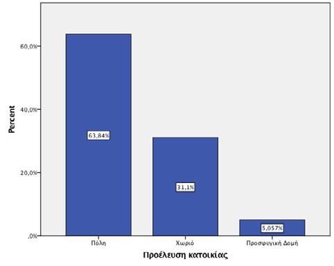
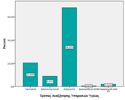
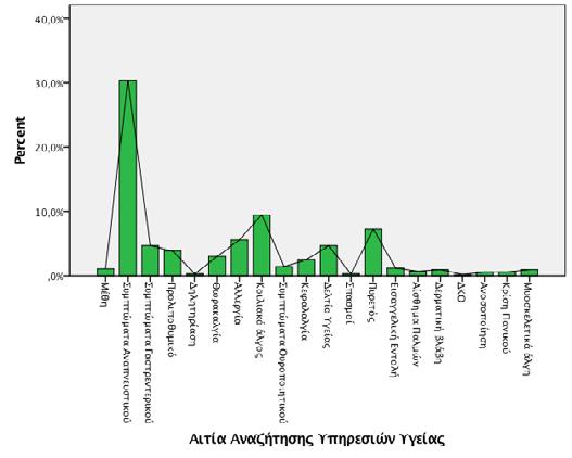
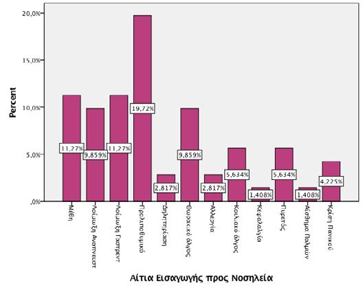
Results: During the study period, 791 adolescents (48.4% boys) aged 14.81±0.6 years, sought health services. The majority of adolescents lived in cities (63.8%), while 5.1% were adolescent-refugees. Among them 76% (n=608/791) were admitted to the Emergency Departments while only 20.5% of adolescents (n=162/791) came to an Outpatient Clinic after an appointment and 2.6% (n=21/791) were referred by a primary health care center. The most common cause of seeking medical care was the symptoms of a respiratory infection (30.4%), while the less common causes were abdominal pain (9.5%), fever (7.3%) and allergic reactions (5.6%). Among the adolescents who came to the Outpatient Clinic, the most common reason for attending was the issuance of a school health card. Admission to the Paediatric Clinic was recommended to 11.5% of the adolescents who were examined. The most common cause of hospitalization was a fainting/fainting-like episode (19.7%) followed by alcohol intoxication (11.3%), gastrointestinal infection (11.3%) and thoracic pain (9.9%). The mean duration of hospitalization was 2.8±1 days.
Conclusions: The majority of adolescents, aged 14-16, seek health services from the Emergency Departments of our hospital due to infections, as observed in childhood. Causes related to the psychosomatic changes of adolescence and high-risk behaviors are following.
Keywords: adolescent, adolescence, health care, hospitalization, hospital, pediatrician, pediatrics
*Valentina Diamantidou and Alexandra Tsiftsaki have equally contributed to this study (co-first authorship).
2. Blake Κ, Mann Κ, Kutcher M. The structured communication adolescent guide (SCAG). The journal of Teaching and Learning Resources 2008. https://doi.org/10.15766/mep_23748265.798 PMID: 15842682
3. Butragueño Laiseca L, González Martínez F, Oikonomopoulou N, Pérez Moreno J, Toledo del Castillo B, González Sánchez MI, et al. Survey of adolescent perception of hospital admission. The importance of hospital humanisation. Rev Chil Pediatr 2016;87(5):373-379.
4. Pileggi C, Raffaele G, Angelillo IF. Paediatric utilization of an emergency department in Italy. Eur J Public Health 2006;16:565-569.
5. Grove DD, Lazebnik R, Petrack EM. Urban emergency department utilization by adolescents. Clin Pediatr 2000;39:479-483.
6. Marsh RH, Mollen CJ, Shofer FS, Baren JM. Characteristics that distinguish adolescents who present to a children's hospital emergency department from those presenting to a general emergency department. Pediatr Emerg Care. 2009;25:376-379.
7. Coleman J, Brooks F, and Threadgold P. Key Data on Adolescence.“Office of National Statistics,” Census Data, 2011.
8. Homere A, Reddy S, Haller L, Richey J, Gefter L. How do underserved adolescents want to learn about health? An exploration of health concerns, preferences, and resources utilized. J Natl Med Assoc. 2022;114:518-524.
9. Vinagre MDG, Barros L. Adolescents' preferences regarding health care. Cien Saude Colet. 2019 May 30;24(5):1627-1636.
10. Brisson J, Ravitsky V, Williams-Jones B. Fostering autonomy for adolescents to access health services: A need for clarifications. J Adolesc Health. 2021;68:1038-1039.
11. Ziv A, Boulet JR, Slap GB. Emergency department utilization by adolescents in the United States. Pediatrics 1998;101:987-994.
12. Wilson KM, Klein JD. Adolescents who use the emergency department as their usual source of care. Arch Pediatr Adolesc Med 2000;154:361-365.
13. Lehmann CU, Barr J, Kelly PJ. Emergency department utilization by adolescents. JAdolesc Health 1994;15:485-490.
14. Krauss BS, Harakal T, Fleisher GR. The spectrum and frequency of illness presenting to
a pediatric emergency department. Pediatr Emerg Care 1991;7:67-7.
15. KleinJD, McNulty M, Flatau CN. Adolescents' access to care. Arch Pediatr Adolesc Med 1998;152:676-682.
16. Grove DD, Lazebnik R, Petrack EM. Urban emergency department utilization by adolescents. Clin Pediatr (Phila). 2000;39:479-483.
17. Hardin HK, Alchami H, Lee D, Jones MS. Unmet health need and perceived barriers to health care among adolescents living in a rural area. Child Health Care. 2021;50:108-123.
18. Dixon SK, Hoopes AJ, Benkeser D, Grigg A, Grow HM. Characterizing key components of a medical home among rural adolescents. J Adolesc Health. 2016;58:141-7.
Correspondence
Athanasios Tragiannidis
S. Kyriakidi 1, 54636, Thessaloniki
Τ. +302310994804
Ε. atragian@auth.gr atragian@hotmail.com
Role of presepsin and proadrenomedullin in the diagnosis and outcome of infections in neonates and children
Despite the advances in recent years, infections are still a main cause of morbidity and mortality in infants and children. The presence of biomarkers, as C-reactive protein that has widely been used in the last decades and procalcitonin that has been studied in the last 2 decades are helpful for diagnosis and outcome of severe infections in neonates and children. Nevertheless, there is a need for the use of newer biomarkers for screening, diagnosis, course and outcome of infections. Additionally, the role of biomarkers could help in the stratification of patients in risk groups and as monitoring of therapy response and rational use of antibiotics. Therefore, aim of the present review was the study of current literature regarding the newer biomarkers as presepsin and prodrenomedullin. Presepsin is a glycoprotein expressed in monocytes and phagocytes and its levels are increased within 2 hours after initiation of inflammation. Its role has been studied in neonates and children with sepsis and in febrile neutropenia episodes of children with cancer and has been shown that is a reliable marker of early sepsis in neonates. Proadrenomedullin is the precursor peptide of adrenomedullin released from various tissues as vascular endothelium as a response to stress and has an anti-inflammatory, antimicrobial and vasoregulatory effect. Its role has been studied in respiratory infections of pediatric patients and in febrile neutropenia episodes of children with malignancy. According to current literature these biomarkers play a role in outcome and course of severe infections in childhood.
2nd Pediatric Department, Aristotle University of Thessaloniki, AHEPA Hospital, Thessaloniki
Lemonia Skoura
University Microbiology Laboratory, AHEPA Hospital, Thessaloniki
ΣΥΝΤΟΜΟΓΡΑΦΙΕΣ
PCT: προκαλσιτονίνη
TNF: παράγοντας
νέκρωσης των όγκων
ADM: αδρενομεδουλλίνη
Pro-ADM:
προαδρενομεδουλλίνη
Baraka
Βιβλιογραφία
1. Ruth A, McCracken CE, Fortenberry JD, Hall M, Simon HK, Hebbar KB. Pediatric severe sepsis: current trends and outcomes from the pediatric health information systems database. Pediatr Crit Care Med. 2014; 15(9): 828–838.
2. Hartman ME, Linde-Zwirble WT, Angus DC, Watson RS. Trends in the epidemiology of pediatric severe sepsis*. Pediatr Crit Care Med. 2013; 14(7): 686–693.
3. Kawasaki T. Update on pediatric sepsis: a review. J Intensive Care. 2017; 5(1): 47.
4. Fleischmann-Struzek C, Goldfarb DM, Schlattmann P, Schlapbach LJ, Reinhart K, Kissoon N. The global burden of paediatric and neonatal sepsis: a systematic review. Lancet Respir Med. 2018; 6(3): 2223–2230.
5. Balamuth F, Weiss SL, Neuman MI, Scott H, Brady PW, Paul R, Farris RW, McClead R, Hayes K, Gaieski D, et al. Pediatric severe sepsis in U.S. children's hospitals. Pediatr Crit Care Med. 2014; 15(9): 798–805.
6. Carrigan SD, Scott G, Tabrizian M. Toward resolving the challenges of sepsis diagnosis. Clin Chem. 2004; 50(8): 1301–1314.
7. Markanday A. Acute Phase Reactants in Infections: Evidence-Based Review and a Guide for Clinicians. Open Forum Infectious Dis. 2015; 2(3): ofv098.
8. Henriquez-Camacho C, Losa J. Biomarkers for sepsis. Biomed Res Int. 2014; 2014: 547818.
9. Standage SW, Wong HR. Biomarkers for pediatric sepsis and septic shock. Expert Rev Anti-Infect Ther. 2011; 9(1): 71–79.
10.Sanders S, Barnett A, Correa-Velez I, Coulthard M, Doust J. Systematic review of the diagnostic accuracy of C-reactive protein to detect bacterial infection in nonhospitalized infants and children with fever. J Pediatr. 2008; 153(4): 570–574.
11.Pontrelli G, De Crescenzo F, Buzzetti R, Jenkner A, Balduzzi S, Calo Carducci F, Amodio D, De Luca M, Chiurchiu S, Davies EH, et al. Accuracy of serum procalcitonin for the diagnosis of sepsis in neonates and children with systemic inflammatory syndrome: a meta-analysis. BMC Infect Dis. 2017; 17(1): 302.
12.Rowland T, Hilliard H, Barlow G. Procalcitonin: potential role in diagnosis and management of sepsis. Adv Clin Chem. 2015; 68: 71–86.
13.van Rossum AM, Wulkan RW, Oudesluys-Murphy AM. Procalcitonin as an early marker of infection in neonates and children. Lancet Infect Dis. 2004; 4(10): 6620–6630.
14.Lin SG, Hou TY, Huang DH, He SY, Lin YD, Zhang LY, Hsieh PS. Role of procalcitonin in the diagnosis of severe infection in pediatric patients with fever and neutropenia--a systemic review and meta-analysis. Pediatr Infect Dis J. 2012; 31(10): e182–188.
15.Yo C-H, Hsieh P-S, Lee S-H, Wu J-Y, Chang S-S, Tasi K-C, Lee C-C. Comparison of the test characteristics of Procalcitonin to C-reactive protein and leukocytosis for the detection of serious bacterial infections in children presenting with fever without source: a systematic review and metaanalysis. Ann Emerg Med. 2012; 60(5): 591–600.
16. Volanakis JE. Human C-reactive protein: expression, structure, and function. Mol Immunol. 2001; 38(2–3): 1189–97.
17.Okamura Y, Yokoi H. Development of a point-of-care assay system for measurement of presepsin (sCD14-ST). Clin Chim Acta. 2011; 412(23–24): 2157–2161.
18.Lanziotti VS, Póvoa P, Soares M, Silva JRLE, Barbosa AP, Salluh JIF. Use of biomarkers in pediatric sepsis: literature review. Revista Brasileira de terapia intensiva. 2016; 28(4): 472–482.
19.Meisner M. Update on procalcitonin measurements. Ann Lab Med. 2014; 34(4): 263–273.
20.Becker KL, Snider R, Nylen ES. Procalcitonin in sepsis and systemic inflammation: a harmful biomarker and a therapeutic target. Br J Pharmacol. 2010; 159(2): 2253–64.
21.Zou Q, Wen W, Zhang XC. Presepsin as a novel sepsis biomarker. World J Emerg Med.
2014; 5(1): 16–19.
22.Mussap M, Noto A, Fravega M, Fanos V. Soluble CD14 subtype presepsin (sCD14-ST) and lipopolysaccharide binding protein (LBP) in neonatal sepsis: new clinical and analytical perspectives for two old biomarkers. J Matern Fetal Neonatal Med. 2011; 24(Suppl 2): 12–14.
23.Wu J, Hu L, Zhang G, Wu F, He T. Accuracy of Presepsin in Sepsis diagnosis: a systematic review and meta-analysis. PLoS One. 2015; 10(7): e0133057.
24.Önal U, Valenzuela-Sánchez F, Vandana KE, Rello J. Mid-Regional Pro-Adrenomedullin (MR-proADM) as a Biomarker for Sepsis and Septic Shock: Narrative Review. Healthcare (Basel). 2018; 6(3): 110.
25.Bellia C, Agnello L, Lo Sasso B, Bivona G, Raineri MS, Giarratano A, Ciaccio M. Midregional pro-adrenomedullin predicts poor outcome in non-selected patients admitted to an intensive care unit. Clin Chem Lab Med. 2019; 57(4): 549-555.
26.Jougasaki M, Burnett JC Jr. Adrenomedullin: potential in physiology and pathophysiology. Life Sci. 2000; 66(10): 855-872.
27.Pizzolato E, Ulla M, Galluzzo C, Lucchiari M, Manetta T, Lupia E, Mengozzi G, Battista S. Role of presepsin for the evaluation of sepsis in the emergency department. Clin Chem Lab Med. 2014; 52(10): 1395-400.
28.Zou Q, Wen W, Zhang XC. Presepsin as a novel sepsis biomarker. World J Emerg Med. 2014; 5(1): 16-19.
29.Velissaris D, Zareifopoulos N, Karamouzos V, Karanikolas E, Pierrakos C, Koniari I, Karanikolas M. Presepsin as a Diagnostic and Prognostic Biomarker in Sepsis. Cureus. 2021; 13(5): e15019.
30.Hincu MA, Zonda GI, Stanciu GD, Nemescu D, Paduraru L. Relevance of Biomarkers Currently in Use or Research for Practical Diagnosis Approach of Neonatal Early-Onset Sepsis. Children (Basel). 2020; 7(12): 309.
31.Mohammad Yousef N, Hossein Bannazadeh Baghi, Presepsin: A promising biomarker for the detection of bacterial infections, Biomedicine & Pharmacotherapy. 2019; 11: 649-656.
32.Galliera, E.; Massaccesi, L.; de Vecchi, E.; Banfi, G.; Romanelli, M. Clinical application of presepsin as diagnostic biomarker of infection: Overview and updates. Clin. Chem. Lab. Med. 2019; 44: 21–27.
33.Nagata, T.; Yasuda, Y.; Ando, M.; Abe, T.; Katsuno, T.; Kato, S.; Tsuboi, N.; Matsuo, S.; Maruyama, S. Clinical impact of kidney function on presepsin levels. PLoS ONE 2015; 10: e0129159.
34.Handke, J.; Piazza, O.; Larmann, J.; Tesoro, S.; De Robertis, E. Presepsin as a biomarker in perioperative medicine. Minerva Anestesiol. 2020; 86: 768–776.
35.Nakamura, Y.; Ishikura, H.; Nishida, T.; Kawano, Y.; Yuge, R.; Ichiki, R.; Murai, A. Usefulness of presepsin in the diagnosis of sepsis in patients with or without acute kidney injury. BMC Anesthesiol. 2014; 14: 88.
36.Nakamura, Y.; Murai, A.; Mizunuma, M.; Ohta, D.; Kawano, Y.; Matsumoto, N.; Nishida, T.; Ishikura, H. Potential use of procalcitonin as biomarker for bacterial sepsis in patients with or without acute kidney injury. J. Infect. Chemother. 2015; 21: 257–263.
37.Endo, S.; Suzuki, Y.; Takahashi, G.; Shozushima, T.; Ishikura, H.; Murai, A.; Nishida, T.; Irie, Y.; Miura, M.; Iguchi, H.; et al. Usefulness of presepsin in the diagnosis of sepsis in a multicenter prospective study. J Infect Chemother. 2012; 18: 891–897.
38.Masson, S.; Caironi, P.; Fanizza, C.; Thomae, R.; Bernasconi, R.; Noto, A.; Oggioni, R.; Pasetti, G.S.; Romero, M.; Tognoni, G.; et al. Circulating presepsin (soluble CD14 subtype) as a marker of host response in patients with severe sepsis or septic shock: Data from the multicenter, randomized ALBIOS trial. Intensive Care Med. 2015: 41: 12–20.
39.Ulla, M.; Pizzolato, E.; Lucchiari, M.; Loiacono, M.; Soardo, F.; Forno, D.; Morello, F.; Lupia, E.; Moiraghi, C.; Mengozzi, G.; et al. Diagnostic and prognostic value of presepsin in the management of sepsis in the emergency department: A multicenter prospective study. Crit. Care 2013: 17, R168.
40.Endo S, Suzuki Y, Takahashi G, Shozushima T, Ishikura H, Murai A, Nishida T, Irie Y, Miura M, Iguchi H, Fukui Y, Tanaka K, Nojima T, Okamura Y. Usefulness of presepsin in the diagnosis of sepsis in a multicenter prospective study. J Infect Chemother. 2012; 18(6): 891-897.
41.Maddaloni C, De Rose DU, Santisi A, Martini L, Caoci S, Bersani I, Ronchetti MP, Auriti C. The Emerging Role of Presepsin (P-SEP) in the Diagnosis of Sepsis in the Critically Ill Infant: A Literature Review. Int J Mol Sci. 2021; 22(22): 12154.
42.Chenevier-Gobeaux C, Borderie D, Weiss N, Mallet-Coste T, Claessens YE. Presepsin (sCD14-ST), an innate immune response marker in sepsis. Clin Chim Acta. 2015; 450: 97103.
43.Sakyi S.A., Enimil A., Adu D.K., Ephraim R.D., Danquah K.O., Fondjo L., Baidoe-Ansah D., Adoba P., Toboh E., Afranie B.O. Individual and combined bioscore model of presepsin, procalcitonin, and high sensitive C-reactive protein as biomarkers for early diagnosis of paediatric sepsis. Heliyon. 2020; 6: e04841.
44.Yoon S.H., Kim E.H., Kim H.Y., Ahn J.G. Presepsin as a diagnostic marker of sepsis in children and adolescents: A systemic review and meta-analysis. BMC Infect. Dis. 2019; 19: 760.
45.Sharma, A.; Thakur, A.; Bhardwaj, C.; Kler, N.; Garg, P.; Singh, M.; Choudhury, S. Potential biomarkers for diagnosing neonatal sepsis. Curr. Med. Res. Pract. 2020; 10: 12–17.
46.Ruan, L.; Chen, G.Y.; Liu, Z.; Zhao, Y.; Xu, G.Y.; Li, S.F.; Li, C.N.; Chen, L.S.; Tao, Z. The combination of procalcitonin and C-reactive protein or presepsin alone improves the accuracy of diagnosis of neonatal sepsis: A meta-analysis and systematic review. Crit. Care 2018; 22: 1–9.
47.Van Maldeghem, I.; Nusman, C.M.; Visser, D.H. Soluble CD14 subtype (sCD14-ST) as biomarker in neonatal early-onset sepsis and late-onset sepsis: A systematic review and meta-analysis. BMC Immunol. 2019: 20: 17.
48.Bellos, I.; Fitrou, G.; Pergialiotis, V.; Thomakos, N.; Perrea, D.N.; Daskalakis, G. The diagnostic accuracy of presepsin in neonatal sepsis: A meta-analysis. Eur. J. Pediatrics 2018: 177: 625–632.
49.Memar, M.Y.; Baghi, H.B. Presepsin: A promising biomarker for the detection of bacterial infections.Biomed. Pharmacother. 2019: 111: 649–656.
50.Tzialla, C.; Manzoni, P.; Achille, C.; Bollani, L.; Stronati, M.; Borghesi, A. New Diagnostic Possibilities for Neonatal Sepsis. Am. J. Perinatol. 2018: 35: 575–577.
51.Montaldo P, Rosso R, Santantonio A, Chello G, Giliberti P. Presepsin for the detection of early-onset sepsis in preterm newborns. Pediatr Res. 2017; 81(2): 329-334.
52.Delanghe, J.R.; Speeckaert, M.M. Translational research and biomarkers in neonatal sepsis. Clin. Chim. Acta 2015: 451: 46–64.
53.Memar, M.Y.; Alizadeh, N.; Varshochi, M.; Kafil, H.S. Immunologic biomarkers for diagnostic of early-onset neonatal sepsis. J. Matern. Neonatal Med. 2019; 32: 143–153.
54.Parri, N.; Trippella, G.; Lisi, C.; De Martino, M.; Galli, L.; Chiappini, E. Accuracy of presepsin in neonatal sepsis: Systematic review and meta-analysis. Expert Rev. Anti. Infect. Ther. 2019: 17: 223–232.
55.Yoon SH, Kim EH, Kim HY, Ahn JG. Presepsin as a diagnostic marker of sepsis in children and adolescents: a systemic review and meta-analysis. BMC Infect Dis. 2019; 19(1): 760.
56.Santolaya ME, Alvarez AM, Avilés CL, Becker A, Mosso C, O'Ryan M, Payá E, Salgado C, Silva P, Topelberg S, Tordecilla J, Varas M, Villarroel M, Viviani T, Zubieta M. Admission clinical and laboratory factors associated with death in children with cancer during a febrile neutropenic episode. Pediatr Infect Dis J. 2007; 26(9): 794-798.
57.Phillips RS, Wade R, Lehrnbecher T, Stewart LA, Sutton AJ. Systematic review and meta-analysis of the value of initial biomarkers in predicting adverse outcome in febrile neutropenic episodes in children and young people with cancer. BMC Med. 2012; 10: 6.
58.Olad E, Sedighi I, Mehrvar A, Tashvighi M, Fallahazad V, et al. Presepsin (scd14) as a marker of serious bacterial infections in chemotherapy induced severe neutropenia. Iran J Pediatr 2014; 24: 715–722.
59.Poggi C, Bianconi T, Gozzini E, Generoso M, Dani C. Presepsin for the detection of lateonset sepsis in preterm newborns. Pediatrics 2015; 135: 68–75.
60.Stubjar D, Kopitar A, Groselj-Grenc M, Suhadolc K, Fabjan T, et al. Diagnostic accuracy of presepsin (sCD14-ST) for prediction of bacterial infection in cerebrospinal fluid samples from children with suspected bacterial meningitis or ventriculitis. J Clin Microbiol 2015; 53: 1239–1244.
61.Urbonas V, Eidukaite A, Tamuliene A, Tamuliene I. The predictive value of soluble biomarkers (CD14 subtype, interleukin-2 receptor, human leucocyte antigen-G) and procalcitonin in the detection of bacteremia and sepsis in pediatric oncology patients with chemotherapy-induced febrile neutropenia. Cytokine 2013; 62: 34–37.
62.Osman AS, Awadallah M, El-Mageed T, Abed NT, Goudah E. Presepsin as a Novel Diagnostic Marker in Neonatal Septicemia. Egypt J Med Microbiol 2015; 24: 21–26.
63.Plesko M, Suvada J, Makohusova M, Waczulikova I, Behulova D, Vasilenkova A, Vargova M, Stecova A, Kaiserova E, Kolenova A. The role of CRP, PCT, IL-6 and presepsin in early diagnosis of bacterial infectious complications in paediatric haemato-oncological patients. Neoplasma. 2016; 63(5): 752-760.
64.Arıkan K, Karadag-Oncel E, Aytac S, Cetin M, Cengiz AB, Gümrük F, Kara A, Ceyhan M. Usage of Plasma Presepsin, C-Reactive Protein, Procalcitonin and Proadrenomedullin to Predict Bacteremia in Febril Neutropenia of Pediatric Hematological Malignancy Patients. Lab Med. 2021; 52(5): 477-484.
65.Baraka A, Zakaria M. Presepsin as a diagnostic marker of bacterial infections in febrile neutropenic pediatric patients with hematological malignancies. Int J Hematol. 2018; 108(2): 184-191.
66.Temmesfeld-Wollbruck B, Brell B, David I, et al. Adrenomedullin reduces vascular hyperpermeability and improves survival in rat septic shock. Intensive Care Med. 2007; 33: 703–710.
67.Carrizo GJ, Wu R, Cui X, Dwivedi AJ, Simms HH, Wang P. Adrenomedullin and adrenomedullin-binding protein-1 downregulate inflammatory cytokines and attenuate tissue injury after gut ischemia-reperfusion. Surgery. 2007; 141:245–253.
68.Vigue B, Leblanc PE, Moati F, et al. Mid-regional pro-adrenomedullin (MR- proADM), a marker of positive fluid balance in critically ill patients: results of the ENVOL study. Crit Care. 2016; 20:363.
69.Gille J, Ostermann H, Dragu A, Sablotzki A. MR-proADM: A new biomarker for early diagnosis of sepsis in burned patients. J Burn Care Res. 2017; 38:290–298.
70.Bustamante A, García-Berrocoso T, Penalba A, et al. Sepsis biomarkers reprofiling to predict stroke-associated infections. J Neuroimmunol. 2017; 312: 19–23.
71.Decker SO, Sigl A, Grumaz C, et al. Immune-response patterns and next generation sequencing diagnostics for the detection of mycoses in patients with septic shock - results of a combined clinical and experimental investigation. Int J Mol Sci. 2017;18: 1796
72.Albrich WC, Dusemund F, Ruegger K, et al. Enhancement of CURB65 score with proadrenomedullin (CURB65-A) for outcome prediction in lower respiratory tract infections: derivation of a clinical algorithm. BMC Infect Dis. 2011; 11:112.
73.Albrich WC, Ruegger K, Dusemund F, et al. Optimised patient transfer using an innovative multidisciplinary assessment in Kanton Aargau (OPTIMA I): an observational survey in lower respiratory tract infections. Swiss Med Wkly. 2011;141: w13237.
74.van der Starre WE, Zunder SM, Vollaard AM, van NC, Stalenhoef JE, Delfos NM, van't Wout JW, Spelt IC, Blom JW, Leyten EM, et al. Prognostic value of pro-adrenomedullin, procalcitonin and C-reactive protein in predicting outcome of febrile urinary tract infection. Clin Microbiol Infect. 2014;20(10): 1048–1054.
75.Elke G, Bloos F, Cameron D, Wilson DC, Brunkhorst FM, Briege J, Reinhart K, Loeffler M, Kluge S, Nierhaus A, Jaschinski U, Moerer O, Weyland A, Meybohmand P and the SepNet Critical Care Trials Group. The use of mid-regional proadrenomedullin to identify disease severity and treatment response to sepsis - a secondary analysis ofa large randomised controlled trial. Critical Care 2018; 22: 79
76.Al Shuaibi M, Bahu RR, Chaftari AM, Al. Wohoush I. Shomali W, Jiang Y, Debiane L, Raad, Jabbour J, Al Akhrass F, Hachem RY, Raad I. Pro-adrenomedullin as a novel biomarker for predicting infections and response to antimicrobials in febrile patients with hematologic malignancies. Clin Infect Dis. 2013; 56(7): 943-950.
77.Alcoba G , Manzano S, Lacroix L, Galetto-Lacour A, Gervaix A. Proadrenomedullin and copeptin in pediatric pneumonia: a prospective diagnostic accuracy study. BMC Infectious Diseases 2015; 15: 347.
78.Sarda Sanchez M, Hernandez JC, Hernandez-Bou S, Teruel GC, Rodriguez JV, Cubells CL. Pro-adrenomedullin usefulness in the management of children with community-acquired pneumonia, a preliminar prospective observational study. BMC Res Notes. 2012; 5:363.
79.Korkmaz MF, Güzel A, Açıkgöz M, Okuyucu A, Alaçam H. Reliability of Pro-adrenomedullin and Interleukin 1β in Predicting Severity of Community-Acquired Pneumonia in Pediatric Patients. Ann Clin Lab Sci. 2018 Jan;48(1):81-89.
80.Baumann P, Fuchs A, Gotta V, Ritz N, Baer G, Bonhoeffer JM, Buettcher M, Heininger U, Szinnai G, Bonhoeffer J; ProPAED study group. The kinetic profiles of copeptin and mid regional proadrenomedullin (MR-proADM) in pediatric lower respiratory tract infections. PLoS One. 2022; 17(3): e0264305.
81.Debiane L, Hachem RY, Al Wohoush I, et al. The utility of proadrenomedullin and procalcitonin in comparison to C-reactive protein as predictors of sepsis and bloodstream infections in critically ill patients with cancer. Crit Care Med. 2014; 42: 2500–2507.
82.Oncel MY, Dilmen U, Erdeve O, Ozdemir R, Calisici E, Yurttutan S. Canpolat FE, Oguz SS, Uras N. Proadrenomedullin as a prognostic marker in neonatal sepsis. Pediatric Res. 2012; 72(5): 507-512
83.Demirkaya M, Tugcu D, Akcay A, Aydogan G, Akıcı F, Salcioglu Z, et al. Adrenomedullin-A new marker in febrile neutropenia: comparison with CRP and procalcitonin. Pediatr Hematol Oncol. 2015; 32(7): 482-489.
84.Agnello L, Bivona G, Parisi E, Lucido GD, Iacona A, Ciaccio AM, Giglio RV, Ziino O, Ciaccio M. Presepsin and Midregional Proadrenomedullin in Pediatric Oncologic Patients with Febrile Neutropenia. Lab Med. 2020; 51(6): 585-591.
Correspondence
Koutsaftiki Chrysi
Sof. Venizelou 77, Halandri, PC 15232
T. +306970061238
E. kochrysa@yahoo.com
"Long-Post COVID-19" syndrome in children and adolescents
Background: COVID-19 infection in the vast majority of children is of mild or moderate severity and mortality is almost zero. However, symptoms from various systems may persist, re-emerge, or recur for months after the acute phase, representing the Long COVID-19 syndrome that has been described primarily in adults.
Purpose-Objectives: The present review aims to evaluate the symptoms-conditions described and defined in the international literature as Long-Post COVID (LPC), as well as to capture the frequency, in different age groups of children. In addition, an attempt is made to record the major risk factors, the possible pathophysiological mechanisms and to highlight ways of management.
Method: Studies of the international literature, published in PubMed database, as well as articles from other sources were reviewed. Particular emphasis was placed on systematic reviews and meta-analyses published in the year 2022, reflecting accumulated experience. The search was made in September 2022.
Results: The most common symptoms are: headache, fatigue, cognitive disorders as well as myalgias/arthralgias. The frequency varies a lot, depending on the study, while it seems that almost all studies presented many methodological problems. Pathophysiologically, the activation of cytokines and other immunologic mechanisms seem to dominate. At the same time, many children with a negative history of infection also showed symptoms such as anxiety, sleep disturbances, headaches and decline in mental functions, attributed to the pandemic itself.
Conclusions: It is necessary to clarify the syndrome, in order to assess the actual risk that children face, so as to prepare health workers for its best management, but also to assess the necessity of universal vaccination of children against the new coronavirus as well as the evaluation of measures to safeguard public health.
Paediatric department, “Rafina” Health Care Centre, Rafina-Attika, Greece
Sergentanis Theodoros
Merakou Kyriakoula
Kornarou Eleni
Department of Public Health Policy, School of Public Health, University of West Attica, Athens, Greece
Μεθοδολογία
((long-Covid) OR (long-Covid symptoms) OR (long Hauler*) OR (post-Covid-19) OR (postacute Covid-19 syndrome) OR (post-acute Covid-19 symptoms) OR (Covid-19 sequelae) OR (chronic Covid-19 syndrome) OR (post-infectious Covid-19) OR (post Covid-19 recovery syndrome) OR (Post-acute sequelae of Sars-CoV-2 infection) OR (post Covid-19 recovery syndrome)) AND (pediatr* OR paediat* OR kids OR young OR infant OR adolesc*).
οργανισμούς WHO, Centers for Disease Control and Prevention (CDC), και τους βρετανικούς National Institute for Health and Care Excellence (NICE), UK National Institute for Health Research (NIHR) καθώς και CLoCk Consortium όπως
World Health Organization
Centers of Disease Control and Prevention
National Institute for Health and Care Excellence (NICE)
National Institute for Health Research (NIHR)
ως Long-Covid τα σημεία και τα συμπτώματα εκείνα που συνεχίζουν ή αναπτύσσονται μετά την οξεία COVID-19. Δηλαδή επίμονα συμπτώματα της λοίμωξης (ongoing symptomatic COVID-19) που διαρκούν από 4 έως 12 εβδομάδες αλλά και συμπτώματα μετά τη λοίμωξη (post-COVID-19 syndrome) που
1. Lopez-Leon S, Wegman-Ostrosky T, Cipatli Ayuzo del Valle N, Perlman C, Sepulveda R, et al. Long COVID in children and adolescents: asystematic review and meta-analyses Scientific Reports (2022) 12:9950 https://doi.org/10.1038/ss41598-022-13495-5
2. Merzon E, Weiss M, Krone B, Cohen S, Ilani G, Vinker S, et al. Clinical and socio-demographic variables associated with the diagnosis of Long COVID syndrome in youth: a population-based study. Int.J.Environ.Res.Public Health 2022,19,5993. https:// doi.org/10.3390/ ijerph19105993
3. Pavli A, Theodoridou M, Maltezou H. Post-COVID Syndrome: Incidence, clinical spectrum and challenges for Primary Healthcare Professionals. Archives of Medical Research 52 (2021) 575-581.
4. Callard F, P E. How and why patients made long COVID. Soc Sci Med 2021; 268:113426
5. Soriano JB, Murthy S, Marshall JC, Relan P, Diaz JV, on behalf of the WHO Clinical Case Definition Working Group on Post-COVID-19 condition. A clinical case definition of postCOVID-19 condition by a Delphi consensus. Lancet Infect Dis 2022; 22: e102-07. https://doi. org/10.1016/S1473-3099(21)00703-9
6. Centers for Disease Control and Prevention Post-COVID Conditions: Information for Healthcare Providers. U.S. Dep. Heal. Hum. Serv.2021, 1–3
7. Overview| Covid-19 rapid guideline: managing the long-term effects of COVID-19 | Guidance| NICE [Internet]. Nice.org.uk.2021 [cited 07 June 2021]. Available from: https://www. nice.org.uk/guidance/NG 188.)
8. NIHR Evidence. 2021 [cited 02 October 2021]. Living with Covid-19 Second Review [Internet]. Available from: https://evidence.nihr.ac.uk/themedreview/living-with-covid19-sec-
ond-review/ (accessed 02 October 2021)
9. Stephenson T, Allin B, Nugawela M, Rojas N, Dalrymple E, et al. Long COVID (post-COVID-19 condition) in children: a modified Delphi process Arch Dis Child 2022;107:674-680)
10.Nittas V, Gao M, West EA, Ballouz T, Menges D, et al Long COVID through a public health lens: an umbrella review Public Health Rev 2022, 43:1604501. Doi:10.3389/phrs.2022.1604501
11.Deer RR, Rock MA, Vasilevsky N, Carmody L, Rando H, Anzalone AJ, et al. Characterizing Long COVID: Deep phenotype of a complex condition. EBioMedicine 74(2021)103722
12.Ludvigsson JF. Case report and systematic review suggest that children may experience similar long-term effects to adults after clinical COVID-19. Acta Paediatr. 2021,110,914-921.
13.Zimmermann P, Pittet LF, Curtis N. How common is Long COVID in children and adolescents? Pediatr nfect Dis J 2021; 40: e482-e487
14.Zimmermann P, Pittet LF, Curtis N. Long covid in children and adolescents. Risk appears low, but many questions remain. BMJ 2022;376:o143 http://dx.doi.org/10.1136/bmg.o143
15.Zimmermann P, Pittet LF, Curtis N. The challenge of studying Long COVID: an updated review. Pediatr Infect Dis J 2022; 41:424-426
16.Kikkenborg-Berg S, Palm P, Nygaard U, Bundgaard H, Schmidt-Petersen MN, Rosenkilde S, et al. Long COVID symptoms in SARS-CoV-2-positive children aged 0-14 years and matched controls in Denmark (LongCOVIDKidsDK): a national, cross-sectional study. Lancet Child Adolesc Health 2022; 6:614-23 https://doi.org/10.1016/S2352-4642(22)00154-7
17.Kikkenborg-Berg S, Dam Nielsen S, NygaardU, Bundgaard H, Palm P, et al. Long COVID symptoms in SARS-CoV-2-positive adolescents and matched controls (LongCOVODKidsDK): a national, cross-sectional study. Lancet Child Adolesc Health 2022; 6:240-48.
18.Morrow AK, Malone LA, Kokorelis C, Petracek LS, et al. Long-term COVID 19 sequelae in adolescents: the overlap with orthostatic intolerance and ME/CFS Current Pediatric Reports (2022) 10:31-44 https://doi.org/10.1007/s40124-022-00261-4
19.Kompaniyets L, Bull-Otterson L, Boehmer TK, et al Post-COVID-19 symptoms and conditions among children and adolescents-U.S. March 1, 2020–January 31, 2022. MMWR Morb Mortal Wkly Rep 2022; 71:993–999. DOI: http://dx.doi.org/10.15585/mmwr.mm7131a3.
20.Barret CE, Koyama AK, Alvarez AK, Alvarez P, Chow W, Lundeen EA, et al Risk of newly diagnosed diabetes >30 days after SARS-CoV-2 infection among persons aged<18 years United States, March 1, 2020-June 28,2021 MMWR Morb Mortal Wkly Rep. 2022 Jan 14;71(2): 59-65.doi: 10.15585/mmwr.mm7102e2.
21.Ludvigsson JF. Convulsions in children with COVID-19 during the Omicron wave. Acta Paediatr. 2022 May;111(5):1023-1026. doi: 10.1111/apa.16276. Epub 2022 Feb 10. PMID: 35098577
22.Thallapureddy K, Thallapureddy K, Zerda E, Suresh N, et al. Current Pediatrics Reports (2022) 10:11-17 https://doi.org/10.1007/s40124-021-00260-x
23.Benhood SA, Shafran R, Bennet SD, Zhang AXD, O’Mahoney LL, Stephenson TJ, et al. Persistent symptoms following SARS-CoV-2 infection amongst children and young people: A meta-anlysis of controlled and uncontrolled studies. Journal of infection 84 (2022) 158170.
24.Esposito S, Principi N, Azzari C, Cardinale F, Di Mauro G, Galli L, et al. Italian intersociety consensus on management of long covid in children. Italian Journal of Pediatrics (2022) 48:42 https://doi.org/10.1186/s13052-022-01233-6
25.Izquierdo-Pujol J, Moron-Lopez S, Dalmau J, Gonzalez-Aumatell A, Carreras-Adab C, Mendez-Picardo J Post-COVID-19 condition in children and adolescents: an emerging problem. Front. Pediatr. (2022) 10:894294 doi: 10.3389/fped.2022.894294
26.Osmanov IM, Spiridonova E, Bobkova P, Gamirova A, Shikhaleva A, Andreeva M, et al. Risk factors for post -COVID-19 condition in previously hospitalized children using the ISARIC Global follow-up protocol: a prospective cohort study. Eur Respir J 2022; 59: 2101341 [DOI: 10.1183/13993003.01341-2021]
27.Greenhalgh T, Sivan M, Delaney B, Evans R, Milne R Long covid-an update for primary care BMJ 2022; 378: e072117 http://dx.doi.org/10.1136/bmj-2022-072117
28.Deville JG, Song E, Ouellette CP COVID-19: Clinical manifestations and diagnosis in children-UpToDate https://www.uptodate.com/contents/covid-19-clinical-manifestations-...2022
29.Piazza M, Di Cicco M, Pecoraro L, Ghezzi M, Peroni D, Comberiati P. Long COVID-19 in children: from the pathogenesis to the biologically plausible roots of the syndrome. Bio-
30.Fainardi V, Meoli A, Chiopris G, Motta M, Skenderaij K, et al Long COVID in children and adolescents Life 2022,12,285. htpps://doi.org/10.3390/life12020285
31.Crook H, Raza S, Nowell J, Young M, Edison P. Long covid-mechanisms, risk factors and management. BMJ 2021, 374, n1648,
32.Glynne P, Tahmasebi N, Gant V, Gupta R Long COVID following mild SARS-CoV-2 infection: characteristic T cell alterations and response to antihistamines. J Investig Med 2022; 70:61-67. (http://dx.doi.org/10.1136/jim-2021-002051)
33.Sante GD, Buonsenso D, De Rose C, Valentini P, Ria F, Sanguinetti M, et al. Immune profile of children with post-acute sequelae of SARS-CoV-2infection (LONG COVID). medRxiv. (2021) [Preprint]. doi: 10.1101/2021.05.07.21256539
34. Simani L, Ramezani M, Darazam IA et al. Prevalence and correlates of chronic fatigue syndrome and post-traumatic stress disorder after the outbreak of the COVID-19. J neurovirol 2021;27: 154-159
35.Bronson SC. Practical scenarios and day-to-day challenges in the management of diabetes in COVID-19—dealing with the ‘double trouble’. Prim Care Diabetes 2021; 15:737-9. PMID: 34039524 https://doi.org/10.1016/j.pcd.2021.05.007
36.Munblit D, Simpson F, Mabbitt J, Dunn-Galvin A, Semple C, O Warner J. Legacy of COVID-19 infection in children: long-COVID will have a lifelong health/economic impact Arch Dis Child 2022; 107: e2.
37.Borch L, Holm M, Knudsen M, Ellermann-Eriksen S, Hagstroem S. Long COVID symptoms and duration in SARS-CoV-2 positive children―a nationwide cohort study. European Journal of Pediatrics (2022) 181:1597-1607 https://doi.org/10.1007/s00431-021-04345-z
38.Hoang A, Chorath K, Moreira A, et al. COVID-19 in 7780 pediatric patients: A systematic review. EClinicalMedicine 2020; 24:100433.
39.Cozzi G, Marchetti F, Barbi E. Clinicians need to be careful that they do not confuse mental health issues and long COVID in children and adolescents. Acta Paediatr. 2022; 00:1-3. Doi:10.1111/apa.16332
40.Notarte KI, Catahay JA, Velasco JV, Pastrana A, Ver AT, et al. Impact of COVID-19 vaccination on the risk of developing long-COVID and on existing long-COVID symptoms: a systematic review. EClinicalMedicine. 2022; 53:101624. doi: 10.1016/j.eclinm.2022.101624. eCollection 2022 Nov.
41.Spiliopoulos L, Vedel Sørensen AI, Bager P, Munk Nielsen N, Vinsløv Hansen J, Koch A, et al. Post-acute symptoms four months after SARS-CoV-2 infection during the Omicron period: a nationwide Danish questionnaire study. medRxiv preprint doi: https://doi.org/10.1101/2022. 10.12.22280990; this version posted October 12, 2022
42.Goldman RD. Long COVID in children. Can Fam Physician 2022; 68:263-5. DOI:10.46747/ cfp.6804263
"pathophysiology".
E.
Correspondence
Georgios Katsaras
End of Egnatia Str, 58200, Edessa T. +306939193901
E. gkatsaras84@gmail.com
Lithiasis of the urinary system in childhood
Anastasios Vladikas, Anastasia Anastasiou, Miguel Shkira, Georgios Katsaras
Abstract
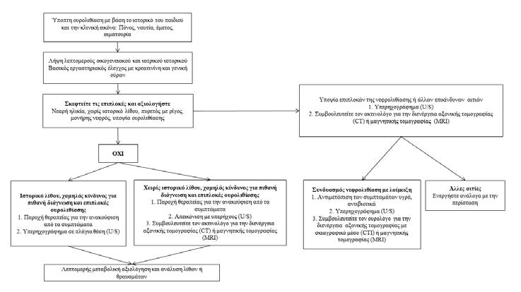
Urolithiasis in children is not a common disease in developed countries. However, in recent years its incidence has been steadily increasing, while wrong diagnosis can lead to chronic kidney disease.
Methods: Extensive literature review in the international databases PubMed and Scopus took place, in which the key terms used were "pediatric urolithiasis", "epidemiology", "metabolic factors", "pathophysiology".
Results: Metabolic, genetic, anatomical, dietary, and environmental factors are to blame for this increase. The typical clinical symptomatology of renal colic that characterizes lithiasis in adults is present in only 50% of children with urolithiasis. Microscopic or macroscopic hematuria is the most common finding and affects up to 90% of patients. In younger children the main symptom may be only recurrent urinary tract infections. The majority of renal stones in children are calcium oxalate or calcium phosphate.
Conclusions: Urolithiasis is a significant problem with increasing frequency in childhood, which requires the pediatrician to be alert for early diagnosis. Investigation of possible anatomical abnormalities is required, as well as a thorough metabolic control to avoid recurrences. Patients should be monitored frequently by specialists and depending on the underlying cause, the appropriate intervention should be performed.
Paediatric Department, General Hospital of Pella - Hospital Unit of Edessa, Edessa
Anastasia Anastasiou
Health Care Center of Edessa, Edessa
Pediatric Nephrology Department, 4th Pediatric Department, Aristotle University of Thessaloniki, Papageorgiou General Hospital of Thessaloniki, Thessaloniki
Tamm-Horsfall (THP), οι γλυκοζαμινογλυκάνες (GAGs), οι
(Bikunin), η καλγκρανουλίνη και η οστεοποντίνη (OPN) (9–11).
Μεταβολικοί παράγοντες κινδύνου Οι κύριοι μεταβολικοί παράγοντες κινδύνου περιλαμβάνουν την υπερασβεστιουρία, την υπεροξαλουρία, την υπερουρικοζουρία, την κυστινουρία, την ξανθινουρία, την υποκιτρουρία και την υπομαγνησιαιμία (Πίνακας 1) (4,5).
Πίνακας 1: Μεταβολικοί παράγοντες κινδύνου
Υπερασβεστιουρία
Νορμοασβεστιαιμική υπερασβεστιουρία
Ιδιοπαθής υπερασβεστιουρία
Άπω σωληναριακή νεφρική οξέωση
Χρήση διουρητικών
Μονογονιδιακές διαταραχές
Υπερδιέγερση
Μυελώδης σπογγοειδής νεφρός
Υπερασβεστιαιμική υπερασβεστιουρία
Πρωτοπαθής υπερπαραθυρεοειδισμός
Υπό/υπέρ θυρεοειδισμός
Ακινητοποίηση
Μεταστατική νόσος των οστών
Σύνδρομο Cushing
Υπερβιταμίνωση D ή A
Επινεφριδιακή ανεπάρκεια
Nature Reviews Urology, 2012;9(3), 138–146.(5)
PediatrNephrol. 2010;25:403-413.(12)
Pediatr Clin North Am. 2012 August; 59(4): 881–896.(4)
1. Bitsori M, Galanakis E. Epicurus? death. World J Urol. 2004 Dec 15;22(6):466–9.
2. Tasian GE, Copelovitch L. Evaluation and Medical Management of Kidney Stones in Children. J Urol. 2014 Nov;192(5):1329–36.
3. Rellum DM, Feitz WF, van Herwaarden AE, Schreuder MF. Pediatric urolithiasis in a nonendemic country: A single center experience from The Netherlands. J Pediatr Urol. 2014 Feb;10(1):155–61.
4. Copelovitch L. Urolithiasis in Children. Pediatr Clin North Am. 2012 Aug;59(4):881–96.
5. Baştuğ F, Düşünsel R. Pediatric urolithiasis: causative factors, diagnosis and medical management. Nat Rev Urol. 2012 Mar 7;9(3):138–46.
6. Clayton DB, Pope JC. The increasing pediatric stone disease problem. Ther Adv Urol. 2011 Feb 8;3(1):3–12.
7. Abhishek, Kumar J, Mandhani A, Srivastava A, Kapoor R, Ansari MS. Pediatric urolithiasis: Experience from a tertiary referral center. J Pediatr Urol. 2013 Dec;9(6):825–30.
8. Novak TE, Lakshmanan Y, Trock BJ, Gearhart JP, Matlaga BR. Sex Prevalence of Pediatric Kidney Stone Disease in the United States: An Epidemiologic Investigation. Urology. 2009 Jul;74(1):104–7.
9. Stechman MJ, Loh NY, Thakker R V. Genetic causes of hypercalciuric nephrolithiasis. Pediatr Nephrol. 2009 Dec 30;24(12):2321–32.
10. Sliman GA, Winters WD, Shaw DW, Avner ED. Hypercalciuria and nephrocalcinosis in the oculocerebrorenal syndrome. J Urol. 1995 Apr;153(4):1244–6.
11. Coe FL, Evan AP, Worcester EM, Lingeman JE. Three pathways for human kidney stone formation. Urol Res. 2010 Jun 22;38(3):147–60.
12. Hoppe B, Kemper MJ. Diagnostic examination of the child with urolithiasis or nephrocalcinosis. Pediatr Nephrol. 2010 Mar 23;25(3):403–13.
13. Pak CY, Kaplan R, Bone H, Townsend J, Waters O. A simple test for the diagnosis of absorptive, resorptive and renal hypercalciurias. N Engl J Med. 1975 Mar 6;292(10):497–500.
14. Devuyst O, Thakker R V. Dent’s disease. Orphanet J Rare Dis. 2010 Oct 14;5:28.
15. Thakker RV. Molecular Pathology of Renal Chloride Channels in Dent’s Disease andBartter’ s Syndrome. Nephron Exp Nephrol. 2000 Sep 15;8(6):351–60.
16. Paunier L, Radde IC, Kooh SW, Conen PE, Fraser D. Primary hypomagnesemia with secondary hypocalcemia in an infant. Pediatrics. 1968 Feb;41(2):385–402.
17. Konrad M, Schaller A, Seelow D, Pandey A V., Waldegger S, Lesslauer A, et al. Mutations in the Tight-Junction Gene Claudin 19 (CLDN19) Are Associated with Renal Magnesium Wasting, Renal Failure, and Severe Ocular Involvement. Am J Hum Genet. 2006 Nov;79(5):949–57.
18. Simon DB, Lu Y, Choate KA, Velazquez H, Al-Sabban E, Praga M, et al. Paracellin-1, a renal tight junction protein required for paracellular Mg2+ resorption. Science. 1999 Jul 2;285(5424):103–6.
19. Vall-Palomar M, Madariaga L, Ariceta G. Familial hypomagnesemia with hypercalciuria and nephrocalcinosis. Pediatr Nephrol. 2021;36(10):3045–55.
20. Smith AN, Skaug J, Choate KA, Nayir A, Bakkaloglu A, Ozen S, et al. Mutations in ATP6N1B, encoding a new kidney vacuolar proton pump 116-kD subunit, cause recessive distal renal tubular acidosis with preserved hearing. Nat Genet. 2000 Sep;26(1):71–5.
21. von Vigier RO, Ortisi MT, La Manna A, Bianchetti MG, Bettinelli A. Hypokalemic rhabdomyolysis in congenital tubular disorders: a case series and a systematic review. Pediatr Nephrol. 2010 May;25(5):861–6.
22. Baroncelli GI, Mora S. X-Linked Hypophosphatemic Rickets: Multisystemic Disorder in Children Requiring Multidisciplinary Management. Front Endocrinol (Lausanne). 2021;12(August):688309.
23. Harambat J, van Stralen KJ, Espinosa L, Groothoff JW, Hulton S-A, Cerkauskiene R, et al. Characteristics and Outcomes of Children with Primary Oxalosis Requiring Renal Replacement Therapy. Clin J Am Soc Nephrol. 2012 Mar;7(3):458–65.
24. Salido E, Pey AL, Rodriguez R, Lorenzo V. Primary hyperoxalurias: disorders of glyoxylate detoxification. Biochim Biophys Acta. 2012 Sep;1822(9):1453–64.
25. Beck BB, Baasner A, Buescher A, Habbig S, Reintjes N, Kemper MJ, et al. Novel findings in patients with primary hyperoxaluria type III and implications for advanced molecular testing strategies. Eur J Hum Genet. 2013 Feb 11;21(2):162–72.
26. Polinsky MS, Kaiser BA, Baluarte HJ. Urolithiasis in childhood. Pediatr Clin North Am. 1987 Jun;34(3):683–710.
27. Monico CG, Rossetti S, Belostotsky R, Cogal AG, Herges RM, Seide BM, et al. Primary Hyperoxaluria Type III Gene HOGA1 (Formerly DHDPSL ) as a Possible Risk Factor for Idiopathic Calcium Oxalate Urolithiasis. Clin J Am Soc Nephrol. 2011 Sep 6;6(9):2289–95.
28. Tekin A, Tekgul S, Atsu N, Sahin A, Ozen H, Bakkaloglu M. A study of the etiology of idiopathic calcium urolithiasis in children: hypocitruria is the most important risk factor. J Urol. 2000 Jul;164(1):162–5.
29. López M, Hoppe B. History, epidemiology and regional diversities of urolithiasis. Pediatr Nephrol. 2010 Jan 1;25(1):49–59.
30. Becker MA, Puig JG, Mateos FA, Jimenez ML, Kim M, Simmonds HA. Inherited superactivity of phosphoribosylpyrophosphate synthetase: Association of uric acid overproduction and sensorineural deafness. Am J Med. 1988 Sep;85(3):383–90.
31. Park JH, Jo Y-I, Lee J-H. Renal effects of uric acid: hyperuricemia and hypouricemia. Korean J Intern Med. 2020;35(6):1291–304.
32. Riches PL, Wright AF, Ralston SH. Recent insights into the pathogenesis of hyperuricaemia and gout. Hum Mol Genet. 2009 Oct 15;18(R2):R177–84.
33. Clark CS, Gnanappiragasam S, Thomas K, Bultitude M. Cystinuria: An Overview of Challenges and Surgical Management. Front Surg. 2022;9:812226.
34. Cochat P, Pichault V, Bacchetta J, Dubourg L, Sabot J-F, Saban C, et al. Nephrolithiasis related to inborn metabolic diseases. Pediatr Nephrol. 2010 Mar 21;25(3):415–24.
35. Serra-Majem L, Bartrina JA, Pérez-Rodrigo C, Ribas-Barba L, Delgado-Rubio A. Prevalence and deteminants of obesity in Spanish children and young people. Br J Nutr. 2006 Aug 8;96(S1):S67–72.
36. Peretz H, Korostishevsky M, Steinberg DM, Kabha M, Usher S, Krause I, et al. An ancestral variant causing type I xanthinuria in Turkmen and Arab families is predicted to prevail in the Afro-Asian stone-forming belt. JIMD Rep. 2020 Jan 5;51(1):45–52.
37. Akıncı N, Cakıl A, Oner A. Classical xanthinuria: a rare cause of pediatric urolithiasis. Türk Üroloji Dergisi/Turkish J Urol. 2013 Oct 3;39(4):274–6.
38. Hess B, Michel R, Takkinen R, Ackermann D, Jaeger P. Risk factors for low urinary citrate in calcium nephrolithiasis: low vegetable fibre intake and low urine volume to be added to the list. Nephrol Dial Transplant. 1994;9(6):642–9.
39. Gürgöze MK, Sarı MY. Results of medical treatment and metabolic risk factors in children with urolithiasis. Pediatr Nephrol. 2011 Jun 1;26(6):933–7.
40. Curhan GC, Willett WC, Speizer FE, Spiegelman D, Stampfer MJ. Comparison of dietary calcium with supplemental calcium and other nutrients as factors affecting the risk for kidney stones in women. Ann Intern Med. 1997 Apr 1;126(7):497–504.
41. Domrongkitchaiporn S, Stitchantrakul W, Kochakarn W. Causes of Hypocitraturia in Recurrent Calcium Stone Formers: Focusing on Urinary Potassium Excretion. Am J Kidney Dis. 2006 Oct;48(4):546–54.
42. Spivacow FR, Negri AL, del Valle EE, Calviño I, Zanchetta JR. Clinical and metabolic risk factor evaluation in young adults with kidney stones. Int Urol Nephrol. 2010 Jun 4;42(2):471–5.
44. Garcia-Nieto V, Negrete-Pedraza F, Lopez-Garcia M, Luis-Yanes MI. Are simple renal cysts in childhood associated with kidney stones? Nephrourol Mon. 2012;4(4):596–8.
45. Firinci F, Soylu A, Kasap Demir B, Turkmen M, Kavukcu S. An 11-year-old child with autosomal dominant polycystic kidney disease who presented with nephrolithiasis. Case Rep Med. 2012;2012:428749.
46. Cameron MA, Sakhaee K, Moe OW. Nephrolithiasis in children. Pediatr Nephrol. 2005 Nov 23;20(11):1587–92.
47. Terry RS, Preminger GM. Metabolic evaluation and medical management of staghorn calculi. Asian J Urol. 2020 Apr;7(2):122–9.
48. Milliner DS, Murphy ME. Urolithiasis in pediatric patients. Mayo Clin Proc. 1993 Mar;68(3):241–8.
49. Siener R, Strohmaier WL, Neisius A. [Urolithiasis-Therapy and recurrence prevention taking into account gender-specific aspects]. Urol (Heidelberg, Ger. 2022 Oct;61(10):1076–82.
51. Cohen-Solal F, Abdelmoula J, Hoarau MP, Jungers P, Lacour B, Daudon M. [Urinary lithiasis of medical origin]. Therapie. 2001;56(6):743–50.
52. Safaei Asl A, Heidarzadeh A, Maleknejad S, Moradi B. Hypercalciuria in school-aged children of Rasht: a single-center study. Iran J Kidney Dis. 2013 Jul;7(4):265–7.
53. Evans K, Costabile RA. Time to development of symptomatic urinary calculi in a high risk environment. J Urol. 2005 Mar;173(3):858–61.
54. Brikowski TH, Lotan Y, Pearle MS. Climate-related increase in the prevalence of urolithiasis in the United States. Proc Natl Acad Sci U S A. 2008 Jul 15;105(28):9841–6.
55. Malieckal DA, Goldfarb DS. Occupational kidney stones. Curr Opin Nephrol Hypertens. 2020 Mar;29(2):232–6.
56. Polito C, Manna AL, Signoriello G, Marte A. Recurrent Abdominal Pain in Childhood Urolithiasis. Pediatrics. 2009 Dec 1;124(6):e1088–94.
57. Habbig S, Beck BB, Hoppe B. Nephrocalcinosis and urolithiasis in children. Kidney Int. 2011 Dec;80(12):1278–91.
58. Coward RJM, Peters CJ, Duffy PG, Corry D, Kellett MJ, Choong S, et al. Epidemiology of paediatric renal stone disease in the UK. Arch Dis Child. 2003 Nov;88(11):962–5.
59. Reusz GS, Hosszu A, Kis E. Evaluation of a child with suspected nephrolithiasis. Curr Opin Pediatr. 2020 Apr;32(2):265–72.
60. Safdar OY, Alblowi SS, Aboulola NA, Alharazy DT. Renal Stones and Risk Factors in Jeddah and Riyadh. Saudi J Kidney Dis Transpl. 2021;32(1):191–8.
61. Penbegul N, Hatipoglu NK, Bodakci MN, Atar M, Bozkurt Y, Sancaktutar AA, et al. Role of ultrasonography in percutaneous renal access in patients with renal anatomic abnormalities. Urology. 2013 May;81(5):938–42.
62. Chua ME, Gatchalian GT, Corsino MV, Reyes BB. Diagnostic utility of attenuation measurement (Hounsfield units) in computed tomography stonogram in predicting the radio-opacity of urinary calculi in plain abdominal radiographs. Int Urol Nephrol. 2012 Oct;44(5):1349–55.
63. Castle SM, Cooperberg MR, Sadetsky N, Eisner BH, Stoller ML. Adequacy of a single 24-hour urine collection for metabolic evaluation of recurrent nephrolithiasis. J Urol. 2010 Aug;184(2):579–83.
64. Pricop C, Ivănuță M, Puia D. Active metabolic lithiasis: A condition that requires proper evaluation and monitoring. Exp Ther Med. 2022 Oct 10;24(6):715.
65. Borghi L, Meschi T, Amato F, Briganti A, Novarini A, Giannini A. Urinary volume, water and recurrences in idiopathic calcium nephrolithiasis: a 5-year randomized prospective study. J Urol. 1996 Mar;155(3):839–43.
66. Moe OW, Pearle MS, Sakhaee K. Pharmacotherapy of urolithiasis: evidence from clinical trials. Kidney Int. 2011 Feb;79(4):385–92.
67. Miller OF, Kane CJ. Time to stone passage for observed ureteral calculi: a guide for patient education. J Urol. 1999 Sep;162(3 Pt 1):688–90; discussion 690-1.
68. Caione P, Matarazzo E, Battaglia S. Stone treatment in children: where we are today? Arch Ital di Urol Androl organo Uff [di] Soc Ital di Ecogr Urol e Nefrol. 2010 Mar;82(1):45–8.
69. Naseri M, Sadeghi R. Role of high-dose hydrochlorothiazide in idiopathic hypercalciuric urolithiasis of childhood. Iran J Kidney Dis. 2011 Jul;5(3):162–8.
70. Alon US. Medical treatment of pediatric urolithiasis. Pediatr Nephrol. 2009 Nov;24(11):2129–35.
71. McNally MA, Pyzik PL, Rubenstein JE, Hamdy RF, Kossoff EH. Empiric use of potassium citrate reduces kidney-stone incidence with the ketogenic diet. Pediatrics. 2009 Aug;124(2):e300-4.
Correspondence
Elpidoforos Mantadakis
Department of Pediatrics, University General Hospital of Alexandroupolis
T. +302551351424
Ε. emantada@med.duth.gr
Symptomatic thrombocytopenia as the initial manifestation of infectious mononucleosis: A case report and literature review
Infectious mononucleosis is an acute infectious syndrome that is usually caused by the Epstein-Barr virus (EBV). It usually manifests with fever, sore throat, cervical lymphadenopathy, and myalgias, while the discovery of hepatosplenomegaly during clinical examination is common. The presence of mild to moderate thrombocytopenia is a frequent complication of the disease, as it has been observed in up to one-third of children with infectious mononucleosis. The case of a 6.5-year-old boy with symptomatic thrombocytopenia (ecchymoses, petechiae, wet purpura), with a platelet count of 5,000/μL, as the initial manifestation of EBV-related infectious mononucleosis is presented, and the relevant medical literature is reviewed.
Department of Pediatrics, University General Hospital of Alexandroupolis
(polymerase chain reaction, PCR) (4).
Gaucher (5).
1. Dunmire SK, Hogquist KA, Balfour HH. Infectious Mononucleosis. Curr Top Microbiol Immunol. 2015;390(Pt1):211-240. doi: 10.1007/978-3-319-22822-8_9.
2. Tilden W, Valliani S. Severe thrombocytopenia and recurrent epistaxis associated with primary Epstein-Barr virus infection. BMJ Case Rep. 2015; 9:2015:bcr2014208018. doi: 10.1136/bcr-2014-208018.
3. Naughton P, Healy M, Enright F, Lucey B. Infectious Mononucleosis: diagnosis and clinical interpretation. Br J Biomed Sci. 2021;78(3):107-116. doi: 10.1080/09674845.2021.1903683.
4. Tilden W, Valliani S. Severe thrombocytopenia and recurrent epistaxis associated with primary Epstein-Barr virus infection. BMJ Case Rep. 2015; 2015:bcr2014208018. doi: 10.1136/ bcr-2014-208018.
9. Minot GR. Purpura hemorrhagica with lymphocytosis. Am J M Sc. 1936;192:445.
10. Radel EG, Schorr JB. Thrombocytopenic purpura with infectious mononucleosis. Report of 2 cases and a review of the literature. J Pediatr. 1963;63:46-60. doi: 10.1016/s00223476(63)80301-7.
11. Carter RL. Platelet levels in infectious mononucleosis. Blood. 1965;25:817-821.
12. Likic R, Kuzmanic D. Severe thrombocytopenia as a complication of acute Epstein-Barr virus infection. Wien Klin Wochenschr. 2004;116(1-2):47-50. doi: 10.1007/BF03040424. PMID: 15030124.
13. Sumaya CV, Ench Y. Epstein-Barr virus infectious mononucleosis in children. I. Clinical and general laboratory findings. Pediatrics. 1985;75(6):1003-1010.
14. Pipp ML, Means ND, Sixbey JW, Morris KL, Gue CL, Baddour LM. Acute Epstein-Barr virus infection complicated by severe thrombocytopenia. Clin Infect Dis. 1997;25(5):12371239. doi: 10.1086/516114.
15. Hsiao CC. Epstein-Barr virus associated with immune thrombocytopenic purpura in childhood: a retrospective study. J Paediatr Child Health. 2000;36(5):445-448. doi: 10.1046/ j.1440-1754.2000.00539
16. Smalisz-Skrzypczyk K, Romiszewski M, Matysiak M, Demkow U, Pawelec K. The Influence of Primary Cytomegalovirus or Epstein-Barr Virus Infection on the Course of Idiopathic Thrombocytopenic Purpura. Adv Exp Med Biol. 2016;878:83-88. doi: 10.1007/5584_2015_162.
17. Tanaka M, Kamijo T, Koike K, Ueno I, Nakazawa Y, Kurokawa Y, Sakashita K, Komiyama A, Fujisawa K. Specific autoantibodies to platelet glycoproteins in Epstein-Barr virus-associated immune thrombocytopenia. Int J Hematol. 2003;78(2):168-170. doi: 10.1007/ BF02983388.
18. Vrbensky JR, Moore JE, Arnold DM, Smith JW, Kelton JG, Nazy I. The sensitivity and specificity of platelet autoantibody testing in immune thrombocytopenia: a systematic review and meta-analysis of a diagnostic test. J Thromb Haemost. 2019;17(5):787-794. doi:
10.1111/jth.14419.
2019;82(3):216-229.
Klinefelter
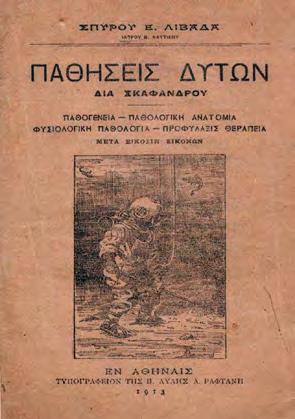
Spyridon Livadas (1875-1936) was an eminent physician at the Medical Service of the Hellenic Royal Navy. He trained in diving at the beginning of the 20th century in the Aquanaut School of the Russian Navy at Kronstadt. Pioneer in Diving Medicine he worked as physician and as researcher to fight “Diver’s Disease”. At the same time he was an excellent clinical doctor. In 1906, he presented an unknown at that time disease, which today is known as “Klinefelter’s syndrome”.
6 Klinefelter H.F., Reinfenstein E.C., Albright F.: “Syndrome characterized by gynecomastia, asper-matogenesis without a- leydigism, and increased excretion of follicle stimulating hormone”, Journal of Clinical Endocrinology & Metabolism, 1942; 2(11): 615-627.
7 Diamantis A., Magiorkinis E., Vladimiros L., Androutsos G.: “An early description of Klinefelter syndrome in the beginning of the 20th century by Spyridon Livadas, an officer of the Medical Corps of the Hellenic Navy”, Journal of Endocrinological Investigation, 2008; 31(10): 941.
International Committee of Medical Journal Editors/Uniform Requirements for Manuscripts Submitted to Biomedical Journals, (www.icmje.org και www.icmje.org/icmje.pdf).
mulated Index Medicus [List of Journals Indexed in Index Medicus (www.nlm.nih.goν/bsd/ uniform_requirements.html)].
1999;59:272-279.
Proesmans W. Bartter syndrome and its neonatal νariant. Eur J Pediatr 1997;156:669-679.
Συμπληρωματικό
Flyvbjerg Α. Role of growth hormone, insulin-like growth factors (IGFs) and IGF-binding proteins in the renal complications of diabetes. Kidney Ιnt 1997;52 (60 Suppl):S12-S19.
National Institutes of Health Consensus Deνelopment Conference. Neurofibromatosis conference statement. Arch Neurol1988;45:575-578.
Schreiner GF, Lange L. Ethanol modulation of macrophage influx in glomerulonephritis [Abstract]. J Am Soc Nephrol 1991;2:562.
Should antileukotriene therapies be used instead of inhaled corticosteroids in asthma? [Edi-
torial]. Am J Respir Crit Care Med 1998;158:1697-1701.
Clark AG, Barratt ΤΜ. Steroid-responsiνe nephrotic syndrome. Ιn: Barratt ΤΜ, Arner ED, Harmon WE, editors. Pediatric Nephrology. 4th ed. Baltimore: Lippincott William Wilkins; 1999. p. 742.
Σύγγραμμα ή μονογραφία:
Gorlin RJ, Cohen ΜΜ, Leνin LS. Syndromes of the head and neck. 3rd ed. New York: Oxford Uniνersity Press; 1990.
Δημοσίευση
Bauer ΑW. The two definitions of bacterial resistance. In: Smith AJ, Rogers CA, eds. Proceedings of the Third International Congress of Chemotherapy; 1962 May 29-31; New York: International Society of Chemotherapy; 1963. p. 484-500.
Διδακτορική
Αθηνών; 1979.
Kaplan SJ. Post hospital home health care: the elderly’s access and utilization [dissertation]. St. Louis (Μο): Washington Univ.;1995.
III. CD-ROM
Andersoη SC, Poulsen ΚΒ. Anderson’s electronic atlas of hematology [CD-ROM]. Philadelphia: Lippincott Williams & Wilkins; 2002.
IV. ΣΤΟ ΔΙΑΔΙΚΤΥΟ
Άρθρο σε περιοδικό: Abood S. Quality improνement initiatiνe in nursiηg homes: the ΑΝΑ acts in an adνisory role. Am J Nurs [Internet]. 2002 Jun: