
Four monthly scientific journal of the Greek Paediatric Society

010
016
026
03
042
Υποβολή
e-mail: grammateia@e-child.gr
Οδηγίες
http://e-child.gr/publications/ instructions-to-authors
Iδιοκτήτης
e-mail: grammateia@e-child.gr Eτήσια
048
054
056
058

Four monthly scientific journal of the Greek Paediatric Society
010
016
026
03
042
Υποβολή
e-mail: grammateia@e-child.gr
Οδηγίες
http://e-child.gr/publications/ instructions-to-authors
Iδιοκτήτης
e-mail: grammateia@e-child.gr Eτήσια
048
054
056
058
Volume 83 | Number 1 | January - February - March - April 2020
Four monthly publication of the Greek Paediatric Society
008
EDITORIAL
Stelios Antoniadis
010
AWARDED PAPER
Expression of CD45 antigen as a prognostic factor in children with acute lymphoblastic leukaemia
Athanasios Tragiannidis, Eleni Vasileiou, Anna Taparkou, Evangelia Farmaki, Evdoxia Gounari, Emmanouil Hatzipantelis, Maria Kourti, Maria Lamprou, Vasiliki Tsavdaridou, Theodotis
Papageorgiou, Lemonia Skoura, Eygenia Papakonstantinou, Andreas Giannopoulos, Dimitrios
Zafeiriou
016
REVIEW ARTICLES
Abusive head trauma in infants and young children
Dimitra Zisi, Alexandros Makis, Alexandra Soldatou
026
New treatment approaches in Hemophilia. Gene therapy
Moschovi Maria
034
CASE REPORTS
Narcolepsy with cataplexy in a female adolescent patient: a rare sleep disorder with significant clinical implications
Pinelopi Dragoumi, StylianiGlykou, Maria Milioudi, Maria Kyriazi, Eythimia Vargiami, Dimitrios I. Zafeiriou
President
A. Constantopoulos
Editorial board
Editor- in- Chief
S. Antoniadis
Members
S. Andronikou
E. Galanakis
A. Evangeliou
L. Thomaidou
M. Kanariou
A. Kapogiannis
S. Kitsiou-Tzeli
E. Mantadakis
P. Panagiotopoulou-Gartagani
A. Papadopoulou
V. Papaevagelou
A. Papathanassiou
A. Siamopoulou-Mavridou
A. Syrigou-Papavasiliou
Manuscript submission
e-mail: grammateia@e-child.gr
Instructions to authors: http://e-child.gr/publications/ instructions-to-authors
Owner Greek Paediatric Society 15, Mpakopoulou st. GR - 15451, Ν. Psychiko Tel.: +302107771140
e-mail: grammateia@e-child.gr
Annual subscription All foreign countries: US$50
042
Erythema Multiforme associated with Mycoplasma Pneumoniae infection: case presentation Ioannis Chastas, Charis Lampada, Aikaterini Liana, Eleni Papastergiou, Chrysoula Pistiola, Fotios Zarkinos
048
An 11-month-old infant with cannabinoid poisoning Kyrochristou Electra, Vlachou Georgia, Mpoumpa Vasiliki, Kyrochristou Gerasimia, Gketsi Vasiliki
054
BETWEEN COLLEAGUES
Stelios Antoniadis
056 BOOK PRESENTATION
Stelios Antoniadis
058
INSTRUCTIONS TO AUTHORS
e-mail: atragian@auth.gr atragian@hotmail.com
Correspondence
Athanasios Tragiannidis
S. Kiriakidi1, 54636, Thessaloniki
Τ. +302313303944, +306944944777
e-mail: atragian@auth.gr atragian@hotmail.com
Athanasios Tragiannidis
Eleni Vasileiou
Emmanouil Hatzipantelis
Theodotis Papageorgiou
Pediatric Hematology and Oncology Unit, 2nd Department of Pediatrics, Aristotle University of Thessaloniki, AHEPA University Hospital, Thessaloniki, Greece
Anna Taparkou
Evangelia Farmaki
Dimitrios Zafeiriou
1st Department of Pediatrics, Aristotle University of Thessaloniki, Hippokration General Hospital, Thessaloniki, Greece
Evdoxia Gounari
Vasiliki Tsavdaridou
Lemonia Skoura Department of Immunology, Microbiology Laboratory, AHEPA University Hospital, Thessaloniki, Greece
Maria Kourti
Maria Lamprou
Eygenia Papakonstantinou Department of Pediatric Hematology and Oncology, Hippokration General Hospital, Thessaloniki, Greece
Andreas Giannopoulos 2nd Department of Pediatrics, Aristotle University of Thessaloniki, AHEPA University Hospital, Thessaloniki, Greece
Athanasios Tragiannidis, Eleni Vasileiou, Anna Taparkou, Evangelia Farmaki, Evdoxia Gounari, Emmanouil Hatzipantelis, Maria Kourti, Maria Lamprou, Vasiliki Tsavdaridou, Theodotis Papageorgiou, Lemonia Skoura, Eygenia Papakonstantinou, Andreas Giannopoulos, Dimitrios Zafeiriou
Abstract
CD45 antigen, also known as Protein Tyrosine Phosphatase, receptor type, C (PTPRC), is expressed in hematopoietic cells and its expression is commonly quantified at diagnostic immunophenotyping. Overexpression of CD45 has been previously linked to poor prognosis in children with B- and T-cell ALL. Our goal is to study the possible use of CD45 expression as a prognostic factor.
Methods: Bone marrow samples of children with B- and T-cell ALL were studied at the time of diagnosis before treatment initiation according to ALL-BFM 2009. All patients that were admitted to thetwopediatric oncology departments in Thessaloniki from January 2013 to December 2017 were enrolled in the study (79 patients in total). CD45 expression was measured both in normal lymphocytes and leukemic blasts and a relative ratio of its expression (leukemic versus normal) was calculated. Statistical analysis was conducted with the Statistical Package for the Social Science (IBM SPSS) for Windows, version 23.
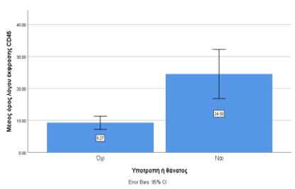
Results: We divided the sample into two groups according to the cell line affected (B- and T-cell leukemia). In our study group 71 children were diagnosed with B-ALL and only 8 with T-ALL. The expression of CD45 was assumed to be high when it exceeded the 75th percentile (relative ratio 12.5% for B-ALL and 36.08% for T-ALL). It was observed that the mean expression of CD45 in patients with B-ALL who had a relapse or died than in patients without an event was higher (Figure 1). We also found that there is a statistically important correlation between relapse or death and the expression of CD45 above the 75th percentile in patients with B-ALL, p<0.05. Moreover, we calculated that the probability of an event (relapse or death) in patients with B-ALL was 20.4 times higher when the expression of CD45 was above the 75th percentile. Due to the small sample of patients with T-ALL we could not calculate statistically important correlations in that group.
Conclusion: The high expression of CD45 could be used as a prognostic factor in patients with acute lymphoblastic leukemia.
Key words: acute lymphoblastic leukemia, CD45, prognosis, survival, childhood
SPSS (“Statistical Package for the Social Science IBM SPSS for Windows” έκδοση 25).
8% versus 78 ± 4%, p=0.02),
(Β-ΟΛΛ: 22 ± 3% versus 11 ± 1%, p<0.0001;
8% versus 11 ± 3%, p=0.003).
1. Hunger S and Mullighan C. Acute Lymphoblastic Leukemia in Children. N Engl J Med. 2015;373(16):1541-1552.
2. Douer D and Thomas D. New developments in acute lymphoblastic leukemia. Clin Adv Hematol Oncol. 2014;12(6 Suppl 12):13-22.
3. Iacobucci I and Mullighan C. Genetic Basis of Acute Lymphoblastic Leukemia. J Clin Oncol. 2017;35(9):975-983.
4. Pui C, Yang J, Hunger S, Pieters R, Schrappe M, Biondi A, Vora A, et al. Childhood Acute Lymphoblastic Leukemia: Progress Through Collaboration. J Clin Oncol. 2015;33(27):293848.
5. Hermiston M, Xu Z and Weiss A. CD45: a critical regulator of signaling thresholds in immune cells. Annu Rev Immunol. 2003;21:107-137.
6. Borowitz M, Shuster J, Carroll A, Nash M, Look T, Camitta B, Mahoney D, Lauer S and Rullen J. Prognostic significance of fluorescence intensity of surface marker expression in childhood B-precursor acute lymphoblastic leukemia. A Pediatric Oncology Group Study. Blood. 1997; 89(11):3960-3966.
7. Cario G, Rhein P, Mitlohner R, Zimmermann M, Bandapalli O, Romey R, Moericke A, et al. High CD45 surface expression determines relapse risk in children with precursor B-cell and T-cell acute lymphoblastic leukemia treated according to the ALL-BFM 2000 protocol. Haematologica. 2014;99(1):103-110.
8. Moorman A. New and emerging prognostic and predictive genetic biomarkers in B-cell precursor acute lymphoblastic leukemia. Haematologica. 2016;101(4):407-416.
9. Ratei R, Karawajew L, Lacombe F, Jagoda K, Del Poeta G, Kraan J, De Santiago M, et al. Normal lymphocytes from leukemic samples as an internal quality control for fluorescence intensity in immunophenotyping of acute leukemias. Cytometry B Clin Cytom. 2005;70Β(1):1-
9.
10. Iyer N, Balsamo L, Bracken M and Kadan-Lottick N. Chemotherapy-only treatment effects on long-term neurocognitive functioning in childhood ALL survivors: a review and metaanalysis. Blood. 2015;126(3):346-353.
11. Patrick K and Vora A. Update on biology and treatment of T-cell acute lymphoblastic leukaemia. Curr Opin Pediat. 2015;27(1):44-49.
12. Thomas X. Novel approaches to pediatric leukemia treatment. Expert Rev Anticancer Ther. 2015; 15(7):811-28.
13. Pierro J, Hogan L, Bhalta T and Carroll W. New targeted therapies for relapsed pediatric acute lymphoblastic leukemia. Expert Rev Anticancer Ther. 2017;17(8):725-736.
14. Pui C. Genomic and pharmacogenetic studies of childhood acute lymphoblastic leukemia. Front Med. 2015;9(1):1-9.
15. William C and Teena B. Acute Lymphoblastic Leukemia in Lanzkowsky's Manual of Pediatric Hematology and Oncology. Academic Press. 2016:367-389.
16. Bene M, Nebe T, Bettelheim P, Buldini B, Bumbea H, Kern W, et al. Immunophenotyping of acute leukemia and lymphoproliferative disorders: a consensus proposal of the European LeukemiaNet Work Package 10. Leukemia. 2011;25(4):567-574.
e-mail:
com
Correspondence
Ζisi Dimitra
Ad. Kasioumi 19, Stavraki, Ioannina
Τ. +306972867596
e-mail: deni_denni@hotmail. com
Dimitra Zisi, Alexandros Makis, Alexandra Soldatou
Abstract
The term child abuse includes all forms of abuse and neglect. Abusive head trauma (AHT) is a type of physical abuse and is the leading cause of fatal head injuries for children younger than 2 years of age. The purpose of this article is to describe the syndrome, referring to the clinical characteristics, risk factors, effects and prevention strategies.
In addition, the needs of abused infants/children require interdisciplinary evaluation and management. It is important that all child health care providers be careful in order to make a timely diagnosis relying on a detailed history, meticulous clinical examination, imaging and laboratory findings and then follow the procedure for their protection. Abusive head trauma, like all forms of abuse, is preventable. New parents/caregivers need information, education and support to avoid the serious problems that come with violent behavior.
Informing and raising awareness of the catastrophic effects of infant/child health and the huge financial cost to society are issues that must be addressed by the citizens but also by the state which must safeguard and claim a safe environment for all children free from all forms of violence.
Key words: Child, toddler, abuse, traumatic brain injury
Dimitra Zisi
Alexandros Makis
Child Health Dept., Faculty of Medicine, School of Health Sciences, University of Ioannina
Alexandra Soldatou
2nd Department of Pediatrics, National and Kapodistrian University of Athens (NKUA)
(5). Έκτοτε
‘Whiplash-Shaken Infant Syndrome',
και
‘Non Accidental Head Injury’,
‘ShakenBabySyndrome’ (σύνδρομο
(American Academy of Pediatrics,
(Abusive Head Trauma)
3.
4.
1. W.H.O. World Health Organization 2006. Preventing child maltreatment: A guide to taking action and generating evidence. Geneva: WHO. Webpage http://www.who.int/violence_injury_ prevention/publications/violence/child_maltreatment/en/.
2. Frasier LD, Kelly P, Al-Eissa M, Otterman GJ. International issues in abusive head trauma. Pediatr radiol. 2014;44 Suppl 4:S647-53.
3. Duhaime AC, Christian CW, Rorke LB, Zimmerman RA. Nonaccidental head injury in infants--the "shaken-baby syndrome". N Engl J Med. 1998;338(25):1822-9.
4. Matschke J, Herrmann B, Sperhake J, Körber F, Bajanowski T, Glatzel M. Shaken baby syndrome: a common variant of non-accidental head injury in infants. Dtsch Arztebl Int. 2009;106(13):211-7.
5. Caffey J. On the theory and practice of shaking infants. Its potential residual effects of permanent brain damage and mental retardation. Am J Dis Child. (1960). 1972;124(2):161-9.
6. Christian CW, Block R. Abusive head trauma in infants and children. Pediatrics. 2009;123(5):1409-11.
7. AAP. American Academy of Pediatrics (2015) Understanding abusive head trauma in infants and children: answers from America’s pediatricians. Webpage https://www.aap.org/en-us/Documents/cocan_understanding_aht_in_infants_children.pdf
8. Joyce T, Huecker MR. Pediatric Abusive Head Trauma (Shaken Baby Syndrome). StatPearls. Treasure Island (FL): StatPearls Publishing StatPearls Publishing LLC.; 2019.
9. Kato N. Prevalence of Infant Shaking Among the Population as a Baseline for Preventive Interventions. J Epidemiol. 2016;26(1):2-3.
10. King WJ, MacKay M, Sirnick A. Shaken baby syndrome in Canada: clinical characteristics and outcomes of hospital cases. CMAJ. 2003;168(2):155-9.
11. Kelly P, John S, Vincent AL, Reed P. Abusive head trauma and accidental head injury: a 20-year comparative study of referrals to a hospital child protection team. Arch Dis Child. 2015;100(12):1123-30.
12. Palusci VJ, Covington TM. Child maltreatment deaths in the U.S. National Child Death Review Case Reporting System. Child abuse & neglect. 2014;38(1):25-36.
13. Davies WH, Garwood MM. Chapter Four: Who Are the Perpetrators and Why Do They Do It? J. Aggress. Maltreatment Trauma. 2001;5(1):41-54.
14. Hennes H, Kini N, Palusci VJ. Chapter Three: The Epidemiology, Clinical Characteristics and Public Health Implications of Shaken Baby Syndrome. J. Aggress. Maltreatment Trauma. 2001;5(1):19-40.
15. Leventhal JM, Asnes AG, Pavlovic L, Moles RL. Diagnosing abusive head trauma: the challenges faced by clinicians. Pediatr Radiol. 2014;44 Suppl 4:S537-42.
16. Lynoe N, Elinder G, Hallberg B, Rosen M, Sundgren P, Eriksson A. Insufficient evidence for 'shaken baby syndrome' - a systematic review. Acta Paediatr (Oslo, Norway : 1992). 2017;106(7):1021-7.
17. Wester K. Has a 'shaken baby' always been shaken? Tidsskr Nor Laegeforen. 2018;138(15).
18. Louwers EC, Affourtit MJ, Moll HA, de Koning HJ, Korfage IJ. Screening for child abuse at
emergency departments: a systematic review. Arch Dis Child. 2010;95(3):214-8.
19.
29: σελ. 367-372.
Leventhal JM.
2019:
20. Vester ME, Bilo RA, Karst WA, Daams JG, Duijst WL, van Rijn RR. Subdural hematomas: glutaric aciduria type 1 or abusive head trauma? A systematic review. Forensic Sci Med Pathol. 2015;11(3):405-15.
21. Listman DA, Bechtel K. Accidental and abusive head injury in young children. Curr Opin Pediatr. 2003;15(3):299-303.
22. Choudhary AK, Servaes S, Slovis TL, Palusci VJ, Hedlund GL, Narang SK, et al. Consensus statement on abusive head trauma in infants and young children. Pediatr Radiol. 2018;48(8):1048-65.
23. AAP. Shaken baby syndrome: rotational cranial injuries-technical report. Pediatrics. 2001;108(1):206-10.
24. Bechtel K, Stoessel K, Leventhal JM, Ogle E, Teague B, Lavietes S, et al. Characteristics that distinguish accidental from abusive injury in hospitalized young children with head trauma. Pediatrics. 2004;114(1):165-8.
25. Morad Y, Kim YM, Armstrong DC, Huyer D, Mian M, Levin AV. Correlation between retinal abnormalities and intracranial abnormalities in the shaken baby syndrome. Am J Ophthalmol. 2002;134(3):354-9.
26. Laghmari M, Skiker H, Handor H, Mansouri B, Ouazzani Chahdi K, Lachkar R, et al. [Birth-related retinal hemorrhages in the newborn: incidence and relationship with maternal, obstetric and neonatal factors. Prospective study of 2,031 cases]. Journal francais d'ophtalmologie. 2014;37(4):313-9.
27. Barr RG, Barr M, Rajabali F, Humphreys C, Pike I, Brant R, et al. Eight-year outcome of implementation of abusive head trauma prevention. Child abuse & neglect. 2018;84:106-14.
28. Lind K, Toure H, Brugel D, Meyer P, Laurent-Vannier A, Chevignard M. Extended followup of neurological, cognitive, behavioral and academic outcomes after severe abusive head trauma. Child abuse & neglect. 2016;51:358-67.
29. Goulet C, Frappier JY, Fortin S, Deziel L, Lampron A, Boulanger M. Development and evaluation of a shaken baby syndrome prevention program. Journal of obstetric, gynecologic, and neonatal nursing : JOGNN. 2009;38(1):7-21.
30. Duffee JH, Mendelsohn AL, Kuo AA, Legano LA, Earls MF. Early Childhood Home Visiting. Pediatrics. 2017;140(3).
31. Showers J. "Don't shake the baby": the effectiveness of a prevention program. Child abuse & neglect. 1992;16(1):11-8.
32. Barr RG. National Center on Shaken Baby Syndrome. Period of PURPLE crying. Ogden (UT): National Center on Shaken Baby Syndrome. 2004.
33. Ludvigsson JF. Extensive shaken baby syndrome review provides a clear signal that more research is needed. Actapaediatrica (Oslo, Norway : 1992). 2017;106(7):1028-30.
34.
. Webpage: http://dkday-athin.att.sch.gr/autosch/joomla15/images/UpLoad/odigos%20efarmogis.pdf
35. Ilias F, Leventhal JM, Magalhaes T, Oliveira M, Oral R, Soldatou A, Stathi A, Zafar N. Building Systems to Address Child Abuse And Neglect: Successful Collaborations With International Partners. 13th ed. World Perspectives on Child Abuse, 2018.
6932684622
e-mail: mmoschov@med. uoa.gr
mmoschovi04@gmail.com
Correspondence
Maria Moschovi, MD, PhD
Levadias 8, Goudi, 11527, Athens, Greece
T. +302107452112
+306932684622
e-mail: mmoschov@med. uoa.gr mmoschovi04@gmail.com
Moschovi Maria
Summary
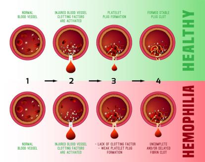
Hemophilia A and B are X-linked recessive disorders resulting from mutations in the gene for blood clotting factor VIII and factor IX, respectively. Hemophilia is rare disease and collectively, they are among the most common inherited bleeding disorders in the world. Therefore, hemophilia patients have to carefully plan periods of increased physical activities, such as sports. The current standard of care for severely affected hemophilia patients consists of regularly administered prophylaxis with factor concentrates intended to maintain the factor level >1% of normal.
Gene therapy offers the potential for a cure for patients with hemophilia by establishing continuous endogenous expression of factor VIII or factor IX (FIX) following transfer of a functional gene to replace the hemophilic patient's own defective gene. The hemophilias are ideally suited for gene therapy because a small increment in blood factor levels (≥5% of normal) is associated with significant amelioration of bleeding phenotype in severely affected patients. In 2011, the St. Jude/UCL phase 1/2 trial was the first to provide clear evidence of a stable dose-dependent increase in FIX levels in patients with severe hemophilia B following a single administration of adeno-associated viral (AAV) vectors. Transgenic FIX expression has remained stable at ~5% of normal in the high-dose cohort over a 7-year follow-up period, resulting in a substantial reduction in spontaneous bleeding and FIX protein usage without toxicity. This study has been followed by unparalleled advances in gene therapy for hemophilia A and B, leading to clotting factor activity approaching normal or near-normal levels associated with a "zero bleed rates" in previously severely affected patients following a single administration of AAV vectors.
Key words: hemophilia, bispecific monoclonal antibody, gene therapy
Maria Moschovi, MD, PhD
Division of Hematology - Oncology, First Dept of Pediatrics, National and Kapodistrian University of Athens "Marianna V Vardinoyannis-Elpida" Oncology Unit, 'Agia Sofia' Children's Hospital
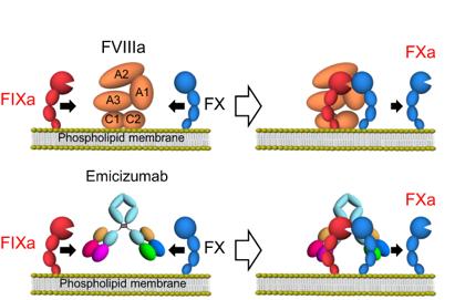
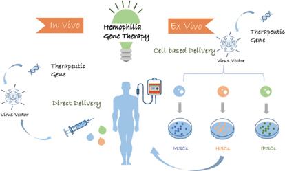
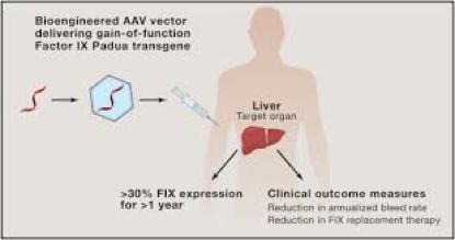
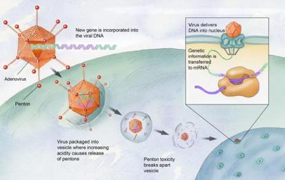
(6).
1. Hemophilia A. National Hemophilia Foundation https://www.hemophilia.org/BleedingDisorders/Types-of-Bleeding-Disorders/Hemophilia-A
2. Moschovi M, Aronis S, Trimis G, Platokouki H, Salavoura K, Tzortzatou-Stathopoulou F Rituximab in the treatment of high responding inhibitors in severe haemophilia A. Haemophilia. 2006 Jan;12(1):95-9.
3. Pipe SW, Shima M, Lehle M, Shapiro A, Chebon S, Fukutake K, et al. Efficacy, Safety, and Pharmacokinetics of Emicizumab Prophylaxis Given Every 4 Weeks in People WithHaemophilia A (HAVEN 4): A Multicentre, Open-Label, Non-Randomised Phase 3 Study. Lancet Haematol, 2019, 6 (6), e295-e305
4. KandaceL.GollompaBhavyaS.Doshi, ValderR.Arruda. Gene therapy for hemophilia: Progress to date and challenges moving forward. Transfusion and Apheresis Science 2019, 58: 602-612
5. Nathwani AC. Breakthroughs in Gene Therapy for Hemophilia. American Society of Hematology Education Program. 2019 Dec 6;2019(1):1-8. doi: 10.1182/hematology.2019000007.
6. Machin N, Ragni MV, Smith KJ. Gene therapy in hemophilia A: a cost-effectiveness analysis. Blood Adv. 2018;2:1792-98.
e-mail:
Correspondence
Dragoumi Pinelopi
49 Konstantinoupoleos St. 54642, Thessaloniki
T. +302313312501
+306944913556
e-mail: ezlouis@gmail.com
Pinelopi Dragoumi, Styliani Glykou, Maria Milioudi, Maria Kyriazi, EythimiaVargiami, Dimitrios I. Zafeiriou
Abstract
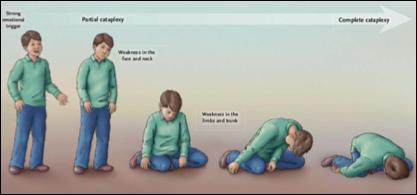
Narcolepsy is a rare sleep disorder, with significant impact on the quality of life of each patient, often diagnosed with great delay. Herein we describe the case of 15 year-old teenage girl diagnosed with narcolepsy with cataplexy following gradually worsening sleepiness during the day and disturbed night sleep for about 2 years, and also episodic loss of muscle tone during emotional stress the past 6 months. Polysomnography and Multiple Sleep Latency Test (MSLT) revealed early onset of REM phase of sleep (in less than 9 minutes) and low levels of hypocretin were found in the CSF (<50 ng/L). The patient was treated with gradually increasing doses of modafinil combined with cognitive behavioral therapy, which lead to amelioration of her symptoms. Awareness of narcolepsy is required among pediatric specialists for the timely diagnosis and referral to specialized medical centers, where all necessary investigations and individualized treatment can be provided.
Key words: narcolepsy, cataplexy, sleep disorder, hypocretin, multiple sleep latency test.
Pinelopi Dragoumi
Styliani Glykou
Maria Milioudi
Maria Kyriazi
EythimiaVargiami
Dimitrios I. Zafeiriou
1st Department of Paediatrics, Aristotle
University of Thessaloniki, Hippokration General Hospital of Thessaloniki, Greece
Εικόνα
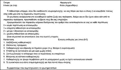
Tsara V.et al. Greek version of the Epworth Sleepiness Scale. Sleep Breath. 2004
1.
2.
2
3.
4.
SOREMP (sleeponset REM period)
(multiplesleeplatencytest)
1. Postiglione E, Antelmi E, Pizza F, Lecendreux M, Dauvilliers Y, Plazzi G. The Clinical Spectrum Of Childhood Narcolepsy. Sleep Medicine Reviews. 2018;38:70-85.doi:10.1016/J. Smrv.2017.04.003
2. Babiker MOE, Prasad M. Narcolepsy In Children: A Diagnostic And Management Approach. Pediatric Neurology. 2015;52(6):557-565.doi:10.1016/J. Pediatrneurol.2015.02.020
3. Mahoney CE, Cogswell A, Koralnik IJ, Scammell TE. The neurobiological basis of narcolepsy. Nature Reviews Neuroscience. 2019;20(2):83-93.doi: 10.1038/s41583-018-0097-x
4. Scammell TΕ. Narcolepsy. New England Journal Of Medicine 2015;373(27):2654-2662. doi:10.1056/Nejmra1500587
5. Szabo ST, Thorpy M, Mayer G, Peever J, Kilduff T. Neurobiological And Immunogenetic Aspects Of Narcolepsy: Implications For Pharmacotherapy. Sleep Medicine Reviews. 2019;43:23-36. doi:10.1016/J.Smrv.2018.09.006
6 Abad VC, Guilleminault C. New developments in the management of narcolepsy. Nature and Science of Sleep 2017 Mar.3;9:35-57. doi:10.2147/NSS.S103467




e-mail:
Correspondence
Ioannis Chastas, Ellis 2
50100, Kozani
Τ. +6972824821
e-mail: chastasj@gmail.com
Ioannis Chastas, Charis Lampada, Aikaterini Liana, Eleni Papastergiou, Chrysoula Pistiola, Fotios
Zarkinos
Abstract
Erythema multiforme is an acute immunologically mediated reaction with skin and mucous membranes effects. It used to belong to the mucocutaneous syndromes group with the first reference to the term by Hebra (1866). References to m.pneumoniae as a trigger has existed since the 1970s (1). Its prevalence in the general population is estimated <1% and mainly affects young adults, while the frequency in the pediatric population remains unknown.
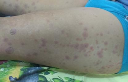
We present a case of a patient with febrile infection and skin lesions typical of EM. Infection was attributed to m.pneumoniae following confirmation by imaging and serological studies. Treatment of the infectious agent resulted in remission of symptoms.
The importance of inclusion of M.pneumoniae in differential diagnosis of erythema multiforme is highlighted as early diagnosis and treatment may lead to reduction of skin lesions.
Key words: erythema multiforme, mycoplasma pneumoniae
HSV
EBV, CMV, VZV, enteroviruses, coxsackie B5, echoviruses (2), parainfluenza virus (7) και
όπως M.pneumoniae, Mycobacterium leprae, Legionella pneumophila, Salmonella, Streptococcus, Pneumococcus (3,7,8,9,10).
ερυθρομυκίνη, καρβαμαζεπίνη, βαλπροϊκό,
και εμβόλια (HepB, DTP). Η νόσος εκδηλώνεται 2 - 6 εβδομάδες
Ioannis Chastas
Charis Lampada
Aikaterini Liana
Eleni Papastergiou
Chrysoula Pistiola
Fotios Zarkinos
Pediatric clinic, General Hospital of Kozani, "Mamatseio"
Συντομογραφίες:
M.pneumoniae: Mycoplasma pneumoniae
HSV: Herpes simplex virus
EBV: Epstein Barrvirus
CMV: Cytomegalovirus
VZV: Varicella Zostervirus
ΜΣΑΦ:
HepB: Hepatitis B
DTP: Diphtheria Tetanus Pertussis
IgM: immunoglobulin M
IgG: immunoglobulin G
C3: Complement component 3
C4: Complement component 4
1. H. J. Harwock, G. M. Kalmanson, andL. B. Guze, “Human diseases associated with mycoplasms. With an appendix on simple culture techniques.,” Calif. Med., vol. 116, no. 5, pp. 1–7, 1972.
2. F. Samim, A. Auluck, C. Zed, and P. M. Williams, “Erythema multiforme. A review of epidemiology, pathogenesis, clinical features, and treatment.,” Dental Clinics of North America, vol. 57, no. 4. pp. 583–596, Oct-2013.
3. N. Rock, D. Belli, and N. Bajwa, “Erythema bullous multiforme: A complication of mycoplasma pneumoniae infection,” J. Pediatr., vol. 164, no. 2, p. 421, 2014.
4. A. Langley, N. Anooshiravani, S. Kwan, J. Zeller, and E. Pope, “Erythema multiforme in children and mycoplasma pneumoniae aetiology,” J. Cutan. Med. Surg., vol. 20, no. 5, pp. 453–457, 2016.
5. R. D. G. Alfred K. Yeung, “Use of steroids for erythema multiforme in children,” Can. Fam. Physician, vol. 51, no. 11, pp. 1481–1483, 2005.
6. R. Amode et al., “Clinical and histologic features of Mycoplasma pneumoniae–related erythema multiforme: A single-center series of 33 cases compared with 100 cases induced by other causes,” J. Am. Acad. Dermatol., vol. 79, no. 1, pp. 110–117, Jul. 2018.
7. D. Kahvecioglu, D. Yildiz, B. Atasay, and O. Erdeve, “Erythema multiforme due to parainfluenza virus in a newborn: A case report and review of the literature,” Indian J. Paediatr. Dermatology, vol. 15, no. 3, p. 117, 2014.
8. M. Martínez-Pérez, A. Imbernón-Moya, A. Lobato-Berezo, and M. Churruca-Grijelmo, “Mycoplasma pneumoniae -Induced Mucocutaneous Rash: A New Syndrome Distinct from Erythema Multiforme? Report of a New Case and Review of the Literature,” Actas DermoSifiliográficas (English Ed., 2016.
9. Y. Siedner-Weintraub, I. Gross, A. David, S. Reif, and V. Molho-Pessach, “Paediatric erythema multiforme: Epidemiological, clinical and laboratory characteristics,” Acta Derm. Venereol., vol. 97, no. 4, pp. 489–492, 2017.
10. J. A. Vargas-Hitos, M. V. Manzano-Gamero, and J. Jiménez-Alonso, “Erythema multiforme associated with Mycoplasma pneumoniae,” Infection, vol. 42, no. 4, pp. 797–798, 2014.
11. Z. H. Tan, K. C. Thoon, and M. J. A. Koh, “Case series of three infants with erythema multiforme following hepatitis B vaccination,” Pediatr. Neonatol., vol. 57, no. 1, pp. 72–75, 2016.
12. M. Lerch, C. Mainetti, B. Terziroli Beretta-Piccoli, and T. Harr, “Current Perspectives on Erythema Multiforme,” Clinical Reviews in Allergy and Immunology, vol. 54, no. 1. Humana
Press Inc., pp. 177–184, 01-Feb-2018.
13. L. Terraneo et al., “Unusual Eruptions Associated with Mycoplasma pneumoniae Respiratory Infections: Review of the Literature,” Dermatology, vol. 231, no. 2, pp. 152–157, Aug. 2015.
14. D. Poddighe and P. Bruni, “Mycoplasma pneumoniae -induced rash and mucositis (MIRM): An unusual mild skin rash associated with severe mucosal involvement,” BMJ Case Reports, vol. 2017. BMJ Publishing Group, 2017.
15. M. A. Rodríguez Pérez and J. F. Delgado Álvarez, “Erythema multiforme minor. From mouth to skin,” Med. Clínica (English Ed., vol. 148, no. 4, p. 195, 2017.
16. J. Read and G. B. Keijzers, “Pediatric Erythema Multiforme in the Emergency Department More Than ‘Just a Rash,’” 2015.
17. A. Heinze, M. Tollefson, K. E. Holland, and Y. E. Chiu, “Characteristics of pediatric recurrent erythema multiforme,” Pediatr. Dermatol., vol. 35, no. 1, pp. 97–103, Jan. 2018.
18. M. A. Jason J. Emer, Sebastian G. Bernardo, Olga Kovalerchik, “Urticaria Multiforme,” J. Clin. Aesthet. Dermatol., vol. Volume 6, no. (3), pp. 34–39, 2013.
19. H. Song, J. T. Huang, and J. K. Tan, “Mycoplasma-Induced Rash and Mucositis in a Father and Son: A Case Report,” Pediatr. Infect. Dis. J., vol. 37, no. 7, pp. e205–e206, Jul. 2018.
Correspondence
Gketsi Vasiliki
Neochoropoulos of Ioannina, PO Box 339, 45500
Τ. +306945398479
e-mail: vikigetsi@gmail.com
Kyrochristou Electra, Vlachou Georgia, Mpoumpa Vasiliki, Kyrochristou Gerasimia, Gketsi Vasiliki
Abstract
Introduction: In children, as in adults, cannabinoids can cause a variety of symptoms, mainly from the CNS, such as hypotonia, agitation, seizures, coma.
Methods: We report the case of an 11-month-old male infant, in a family of Gypsies, who was presented to the Emergency Department after two episodes of seizures.
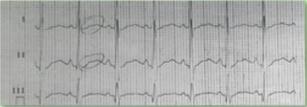
Results: On admission he was lethargic, with generalized hypotonia. Hematological and blood chemistry investigations showed leukocytosis, acidosis, hypokalemia and hyponatremia. The electrocardiogram showed prolonged QT and U waves. He was treated symptomatically with intravenous hydration and correction of electrolyte imbalances. The clinical and laboratory image were fully restored at the 5th day of hospitalization. In the context of investigation of seizures and due to family environment, a urine specimen was sent for toxicological analysis by immunological (EMIT), spectrophotometric (UV-VIS) and chromatographic (TLC, GLC, GC / MS) methods, which were positive for cannabinoids.
Conclusion: The possibility of cannabis poisoning should be considered in cases of unexplained acute onset of neurological symptoms in previously healthy children and infants.
Key words: cannabis intoxication, convulsions, hypokalemia, hyponatremia
Kyrochristou Electra
General Hospital of NikaiaAgios Panteleimon
Vlachou Georgia
Gketsi Vasiliki
Pediatric Clinic of the General Hospital of Ioannina “G. Hatzicosta”
Mpoumpa Vasiliki
Kyrochristou Gerasimia
Medical School of the University of Ioannina
Συντομογραφίες:
UV
GC/MS:
1. Rachel J.Levene, Elza Pollak-Christian, Sigrid Wolfram “A 21st century problem: Cannabis toxicity in a 13month old child’’, The Journal of Emergency Medicine, Jan2019, volume 56, issue 1, pages 94-96
2. Cipriani F, Mancino A, Pulitanò SM, Piastra M, Conti G., “A cannabinoid-intoxicated child treated with dexmedetomidine: a case report.”, Journal of Medical Case Reports, (2015) 9:152
3. Mark L. Feldman ,Sarah Hadfield ‘‘Pot paresis: marijuana and a case of hypokalemic periodic paralysis’’, The Journal of Emergency Medicine, Apr 2009, volume 36, issue 3, pages 236-238
4. George Sam Wang, Genie Roosevelt, Kennon Heard, “Pediatric Marijuana Exposures in a Medical Marijuana State”, JAMA Pediatrics2013;167(7):630-633
5. The health effects of cannabis and cannabinoids. National Academy of sciences.2017
6. Isabelle Claudet, Mathilde Le Breton, Camille Brehin, Nicolas Franchitto “A 10-year review of cannabis expossure in children under 3-years of age: do we need a more global approach? ”, European Journal of Pediatrics, Apr 2017, volume 176, issue 4, pages 553-556
7.Croche Santander B Alonso Salas MT, Loscertales Abril M, “Accidental cannabis poisoning in children: report of four cases in a tertiary care center from southern Spain”, Archives Argent Pediatrics. 2011 Feb;109(1):4-7
2.
3.
4.
5.
6.
7.
8.
9. Ενδιαφέρουσες
10. Σύντομα
11. Βραχείες
12.
13.
14.
(Uniform Requirements for Manuscripts Submitted to Biomedical Journals),
International Committee of Medical Journal Editors/Uniform Requirements for Manuscripts Submitted to Biomedical Journals, (www.icmje.org και www.icmje.org/icmje. pdf).
Cumulated Index Medicus [List of Journals Indexed in Index Medicus (www.nlm.nih.goν/bsd/uniform_requirements.html)].
1999;59:272-279.
Proesmans W. Bartter syndrome and its neonatal νariant. Eur J Pediatr 1997;156:669-679.
Flyvbjerg Α. Role of growth hormone, insulin-like growth factors (IGFs) and IGF-binding proteins in the renal complications of diabetes. Kidney Ιnt 1997;52 (60 Suppl):S12-S19.
Χωρίς συγγραφέα:
National Institutes of Health Consensus Deνelopment Conference. Neurofibromatosis conference statement. Arch Neurol1988;45:575-578.
Προσδιορισμός τύπου άρθρου: Schreiner GF, Lange L. Ethanol modulation of macrophage influx in glomerulonephritis [Abstract]. J Am Soc Nephrol 1991;2:562.
Should antileukotriene therapies be used instead of inhaled corticosteroids in asthma? [Editorial]. Am J Respir Crit Care Med 1998;158:1697-1701.
Laux-End R, Inaebnit D, Gerber ΗΑ, Bianchetti MG. Vasculitis associated with leνamisole and circulating autoantibodies [Let ter]. Arch Dis Child 1996;75:355-356.
II. ΒΙΒΛΙΑ
Clark AG, Barratt ΤΜ. Steroid-responsiνe nephrotic syndrome. Ιn: Barratt ΤΜ, Arner ED, Harmon WE, editors. Pediatric Nephrology. 4th ed. Baltimore: Lippincott William Wilkins; 1999. p. 742.
Σύγγραμμα
Gorlin RJ, Cohen ΜΜ, Leνin LS. Syndromes of the head and neck. 3rd ed. New York: Oxford Uniνersity Press; 1990.
Δημοσίευση
Bauer ΑW. The two definitions of bacterial resistance. In: Smith AJ, Rogers CA, eds. Proceedings of the Third International Congress of Chemotherapy; 1962 May 29-31; New York: International Society of Chemotherapy; 1963. p. 484-500.
Διδακτορική
Αθηνών; 1979.
Kaplan SJ. Post hospital home health care: the elderly’s access and utilization [dissertation]. St. Louis (Μο): Washington Univ.;1995.
III. CD-ROM
Andersoη SC, Poulsen ΚΒ. Anderson’s electronic atlas of hematology [CD-ROM]. Philadelphia: Lippincott Williams & Wilkins; 2002.
IV. ΣΤΟ
Abood S. Quality improνement initiatiνe in nursiηg homes: the ΑΝΑ acts in an adνisory role. Am J Nurs [Internet]. 2002 Jun:
Webpage: http://www.nursingworld.org/AJN/2002/june/Wawatch.htm
Μονογραφία: Foley ΚΜ, Gelbaηd Η, editors. Improving palliative care for cancer [Monograph, Internet]. Washington: National Academy Press; 2001.
Webpage: http://www.nap.edu/books/0309074029/html
Ιστοσελίδες: Cancer-Pain.org [Webpage, Internet]. New York: Association of Cancer Online Resources, Ιnc.; 2002: http://www.cancer-pain.org/
Πίνακες
2.
3.
4.
5.
7.
•
• επιστημονικό/ά κέντρο/α
•