
Four monthly scientific journal of the Greek Paediatric Society


216
230
238
Υποβολή
e-mail: grammateia@e-child.gr
Οδηγίες
http://e-child.gr/publications/ instructions-to-authors
Iδιοκτήτης
Tηλ.:
e-mail: grammateia@e-child.gr Eτήσια

Four monthly scientific journal of the Greek Paediatric Society
216
230
238
Υποβολή
e-mail: grammateia@e-child.gr
Οδηγίες
http://e-child.gr/publications/ instructions-to-authors
Iδιοκτήτης
Tηλ.:
e-mail: grammateia@e-child.gr Eτήσια
Volume 82 | Number 3 | September - October - November - December 2019
Four monthly publication of the Greek Paediatric Society
124
EDITORIAL
Stelios Antoniadis
206
RESEARCH STUDY
Epidemiology data for paediatric heart failure due to cardiomyopathy - myocarditis. Single tertiary centre retrospective study.
Georgios Kourelis, Georgios Vagenakis, Felicia Kakava, Cleo Laskari, Alexandros Tsoutsinos, Konstantinos Kyriakoulis, Sotiria Apostolopoulou, Spyridon Rammos
216
REVIEW ARTICLES
Haemophagocyticlymphohistiocytosis: Diagnosis and management
Elpidoforos Mantadakis, Vasileios Papadakis
230
Factors that predict response to colchicine in children with familial Mediterranean fever
Asimenia Athousaki, Fani Ladomenou
238
The role of Technology in the Development of Social Skills of Students with Autism Spectrum Disorders (ASD)
Christina Syriopoulou-Delli, Eleni Gkiolnta
250
Breast diseases: Risk factors from childhood and adolescence
Thanou Chrissanthi, Christopoulos Panagiotis, Dimitroulis Dimitrios, Vlachos Nikolaos
President
A. Constantopoulos
Editorial board
Editor- in- Chief
S. Antoniadis
Members
S. Andronikou
E. Galanakis
A. Evangeliou
L. Thomaidou
M. Kanariou
A. Kapogiannis
S. Kitsiou-Tzeli
E. Mantadakis
P. Panagiotopoulou-Gartagani
A. Papadopoulou
V. Papaevagelou
A. Papathanassiou
A. Siamopoulou-Mavridou
A. Syrigou-Papavasiliou
Manuscript submission
e-mail: grammateia@e-child.gr
Instructions to authors: http://e-child.gr/publications/ instructions-to-authors
Owner Greek Paediatric Society 15, Mpakopoulou st. GR - 15451, Ν. Psychiko Tel.: +302107771140
e-mail: grammateia@e-child.gr
Annual subscription All foreign countries: US$50
Infantile pyknocytosis: A rare cause of newborn hemolytic anemia - 2 case reports Evangelos Christou, Konstantina Avgerinou, Theodora Bachou, Varvara Douna, Mersini Mavrikou, Dimitris Delis
266
Late presenting diaphragmatic hernia in childhood - case report
Eftychia Kanioura, Ioannis Skondras
272 BETWEEN COLLEAGUES
Stelios Antoniadis
274 BOOK PRESENTATION
Stelios Antoniadis
276 NEW PUBLICATION
278
INSTRUCTIONS TO AUTHORS
Τ.
e-mail: gkourelis@yahoo.gr
Correspondence
Georgios Kourelis
Dimokratias 35 Zografou, 15772
T. +306973815395
e-mail: gkourelis@yahoo.gr
Georgios Kourelis, Georgios Vagenakis, Felicia Kakava, Cleo Laskari, Alexandros Tsoutsinos, Konstantinos Kyriakoulis, Sotiria Apostolopoulou, Spyridon Rammos
Abstract
Introduction: Cardiomyopathies and myocarditis are diseases of the heart muscle leading to significant morbidity and mortality. Epidemiologic data available in Greece regarding children with heart failure (HF) secondary to cardiomyopathies or myocarditis are scarce.
Objective: To present demographics and epidemiologic data regarding diagnosis and clinical presentation of children with HF due to cardiomyopathy or myocarditis.
Materials - methods: Retrospective data analysis of children with HF who were hospitalized in our institution between 2004 and 2017.
Results: 75 patients were included in the study. Median age at presentation was 5.5 years (IQR 1.6-12.2). Male sex predominated [44/75 (59%)]. Fifty six patients (74%) were diagnosed with dilated cardiomyopathy, 5 (7%) with hypertrophic, 3 (4%) with restrictive and 2 (3%) with arrhythmogenic. Histologic evidence of acute myocarditis was present in 9 patients (12%), while 23 patients (32%) had fibrosis on endomyocardial biopsy. Forty nine (71%) patients had HF stage III/IV at presentation, while 43 (57%) needed intensive care unit (ICU) admission. Thirty six out of the ICU patients (84%) needed intubation and / or intravenous inotropic / vasopressor agents.
Conclusions: Dilated cardiomyopathy was the predominant diagnosis in our studied population. Most of our patients presented with significant heart failure (stage III/IV), requiring ICU admission.
Georgios Kourelis
Felicia Kakava
Konstantinos Kyriakoulis
Paediatric Intensive Care
Unit and Adults with Congenital Heart Disease, Onassis Cardiac Surgery Centre, Athens
Georgios Vagenakis
Cleo Laskari
Alexandros Tsoutsinos
Sotiria Apostolopoulou
Spyridon Rammos
Department of Paediatric
Cardiology and Adults with Congenital Heart Disease, Onassis Cardiac Surgery Centre, Athens
Key words: children, heart failure, cardiomyopathies, myocarditis, intensive care unit.
Left Ventricular Fractional Shortening - LVFS,
- Left Ventricular End Diastolic Pressure - LVEDP)
Συντομογραφίες:
ΕΣΥ:
ΚΑ:
ΜΕΘ:
ΥΜ:
HF: Heart Failure
ICU: Intensive Care
IQR: Interquartile Range
LVEDP: Left Ventricular End Diastolic Pressure
LVFS: Left Ventricular Fractional Shortening (κλάσμα βράχυνσης
NT-proBNP: N-terminal probrain natriuretic peptide (αμινοτελικό
NYHA: New York Heart Association
WPW: Wolf-ParkinsonWhite
αταξία Friedreich (n=1)
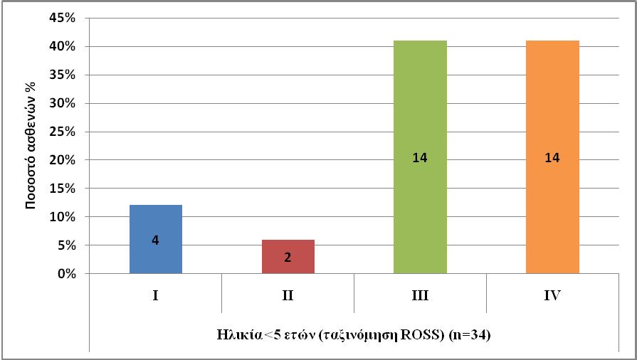
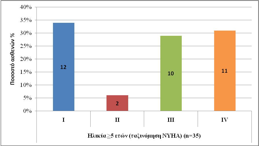
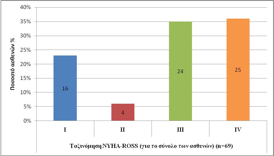
NYHA = New York Heart Association
1. Elliott P, Andersson B, Arbustini E, Bilinska Z, Cecchi F, Charron P, etal. Classification of the cardiomyopathies: apposition statement from the European Society Of Cardiology Working Group on Myocardial and Pericardial Diseases. Eur Heart J 2008; 29:270.
2. Wilkinson JD, Landy DC, Colan SD, Towbin JA, Sleeper LA, Orav EJ, et al. The pediatric cardiomyopathy registry and heart failure: key results from the first 15 years. Heart Fail Clin. 2010 Oct;6(4):401-13, vii.
3. Lipshultz SE, Sleeper LA, Towbin JA, Lowe AM, Orav EJ, Cox GF, et al. The incidence of pediatric cardiomyopathy in two regions of the United States. N Engl J Med. 2003 Apr 24;348(17):1647-55.
4. Nugent AW, Daubeney PE, Chondros P, Carlin JB, Cheung M, Wilkinson LC, et al. The epidemiology of childhood cardiomyopathy in Australia. N Engl J Med. 2003;348(17):1639.
5. Kantor PF, Lougheed J, Dancea A, McGillion M, Barbosa N, Chan C, et al. Presentation, diagnosis, and medical management of heart failure in children: Canadian Cardiovascular Society guidelines. Can J Cardiol. 2013 Dec;29(12):1535-52.
6. Caforio AL, Pankuweit S, Arbustini E, Basso C, Gimeno-Blanes J, Felix SB, et al. Current state of knowledge on aetiology, diagnosis, management, and therapy of myocarditis: a position statement of the European Society of Cardiology Working Group on Myocardial and Pericardial Diseases. Eur Heart J 2013; 34:2636.
7. Towbin JA, Lowe AM, Colan SD, Sleeper LA, Orav EJ, Clunie S, et al. Incidence, Causes, and Outcomes of Dilated Cardiomyopathy in Children. JAMA. 2006;296(15):1867–1876.
8. Sagar S, Liu PP, Cooper LT Jr. Myocarditis. Lancet 2012; 379:738-47.
9. Rosenthal D, Chrisant MR, Edens E, Mahony L, Canter C, Colan S, et al. International Society for Heart and Lung Transplantation: Practice guidelines for management of heart failure in children. J Heart Lung Transplant. 2004 Dec;23(12):1313-33.
10. Yancy CW, Jessup M, Bozkurt B, Butler J, Casey DE Jr, Drazner MH, et al. 2013 ACCF/ AHA guideline for the management of heart failure: a report of the American College of Cardiology Foundation/American Heart Association Task Force on Practice Guidelines. J Am Coll Cardiol. 2013 Oct;62(16):e147-239.
11. Andrews RE, Fenton MJ, Ridout DA, Burch M. New-onset heart failure due to heart muscle disease in childhood: a prospective study in the United Kingdom and Ireland. Circulation. 2008 Jan 1;117(1):79-84.
12. Bakeet MA, Mohamed MM, Allam AA, Gamal R.Childhood Cardiomyopathies: A Study in Tertiary Care Hospital in Upper Egypt. Electron Physician. 2016 Nov 25;8(11):3164-3169.
13. Arola A, Jokinen E, Ruuskanen O, Saraste M, Pesonen E, Kuusela AL, et al. Epidemiology of idiopathic cardiomyopathies in children and adolescents. A nationwide study in Finland.Am J Epidemiol. 1997 Sep 1;146(5):385-93.
14. Richardson P, McKenna W, Bristow M, Maisch B, Mautner B, O'Connell J, et al. Report of the 1995 World Health Organization/International Society and Federation of Cardiology Task Force on the Definition and Classification of cardiomyopathies. Circulation 1996; 93:841.
Τ.
Κ.
F.25510-30340
e-mail: emantada@med. duth.gr
Correspondence
Elpidoforos Mantadakis, MD, PhD
6th Kilometer Alexandroupolis-Makris 68100, Alexandroupolis
T. +302551351411
M. +306946064690
F. +302551030340
e-mail: emantada@med. duth.gr
Elpidoforos Mantadakis, Vasileios Papadakis
Elpidoforos Mantadakis
Department of Pediatrics
University General Hospital of Alexandroupolis, Democritus University of Thrace
Vasileios Papadakis
Department of Pediatric Hematology - Oncology, Unit B VardinogiannisELPIDA, Aghia Sophia Children’s Hospital, Athens
Summary
Haemophagocytic lymphohistiocytosis (HLH) is a hyperin flammatory syndrome characterized by high fever, cytopenias and hepatosplenomegaly. The development of the syndrome usually requires the presence of genetic susceptibility along with an exogenous triggering factor, usually an infection. A characteristic, but not necessary finding for diagnosing HLH is haemophagocytosis by activated macrophages in various body compartments. HLH has been described in all races and ethnic groups, while familial HLH is an autosomal recessive disease that is more common in populations where inbreeding is prevalent. A molecular diagnosis or fulfillment of 5 out of 8 criteria is required for diagnosis according to the Histiocyte Society (HLH 2004 criteria). Secondary HLH is associated with viral, bacterial, parasitic or fungal infections, neoplasias, rheumatologic conditions or medical interventions and is more common in adults. The incidence of HLH varies from 1 to 225 cases per 300.000 livebirths, while the prevalence of the syndrome is unknown, as it is frequently underdiagnosed. The main differential diagnosis of HLH includes sepsis, metabolic diseases that cause organomegaly and/or hypertriglyceridemia and various hematologic malignancies, such as leukemias, lymphomas and multiple myeloma. Primary HLH is a deadly disease that requires prompt therapy initiation and then haematopoietic stem cell transplantation. Initial HLH therapy is based on the HLH-94 protocol of the Histiocyte Society that uses dexamethasone, etoposide, cyclosporine and intrathecal methotrexate with the goal of eventually transplanting the patient. The recent approval in the US of emapalumab, a human monoclonal antibody against interferon-γis expected to improve the prognosis of patients with resistant or refractory disease.
Key words: Haemophagocytic lymphohistiocytosis, MAS, immune deficiencies, inflammation, fibrinogen, infections, haematopoietic stem cell transplantation, ferritin
(11).
HLH (familial HLH, FHLH),
(malignancy-associated HLH, M-HLH), γ)
(rheumatologic HLH, R-HLH, γνωστό
macrophage activation syndrome, MAS-HLH), δ)
(iatrogenic HLH, Rx-HLH) (12).
HLH (immune compromise HLH, IC-HLH)
PRF1 (οικογενές HLH
2, FHLH2), το UNC13D (οικογενές HLH τύπου 3, FHLH3), το STX11 (οικογενές HLH τύπου 4, FHLH4) και το STXBP2 (οικογενές HLH τύπου 5, FHLH5) (13).
Συντομογραφίες:
1. Πυρετός >38,3°C
2. Σπληνομεγαλία
3. Κυτταροπενίες
(ινωδογόνο<150mg/dL)
5.
Οικογενές HLH τύπου 2 (PRF1)
Οικογενές HLH
3 (UNC13D)
Οικογενές HLH τύπου 4 (STX11)
Οικογενές HLH τύπου 5 (STXBP2)
Λεμφοϋπερπλαστικό σύνδρομο τύπου1 (SH2D1A)
Σύνδρομο Griscelli τύπου 2 (RAB27A)
Σύνδρομο Chediak-Higashi (LYST)
Σύνδρομο Hermansky-Pudlak τύπου 2
HLH που συνδέεται με διαταραχές
2 (BIRC4)
Mutation of Nod-like receptor family, caspase recruitment domain-containing 4 (NLRC4)
HLH
Ιοί: EBV, CMV, VZV, HSV-1 &HSV-2 (30% των νεογνικών περιπτώσεων HLH στην Ιαπωνία), HHV-6, HHV-7, HHV8, γρίπηςA, γρίπης Β, HIV, HTLV, ηπατίτιδας Α, B και C, adenovirus, ιλαράς, παρωτίτιδας, ερυθράς, παρβοϊός Β19, Eastern equine encephalitis virus, φλαβοϊοί (π.χ., δάγκειος), hanta virus, BKV, εντεροϊοί, JCvirus, RSV
Παράσιτα: Toxoplasma gondii, Plasmodium falciparum, Plasmodium vivax, Leishmania spp., Strongy loides spp., Babesia spp.
Βακτήρια: Mycobacterium tuberculosis, Brucella melitensis, Rickettsia spp., Campylobacter spp., Neisseria meningitidis, Staphylococcus aureus, Streptococcus spp., Haemophilus spp., Serratia spp., Salmonella typhi, Ehrlichiach affeensis, Chlamydiae, Mycoplasma spp., Borrelia spp., Bartonella henselae, Legionnella spp., Fusobacterium spp., Acinetobacter baumannii
Μύκητες: Candida spp., Trichosporon asahii, Cryptococcus spp., Pneumocystis spp., Histoplasma spp., Penicillium marneffei, υφομύκητες (Aspergillus spp., Fusarium spp.)
-γ, IL-1β, TNF-α, IL-6, IL-10, IL-12, IL-16, IL-18, sCD25, sCD163 (soluble hemoglobin scavenger receptor), δηλαδή κυτοκινών
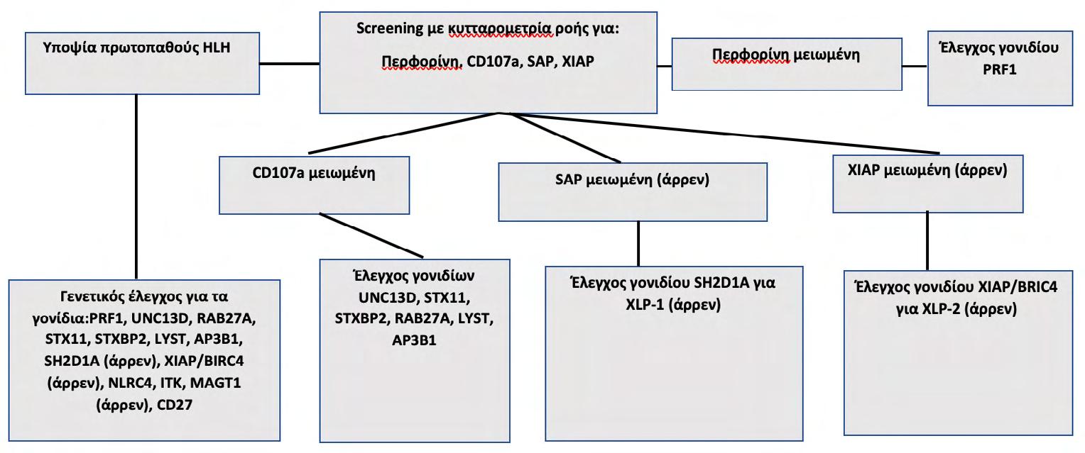
X-linkedin hibitor of apoptosis (XIAP,
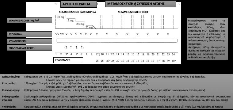
(https://clinicaltrials.gov/ct2/ show/NCT01818492).
Βιβλιογραφία
1. Chandrakasan S, Filipovich AH. Hemophagocytic lymphohistiocytosis: advances in pathophysiology, diagnosis, and treatment. J Pediatr. 2013;163(5):1253-1259.
2. Allen CE, McClain KL. Pathophysiology and epidemiology of hemophagocytic lymphohistiocytosis. Hematology Am Soc Hematol Educ Program. 2015;2015:177-182.
3. Ishii E. Hemophagocytic lymphohistiocytosis in children: pathogenesis and treatment. Front Pediatr. 2016;4:47.
4. Παπαδάκης
2011;28(2):175181.
5. Kogawa K, Sato H, Asano T, Ohga S, Kudo K, Morimoto A, et al. Prognostic factors of Epstein-Barr virus-associated hemophagocytic lymphohistiocytosis in children: report of the Japan Histiocytosis Study Group. Pediatr Blood Cancer. 2014;61(7):1257-1262.
6. Horne A, Wickström R, Jordan MB, Yeh EA, Naqvi A, HenterJI,et al. How to treat involvement of the central nervous system in hemophagocytic lymphohistiocytosis? Curr Treat Options Neurol. 2017;19(1):3.
7. Gupta A, Tyrrell P, Valani R, Benseler S, Weitzman S, Abdelhaleem M. The role of the initial bone marrow aspirate in the diagnosis of hemophagocytic lymphohistiocytosis. Pediatr Blood Cancer. 2008;51(3):402-404.
8. Emile JF, Abla O, Fraitag S, Horne A, Haroche J, Donadieu J, et al; Histiocyte Society. Revised classification of histiocytoses and neoplasms of the macrophage-dendritic cell lineages.
Blood. 2016;127(22):2672-2681.
9. Henter JI, Elinder G, Ost A. Diagnostic guidelines for hemophagocytic lymphohistiocytosis. The FHL Study Groupof the Histiocyte Society. Semin Oncol. 1991;18(1):29-33.
10. Henter JI, Horne A, Aricό M, Egeler RM, Filipovich AH, Imashuku S, et al. HLH-2004: Diagnostic and therapeutic guidelines for hemophagocytic lymphohistiocytosis. Pediatr Blood Cancer. 2007;48(2):124-131.
11. Filipovich AH. Hemophagocytic lymphohistiocytosis (HLH) and related disorders. Hematology Am Soc Hematol Educ Program. 2009:127-131.
12. Jordan MB, Allen CE, Greenberg J, Henry M, Hermiston ML, Kumar A, et al. Challenges in the diagnosis of hemophagocytic lymphohistiocytosis: recommendations from the North American Consortium for Histiocytosis (NACHO). Pediatr Blood Cancer. 2019:e27929.
13. Al-Samkari H, Berliner N. Hemophagocytic lymphohistiocytosis. Annu Rev Pathol. 2018;13:27-49.
14. Chinn IK, Eckstein OS, Peckham-Gregory EC, Goldberg BR, Forbes LR, Nicholas SK, et al. Genetic and mechanistic diversity in pediatric hemophagocytic lymphohistiocytosis. Blood. 2018;132(1):89-100.
15. Maakaroun NR, Moanna A, Jacob JT, Albrecht H. Viral infections associated with haemophagocytic syndrome. Rev Med Virol. 2010;20(2):93-105.
16. Strenger V, Merth G, Lackner H, Aberle SW, Kessler HH, SeidelMG, et al.Malignancy and chemotherapy induced haemophagocytic lymphohistiocytosis in children and adolescents-a single centre experience of 20 years. Ann Hematol. 2018;97(6):989-998.
17. Neelapu SS, Tummala S, Kebriaei P, Wierda W, Gutierrez C, LockeFL, et al. Chimeric antigen receptor T-cell therapy - assessment and management oftoxicities. Nat Rev Clin Oncol. 2018;15(1):47-62.
18. Kaufman KM, Linghu B, Szustakowski JD, Husami A, Yang F, Zhang K, et al. Wholeexome sequencing reveals overlap between macrophage activation syndrome in systemic juvenile idiopathic arthritis and familial hemophagocytic lymphohistiocytosis. Arthritis Rheumatol. 2014;66(12):3486-3495.
19. Cetica V, Sieni E, Pende D, Danesino C, De Fusco C, Locatelli F, et al. Genetic predisposition to hemophagocytic lymphohistiocytosis: report on 500 patients from the Italian registry. J Allergy Clin Immunol. 2016;137(1):188-196.e4.
20. Wang Y, Wang Z, Zhang J, Wei Q, Tang R, Qi J, et al. Genetic features of late onset primary hemophagocytic lymphohistiocytosis in adolescence or adulthood. PLoS One. 2014;9(9):e107386.
21. Schram AM, Berliner N. How I treat hemophagocytic lymphohistiocytosis in the adult patient. Blood. 2015;125(19):2908-2914.
22. Henter JI, Elinder G, Söder O, Ost A. Incidence in Sweden and clinical features of familial hemophagocytic lymphohistiocytosis. Acta Paediatr Scand. 1991;80(4):428-435.
23. Meeths M, Horne A, Sabel M, Bryceson YT, Henter JI. Incidence and clinical presentation of primary hemophagocytic lymphohistiocytosis in Sweden. Pediatr Blood Cancer. 2015;62(2):346-352.
24. Niece JA, Rogers ZR, Ahmad N, Langevin AM, McClain KL. Hemophagocytic lymphohistiocytosis in Texas: observations on ethnicity and race. Pediatr Blood Cancer. 2010;54(3):424428.
25. Janka GE. Familial and acquired hemophagocytic lymphohistiocytosis. Eur J Pediatr. 2007;166(2):95-109.
26. Gholam C, Grigoriadou S, Gilmour KC, Gaspar HB. Familial haemophagocytic lymphohistiocytosis: advances in the genetic basis, diagnosis and management. Clin Exp Immunol. 2011;163(3):271-283.
27. Filipovich AH. The expanding spectrum of hemophagocytic lymphohistiocytosis. CurrOpin Allergy Clin Immunol. 2011;11(6):512-516.
28. Filipovich AH, Chandrakasan S. Pathogenesis of hemophagocytic lymphohistiocytosis. Hematol Oncol Clin North Am. 2015;29(5):895-902.
29. Marsh RA, Haddad E. How I treat primary haemophagocytic lymphohistiocytosis. Br J Haematol. 2018;182(2):185-199.
30. Marsh RA, Bleesing JJ, Filipovich AH. Using flow cytometry to screen patients for X-linked lymphoproliferative disease due to SAP deficiency and XIAP deficiency. J Immunol Methods. 2010;362(1-2):1-9.
31. Degar B. Familial hemophagocytic lymphohistiocytosis. Hematol Oncol Clin North Am. 2015;29(5):903-913.
32. Rosado FG, Kim AS. Hemophagocytic lymphohistiocytosis: an update on diagnosis and pathogenesis. Am J Clin Pathol. 2013;139(6):713-727.
33. Yasumi T, Hori M, Hiejima E, Shibata H, Izawa K, Oda H, et al. Laboratory parameters identify familial haemophagocytic lymphohistiocytosis from other forms of paediatric haemophagocytosis. Br J Haematol. 2015;170(4):532-538.
34. Cui Y, Xiong X, Ren Y, Wang F, Wang C, Zhang Y. CD163 as a valuable diagnostic and prognostic biomarker of sepsis-associated hemophagocytic lymphohistiocytosis in critically ill children. Pediatr Blood Cancer. 2019:e27909.
35. Bryceson YT, Pende D, Maul-Pavicic A, Gilmour KC, Ufheil H, Vraetz T, et al. A prospective evaluation of degranulation assays in the rapid diagnosis of familial hemophagocytic syndromes. Blood. 2012;119(12):2754-2763.
36. Ammann S, Lehmberg K, Zur Stadt U, Janka G, Rensing-Ehl A, Klemann C, et al; HLH study of the GPOH. Primary and secondary hemophagocytic lymphohistiocytosis have different patterns of T-cell activation, differentiation and repertoire. Eur J Immunol. 2017;47(2):364373.
37. McCall CM, Mudali S, Arceci RJ, Small D, Fuller S, Gocke CD, et al. Flow cytometric findings in hemophagocytic lymphohistiocytosis. Am J Clin Pathol. 2012;137(5):786-794.
38. Sumegi J, Nestheide SV, Barnes MG, Villanueva J, Zhang K, Grom AA, et al. Gene-expression signatures differ between different clinical forms of familial hemophagocytic lymphohistiocytosis. Blood. 2013;121(7):e14-24.
39. Allen CE, Yu X, Kozinetz CA, McClain KL. Highly elevated ferritin levels and the diagnosis of hemophagocytic lymphohistiocytosis. Pediatr Blood Cancer. 2008;50(6):1227-1235.
40. Lin TF, Ferlic-Stark LL, Allen CE, Kozinetz CA, McClain KL. Rate of decline of ferritin in patients with hemophagocytic lymphohistiocytosis as a prognostic variable for mortality. Pediatr Blood Cancer. 2011;56(1):154-155.
41. Ravelli A, Minoia F, Davì S, Horne A, Bovis F, Pistorio A, et al; Paediatric Rheumatology International Trials Organization; Childhood Arthritis and Rheumatology Research Alliance; Pediatric Rheumatology Collaborative Study Group; Histiocyte Society. 2016 classification criteria for macrophage activation syndrome complicating systemic juvenile idiopathic arthritis: a European League Against Rheumatism/American College of Rheumatology/Paediatric Rheumatology International Trials Organization Collaborative Initiative. Ann Rheum Dis. 2016;75(3):481-489.
42. Minoia F, Bovis F, Davì S, Insalaco A, Lehmberg K, Shenoi S, et al; Pediatric Rheumatology International Trials Organization, the Childhood Arthritis and Rheumatology Research Alliance, the Pediatric Rheumatology Collaborative Study Group, and the Histiocyte Society. Development and initial validation of the macrophage activation syndrome/primary hemophagocytic lymphohistiocytosis score, a diagnostic tool that differentiates primary hemophagocytic lymphohistiocytosis from macrophage activation syndrome. J Pediatr. 2017;189:72-78.e3.
43. Fardet L, Galicier L, Lambotte O, Marzac C, Aumont C, Chahwan D, et al. Development and validation of the HScore, a score for the diagnosis of reactive hemophagocytic syndrome. Arthritis Rheumatol. 2014;66(9):2613-2620.
44. Gagnaire MH, Galambrun C, Stéphan JL. Hemophagocytic syndrome: a misleading complication of visceral leishmaniasis inchildren--a series of 12 cases. Pediatrics. 2000;106(4):E58.
45. Singh G, Shabani-Rad MT, Vanderkooi OG, Vayalumkal JV, Kuhn SM, Guilcher GM,et al. Leishmania in HLH: a rare finding with significant treatment implications. J Pediatr Hematol Oncol. 2013;35(3):e127-129.
46. Nikiforow S. The role of hematopoietic stem cell transplantation in treatment of hemophagocytic lymphohistiocytosis. Hematol Oncol Clin North Am. 2015;29(5):943-959.
47. Henter JI, Aricò M, Egeler RM, Elinder G, Favara BE, Filipovich AH, et al. HLH-94: a treatment protocol for hemophagocytic lymphohistiocytosis. HLH study Group of the Histio-
cyte Society. Med Pediatr Oncol. 1997;28(5):342-347.
48. Jordan MB, Allen CE, Weitzman S, Filipovich AH, McClain KL. How I treat hemophagocytic lymphohistiocytosis. Blood. 2011;118(15):4041-4052.
49. Henter JI, Samuelsson-Horne A, Aricò M, Egeler RM, Elinder G, Filipovich AH, et al; Histocyte Society. Treatment of hemophagocytic lymphohistiocytosis with HLH-94 immuno chemotherapy and bone marrow transplantation. Blood. 2002;100(7):2367-2373.
50. Trottestam H, Horne A, Aricò M, Egeler RM, Filipovich AH, Gadner H, et al; Histiocyte Society. Chemoimmunotherapy for hemophagocytic lymphohistiocytosis: long-term results of the HLH-94 treatment protocol. Blood. 2011;118(17):4577-4584.
51. Bergsten E, Horne A, Aricó M, Astigarraga I, Egeler RM, Filipovich AH, et al. Confirmed efficacy of etoposide and dexamethasone in HLH treatment: long-term results of the cooperative HLH-2004 study. Blood. 2017;130(25):2728-2738.
52. Imashuku S, Kuriyama K, Teramura T, Ishii E, Kinugawa N, Kato M, et al. Requirement for etoposide in the treatment of Epstein-Barr virus-associated hemophagocytic lymphohistiocytosis. J Clin Oncol. 2001;19(10):2665-2673.
53. Arca M, Fardet L, Galicier L, Rivière S, Marzac C, Aumont C, et al. Prognostic factors of early death in a cohort of 162 adult haemophagocytic syndrome: impact of triggering disease and early treatment with etoposide. Br J Haematol. 2015;168(1):63-68.
54. Mahlaoui N, Ouachée-Chardin M, de Saint Basile G, Neven B, Picard C, BlancheS, et al. Immunotherapy of familial hemophagocytic lymphohistiocytosis with antithymocyte globulins: a single-center retrospective report of 38 patients. Pediatrics. 2007;120(3):e622-628.
55. Marsh RA, Jordan MB, Talano JA, Nichols KE, Kumar A, Naqvi A, et al; Histiocyte Society Salvage Therapy Working Group. Salvage therapy for refractory hemophagocytic lymphohistiocytosis: a review of the published experience. Pediatr Blood Cancer. 2017;64(4).
56. Marsh RA, Allen CE, McClain KL, Weinstein JL, Kanter J, Skiles J, et al. Salvage therapy of refractory hemophagocytic lymphohistiocytosis with alemtuzumab. Pediatr Blood Cancer. 2013;60(1):101-109.
57. Marsh RA, Vaughn G, Kim MO, Li D, Jodele S, Joshi S, et al. Reduced-intensity conditioning significantly improves survival of patients with hemophagocytic lymphohistiocytosis undergoing allogeneic hematopoietic cell transplantation. Blood. 2010;116(26):5824-5831.
58. Messina C, Zecca M, Fagioli F, Rovelli A, Giardino S, Merli P, et al. Outcomes of children with hemophagocytic lymphohistiocytosis given allogeneic hematopoietic stem cell transplantation in Italy. Biol Blood Marrow Transplant. 2018;24(6):1223-1231.
59. Al-Salama ZT. Emapalumab: first global approval. Drugs. 2019;79(1):99-103.
60. Lounder DT, Bin Q, de Min C, Jordan MB. Treatment of refractory hemophagocytic lymphohistiocytosis with emapalumab despite severe concurrent infections. Blood Adv. 2019;3(1):47-50.
e-mail: fladomenou@gmail. com
Correspondence
Ladomenou Fani, MD, PhD T. +306976311544
e-mail: fladomenou@gmail. com
Asimenia Athousaki, Fani Ladomenou
Abstract
Introduction: Familiar Mediterranean Fever (FMF) is an auto inflammatory disease which is inherited with an autosomal recessive pattern and characterized by recurrent episodes of fever with polyserositis or/and arthritis. Colchicine is the first-line drug in treating this disease and preventing the occurrence of secondary amyloidosis. However, 5-10% of patients do not respond to colchicine and are candidates to receive biological agents. The purpose of this study was to determine the factors that affect the response to colchicine in pediatric patients with FMF.
Methods: Articles from the PubMed database were searched without time limitation in the bibliography. Only studies with pediatric patients were included.
Results: Non-response to treatment has been associated with higher disease activity associated with specific mutations in the MEFV gene (eg M694V / M694V). Moreover, environmental factors and co-inflammatory diseases (eg, spinal arthritis) contribute to the severity of the disease. Other factors are related to the bioavailability of colchicine with emphasis on genetic polymorphisms in the ABCB1 transporter gene (MDR1) and interactions with drugs that inhibit the cytochrome P450 protein as well as the Pgp transporter protein (ABCB1). Finally, non-compliance to treatment should be considered in all patients with a poor response to colchicine.
Conclusions: Given the side effects and the high cost of biological factors, it is crucial to accurately identify those patients who do not respond to colchicine and to determine the causes of ineffectiveness of treatment in this patient group.
Key words: FMF, children, colchicine, resistance
Asimenia Athousaki School of Medicine, University of Crete
Fani Ladomenou Department of Paediatrics, Venizeleion General Hospital, Heraklion, Crete
ασθενών.
Βιβλιογραφία
1. Ben-ChetritE, LevyM. Does the lack of the P glycoprotein efflux pump in neutrophils explain the efficacyof colchicine in Familiar Mediterranean Fever? Med Hypotheses. 1998;51:377-80.
2. Lidar M, Scherrmann JM, Shinar Y. Colchicine non-responsiveness in familial Mediterranean fever: clinical, genetic, pharmacokinetic, and socioeconomic characterization. Semin Arthritis Rheum. 2004; 33:273–82.
3. Ozen S, Demirkaya E, Erer B, Livneh A, Ben-Chetrit E, Giancane G. EULAR recommendations for the management of familial Mediterranean fever. Ann Rheum Dis. 2016; 75:644–51.
4. Dode C, Pecheux C, Cazeneuve C, Cattan D, Dervichian M, Goossens M et al. Mutations in the MEFV gene in a large series of patients with a clinical diagnosis of familial Mediterranean fever. Am J Med Genet. 2000; 92:241-246.
5. Ozen F, Silan C, UludagΑ, Candan F, Silan F, Ozdemir S et al.Association between ABCB1 (MDR1) Gene 3435 C>T Polymorphism and Colchicine Unresponsiveness of FMF Patients. Renal Failure. 2011;33: 899–903.
6. Mansfield E1, Chae JJ, Komarow HD, Brotz TM, Frucht DM, Aksentijevich I et al.The familial Mediterranean fever protein, pyrin, associates with microtubules and colocalizes with actin filaments. Blood. 2001Aug 1;98(3):851-9.
7. Sohar E, Gafni J, Pras M, Heller H. Familial Mediterranean fever. A survey of 470 cases and review of the literature. Am J Med 1967;43:227-53.
8. Touitou I. The spectrum of Familial Mediterranean Fever (FMF) mutations.Eur J Hum Genet 2001; 9:473-83.
9. Ben-Chetrit E, Ben-Chetrit A. Familial Mediterranean fever and menstruation. BJOG 2001; 108:403–407
10. Srinivasula SM, Poyet JL, Razmara M, Datta P, Zhang Z, Alnemri ES. The PYRIN-CARD protein ASC is an activating adaptor for caspase-1. J Biol Chem. 2002; 277:21119-22112.
11. Ting JP, Kastner DL, Hoffman HM.Caterpillers,pyrin and hereditary immunological disorders.Nat Rev Immunol. 2006;6:183-95.
12. Dinarello CA, Wolff SM, Goldfinger SE, Dale DC, Alling DW. Colchicine therapyfor Familial Mediterranean Fever. A double-blind trial. NEJM. 1974;291:934–937.
13. Zemer D, Revach M, Pras M, Modan B, Schor S, Sohar E. et al. A controlled trial ofcolchicine in preventing attacks of Familial MediterraneanFever. NEJM. 1974; 291:932–934.
14. Chappey ON, Niel E, Wautier JL, Hung PP, Dervichian M, Cattan D. et al. M. Colchicine disposition in human leukocytes after single and multiple oral administration.Clinical Pharmacology & Therapeutics.1993; 54:360–367.
15. Mansfield E, Chae JJ, Komarow HD, BrotzTM,Frucht DM, Aksentijevich I. et al. The Familial Mediterranean Fever protein, pyrin, associates with microtubules and colocalizes with actin filaments. Blood 2001; 98:851–859.
16. Bar-Eli M, Ehrenfeld M, Levy M, Gallily R, Eliakim M. Leukocyte chemotaxis in recurrentpolyserositis (familial Mediterranean fever). American Journal of the Medical Sciences1981; 281:15–18.
17. Kiraz S, Ertenli I, Arici M, Calguneri M, Haznedaroglu I, Celik I et al. Effects of colchicine on inflammatory cytokines and selectins in familial Mediterraneanfever. Clinical and Experimental Rheumatology 1998; 16:721–724.
18. Hentgen V, Grateau G, Kone-Paut I, Livneh A, Padeh S, Rozenbaum M, et al. Evidencebased recommendations for the practical management of familial Mediterranean fever. Semin Arthritis Rheum 2013; 43:387–91.
19. Meinzer U, Quartier P., Alexandra J. F., Hentgen V., Retornaz F., Kone-Paut I,Interleukin-1 targeting drugs in familial mediterranean fever: a case series and a review ofthe literature, Seminars in Arthritis and Rheumatism, Volume 41, Issue 2,2011 pp. 265– 271
20. Soriano A., Verrecchia E., Afeltra A., Landolfi R., and Manna R., IL-1 biological treatment of familial Mediterranean fever, Clinical Reviews in Allergy & Immunology.2013;A ug;45(1):117-30.
21. Lidar M, Yonath H, Shechter N, Sikron F, Sadetzki S, Langevitz Pet al. Incomplete response to colchicine in M694V homozygote FMF patients. Autoimmun Rev 2012;12:72e6.
22. Ben-Zvi I, Brandt B, Berkun Y,Lidar M, Livneh A.The relative contribution of environmental and genetic factors to phenotypic variation in familial Mediterranean fever (FMF). Gene 2012;491:260e3.
23. Omer Karadag, Abdurrahman Tufan, VeliYazisiz, Kemal Ureten, Sedat Yilmaz, Muhammet Cinar et al. The factors considered as trigger for the attacks in patients with familial Mediterranean fever. Rheumatol Int. 2013 Apr;33(4):893-7.
24. Langevitz P, Livneh A, Zemer D, Shemer J, Pras M. Sero negative spondyloarthropa¬thy in familial Mediterranean fever. Semin Arthritis Rheum 1997; 27: 67-72.
25. Akar S, Soysal O, Balci A, Solmaz D, Gerdan V, Onen F et al. High prevalence of spondyloarthritis and ankylos¬ing spondylitis among familial Mediterranean fever patients and their first-degree relatives: further evidence for the connection. Arthritis Res Ther2013; 15: R21.
26. Eshed I1, Rosman Y, Livneh A, Kedem R, Langevitz P, Ben-Zvi I et al. Exertional leg pain in familial Mediterranean fever: a manifestation of an underlying enthesopathy and a marker of more severe disease. Arthritis Rheumatol2014;66:3221e6.
27. Kaplan E1, Mukamel M, Barash J, Brik R, Padeh S, Berkun Y et al.Protracted febrile myalgia in children and young adults with familial Mediterranean fever: analysis of 15 patients and suggested criteria for working diagnosis. ClinExpRheumatol2007; 25 (Suppl. 45): S114-7.
28. Sissung TM1, Gardner ER, Piekarz RL, Howden R, Chen X, Woo Set al. Impact of ABCB1 allelic variants on QTc interval prolongation. Clin Cancer Res. 2011;17(4):937–946.
29. Tang K, Wong LP, Lee EJ, Chong SS, Lee CG. Genomic evidence for recent positive selection at the human MDR1 gene locus. Hum Mol Genet. 2004;13(8):783–797.
30. Tufan A1, Babaoglu MO, Akdogan A, Yasar U, Calguneri M, Kalyoncu U et al. Association of drug transporter gene ABCB1 (MDR1) 3435C to T polymorphism with colchicine response in familial Mediterranean fever. J Rheumatol. 2007;34(7):1540–1544.
31. Bernal ML, Sinues B, Fanlo A, Mayayo E. Frequency distribution of C3435T mutation in exon 26 of the MDR1 gene in a Spanish population. Ther Drug Monit. 2003;25(1): 107–111.
32. Lidar M1, Yonath H, Shechter N, Sikron F, Sadetzki S, LangevitzP,et al. Incomplete response to colchicine in M694V homozygote FMF patients. Autoimmun Rev 2012;12:72e6.
33. Gül A. Treatment of familial Mediterranean fever: colchicine and beyond. Isr Med Assoc J 2014;16:281e4.
34. Ben-Chetrit E, Ozdogan H. Non-response to colchicine in FMF – definition, causes and suggested solutions Clin Exp Rheumatol 2008; 26 (Suppl. 50): S49-S51.
35. Melikoglu MA, Senel K. Non-response to colchicine in familial Mediterranean fever should be identified accurately. International Journal of Rheumatic Diseases. 2014;20(12):2118-2121.
36. Calligaris L., Marchetti F., Tommasini A., Ventura A. The efficacy of anakinra in an adolescent with colchicine-resistant familial Mediterranean fever. European Journal of Pediatrics 2008;167:695–696.
37. Roldan R., Ruiz A. M., Miranda M. D., Collantes E. Anakinra: new therapeutic approach in children with Familial Mediterranean Fever resistant to colchicine. Joint Bone Spine 2008;75:504–505.
38. Gül Α, Ozdogan H, Erer B, Ugurlu S, Kasapcopur O, Davis N et al. Efficacy and safety of canakinumab in adolescents and adults with colchicine resistant familial Mediterranean fever, Arthritis Research & Therapy;2015;Sep 4;17:243.
39. Eroglu FK, Beşbaş N, Topaloglu R, Ozen S. Treatment of colchicine-resistant Familial Mediterranean fever in children and adolescents. Rheumatology International. 2015;35(10):17331737.
Correspondence
Christina Syriopoulou-Delli
e-mail: csyriop@gmail.com
Christina Syriopoulou-Delli, Eleni Gkiolnta
Αbstract
The use of social robotics could be a very promising method of improving social skills of children with autism. There are many studies that have been conducted in this field during the past years, proving that the use of robots as support may have positive effects in the development of social skills for children of the autism spectrum, especially in those areas where they present great deficits. In this literature review we are attempting to display, organize and evaluate the most important features and results of twelve (12) scientific articles. The analysis of those results is expected to answer our research hypotheses about the effectiveness of robotics in enhancing social skills of children with autism like joint attention, verbal communication, imitation skills etc. From the analysis that is resulting from these variables it is shown that robotic tools have a positive affect and some suggestions for future research is also being provided.
Key words: autism spectrum disorder, robotics, social robotics, social skills
Christina Syriopoulou-Delli
Eleni Gkiolnta
Department of Educational and Social Policy, University of Macedonia
Scopus, Pub
2.3
(M=43.0, SD=19.4)
(M=36.8, SD=19.2, t(23)=1.97, p< 0.05).
Kaspar (15).
“The Gilliam Autism Rating Scale” (GARS-2).
t(27)=.39).
1. Cho, S.-J., & Ahn, D. H. Socially Assistive Robotics in Autism Spectrum Disorder. Hanyang Medical Reviews, 2016;36(1),17.
2.
2016.
3. Bauminger, N., Gal, E., Goren-Bar, D. Enhancing social communication in high functioning children with autism through a colocated interface. 6th International Workshop on Social Intelligence Design, 2007;24(1):75-84.
4. Chaby, L., Chetouani, M., Plaza, M., & Cohen, D. Exploring Multimodal Social-Emotional Behaviors in Autism Spectrum Disorders: An Interface between Social Signal Processing and
Psychopathology. 2012 International Conference on Privacy, Security, Risk and Trust and 2012 International Confernece on Social Computing, 2012;950-954.
5. Bellini, S., McConnell, L. Strength-based educational programming for students with autism spectrum disorders: A case for video modeling. Preventing School Failure,2010;54,220– 227.
6. Robins, B., Dautenhahn, K., Boekhorst, R., Billard, A. Robotic assistants in therapy and education of children with autism: can a small humanoid robot help encourage social interaction skills? Universal Access in the Information Society, 2005;4(2),105–120.
7. Amran, N. A. B., Gunasekaran, S. S., Mahmoud, M. A. Investigating the factors that influence the efficiency of using robots as social skills therapy for children with autism spectrum disorders (ASD). Journal of Fundamental and Applied Sciences, 2018;10(6S), 1779-1792.
8. Kitchenham, B. (2004). Procedures for performing systematic reviews. Keele University, UK and National ICT Australia,2004;33,28.
9. Begum, M., Serna, R. W., &Yanco, H. A. Are Robots Ready to Deliver Autism Interventions? A Comprehensive Review. International Journal of Social Robotics,2016;8(2),157–181.
10. Pop, C. A., Pintea, S., Vanderborght, B., & David, D. O. Enhancing play skills, engagement and social skills in a play task in ASD children by using robot-based interventions. A pilot study. Interaction Studies Interaction Studies Social Behaviour and Communication in Biological and Artificial Systems,2014;15(2),292-320.
11. Conti, D., Nuovo, S. D., Buono, S., Trubia, G., &Nuovo, A. D. Use of robotics to stimulate imitation in children with Autism Spectrum Disorder: A pilot study in a clinical setting. 2015 24th IEEE International Symposium on Robot and Human Interactive Communication (ROMAN),2015;1-6.
12. Costa, S., Lehmann, H., Dautenhahn, K., Robins, B., &Soares, F. Using a Humanoid Robot to Elicit Body Awareness and Appropriate Physical Interaction in Children with Autism. International Journal of Social Robotics, 2015;7(2),265-278.
13. Costa, S., Lehmann, H., Robins, B., Dautenhahn, K., &Soares, F. “Where is Your Nose?” - Developing Body Awareness Skills Among Children With Autism Using a Humanoid Robot. ACHI 2013, The Sixth International Conference on Advances in Computer-Human Interactions, 2013;117–122 (ανακτήθηκε από https://www.researchgate.net/publication/235350482).
14. Hanafiah, F. A., Zahari, N. I., Shamsuddin, S., Yussof, H., Ismail, L. I., & Mohamed, S. Initial Response in HRI- a Case Study on Evaluation of Child with Autism Spectrum Disorders Interacting with a Humanoid Robot NAO. Procedia Engineering, 2012;41(Iris), 1448–1455.
15. Wainer, J., Robins, B., Amirabdollahian, F., & Dautenhahn, K. Using the humanoid robot KASPAR to autonomously play triadic games and facilitate collaborative play among children with autism. IEEE Transactions on Autonomous Mental Development, 2014;6(3), 183–199.
16. Huskens, B., Verschuur, R., Gillesen, J., Didden, R., &Barakova, E. Promoting question-asking in school-aged children with autism spectrum disorders: Effectiveness of a robot intervention compared to a human-trainer intervention. DevelopmentalNeurorehabilitation,2013;16(5),345–356.
17. Kim, E. S., Berkovits, L. D., Bernier, E. P., Leyzberg, D., Shic, F., Paul, R., et al. Social robots as embedded reinforcers of social behavior in children with autism. Journal of Autism and Developmental Disorders,2013;43(5),1038–1049.
18. Severson, R. L., Stanton, C. M., Gill, B. T., Ruckert, J. H., & Kahn Jr., P. H. Robotic animals might aid in the social development of children with autism, Proceedings of the 3rd ACM/ IEEE international conference on Human robot interaction,2008;271-278.
19. Yussof, H., Miskam, M. A., Malik, N. A., Hamid, M. A. C., Shamsuddin, S., &Basir, S. N. Study on Social Interaction between Children with Autism and Humanoid Robot NAO. Applied Mechanics and Materials, 2013;393,573–578 (ανακτήθηκεαπόwww.scientific.net/ amm.393.573).
20. Kim, E., Paul, R., Shic, F., &Scassellati, B. Bridging the Research Gap: Making HRI Useful to Individuals with Autism. JournalofHuman-RobotInteraction, 2012;1(1),26–54.
21. Feil-Seifer, D., &Mataric, M. (2011). Automated detection and classification of positive vs. negative robot interactions with children with autism using distance-based features, Proceedings of the 6th international conference on Human-robot interaction, 2011;323-330.
e-mail: info@healthylady.gr
Correspondence
Panagiotis Christopoulos
Mouson 1 Athens, Greece
T. +3021069192129
e-mail: info@healthylady.gr
Thanou Chrissanthi, Christopoulos Panagiotis, Dimitroulis Dimitrios, Vlachos Nikolaos
Abstract
Background: Definite factors during childhood and adolescence may lead to the development of proliferative benign diseases (P-BD) and breast cancer (BC) in adulthood, as breast tissue is vulnerable in childhood and adolescence because cells in this crucial period multiply. Therefore any exposure during this period may lead to the manifestation of benign diseases and BC in adulthood. The aim of the review is to analyze the importance of possible predisposing factors that may form the basis for the development of adult breast pathology.
Methods: Review of international literature from May 1988 to January 2014 and analysis of factors leading to the emergence of benign diseases and BC.
Results: The analysis of the studies shows that the increase in red meat consumption, alcohol, early menstrual age, body mass index (BMI), and radiation in childhood and adolescence have a clear correlation with proliferative benign breast diseases (P-BBD) and BC during adult life. Conversely, increased fiber consumption is associated with reduced risk for benign breast disorders.
Conclusions: Nutrition, growth rate in childhood and adolescence, menstrual age and radiation are associated with a risk of developing proliferative breast disorders.
Key words: benign breast disorders, breast cancer, adolescence, childhood, risk factors
Thanou Chrissanthi
Postgratuate student, National and Kapodistrian University of Athens
Christopoulos Panagiotis
Vlachos Nikolaos
2nd Department of Obstetrics and Gynecology, National and Kapodistrian University of Athens, Aretaieio Hospital
Dimitroulis Dimitrios
2nd General Surgery Clinic, National and Kapodistrian University of Athens, Laiko Hospital
ΚΜ:
P-BD: Proliferative benign diseases
BC: Breast cancer
BMI: Body mass index
P-BBD: Proliferative benign breast diseases
RR: Relative Risk:
CI: Confidence Interval:
ER: Estrogen receptor:
PR: Progesterone
receptor:
OR: Odds ratio:
IGF1: Insulin Growth
Factor:
NHS II: Nurses’ Health Study II
GUTS: Growing Up Today Study
1. Frazier AL and Rosenberg SM. Preadolescent and adolescent risk factors for benign breast disease. Journal of adolescent Health 2013; 52 S36-S40
2. Berkey CS, Willett WC, Tamimi RM, Rosner B, et al. Diary intakes in older girls and risk of benign breast disease in young women. Cancer Epidemiol Biomarkers Prev. 2013; 22(4): 670674
3. Baer HJ, Tworoger SS, Hankinson SE, Willett WC. Body fatness at young ages and risk of breast cancer throughout life. Am J Epidemiol 2010; 171:1183-94
4. deWaard F, Trichopoulos D. A unifying concept of the etiology of breast cancer. Int J Cancer. 1988;41:666–669
5. Berkey CS, Willett WC, Tamimi RM, et al. Vegetable protein and vegetable fat intakes in pre-aolescence and adolescent girls, and risk for benign breast disease in young women. Breast Cancer Res Treat. 2013; 141: 299-306
6. Baer HJ, Schnitt SJ Connolly JL, et al. Adolescent diet and incidence of proliferative benign breast disease. Cancer Epidemiol Biomarkers Prev 2003; 12: 1159-67
7. Linos E, Willett WC, Cho E, Colditz G, and Frazier L. Red meat consumption during adolescence among premenopausal women and risk of breast cancer. Cancer Epidemiol Biomarkers Prev. 2008; 17(8): 2146-51
8. Linos E, Willett WC, Cho E, Frazier L. Adolescent diet in relation to breast cancer risk among premenopausal women. Cancer Epidemiol Biomarkers Prev 2010; 19:689-96
9. Su X, Tamimi RM, Colins LC, et al. Intake of fiber and nuts during adolescence and incidence of proliferative benign breast disease. Cancer Causes Control. 2010; 21(7):1033-1046
10. Liu Y, Colditz GA, Cotterchio M, Boucher BA, Kreiger N. Adolescent dietary fiber, vegetable fat, vegetable protein, and nut intakes and breast cancer risk. Breast cancer research and treatment. 2014 doi: 10.1007/s10549-014-2953-3.
11. Korde LA, Wu AH, Fears T, Nomura AM, West DW, Kolonel LN, Pike MC, Hoover RN, Ziegler RG. Childhood soy intake and breast cancer risk in Asian American women. Cancer Epidemiol Biomarkers Prev. 2009;18(4):1050–1059. doi: 10.1158/1055-9965.EPI-08-0405.
12. Wu AH, Yu MC, Tseng CC, Pike MC. Epidemiology of soy exposures and breast cancer risk. Br J Cancer. 2008;98(1):9–14. doi: 10.1038/sj.bjc.6604145.
13. Lee SA, Shu XO, Li H, Yang G, Cai H, Wen W, Ji BT, Gao J, Gao YT, Zheng W. Adolescent and adult soy food intake and breast cancer risk: results from the Shanghai Women’s Health Study. Am J Clin Nutr. 2009;89(6):1920–1926. doi: 10.3945/ajcn.2008.27361
14. Liu Y, Colditz GA, Rosner B, et al. Alcohol intake between menarche and first pregnancy: a prospective study of breast cancer risk. J Nat Cancer Inst. 2013; 105(20):1571-1576.
15. Garland M, Hunter DJ, Colditz GA, Spiegelman DL, Manson JE, Stampfer MJ, Willett WC. Alcohol consumption in relation to breast cancer risk in a cohort of United States women 25-42 years of age. Cancer epidemiology, biomarkers &prevention : a publication of the American Association for Cancer Research, cosponsored by the American Society of Preventive Oncology. 1999;8(11):1017–1021.
16. Horn-Ross PL, Canchola AJ, West DW, Stewart SL, Bernstein L, Deapen D, Pinder R, Ross RK, Anton-Culver H, Peel D, Ziogas A, Reynolds P, Wright W. Patterns of alcohol consumption and breast cancer risk in the California Teachers Study cohort. Cancer Epidemiol Biomarkers Prev. 2004;13(3):405–411.
17. Berkey CS, Willett WC, Frazier AL, Rosner B, Tamimi RM, Rockett HR, Colditz GA. Prospective study of adolescent alcohol consumption and risk of benign breast disease in young
women. Pediatrics. 2010;125(5):e1081–1087. doi: 10.1542/peds.2009-2347.
18. Liu Y, Tamimi RM, Berkey CS, Willett WC, Collins LC, Schnitt SJ, Connolly JL, Colditz GA. Intakes of alcohol and folate during adolescence and risk of proliferative benign breast disease. Pediatrics. 2012;129(5):e1192–1198. doi: 10.1542/peds.2011-2601.
19. Cui Y, Page DL, Chlebowski RT, Beresford SA, Hendrix SL, Lane DS, Rohan TE. Alcohol and folate consumption and risk of benign proliferative epithelial disorders of the breast. Int J Cancer. 2007;121(6):1346–1351. doi: 10.1002/ijc.22861.
20. Berkey CS, Tamimi RM, Rosner B, Frazier AL, Colditz GA. Young women with family history of breast cancer and their risk factors for benign breast disease. Cancer. 2012;118(11):2796–2803. doi: 10.1002/cncr.26519.
21. Seitz HK, Pelucchi C, Bagnardi V, La Vecchia C. Epidemiology and pathophysiology of alcohol and breast cancer: Update 2012. Alcohol and alcoholism. 2012;47(3):204–212. doi: 10.1093/alcalc/ags011.
22. Monninkhof EM, Elias SG, Vlems FA, van der Tweel I, Schuit AJ, Voskuil DW, van Leeuwen FE. Tfpac. Physical activity and breast cancer: a systematic review. Epidemiology. 2007;18(1):137–157. doi: 10.1097/01.ede.0000251167.75581.98.
23. Peters TM, Moore SC, Gierach GL, Wareham NJ, Ekelund U, Hollenbeck AR, Schatzkin A, Leitzmann MF. Intensity and timing of physical activity in relation to postmenopausal breast cancer risk: the prospective NIH-AARP diet and health study. BMC cancer. 2009;9:349. doi: 10.1186/1471-2407-9-349.
24. Maruti SS, Willett WC, Feskanich D, Rosner B, Colditz GA. A Prospective Study of AgeSpecific Physical Activity and Premenopausal Breast Cancer. J Natl Cancer Inst 2008.
25. Boeke CE, Eliassen AH, Oh H,et al. Adolescent physical activity in relation to breast cancer risk. Breast Cancer Res Treat. 2014; 145(3):715-724.
26. Jung MM, Coldritz GA, Collins LC, et al. Lifetime physical activity and the incidence of proliferative benign breast disease. Cancer Causes Control. 2011; 22:1297-305.
27. Tworoger SS, Rosner BA, Willet WC, Hankinson SE. The combined influence of multiple sex and growth hormons on risk of postmenopausal breast cancer: a nested case-control study. Breast Cancer Res. 2011; 13(5):R99. Doi:10.1186/bcr3040.
28. Van den Brandt PA, Spiegelman D, Yaun SS, Adami HO, et al. Pooled analysis of prospective cohort studies on height, weight, and breast cancer risk. Am J Epidemiol. 2000; 152:514527.
29. Amadou A, Ferrari P, Muwonge R, Moskal A, Biessy C, Romieu I, Hainaut P. Overweight, obesity and risk of premenopausal breat cancer according to ethnicity: review and dose-response meta-analysis. Obes Rev. 2013 doi: 10.1111/obr.12028.
30. Berkey CS, Willet WC, Frazier AL, et al. Prospective study of growth and development in older girls and risk of benign breast disease in young women. Cancer 2011; 117:1612-20.
31. Baer HJ, Schnitt SJ, Connolly JL, et al. Early life factors and incidence of proliferative benign breast disease. Cancer Epidemiol Biomarkers Prev 2005; 14: 2889-97.
32. Preston-Martin S, Pike MC, Ross RK, jones PA, Henderson BE. Increased cell division as a cause of human cancer. Cancer Res. 1990; 50:7415-7421.
33. Ahlgren M, Melbye M, Wohlfahrt J, Sorensen TI. Growth patterns and the risk of breast cancer in women. N Engl J Med. 2004; 351 (16): 1619-1626.
34. De Stavola BL, Dos Santos Silva I, Mc Cormack V, Hardy RJ, Kuh DJ, Wadsworth ME, Childhood growth and breast cancer. American Journal of epidemiology. 2004; 159(7):671-682.
35. Collaborative Group on Hormonal Factors in Breast C, Menarche, menopause, and breast cancer risk: individual participant meta-analysis, including 118 964 women with breast cancer from 117 epidrmiological studies. The lancet oncology. 2012; 13(11):1141-1151.
36. Shi L, Remer T, Buyken AE, Hartman MF, Hoffmann P, wudy SA. Prepubertal urinary estrogen excretion and its relationship with pubertal timing. American journal og physiology Endocrinology and metabolism. 2010; 299(6):E990-997.
37. Madigan MP, Troisi R, Potischman N, et al. Serum hormone levels in relation to reproductive and lifestyle factors in postmenopausal women (United States) Cancer Causes Control. 1998; 9(2):199-207.
38. Setiawan VW, Monroe KR, Wilkens LR, Kolonel LN, Pike MC, Henderson BE, Breast
cancer risk factors defined by estrogen and progesterone receptor status:themultiethnic cohort study. American journal of epidemiology. 2009; 169(10): 1251-1259.
39. Coldritz GA, Bohlke K, Berkey CS. Breast cancer risk accumulation starts early-prevention must also. Breast Cancer Res Treat. 2014; 145(3):567-569.
40. Silvera SA, Rohan TE. Benign proliferative epithelial disorders of the breast: A review of epidemiologic evidence. Breast Cancer Res Treat 2008; 110: 397-409.
41. Tamimi RM, Rosner B, Colditz GA. Evaluation of a breast cancer risk prediction model expanded to include category of prior benign breast disease lesion. Cancer 2010; 116: 4944-53.
42. Karapanou O, Papadimitriou A. Determinants of menarche. Reproductive biology and endocrinology: RB&E. 2010;8:115. Doi: 10.1186/1477-7827-8-115.
43. Morris DH, Jones ME, Schoemaker MJ, Ashoworth A, Swerdlow AJ. Determinants of age at menarche in the UK: analysis from the Breakthrough Generations Study. Br j Cancer. 2010; 103(11):1760-1764.
44. Berkey CS, Gardner JD, Frazier AL, Golditz GA. Relation of childhood diet and body size to menarche and adolescent growth in girls. American journal of epidemiology. 2000; 152(5):446452.
45. Kennedy Raelene D and Boughey Judy C. Management of pediatric and adolescent breast masses. SeminPlast Surg. 2013; 27(1): 19-22.
46. Henderson Tara O, Amsterdam A, Bhatia S, Hudson Melissa M, et al. Surveillance for breast cancer in women treated with chest radiation for a childhood, adolescent or young adult cancer: a report from the children’s oncology group. Ann Intern Med. 2010; 152(7): 444-W154.
47. Inskip PD, Robison LL, Stovall M, Smith SA, Hammond S, Mertens AC, et al. Radiation dose and breast cancer risk in the childhood cancer survivor study. J Clin Oncol. 2009.
48. Moskowitz Chaya S, Chou Joanna F, Wolden Suzanne L, et al. Breast cancer after chest radiation therapy for childhood cancer. J Clin Oncol. 2014; 32(21):2217-2223.
49. De Bruin ML, Sparidans J, Van’t Veer MB, et al. Breast Cancer risk in Female Survivors of Hodgkin’s Lymphoma: Lower Risk After Smaller Radiation Volumes. J Clin Oncol. 2009.
50. Schellong G, Riepenhausen M, Ehlert K, et al, Breast Cancer in Young Women After Treatment for Hodgkin’s Disease During Childhood or Adolescence. DtschArzteblInt. 2014; 111(12):3-9.
e-mail:
Correspondence
Evangelos Christou
Τ. +306982889279
e-mail: va.xristou@gmail.com
Evangelos Christou
Konstantina Avgerinou
Theodora Bachou
Mersini Mavrikou
Dimitris Delis
First Department of Pediatrics, Athens General Children’s Hospital “Panagiotis and Aglaia Kyriakou
Varvara Douna
Laboratory of Hematology, Athens General Children’s Hospital “Panagiotis and Aglaia Kyriakou”
Infantile pyknocytosis: A rare cause of newborn hemolytic anemia.
Evangelos Christou, Konstantina Avgerinou, Theodora Bachou, Varvara Douna, Mersini Mavrikou, Dimitris Delis
Abstract
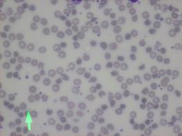
Background: Infantile pyknocytosis (IP) is a rare cause of neonatal hemolytic disease. It was first described by Tuffy et al (1959). Less than 60 reports are described in international literature. The exact etiology of this entity remains unclear. It is characterized by prolonged jaundice and transient hemolytic anemia, combined with increased proportion of pyknocytes in peripheral blood smear. Diagnosis is exclusively based on the detection of a higher proportion of pyknocytes in peripheral blood smear. IP is a self-restricted disease.
Methods: In this case series study we present the diagnostic approach, therapeutic intervention and outcome of two neonates, presented with unconjugated hyperbilirubinemia and major hemolysis, the 17th and 21st day of life respectively, treated with phototherapy and excellent outcome.
Conclusions: Despite the low incidence of this entity, IP should be included in the differential diagnosis in case of prolonged neonatal jaundice not well explained by other common causes of hemolytic anemia.
Key words: Infantile Pyknocytosis, jaundice, hemolytic anemia

Βιβλιογραφια
1. Ackerman B.D., 1969. Infantile pyknocytosis in Mexican-American infants. American Journal of Diseases of Children. 117, 417–423.
2. Amendola G., Di Concilio R., D’Urzo G., Danise P., Parisi G., della Ragione F., Rossi F., Nobili B., Perrotta S., 2008. Erythropoietin treatment can prevent blood transfusion in infantile pyknocytosis. Br J Haematol. 143, 593-595
3. BerardiΑ., Balestri Ε., Bonacorsi G., Chiossi C., Palazzi G., Spaggiari E., Ferrari F., 2017. Neonatal pyknocytosis in a preterm dizygotic twin. World J Clin Pediatr. November 8;6, 176-179
4. Carpentieri U., Gustavson, L., Haggard M.E., 1977. Pyknocytosis in a neonate: an unusual presentation of hereditary elliptocytosis. Clinical Pediatrics. 16, 76–78.
5. Dabbous I.A., Bahlawan L.E., 2002. Infantile pyknocytosis: a forgotten or a dead diagnosis. Journal of Pediatric Hematology and Oncology. 24, 507
6. Dahoui H. A., Abdoud M.R., Saab R., Sinno D., Dabbous I., Muwakkit S.A., 2008. Familial infantile pyknocytosis in association with pulmonary hypertension. Pediatr. Blood Cancer 51, 290–292.
7. Eyssette-Guerreau S., Bader-Meunier B., Garcon L., Guitton C, Cynober T., 2006. Infantile pyknocytosis: a cause of haemolytic anaemia of the newborn. Br J Haematol.133, 439-442
8. Limme B., Dresse M.F., Ketelslegers O., Rigo V., Hoyoux C., 2008. Infantile pyknocytosis: a rare form of neonatal hemolytic anemia. Five case-study. Arch. Pediatr. 15, 1765–1768.
9. Oski F.A., Barness L.A., 1967. Vitamin E deficiency: a previously unrecognized cause of hemolytic anemia in the premature infant. Journal of Pediatrics. 70, 211–220.
10. Tuffy P., Brown A.K., Zuelzer W., 1959. Infantile pyknocytosis; a common erythrocyte abnormality of the first trimester. AMA J Dis Child. 98, 227-241
11. Vos MJ., Martens D., van de Leur S.J., van Wijk R., 2014. Neonatal hemolytic anemia due to pyknocytosis.Eur J Pediatr. December; 173, 1711-4
12. Zannos-Mariolea L., Kattamis C., Paidoucis, M., 1962. Infantile pyknocytosis and glucose6-phosphate dehydrogenase deficiency. British Journal of Haematology. 8, 258–265
e-mail: skondras@yahoo.gr
Correspondence
Ioannis Skondras
Τ. +306932572226, +302132009354
e-mail: skondras@yahoo.gr
Eftychia Kanioura, Ioannis Skondras
Summary
Backround
Congenital diaphragmatic hernia is called the condition where intraabdominal organs are located in the thorax due to a deficit in the diaphragm. It is more usual in the neonatal period, although in a small group of patients the diagnosis is placed after the neonatal period. In these cases symptoms vary from long-lasting, periodical, non-specific symptoms to acute symptoms that could be harmful for the child’s life. In this article a case-report of a Bochdalek type diaphragmatic hernia is presented.
Methods - Case report
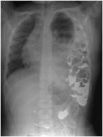
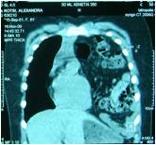
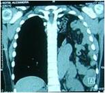
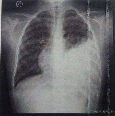
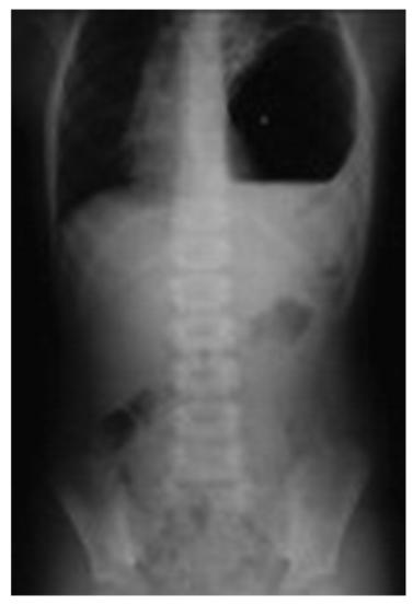
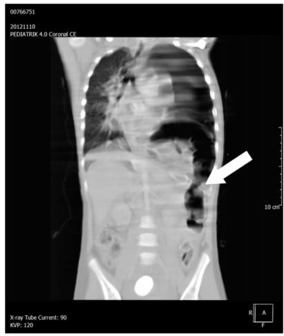
A previously healthy 8-year-old girl presented in the ER due to symptoms of fever during the past 4 days and cough during the past 2 days. The X-Ray revealed a left diaphragmatic hernia.
Results - Discussion
Symptoms of late-presenting diaphragmatic hernia usually involve either the breathing or the gastrointestinal system. This situation leads to misdiagnosing a diaphragmatic hernia hemopneumothorax, pneumothorax, lower lobe pneumonia, pleural effusion, lung cyst.
Conclusions
Lack of typical symptoms may lead to delayed recognition of a diaphragmatic hernia. That is the reason why it should be always part of the differential diagnosis in children presenting with symptoms of breathing disorders or symptoms from the gastrointestinal system, regardless of their age. Surgical treatment of a diaphragmatic hernia should be immediate in order to avoid complications that could be dangerous for a patient’s life.
Key Words
Diaphragmatic hernia, Bochdalek, differential diagnosis
Eftychia Kanioura
Ioannis Skondras
Second Department of Pediatric Surgery GPHA, P. & A. Kyriakou
1. Lu X, Shen Zh, Dong K, Zheng S. Hyper splinism associated with late-presenting congenital diaphragmatic hernia, Medicine (Baltimore) 2016;95(21):1-4.
2. Ozkan A, Cil A, Kaya M, Etcioglu I, Okur M. Late presenting Bochdalek hernia with gastric perforation, Pediatr Emerg Care 2015; 31(1):47-49.
3. Dong J and Jae H. Late-presenting congenital diaphragmatic hernia in children: the experience of single institution in Korea, Yonsei Med J 2013;54(5) 1143-1148.
4. Topor L, Patrancus T, Caragata R, Moga A. Left congenital diaphragmatic hernia – case report, Chirurgia 2015;110(1):84-87.
5. Karaci M, Toroslu E, That D, Unlu M, Piskin I. Late presenting congenital diaphragmatic hernia as tension gastrothorax, Indian J Pediatr, 2015;82(5):483-484.
6. Hamid R, Baba AA, Shera AH, Wani SA, Altaf T, Kant M. Late- presenting congenital diaphragmatic hernia, Afr J Paediatr Surg 2014;11:119-123.
7. Conberiati P, Giacomello L, Camoglio F, Peroni DG. Diaphragmatic hernia in a pediatric emergency department, PediatrEmerg Care 2015;31(5): 354-356.

2.
3.
4.
5.
6.
7.
8.
9. Ενδιαφέρουσες
10. Σύντομα
11. Βραχείες
12.
13.
14.
(Uniform Requirements for Manuscripts Submitted to Biomedical Journals),
International Committee of Medical Journal Editors/Uniform Requirements for Manuscripts Submitted to Biomedical Journals, (www.icmje.org και www.icmje.org/icmje. pdf).
Cumulated Index Medicus [List of Journals Indexed in Index Medicus (www.nlm.nih.goν/bsd/uniform_requirements. html)].
Proesmans W. Bartter syndrome and its neonatal νariant. Eur J Pediatr 1997;156:669-679.
Flyvbjerg Α. Role of growth hormone, insulin-like growth factors (IGFs) and IGF-binding proteins in the renal complications of diabetes. Kidney Ιnt 1997;52 (60 Suppl):S12-S19.
Χωρίς συγγραφέα:
National Institutes of Health Consensus Deνelopment Conference. Neurofibromatosis conference statement. Arch Neurol1988;45:575-578.
Προσδιορισμός
Schreiner GF, Lange L. Ethanol modulation of macrophage influx in glomerulonephritis [Abstract]. J Am Soc Nephrol 1991;2:562.
Should antileukotriene therapies be used instead of inhaled corticosteroids in asthma? [Editorial]. Am J Respir Crit Care Med 1998;158:1697-1701.
Laux-End R, Inaebnit D, Gerber ΗΑ, Bianchetti MG. Vasculitis associated with leνamisole and circulating autoantibodies [Let ter]. Arch Dis Child 1996;75:355-356.
II. ΒΙΒΛΙΑ
Clark AG, Barratt ΤΜ. Steroid-responsiνe nephrotic syndrome. Ιn: Barratt ΤΜ, Arner ED, Harmon WE, editors. Pediatric Nephrology. 4th ed. Baltimore: Lippincott William Wilkins; 1999. p. 742.
Σύγγραμμα
Gorlin RJ, Cohen ΜΜ, Leνin LS. Syndromes of the head and neck. 3rd ed. New York: Oxford Uniνersity Press; 1990.
Δημοσίευση
Bauer ΑW. The two definitions of bacterial resistance. In: Smith AJ, Rogers CA, eds. Proceedings of the Third International Congress of Chemotherapy; 1962 May 29-31; New York: International Society of Chemotherapy; 1963. p. 484-500.
Διδακτορική
Πανεπιστήμιο Αθηνών; 1979.
Kaplan SJ. Post hospital home health care: the elderly’s access and utilization [dissertation]. St. Louis (Μο): Washington Univ.;1995.
III. CD-ROM
Andersoη SC, Poulsen ΚΒ. Anderson’s electronic atlas of hematology [CD-ROM]. Philadelphia: Lippincott Williams & Wilkins; 2002.
IV. ΣΤΟ ΔΙΑΔΙΚΤΥΟ
Abood S. Quality improνement initiatiνe in nursiηg homes: the ΑΝΑ acts in an adνisory role. Am J Nurs [Internet]. 2002 Jun:
Webpage: http://www.nursingworld.org/AJN/2002/june/Wawatch.htm
Μονογραφία: Foley ΚΜ, Gelbaηd Η, editors. Improving palliative care for cancer [Monograph, Internet]. Washington: National Academy Press; 2001.
Webpage: http://www.nap.edu/books/0309074029/html
Ιστοσελίδες: Cancer-Pain.org [Webpage, Internet]. New York: Association of Cancer Online Resources, Ιnc.; 2002: http://www.cancer-pain.org/
Πίνακες
•
•
• επιστημονικό/ά κέντρο/α
•
2.
3.
4.
5.
7.