

Paediatriki
Υποβολή εργασιών e-mail: grammateia@e-child.gr
Οδηγίες
http://e-child.gr/publications/ instructions-to-authors
Iδιοκτήτης
e-mail: grammateia@e-child.gr
372
378
382
Paediatriki
Volume 78 | Number 3 | July - August - September 2015
Trimonthly publication of the Greek Paediatric Society
President
A. Constantopoulos
Editorial board
Director
G. S. Varlamis
Members
S. Andronikou
E. Galanakis
A. Evangeliou
L. Thomaidou
M. Kanariou
A. Kapogiannis
S. Kitsiou-Tzeli
E. Mantadakis
P. Panagiotopoulou-Gartagani
A. Papadopoulou
V. Papaevagelou
A. Papathanassiou
A. Syrigou-Papavasiliou 282
A. Siamopoulou-Mavridou
EDITORIAL
G.S. Varlamis
ABSTRACTS 288
REVIEW ARTICLES
Necrotising enterocolitis
Eftychia Drogouti, Christos Tsakalidis
298
Diagnostic reference levels in pediatric radiology
Triantopoulou Sotiria, Tsapaki Virginia, Triantopoulou Charikleia
318
Review of risk factors for ischaemic stroke in children and imaging modalities
Kourelis George, Galetselli Marianthi
334
Refeeding, menses resumption and improvement of bone density of adolescent girls with anorexia nervosa
Elpis-Athina Vlachopapadopoulou, Artemis Doulgeraki, Artemis Tsitsika
346
ORIGINAL ARTICLES
Familial Μediaterranean Fever in pediatric patiets: A 30-year apraisal
Maria Trachana, Vasiliki Sgouropoulou, Polyxeni Pratsidou-Gertsi, Evangelia Farmaki, Vasiliki Tzimouli, Grigoris Pardalos, Florentia Kanakoudi-Tsakalidou
Manuscript submission
e-mail: grammateia@e-child.gr
Instructions to authors: http://e-child.gr/publications/ instructions-to-authors
Owner Greek Paediatric Society 15, Mpakopoulou st. GR - 15451, Ν. Psychiko Tel.: +302107771140
e-mail: grammateia@e-child.gr
Annual subscription All foreign countries: US$50
358
Polysomnographic study of sleep in children with idiopathic epilepsy
Maria Gogou, Katerina Haidopoulou, Maria Eboriadou, Evangelos Pavlou
372
A decade of experience with gastrostomy placement in infants and children: A critical evaluation of indications, techniques and results
Karetsos Chris, Antoniou Dimitris, Mavridis Georgios
378
CASE REPORT
Benign pancreatic hyperenzymemia or Gullo’s syndrome: a case report
Georgia Zisimopoulou, Nina Manolaki
382
Ketosis and electroencephalographical improvement following viral gastroenteritis in patient with West syndrome
Gkampeta Αnastasia, Pavlidou Εfterpi, Evangeliou Αthanasios, Pavlou Εvangelos
388
Panayiotopoulos syndrome. Case report and review of the literature
Koufopoulou Eleni, Athanassopoulou Panagiota, Dokou Aikaterini, Theodorou Virginia, Manolaki
Nina
SHORT ABSTRACTS
Correspondence
Eftychia Drogouti
59100, Veroia
Τ. +306998424202
e-mail: edrogouti@yahoo. com
Necrotising enterocolitis
Eftychia Drogouti, Christos Tsakalidis
Abstract
Necrotising enterocolitis (ΝEC) is among the most critical disorders of gastrointestinal tract during neonatal period. It is an all too common and devastating disease affecting primarily preterm neonates. Although 90% of infants who develop NEC are born premature, full-term and near-term infants also develop the disease. First described in 1965, the etiology of NEC remains incompletely understood. Several factors appear to trigger the perfect storm of NEC, including gestational age, birth weight, inflammation, ischemia, formula feeding or abnormal gut microbiota. Despite extensive research and advance in medical and surgical treatment over the last six decades, relatively little progress has been made toward improving the outcome of patients with NEC. This article, aims to describe new insights into the pathogenesis of NEC as well as to summarize some potential beneficial strategies in the prevention of NEC.
Keywords: necrotising enterocolitis, premature neonate, pathogenesis
Eftychia Drogouti
Christos Tsakalidis
Second Neonatal Intensive Care Unit, Aristotle University of Thessaloniki, Hospital “Papageorgiou”, Thessaloniki
Lederberg (22).
(Pattern Recognition Receptor-PRR)
TLRs (Toll like receptors), FPRs (formylated peptide receptors)
NODs (nucleotide-binding oligomerization domains) (33).
(mitogen-activated protein kinase
nuclear factor kB NfκB,
(platelet activating factor PAF)
(35).
Ολιγοσακχαρίτες (Human milk oligosaccharides-HMOs):
Λακτοφερρίνη:
1. Josef Neu, M.D., and W. Allan Walker, M.D. Necrotizing Enterocolitis. N Engl J Med 2011; 364:255-264January 20, 2011 DOI: 10.1056/NEJMra1005408
2. Bell MJ, Ternberg JL, Feigin RD, Keating JP, Marshall R, Barton L et al. Neonatal necrotizing enterocolitis. Therapeutic decisions based upon clinical staging. Ann Surg. 1978 Jan;187(1):1-7.
3. Lin PW, Stoll BJ. Necrotising enterocolitis. Lancet. 2006 Oct 7;368(9543):1271-83.
4. Holman RC, Stoll BJ, Curns AT, Yorita KL, Steiner CA, Schonberger LB. Necrotising enterocolitis hospitalisations among neonates in the United States. Paediatr Perinat Epidemiol. 2006 Nov;20(6):498-506.
5. Horbar JD, Badger GJ, Carpenter JH, Fanaroff AA, Kilpatrick S, LaCorte Met al. Trends in mortality and morbidity for very low birth weight infants, 1991-1999. Pediatrics. 2002 Jul;110(1 Pt 1):143-51.
6. Fitzgibbons SC, Ching Y, Yu D, Carpenter J, Kenny M, Weldon Cet al. Mortality of necrotizing enterocolitis expressed by birth weight categories. J Pediatr Surg. 2009 Jun;44(6):1072-5
7. Clark RH, Gordon P, Walker WM, Laughon M, Smith PB, Spitzer AR. Characteristics of patients who die of necrotizing enterocolitis. J Perinatol. 2012 ;32(3):199-204
8. Bisquera JA, Cooper TR, Berseth CL. Impact of necrotizing enterocolitis on length of stay and hospital charges in very low birth weight infants. Pediatrics. 2002 Mar;109(3):423-8.
9. Renu Sharma, MDa and Mark Lawrence Hudak. A Clinical Perspective of Necrotizing Enterocolitis: Past, Present, and Future. Clin Perinatol Clin Perinatol. 2013 Mar; 40(1): 27-51. Published online 2013 Jan 17. doi: 10.1016/j.clp.2012.12.012
10. D K Lambert, R D Christensen, E Henry, G E Besner, V L Baer, S E Wiedmeier et al. Necrotizing enterocolitis in term neonates: data from a multi-hospital health-care system. J Perinatol. 2007 Jul;27(7):437-43. Epub 2007 Mar 29.
11. Maayan-Metzger A, Itzchak A, Mazkereth R, Kuint J. Necrotizing enterocolitis in full-term infants: case-control study and review of the literature. J Perinatol. July 2004 24, 494–499.
12. Manogura AC, Turan O, Kush ML, Kush ML, Berg C, Bhide A, et al. Predictors of necrotizing enterocolitis in preterm growth- restricted neonates. Am J Obstetr Gynecol. 2008 Jun;198(6):638.e1-5 doi: 10.1016/j.ajog.2007.11.048. Epub 2008 Jan 14
13. Neu J, Mihatsch W. Recent developments in necrotizing enterocolitis. JPEN J Parenter Enteral Nutr. 2012 Jan;36(1 Suppl):30S-5S. doi: 10.1177/0148607111422068
14. Sharma R, Tepas JJ. Microecology, intestinal epithelial barrier and necrotizing enterocolitis. Pediatr Surg Int. 2010 Pediatr Surg Int. 2010 Jan; 26(1): 11–21. Published online 2009 Dec 5. doi: 10.1007/s00383-009-2536-2
15. Boccia D, Stolfi I, Lana S, Moro ML. Nosocomial necrotising enterocolitis outbreaks: epidemiology and control measures. Eur J Pediatr. 2001 Jun;160(6):385-91.
16. Van Acker J, de Smet F, Muyldermans G, Bougatef A, Naessens A, Lauwers S. Outbreak of necrotizing enterocolitis associated with Enterobacter sakazakii in powdered milk formula J Clin Microbiol. 2001 Jan; 39(1): 293–297. doi: 10.1128/JCM.39.1.293-297.2001
17. Mehall JR, Kite CA, Saltzman DA, Wallett T, Jackson RJ, Smith SD. Prospective study of the incidence and complications of bacterial contamination of enteral feeding in neonates. J Pediatr Surg. 2002 2002 Aug;37(8):1177-82.
18. Tzialla C, Civardi E, Borghesi A, Sarasini A, Baldanti F, Stronati M. Emerging viral infections in neonatal intensive care unit. J Matern Fetal Neonatal Med. 2011 Oct; 24 Suppl 1:156-8. doi: 10.3109/14767058.2011.607588. Epub 2011 Aug 31.
19. Sharma R, Hudak ML, Premachandra BR, Stevens G, Monteiro CB, Bradshaw JA et al. Clinical manifestations of rotavirus infection in the neonatal intensive care unit. Pediatr Infect Dis J. 2002 Volume 45, Issue 1, February 2012, Pages 15–21doi:10.1016/j.jmii.2011.09.023
20. Sharma R, Garrison RD, Tepas JJ 3rd, Mollitt DL, Pieper P, Hudak ML et al. Rotavirusassociated necrotizing enterocolitis: an insight into a potentially preventable disease? J Pediatr Surg. 2004 Mar;39(3):453-7.
21. Stringer MD, Brereton RJ, Drake DP, Kiely EM, Capps SN, Spitz L. Recurrent necrotizing enterocolitis. J Pediatr Surg. 1993;28(8):979-81.
22. Lederberg J, Mccray A. “Ome sweet” omics: a genealogical treasury of words. Scientist April 2, 2001, http://www.the-scientist.com/?articles.view/articleNo/13313/title/-Ome-Sweet--Omics---A-Genealogical-Treasury-of-Words/
23. Round JL, Mazmanian SK. The gut microbiota shapes intestinal immune responses durin health and disease. Nat Rev Immunol. 2009 May; 9(5): 313–323. doi: 10.1038/nri2515
24. Carlisle EM and Morowitz MJ. The intestinal microbiome and necrotizing enterocolitis Curr Opin Pediatr. 2013 Jun;25(3):382-7. doi: 10.1097/MOP.0b013e3283600e91.
25. Aagaard K, Ma J, Antony KM, Ganu R, Petrosino J, Versalovic J. The placenta harbors a unique microbiome. Sci Transl Med. 2014 May 21;6(237):237ra65. doi: 10.1126/scitranslmed.3008599.
26. Moles L, Gomez M, Heilig H, Bustos, G, Fuentes G, Willem de Vos et al. Bacterial diversity in meconium of preterm neonates and evolution of their fecal microbiota during the first month of life. PLoS June 28, 2013 DOI: 10.1371/journal.pone.0066986
27. Ardissone AN, de la Cruz DM, Davis-Richardson AG, Rechcigl KT, Li N, Drew JC et al. Meconium microbiome analysis identifies bacteria correlated with premature birth. PLoS One 2014; 9(3): e90784. Published online 2014 Mar 10. doi: 10.1371/journal.pone.0090784
28. Johnson CL, Versalovic J. The human microbiome and its potential importance to pediatrics. Pediatrics 2012 May;129(5):950-60. doi: 10.1542/peds.2011-2736. Epub 2012 Apr 2. 29. Torrazza RM, Ukhanova M, Wang X, Sharma R, Hudak ML, Neu J et al. Intestinal microbial ecology and environmental factors affecting necrotizing enterocolitis. PLoS One 2013; Published: December 30, 2013 DOI: 10.1371/journal.pone.0083304
30. Mai V, Young CM, Ukhanova M, Wang X, Sun Y, Casella G et al. Fecal microbiota in premature infants prior to necrotizing enterocolitis. PLoS One 2011Published: June 6, 2011 DOI: 10.1371/journal.pone.0020647
31. Claud EC, Keegan KP, Brulc JM, M Brulc J, Lu L,Bartels D, Glass E al. Bacterial community structure and functional contributions to emergence of health or necrotizing enterocolitis in preterm infants. Microbiome 2013 Microbiome 2013 1:20VDOI: 10.1186/2049-
32. Matsuoka K, Kanai T. The gut microbiota and inflammatory bowel disease. Semin Immunopathol 2015 Semin Immunopathol. 2015; 37: 47–55. Published online 2014 Nov 25. doi: 10.1007/s00281-014-0454-4
33. Sharma R, Young C, Neu J. Molecular modulation of intestinal epithelial barrier: contribution of microbiota. J Biomed Biotechnol. 2010 Journal of Biomedicine and Biotechnology Volume 2010 (2010), Article ID 305879, 15 pages http://dx.doi.org/10.1155/2010/305879
34. Neish AS. Microbes in gastrointestinal health and disease. Gastroenterology. 2009 Jan;136(1):65-80. doi: 10.1053/j.gastro.2008.10.080. Epub 2008 Nov 19.
35.Wu SF, Caplan M, Lin HC. Necrotizing enterocolitis: old problem with new hope. Pediatr Neonatol. 2012 Jun;53(3):158-63. doi: 10.1016/j.pedneo.2012.04.001. Epub 2012 Jun 8.
36. Poltorak A1, He X, Smirnova I, Liu MY, Van Huffel C, Du X, et al, Defective LPS sign-
aling in C3H/HeJ and C57BL/10ScCr mice: mutations in Tlr4 gene.lScience. 1998 Dec 11;282(5396):2085-8.
37. Hotta T, Yoshida N, Yoshikawa T, Sugino S, Kondo M Lipopolysaccharide-induced colitis in rabbits. Res Exp Med (Berl). 1986;186(1):61-9.
38. Feng J, El-Assal ON, Besner GE Heparin-binding EGF-like growth factor (HB-EGF) and necrotizing enterocolitis. Semin Pediatr Surg. 2005 Aug;14(3):167-74.
39. Feng J, Besner GE Heparin-binding epidermal growth factor-like growth factor promotes enterocyte migration and proliferation in neonatal rats with necrotizing enterocolitis. J Pediatr Surg. 2007 Jan;42(1):214-20.
40. Afrazi A, Sodhi C, Richardson W, Neal M, Good M, Siggers R et al.New Insights Into the Pathogenesis and Treatment of Necrotizing Enterocolitis: Toll-Like Receptors and Beyond Pediatr Res. 2011 Mar; 69(3): 183–188. doi: 10.1203/PDR.0b013e3182093280
41. Barclay, A.R.; Stenson, B.; Simpson, J.H.; Weaver, L.T.; Wilson, D.C. Probiotics for necrotizing enterocolitis: A systematic review. J Pediatr Gastroenterol Nutr. 2007 Nov;45(5):56976.
42. Deshpande, G.; Rao., S.; Patole, S. Probiotics for prevention of necrotizing enterocolitis in preterm neonates with very low birth weight: A systematic review of randomised controlled trials. Lancet 2007, 369, 1614–1620. [CrossRef]
43. Alfaleh, K.; Anabrees, J.; Bassler, D. Probiotics reduce the risk of necrotizing enterocolitis in preterm infants: A meta-analysis. Neonatology 2010, 97, 93–99. [CrossRef] [PubMed]
44. Deshpande, G.; Rao, S.; Patole, S.; Bulsara, M. Updated meta-analysis of probiotics for preventing necrotizing enterocolitis in preterm neonates. Pediatrics 2010, 125, 921–930. [CrossRef] [PubMed]
45. Wang, Q.; Dong, J.; Zhu, Y. Probiotic supplement reduces risk of necrotizing enterocolitis and mortality in preterm very low-birth-weight infants: An updated meta-analysis of 20 randomized, controlled trials. J. Pediatr. Surg. 2012, 47, 241–248. [CrossRef] [PubMed]
46. Alfaleh, K.; Anabrees, J.; Bassler, D.; Al-Kharfi, T. Probiotics for prevention of necrotizing enterocolitis in preterm infants. Cochrane Database Syst. Rev. 2014, 4. [CrossRef]
47. Zhou P, Li Y, Ma L and Lin H. The Role of Immunonutrients in the Prevention of Necrotizing Enterocolitis in Preterm Very Low BirthWeight Infants Nutrients. 2015 Sep; 7(9): 7256–7270. Published online 2015 Aug 28. doi: 10.3390/nu7095334
48. Erica M. Carlislea and Michael J. Morowitzb The intestinal microbiome and necrotizing enterocolitis Current Opinion In Pediatrics June 2013 Vol.25-Issue 3:p382-387
49. Bäckhed F, Fraser CM, Ringel Y, Sanders ME, Sartor RB, Sherman PM et al., “Defining a healthy human gut microbiome: current concepts, future directions, and clinical applications,” Cell Host and Microbe 2012 Volume 12, Issue 5, 15 November 2012, Pages 611–622 doi:10.1016/j.chom.2012.10.012
50. T. J. Borody, E. F. Warren, S. Leis, R. Surace, and O. Ashman, “Treatment of ulcerative colitis using fecal bacteriotherapy,”. Journal of Clinical Gastroenterology 08/2003; 37(1):42-7. DOI: 10.1097/00004836-200307000-00012
51. J. S. Bakken, “Fecal bacteriotherapy for recurrent Clostridium difficile infection” Anaerobe. 2009;15:285–9.[PubMed]
52. Terrin G, Scipione A and De Curtis M Update in Pathogenesis and Prospective in Treatment of Necrotizing Enterocolitis BioMed Research International Volume 2014 (2014), Article ID 543765 http://dx.doi.org/10.1155/2014/543765
53. Lucas A, Cole TJ. Breast milk and neonatal necrotising enterocolitis. Lancet. 1990; 336 (8730):1519–1523
54. Newburg DS, Walker WA. Protection of the neonate by the innate immune system of developing gut and of human milk. Pediatric Research (2007) 61, 2–8; doi:10.1203/01. pdr.0000250274.68571.18
55. Meinzen-Derr J, Poindexter B, Wrage L, Morrow AL, Stoll B, Donovan EF. Role of human milk in extremely low birth weight infants’ risk of necrotizing enterocolitis or death J Perinatol. 2009 Jan;29(1):57-62. doi: 10.1038/jp.2008.117. Epub 2008 Aug 21.
56. Petersson J, Phillipson M, Jansson EA, Patzak A, Lundberg JO, Holm L. Dietary nitrate increases gastric mucosal blood flow and mucosal defense American Journal of Physiology. Gas-
trointestinal and Liver Physiology 2007, 292 (3): G718-24
57. Polycarpou E, Zachaki S, Tsolia M, Papaevangelou V, Polycarpou N, Briana DD et al. Enteral L-arginine Supplementation for Prevention of Necrotizing Enterocolitis in Very Low Birth Weight Neonates: A Double-Blind Randomized Pilot Study of Efficacy and Safety. JPEN. Journal of parenteral and enteral nutrition 37:5 2013 Sep pg 617-22
58. Rhoads JM1, Argenzio RA, Chen W, Rippe RA, Westwick JK, Cox AD et al. L-glutamine stimulates intestinal cell proliferation and activates mitogen-activated protein kinases. The American journal of physiology. 1997;272(5 Pt 1):G943–953.[PubMed]
59. Newburg DS, Ruiz-Palacios GM, Morrow AL. Human milk glycans protect infants against enteric pathogens. Annual review of nutrition. 2005 Vol. 25: 37-58 (Volume publication date July 2005) DOI: 10.1146/annurev.nutr.25.050304.092553
60. Bode L. Human milk oligosaccharides: prebiotics and beyond. Nutr Rev. 2009 Nov;67 Suppl 2:S183-91. doi: 10.1111/j.1753-4887.2009.00239.x.
61. Weng M, Walker WA. The role of gut microbiota in programming the immune phenotype. Journal of developmental origins of health and disease. 2013 J Dev Orig Health Dis. 2013 Jun 1; 4(3): 10.1017/S2040174412000712. Published online 2013 Jan 8. doi: 10.1017/ S2040174412000712
62. Valenti P, Antonini G. Lactoferrin: an important host defence against microbial and viral attack.Cellular and molecular life sciences: CMLS. 2005ARTICLE in CELLULAR AND MOLECULAR LIFE SCIENCES CMLS 62(22):2576-87 DECEMBER 2005
63. Haversen L, Ohlsson BG, Hahn-Zoric M, Hanson LA, Mattsby-Baltzer I. Lactoferrin downregulates the LPS-induced cytokine production in monocytic cells via NF-kappa B. Cellular immunology. 2002 Cell Immunol. 2002 Dec;220(2):83-95.
64. Buccigrossi V, de Marco G, Bruzzese E, Ombrato L, Bracale I, Polito G et al. Lactoferrin induces concentration-dependent functional modulation of intestinal proliferation and differentiation. Research (2007) 61, 410–414; doi:10.1203/pdr.0b013e3180332c8d
65. Good M, Sodhi C, and Hackam D Evidence based feeding strategies before and after the development of necrotizing enterocolitis Expert Rev Clin Immunol. 2014 Jul; 10(7): 875–884.
66. Neu J Preterm infant nutrition, gut bacteria, and necrotizing enterocolitis Curr Opin Clin Nutr Metab Care. 2015 May;18(3):285-8. doi: 10.1097/MCO.0000000000000169.
67 .Lima-Rogel et al Mortensen, Grethe, “The Use of Simulated Amniotic Fluid in Preventing Feeding Intolerance and Necrotizing Enterocolitis” (2011). Master of Arts in Nursing Theses. Paper 35, http://sophia.stkate.edu/ma_nursing
68. Siggers JL, Ostergaard MV, Siggers RH, Mette V. Østergaard, Siggers R, Skovgaard K et al. Postnatal amniotic fluid intake reduces gut inflammatory responses and necrotizing enterocolitis in preterm neonates University of Copenhagen. AJP Gastrointestinal and Liver Physiology 03/2013; 304(10). DOI: 10.1152/ajpgi.00278.2012
69. Zani A, Cananzi M, Fascetti-Leon F, Lauriti G, Smith VV, Bollini S,et al. Amniotic fluid stem cells improve survival and enhance repair of damaged intestine in necrotising enterocolitis via a COX-2 dependent mechanism. Gut 2013 Mar 24 doi: 10.1136/gutjnl-2012-303735
70. Siggers J, Ostergaard MV, Siggers RH, Skovgaard K, Mølbak L, Thymann T et al Postnatal amniotic fluid intake reduces gut inflammatory responses and necrotizing enterocolitis in preterm neonates Am J Physiol Gastrointest Liver Physiol. 2013 May 15;304(10):G864-75. doi: 10.1152/ajpgi.00278.2012. Epub 2013 Mar 21.
71.T Sullivan SE, Calhoun DA, Maheshwari A, Ashmeade TL, Auerbach DA, Hudak ML et al Tolerance of simulated amniotic fluid in premature neonates Ann Pharmacother. 2002 Oct;36(10):1518-24.
72. Kim JH Necrotizing enterocolitis: The road to zero Semin Fetal Neonatal Med. 2014 Feb;19(1):39-44. doi: 10.1016/j.siny.2013.10.001. Epub 2013 Nov 21.
73. Maalouf EF, Fagbemi A, Duggan PJ, Jayanthi S, Counsell SJ, Lewis HJ et al. Magnetic resonance imaging of intestinal necrosis in preterm infants. Pediatrics. 2000 Mar;105(3 Pt 1):510-4.
74.Faingold R, Daneman A, Tomlinson G, Babyn PS, Manson DE, Mohanta A et al. Necrotizing enterocolitis:assessment of bowel viability with color doppler US Radiology. 2005 May;235(2):587-94.
75. Muchantef K, Epelman M, Darge K, Kirpalani H, Laje P, Anupindi SA. Sonographic and radiographic imaging features of the neonate with necrotizing enterocolitis: correlating findings with outcomes. Pediatr Radiol. 2013 Nov;43(11):1444-52. doi: 10.1007/s00247-013-2725-y. Epub 2013 Jun 15.
76. Silva CT, Daneman A, Navarro OM, Moore AM, Moineddin R, Gerstle JT et al. Correlation of sonographic findings and outcome in necrotizing enterocolitis.Pediatr Radiol. 2007 Mar;37(3):274-82. Epub 2007 Jan 16
77. Epelman M, Daneman A, Navarro OM, Morag I, Moore AM, Kim JH et al. Necrotizing enterocolitis: review of state-of-the-art imaging findings with pathologic correlation. Radiographics 2007 http://www.ncbi.nlm.nih.gov/pubmed/17374854
78. Muchantef K, Epelman M Darge K, Kirpalani H, Laje P, Anupindi SA et al Sonographic and radiographic imaging features of the neonate with necrotizing enterocolitis: correlating findings with outcomes Pediatric Radiology November 2013, Volume 43, Issue 11, pp 1444-1452
First online: 15 June 2013
79. Staryszak J, Stopa J, Kucharska-Miąsik I, Osuchowska M, Guz W and Witold Błaż W Usefulness of Ultrasound Examinations in the Diagnostics of Necrotizing Enterocolitis Pol J Radiol. 2015 Jan 1;80:1-9. doi: 10.12659/PJR.890539. eCollection 2015.
80. Mai V, Young CM, Ukhanova M, Wang X, Sun Y, Casella G et al. Fecal microbiota in premature infants prior to necrotizing enterocolitis. PLoS. 2011 PLoS One. 2011;6(6):e20647. doi: 10.1371/journal.pone.0020647. Epub 2011 Jun 6.
81. Warner BB, Ryan AL, Seeger K, Leonard AC, Erwin CR, Warner BW. Ontogeny of salivary epidermal growth factor and necrotizing enterocolitis. J Pediatr. 2007 Apr;150(4):358-63.
82. Perrone S, Tataranno ML, Negro S, Cornacchione S, Longini M, Proietti F et al. May oxidative stress biomarkers in cord blood predict the occurrence of necrotizing enterocolitis in preterm infants? J Matern Fetal Neonatal Med. 2012 Apr;25 Suppl 1:128-31. doi: 10.3109/14767058.2012.663197. Epub 2012 Mar 6.
e-mail:
Correspondence
Eftychia Drogouti
59100, Veroia
Τ. +302132057132
F. +302132057158
e-mail: iro.trnt@gmail.com
Diagnostic reference levels in pediatric radiology
Triantopoulou Sotiria, Tsapaki Virginia, Triantopoulou Charikleia
Summary
The existence of diagnostic reference levels (DRL) in pediatric radiology is really important, as children are extremely radiosensitive.
The purpose of this study is to present all the articles in bibliography that define DRLs in pediatric radiology either in a local, national or international level.
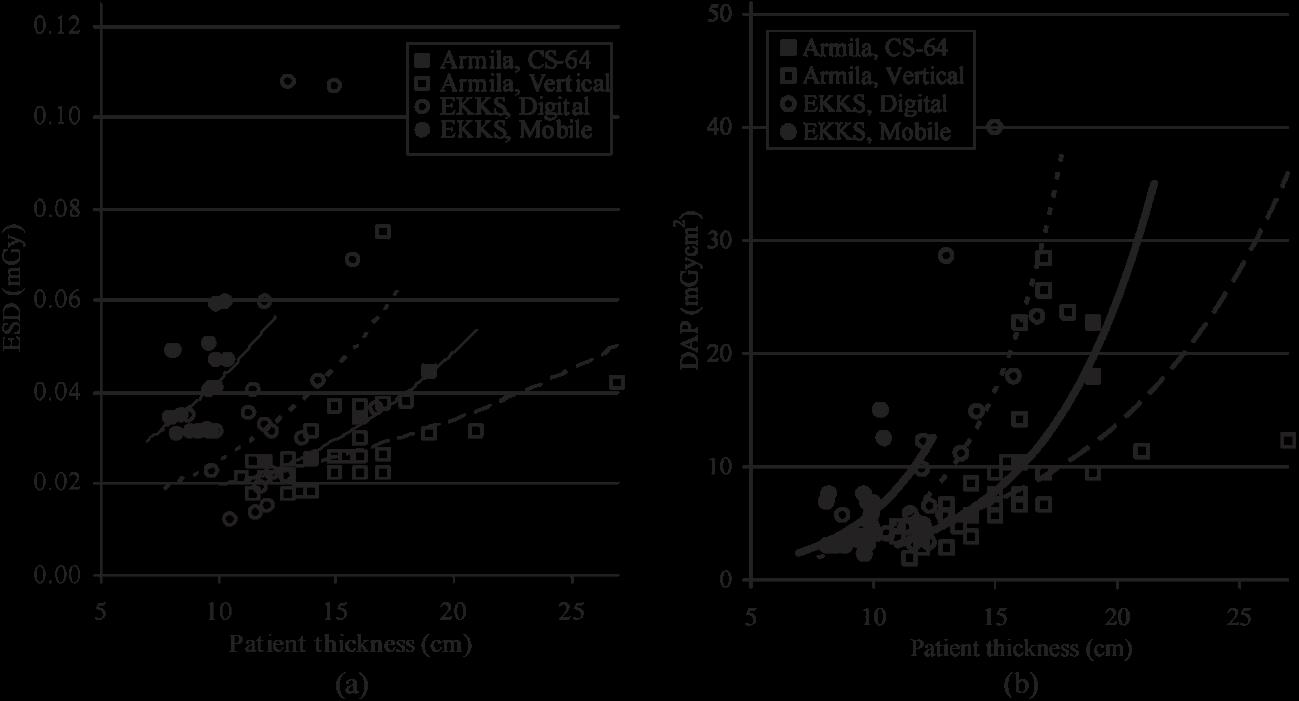
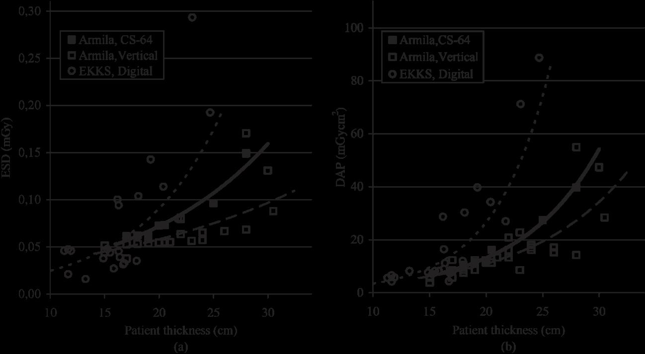
In recent literature the only European countries that have defined national DRLs are Austria, France, England and Finland. Out of the non-European countries only India and Brazil have defined national DRLs and Kenya has defined local DRLs. The main radiographic procedures that are included are chest, skull, pelvis and abdomen X-ray examinations. In most countries the children are divided according to age and the most common expression of DRL is the 75th percentile of Entrance Surface Dose or Dose Area Product value (DAP).
In many studies, the number of patients in some age categories was small and as a result the definition of DRL was not always possible. It is recommended that more health departments should be encouraged to take part in the study so that more data can be collected. In addition more countries should participate in this effort for DRL definition in pediatric radiology, as dose reduction in children undergoing X-rays examinations is of great importance.
Keywords: Diagnostic Reference Levels, Pediatrics, Radiology, Entrance Surface Dose, DAP
Triantopoulou Sotiria
Tsapaki Virginia
Triantopoulou Charikleia
Konstantopouleio General Hospital - Agia Olga
(Ι)
(Ι) / 700 (Α)
(Α)
(Α)
(Ι) / 90 (Α) 53 (Ι) / 150 (Α) 66 (Ι) 88 (Ι) / 100 (Α)
(Α)
(Ι) 475 (Ι)
807 (Ι) / 730 (Α)
892 (Ι) /1320 (Α)
1370 (A) 820 (A)
Smans et al (18)
1. Omolola AM , Bidemi IA, Ademola AJ, Olutayo SA. Entrance Surface Dose from pediatric diagnostic X-ray examinations in a developing world setting: Are we “ALARA Principle” compliant?. Br J Med Med Res 2013; 3:2288-2298.
2. European Commission, Radiation Protection 109, Guidance on Diagnostic Reference Levels (DRLs) for medical exposures. 1999; Directorate-General Environment, Nuclear Safety and civil Protection.
3 Simantirakis G. Koukorava C, Kalathaki M, Pafilis C, Kaisas I, Economides S et al. Reference levels and patient doses in interventional cardiology procedures in Greece. Eur Radiol 2013; 23:2324-2332.
4. IAEA human health series. Dosimetry in diagnostic radiology for paediatric patients. Vienna: International Atomic Energy Agency, 2013; (ISSN 2075–3772; no. 24).
5. Alzen G, Benz-Bohm G. Radiation protection in pediatric radiology. Dtsch Arztebl Int 2011; 108:407–414.
6. Martin L, Ruddlesden R, Makepeace C, Robinson L, Mistry T, Starritt H. Paediatric x-ray radiation dose reduction and image quality analysis. J Radiol Prot 2013; 33:621–633.
7. Diagnostic Reference Levels Position Paper. Medical council 3 September 2004. http://www.hse.ie/eng/about/Who/qualityandpatientsafety/ safepatientcare/ medexpradiatonunit/Diagnostic_Reference_Levels.pdf.
8. Montgomery A, Martin CJ. A study of the application of paediatric reference levels. Br J Radiol 2000; 73:1083-1090.
9. Ελληνική
εξετάσεων [Webpage, Internet]. Αθήνα: 2015: http://eeae.gr/ενημέρωση/ανακοινώσεις/4979-συλλογή-δοσιμετρικών -στοιχείωνπαιδιατρικών-εξετάσεων
10. Billinger J, Nowotny R, Homolka P. Diagnostic Reference Levels in Pediatric Radiology in Austria. Eur Radiol 2010; 20:1572-1579.
11. Roch P, Aubert B. French Diagnostic Reference Levels in Diagnostic Radiology, Computed Tomography and Nuclear Medicine: 2004 – 2008 Review. Radiat Prot Dosimetry 2013; 154:52-75.
12. Hart D, Hillier MC, Wall BF. National reference doses for common radiographic, fluoroscopic and dental X-ray examinations in the UK. Br J Radiol 2009; 82:1-12.
13. Kiljunen T, Järvinen H, Savolainen S. Diagnostic reference levels for thorax X-ray examinations of paediatric patients. Br J Radiol 2007; 80; 452-459.
14. Moyal AE. Nationwide evaluation of X-Ray trends (NEXT): Tabulation and graphical summary of 1998 Pediatric chest survey. In: Conference of Radiation Control Program Directors; Inc., Frankfort, Kentucky: 2004.
15. NCRP Report No.172: Reference Levels and Achievable Doses in Medical and Dental Imaging: Recommendations for the United States. Recommendations of the National Council on Radiation Protection and Measurements. September 30, 2012.
16. Schneider K, Kohn MM, Ernst G. The derivation of Reference Dose values to chest X Rays in Paediatric Radiography. Radiat Prot Dosimetry 1998; 80:199-202.
17. Hart D, Wall BF. Development of Diagnostic Reference Levels in Paediatric Radiology (IAEA-CN-85-56). In: Radiological Protection of Patients in Diagnostic and Interventional Radiology, eds: Nuclear Medicine and Radiotherapy; 2001 March 26-30; Vienna: International Atomic Energy Agency; 2001. p. 483-487.
18. Smans K, Vaño E, Sanchez R, Schultz F.W., Zoetelief J., Kiljunen T., et al. Results of a European survey on patient doses in Paediatric Radiology. Radiat Prot Dosimetry 2008; 129:1-3.
19. http://ddmed.eu/_media/news:ddm2_project_report_part_2_final_rb_16052014.pdf , last updated 16/05/2014, project launched by DG TREN of European Commission.
20. Wambani JS, Korir JK, Korir IK, Kilaha S. Establishment of Local Diagnostic Reference Levels in Paediatric screen-film Radiography at a children’s
21. Sonawane AU, Sunil Kumar JVK, Meghraj Singh, Pradhan AS. Suggested Diagnostic Reference Levels for Paediatric X-ray examinations in India. Radiat Prot Dosimetry 2010; 147:423428.
22. Freitas MB, Yoshimura EM. Diagnostic Reference Levels for the most frequent radiological examinations carried out in Brazil. Rev Panam Salud Publica 2009; 25:95-104.
Correspondence
Kourelis George
e-mail: gkourelis@yahoo.gr
Review of risk factors for ischaemic stroke in children and imaging modalities
Kourelis George, Galetselli Marianthi
Risk factors that have been associated with ischaemic stroke in children include a wide spectrum of disease, such as cardiac disease, vascular abnormalities, coagulation and haematological disorders, infections, disorders of metabolism, genetic syndromes, trauma and drug abuse.
On top of that, the evolution of the imaging modalities offers the clinician several potential regarding diagnosis of ischaemic stroke. Each of these modalities has each own advantages and disadvantages, as expected.
We conducted a review of the literature regarding ischaemic stroke in children, as well as asynopsis of the different imaging modalities available for the clinician.
Keywords: ischaemic stroke, children, risk factors, imaging
Kourelis George Galetselli Marianthi
(π.χ. Marfan, Ehlers-Danlos),
2.
Sjogren,
Henoch–Schonlein,
Kawasaki) (30,60-61) ii.
MELAS (Mitochondrial Encephalopathy, Lactic Acidosis, Stroke - like episodes)
ενήλικες (116-119,123). Το σύνδρομο CADASIL (Cerebral Autosomal Dominant Arteriopathy with Subcortical Infarcts and Leucoence phalopathy)
(MR perfusion techniques -
Βιβλιογραφία
1. K. S. Carvalho and B. P. Garg, “Arterial strokes in children,” Neurologic Clinics, vol. 20, no. 4, pp. 1079–1100, 2002
2. S. Lanthier, L. Carmant, M. David, A. Larbrisseau, and G. de Veber, “Stroke in children: the coexistence of multiple risk factors predicts poor outcome,” Neurology, vol. 54, no. 2, pp. 371–378, 2000
3. A. R. Riela and E. S. Roach, “Etiology of stroke in children,” Journal of Child Neurology, vol. 8, no. 3, pp. 201–220, 1993
4. G. DeVeber, E. S. Roach, A. R. Riela, and M. Wiznitzer, “Stroke in children: recognition, treatment, and future directions,” Seminars in Pediatric Neurology, vol. 7, no. 4, pp. 309–317, 2000
5. V. Ganesan, M. Prengler, M. A. McShane, A. M. Wade, and F. J. Kirkham, “Investigation of risk factors in children with arterial ischemic stroke,” Annals of Neurology, vol. 53, no. 2, pp. 167–173, 2003
6. deVeber G. Canadian Paediatric Ischemic Stroke Study Group. Canadian Paediatric Ischemic Stroke Registry: Analysis of children with arterial ischemic stroke. Ann Neurol. 2000;48:514–514
7. Chabrier S, Husson B, Lasjaunias P, Landrieu P, Tardieu M. Stroke in childhood: outcome and recurrence risk by mechanism in 59 patients. J Child Neurol. 2000;15:290–294
8. Abboud MR, Cure J, Granger S, Gallagher D, Hsu L, Wang W et al; STOP study. Magnetic resonance angiography in children with sickle cell disease and abnormal transcranial Doppler ultrasonography findings enrolled in the STOP study. Blood. 2004;103:2822–2826
9. Golomb MR, Dick PT, MacGregor DL, Armstrong DC, DeVeber GA. Cranial ultrasonography has a low sensitivity for detecting arterial ischemic stroke in term neonates. J Child Neurol. 2003;18:98 –103
10. 158. Childs AM, Cornette L, Ramenghi LA, Tanner SF, Arthur RJ, Martinez D et al. Magnetic resonance and cranial ultrasound characteristics of periventricular white matter abnormalities in newborn infants. ClinRadiol. 2001;56:647– 655
11. DeVeber G. Arterial ischemic strokes in infants and children: an overview of current approaches. SeminThrombHemost. 2003;29:567-73
12. Rollins N, Dowling M, Booth T, Purdy P. Idiopathic ischemic cerebral infarction in childhood: depiction of arterial abnormalities by MR angiography and catheter angiography. AJNR Am J Neuroradiol. 2000;21:549 –556
13. Chung B, Wong V. Pediatric stroke among Hong Kong Chinese subjects. Pediatrics. 2004;114:e206 – e212
14. Ghandehari K, Izadi-Mood Z. Incidence and etiology of pediatric stroke in southern Khorasan. ARYA Atherosclerosis Journal. 2007;3:29-33
15. Salih MA, bdel-Gader AG, Al-Jarallah AA, Kentab AY, Alorainy IA, Hassan HH et al. Stroke in Saudi children. Epidemiology, clinical features and risk factors. Saudi Med J. 2006;27Suppl 1:S12-S20
16. Siddiqui TS, Rehman A, Ahmed B. Etiology of strokes and hemiplegia in children presenting at Ayub Teaching Hospital, Abbottabad. J Ayub Med Coll Abbottabad. 2006;18:60-63
17. Shi KL, Wang JJ, Li JW, Jiang LQ, Mix E, Fang F et al. Arterial ischemic stroke: experience in Chinese children. Pediatr Neurol. 2008;38:186-90
18. Auvichayapat N, Tassniyom S, Hantragool S, Auvichayapat P. The etiology and outcome of cerebrovascular diseases in Northeastern Thai children.JMedAssoc Thai. 2007;90:2058-62
19. Husson B, Lasjaunias P. Radiological approach to disorders of arterial brain vessels associated with childhood arterial stroke: a comparison between MRA and contrast angiography. PediatrRadiol. 2004;34:10–15
20. Lo W, Stephens J, Fernandez S. Pediatric stroke in the United States and the impact of risk factors. J Child Neurol 2009;24:194–203
21. M. W. I. Webster, H. J. Smith, D. N. Sharpe et al., “Patent foramen ovale in young stroke patients,” The Lancet, vol. 2, no. 8601, pp. 11–12, 1988
22. Mas JL, Arquizan C, Lamy C, Zuber M, Cabanes L, Derumeaux G, et al. Recurrent cerebrovascular events associated with patent foramen ovale, atrial septal aneurysm, or both. N Engl J Med. 2001;345:1740–1746
23. Menache CC, du Plessis AJ, Wessel DL, Jonas RA, Newburger JW. Current incidence of acute neurologic complications after open-heart operations in children. Ann Thorac Surg. 2002;73:1752–1758
24. Domi T, Edgell D, McCrindle B.W, William G.W, MacGregor D, Kaplan D, et al. Frequency and predictors of vasoocclusive strokes associated with congenital heart disease. Ann Neurol. 2002;52:S129–133
25. Monagle P, Karl TR. Thromboembolic problems after the Fontan operation. SeminThoracCardiovascSurgPediatr Card SurgAnnu. 2002;5:36–47
26. Husson B, Rodesch G, Lasjaunias P, Tardieu M, Sébire G. Magnetic resonance angiography in childhood arterial brain infarcts: a comparative study with contrast angiography. Stroke. 2002;33:1280–1285
27.Chun DS, Schamberger MS, Flaspohler T, Turrentine MW, Brown JW, Farrell AG et al. Incidence, outcome, and risk for stroke after the Fontan procedure. Am J Cardiol 2004; 93: 117-9
28. Schuchlenz HW, Weihs W, Beitzke A, Stein JI, Gamillscheg A, Rehak P.et al. Transesophageal echocardiography for quantifying size of patent foramen ovale in patients with cryptogenic cerebrovascular events. Stroke 2002; 33: 293-6
29. Steiner MM, Di Tullio MR, Rundek T, Gan R, Chen X, Liguori C, et al. Patent foramen ovale size and embolic brain imaging findings among patients with ischaemic stroke. Stroke 1998; 29: 944-8
30. Roach ES, Golomb MR, Adams R, Biller J, Daniels S, Deveber , et al. Management of stroke in infants and children: a scientific statement from a Special Writing Group of the American Heart Association Stroke Council and the Council on Cardiovascular Disease in the Young. Stroke.2008;39:2644–2691
31. Cottrill CM, Kaplan S. Cerebral vascular accidents in cyanotic congenital heart disease. Am J Dis Child. 1973;125:484–487
32. Phornphutkul C, Rosenthal A, Nadas AS, Berenberg W. Cerebrovascular accidents in infants and children with cyanotic congenital heart disease. Am J Cardiol. 1973;32:329–334
33. Agnetti A, Carano N, Sani E, Tchana B, Allegri V, Bernasconi S et al. Cryptogenic stroke in children: possible role of patent foramen ovale. Neuropediatrics. 2006;37:53–56
34. Butany J, Nair V, Naseemuddin A, Nair GM, Catton C, Yau T. Cardiac tumours: diagnosis and management.LancetOncol. 2005;6:219 –228
35. F. J. Kirkham, “Stroke in childhood,” Archives of Disease in Childhood, vol. 81, no. 1, pp. 85–89, 1999
36. Kirkham FJ, Prengler M, Hewes DK, Ganesan V. Risk factors for arterial ischaemic stroke in children. J Child Neurol 2000; 15: 299-307
37. du Mesnil, de Rochemont R, Heindel W, Wesselmann C, Krüger K, Lanfermann H et al. Nontraumatic subarachnoid hemorrhage: value of repeat angiography. Radiology. 1997;202:798–800
38. Nestoridi E, Buonanno FS, Jones RM, Krishnamoorthy K, Grant PE, Van Cott EM et al. “Arterial ischemic stroke in childhood: the role of plasma-phase risk factors,” Current Opinion in Neurology, vol. 15, no. 2, pp. 139–144, 2002
39. F. J. Kirkham, “Is there a genetic basis for pediatric stroke?” Current Opinion in Pediatrics, vol. 15, no. 6, pp. 547–558, 2003
40. M. Bonduel, G. Sciuccati, M. Hepner, A. F. Torres, G. Pieroni, and J. P. Frontroth, “Prethrombotic disorders in children with arterial ischemic stroke and sinovenous thrombosis,” Archives of Neurology, vol. 56, no. 8, pp. 967–971, 1999
41. Lynch JK. Cerebrovascular disorders in children.CurrNeurolNeurosci Rep. 2004;4:129–138
42. Barnes C, deVeber G. Prothrombotic abnormalities in childhood ischaemicstroke.Thrombosis Research2004
43. DeVeber G, Monagle P, Chan A, MacGregor D, Curtis R, Lee S et al. Prothrombotic disorders in infants and children with cerebralthromboembolism. Arch Neurol. 1998;55:1539-43
44. Zak I, Sarecka-Hujar B, Kopyta I, Emich-Widera E, Marszal E, Wendorff Jet al. T allele of the 677C > T polymorphism of methylenetetrahydrofolatereductase gene is associated with an increased risk of ischemic stroke in Polish children. J Child Neurol. 2009;24:1262–1267
45.Herak DC, Antolic MR, Krleza JL, Pavic M, Dodig S, Duranovic V,et al. Inherited prothrombotic risk factors in children with stroke, transient ischemic attack, or migraine. Pediatrics.2009;123:e653–e660
46. Lidegaard Ø, Kreiner S. Contraceptives and cerebral thrombosis: a five-year national casecontrol study. Contraception. 2002;65:197–205
47. Lidegaard O. Oral contraception and risk of a cerebral thromboembolic attack: results of a case-control study. BMJ. 1993;306:956–963
48. Lidegaard O. Smoking and use of oral contraceptives: impact on thrombotic diseases. Am J Obstet Gynecol. 1999;180:S357–S363
49. Nowak-Göttl U, Sträter R, Heinecke A, Junker R, Koch HG, Schuierer G, von Eckardstein A. Lipoprotein (a) and genetic polymorphisms of clotting factor V, prothrombin, and methylenetetrahydrofolatereductase are risk factors of spontaneous ischemic stroke in childhood. Blood. 1999;94:3678–3682
50. van Beynum IM, Smeitink JA, den Heijer M, tePoelePothoff MT, Blom HJ. Hyperhomocysteinemia: a risk factor for ischemic stroke in children. Circulation. 1999;99:2070–2072
51. Prengler M, Sturt N, Krywawych S, Surtees R, Liesner R, Kirkham F. Homozygous thermolabile variant of the methylenetetrahydrofolatereductase gene: a potential risk factor for hyperhomocysteinaemia, CVD, and stroke in childhood. Dev Med Child Neurol. 2001;43:220–225
52. Sträter R, Becker S, von Eckardstein A, Heinecke A, Gutsche S, Junker R, et al. Prospective assessment of risk factors for recurrent stroke during childhood–a 5-year follow-up study. Lancet. 2002;360:1540–1545
53. Ganesan V, Prengler M, Wade A, Kirkham FJ. Clinical and radiological recurrence after childhood arterial ischemic stroke.Circulation. 2006;114:2170–2177
54. Kumar K. Neurological complications of congenital heart disease.Indian J Pediatr. 2000;67:287–291
55. Sebire G, Fullerton H, Riou E, deVeber G. Toward the definition of cerebral arteriopathies of childhood. CurrOpinPediatr. 2004;16:617–622
56. Fullerton HJ, Johnston SC, Smith WS. Arterial dissection and stroke in children.Neurology. 2001;57:1155–1160
57. Chabrier S, Lasjaunias P, Husson B, Landrieu P, Tardieu M. Ischaemic stroke from dissection of the craniocervical arteries in childhood: report of 12 patients. Eur J Paediatr Neurol. 2003;7:39–42
58. Rafay MF, Armstrong D, DeVeber G, Domi T, Chan A, MacGregor DL. Craniocervical arterial dissection in children: clinical and radiographic presentation and outcome. J Child Neurol. 2006;21:8-16
59. Ishii K, Isono M, Kobayashi H, Kamida T. Temporal profile of angiographical stages of moyamoya disease: when does moyamoya disease progress? Neurol Res. 2003;25:405–410
60. Benseler S, Schneider R. Central nervous system vasculitis in children. CurrOpinRheumatol. 2004;16:43–50
61. Graf WK, Milstein JM, Sherry DD. Stroke and mixed connective tissue disease. J Child Neurol. 1993;8:256–259
62. Lanthier S, Lortie A, Michaud J, Laxer R, Jay V, deVeber G. Isolated angiitis of the CNS in children. Neurology. 2001;56:837–842
63. Gallagher KT, Shaham B, Reiff A, Tournay A, Villablanca JP, Curran J, et al. Primary angii-
tis of the central nervous system in children: 5 cases. J Rheumatol. 2001;28:616–623
64. Sebire G, Meyer L, Chabrier S. Varicella as a risk factor for cerebral infarction in childhood: a case-control study. Ann Neurol. 1999;45:679–680
65. Askalan R, Laughlin S, Mayank S, Chan A, MacGregor D, Andrew M, et al. Chickenpox and stroke in childhood: a study of frequency and causation. Stroke. 2001;32:1257–1262
66. Young N, Dorsch NW, Kingston RJ, Markson G, McMahon J. Intracranial aneurysms: evaluation in 200 patients with spiral CT angiography. EurRadiol. 2001;11:123–130
67. Nabavi DG, Kloska SP, Nam EM, Freund M, Gaus CG, Klotz E, et al. MOSAIC: Multimodal Stroke Assessment Using Computed Tomography: novel diagnostic approach for the prediction of infarction size and clinical outcome. Stroke. 2002;33:2819 –2826.
68.Oelerich M, Stögbauer F, Kurlemann G, Schul C, Schuierer G. Craniocervical artery dissection: MR imaging and MR angiographic findings. EurRadiol 1999; 9: 1385-91.
69. Kilpatrick MM, Yonas H, Goldstein S, Kassam AB, Gebel JM Jr, Wechsler LR et al. CTbased assessment of acute stroke: CT, CT angiography, and xenon-enhanced CT cerebral blood flow. Stroke. 2001;32:2543–2549
70. Silverboard G, Tart R. Cerebrovascular arterial dissection in children and young adults. SeminPediatrNeurol 2000; 7: 289-300.
71.Hasan I, Wapnick S, Tenner MS, Couldwell W. Vertebral artery dissection in children: A comprehensive review. PediatrNeurosurg 2002; 37: 168-77
72. Bonduel M, Hepner M, Sciuccati G, Torres AF, Tenembaum S. Prothrombotic disorders in children with moyamoya syndrome. Stroke 2001; 32: 1786-92
73. Manceau E, Giroud M, Dumas R. Moyamoya disease in children. A review of the clinical and radiological features and current treatment. Child’s NervSyst 1997; 13: 595-600
74. Takeoka M, Takahashi T. Infectious and inflammatory disorders of the circulatory system and stroke in children. CurrOpinNeurol 2002; 15: 159-64
75. Brenner D, Elliston C, Hall E, Berdon W. Estimated risks of radiation-induced fatal cancer from pediatric CT. AJR Am J Roentgenol. 2001;176:289–296
76. Moriuchi H, Rodriguez W. Role of Varicella zoster virus in stroke syndromes. Pediatr Infect Dis J 2000; 19: 648-53
77. Fullerton HJ, Wu YW, Sidney S, Johnston SC. Risk of recurrent childhood arterial ischemic stroke in a population-based cohort: the importance of cerebrovascular imaging. Pediatrics 2007;119:495–501
78. Amlie-Lefond C1, Bernard TJ, Sébire G, Friedman NR, Heyer GL, Lerner NB et al. Predictors of cerebral arteriopathy in children with arterial ischemic stroke: results of the International Pediatric Stroke Study. Circulation 2009;119:1417–1423
79.Rea D, Brandsema JF, Armstrong D, Parkin PC, deVeber G, MacGregor D et al. Cerebral arteriopathyinchildren with neurofibromatosis type 1.Pediatrics 2009
80.Nagel MA, Cohrs RJ, Mahalingam R, Wellish MC, Forghani B, Schiller Aet al. The varicella zoster virus vasculopathies: clinical, CSF, imaging, and virologic features. Neurology 2008;70:853–860
81. Miravet E, Danchaivijitr N, Basu H, Saunders DE, Ganesan V. Clinical and radiological features of childhood cerebral infarction following varicella zoster virus infection. Dev Med Child Neurol 2007;49:417–422
82. Donahue JG, Kieke BA, Yih WK, Berger NR, McCauley JS, Baggs J et al. Varicella vaccination and ischemic stroke in children: is there an association? Pediatrics 2009;123:e228–e234
83. Eastwood JD, Lev MH, Wintermark M, Fitzek C, Barboriak DP, Delong DM et al. Correlation of early dynamic CT perfusion imaging with whole-brain MR diffusion and perfusion imaging in acute hemispheric stroke. AJNR Am J Neuroradiol. 2003;24:1869 –1875
84. Sunshine JL, Bambakidis N, Tarr RW, Lanzieri CF, Zaidat OO, Suarez JI et al. Benefits of perfusion MR imaging relative to diffusion MR imaging in the diagnosis and treatment of hyperacute stroke. AJNR Am J Neuroradiol. 2001;22:915–921
85. Warach S, Gaa J, Siewert B, Wielopolski P, Edelman RR. Acute human stroke studied by whole brain echo planar diffusion-weighted magnetic resonance imaging. Ann Neurol. 1995;37:231–241
86. Benseler SM, deVeber G, Hawkins C, Schneider R, Tyrrell PN, Aviv RI et al. Angiography-
negative primary central nervous system vasculitis in children: a newly recognized inflammatory central nervous system disease. Arthritis Rheum . 2005;52:2159–2167
87. Ikeda H, Sasaki T, Yoshimoto T, Fukui M, Arinami T. Mapping of a familial moyamoya disease gene to chromosome 3p24.2-p26. Am J Hum Genet. 1999;64:533–537
88. Nanba R, Tada M, Kuroda S, Houkin K, Iwasaki Y. Sequence analysis and bioinformatics analysis of chromosome 17q25 in familial moya-moya disease. Childs NervSyst . 2005;21:62–68
89. Inoue TK, Ikezaki K, Sasazuki T, Matsushima T, Fukui M. Linkage analysis of moyamoya disease on chromosome 6. J Child Neurol. 2000;15:179–182
90. Ciceri EF, Cuccarini V, Chiapparini L, Saletti V, Valvassori L (2011) -Paediatric Stroke: Review of the Literature and Possible Treatment Options, including Endovascular Approach Stroke Res Treat ; 2011: 781612
91.Chabrier S, Rodesch G, Lasjaunias P, Tardieu M, Landrieu P, Sébire G. Transient cerebral arteriopathy: a disorder recognized by serial angiograms in children with stroke. J Child Neurol 1998;13:27–32
92. Ganesan V, Chong WK, Cox TC, Chawda SJ, Prengler M, Kirkham FJ. Posterior circulation stroke in childhood: risk factors and recurrence. Neurology 2002;59:1552–1556
93. Bernard TJ, Goldenberg NA, Tripputi M, Manco-Johnson MJ, Niederstadt T, Nowak-Göttl U. Anticoagulation in childhood-onset arterial ischemic stroke with non-moyamoyaarteriopathy: findings from the Colorado and German (COAG) collaboration. Stroke 2009;40:2869–2871
94. Danchaivijitr N, Cox TC, Saunders DE, Ganesan V. Evolution of cerebral arteriopathies in childhood arterial ischemic stroke. Ann Neurol. 2006;59:620–626
95. Benseler SM, Silverman E, Aviv RI, Schneider R, Armstrong D, Tyrrell PNet al. Primary central nervous system vasculitis in children. Arthritis Rheum. 2006;54:1291–1297
96. Diez-Tejedor E, Munoz C, Frank A. Cerebellar infarction in children and young adults related to fibromuscular dysplasia and dissection of the vertebral artery. Stroke 1993; 24: 1096
97. Emparanza JI, Aldamiz-Echevarria L, Perez-Yarza E, Hernandez J, Peña B, Gaztañaga R. Ischaemic stroke due to fibromuscular dysplasia. Neuropaediatrics 1989; 20: 181-2
98.Shields WD, Ziter FA, Osborn AG, Allen J Fibromuscular dysplasia as a cause of stroke in infancy and childhood.Paediatrics 1977; 59: 899-901
99.Earley CJ, Kittner SJ, Feeser BR, Gardner J, Epstein A, Wozniak MA et al. Stroke in children and sickle-cell disease: Baltimore-Washington cooperative young stroke study; Neurology , vol. 51, no. 1, pp. 169–176,1998
100. Prengler M, Pavlakis SG, Prohovnik I, Adams RJ. Sickle cell disease: the neurological complications. Ann Neurol. 2002;51:543–552
101. Ohene-Frempong K, Weiner SJ, Sleeper LA, Miller ST, Embury S, Moohr JW et al. Cerebrovascular accidents in sickle cell disease: rates and risk factors. Blood . 1998;91:288–294
102. G.J.Kato,R.P.Hebbel,M.H. Steinberg,andM.T.Gladwin; Vasculopathy in sickle cell disease: biology, pathophysiology, genetics, translational medicine, and new research directions; American Journal of Hematology ,vol.84,no.9,pp.618–625, 2009
103. Moser FG, Miller ST, Bello JA, Pegelow CH, Zimmerman RA, Wang WC et al. The spectrum of brain MR abnormalities in sickle-cell disease: a report from the Cooperative Study of Sickle Cell Disease. AJNR Am J Neuroradiol. 1996;17:965–972
104. Armstrong FD, Thompson RJ Jr, Wang W, Zimmerman R, Pegelow CH, Miller S, et al. Cognitive functioning and brain magnetic resonance imaging in children with sickle cell disease. Neuropsychology Committee of the Cooperative Study of Sickle Cell Disease. Pediatrics . 1996;97:864–870
105. Miller ST, Macklin EA, Pegelow CH, Kinney TR, Sleeper LA, Bello JA, et al. Cooperative Study of Sickle Cell Disease. Silent infarction as a risk factor for overt stroke in children with sickle cell anemia: a report from the Cooperative Study of Sickle Cell Disease. J Pediatr. 2001;139:385–390
106. Henderson JN, Noetzel MJ, McKinstry RC, White DA, Armstrong M, DeBaun MR. Reversible posterior leukoencephalopathy syndrome and silent cerebral infarcts are associated with severe acute chest syndrome in children with sickle cell disease. Blood. 2003;101:415–419
107. Naran AD, Fontana L. Sickle cell disease with orbital infarction and epidural hematoma. PediatrRadiol. 2001;31:257–259
108. National Heart, Lung, and Blood Institute, National Institutes of Health. Clinical alert from the National Heart, Lung, and Blood Institute September 18, 1997
109. Yenari MA, Palmer JT, Sun GH, de Crespigny A, Mosely ME, Steinberg GK. Time-course and treatment response with SNX-111, an N-type calcium channel blocker, in a rodent model of focal cerebral ischemia using diffusion-weighted MRI. Brain Res. 1996;739:36–45
110. Adams RJ, Brambilla DJ, Granger S, Gallagher D, Vichinsky E, Abboud MR, et al. STOP Study. Stroke and conversion to high risk in children screened with transcranial Doppler ultrasound during the STOP study. Blood. 2004;103:3689–3694
111. Wang WC. The pathophysiology, prevention, and treatment of stroke in sickle cell disease. CurrOpinHematol. 2007;14:191–197
112. Hartfield DS, Lowry NJ, Keene DL, Yager JY. Iron deficiency: a cause of stroke in infants and children. PediatrNeurol 16:50–53, 1997
113. Gunther G, Junker R, Strater R, Schobess R, Kurnik K, Kosch A et al. Symptomatic ischemic stroke in full-term neonates:role of acquired and genetic prothrombotic risk factors. Stroke 31:2437–2441, 2000
114. van Everdingen KJ, van der Grond J, Kappelle LJ, Ramos LM, Mali WP. Diffusion-weighted magnetic resonance imaging in acute stroke.Stroke. 1998;29:1783–1790
115. Cardo E, Campistol J, Caritg J, Ruiz S, Vilaseca MA, Kirkham F, et al. Fatal haemorrhagic infarct in an infant with homocystinuria.Dev Med Child Neurol. 1999;41:132–135
116. Rolfs A, Böttcher T, Zschiesche M, Morris P, Winchester B, Bauer P, et al. Prevalence of Fabry disease in patients with cryptogenic stroke: a prospective study. Lancet. 2005;366:1794–1796
117. Brouns R, Sheorajpanday R, Braxel E, Eyskens F, Baker R, Hughes D, et al. MiddelheimFabry Study (MiFaS): a retrospective Belgian study on the prevalence of Fabry disease in young patients with cryptogenic stroke. ClinNeurolNeurosurg. 2007;109:479–484
118. Eng CM, Guffon N, Wilcox WR, Germain DP, Lee P, Waldek S, et al. International Collaborative Fabry Disease Study Group. Safety and efficacy of recombinant human alphagalactosidase A–replacement therapy in Fabry’s disease. N Engl J Med. 2001;345:9–16
119. Eng CM, Germain DP, Banikazemi M, Warnock DG, Wanner C, Hopkin RJ, et al. Fabry disease: guidelines for the evaluation and management of multi-organ system involvement. Genet Med . 2006;8:539 –548
120. Golomb MR, Sokol DK, Walsh LE, Christensen CK, Garg BP. Recurrent hemiplegia, normal MRI, and NOTCH3 mutation in a 14-year-old: is this early CADASIL? Neurology. 2004;62:2331–2332
121. Herguner MO, Incecik F, Elkay M, Altunbasak S, Baytok V. Evaluation of 39 children with stroke regarding etiologic risk factors and treatment. Turk J Pediatr. 2005;47:116-19
122. Iizuka T, Sakai F, Suzuki N, Hata T, Tsukahara S, Fukuda M et al. Neuronal hyperexcitability in stroke-like episodes of MELAS syndrome. Neurology59:816–824, 2002
123. Desnick RJ, Brady RO. Fabry disease in childhood. J Pediatr 144:S20–S26, 2004
124. Guillamo JS, Creange A, Kalifa C, Grill J, Rodriguez D, Doz F et al. Prognostic factors of CNS tumours in Neurofibromatosis 1 (NF1): a retrospective study of 104 patients. Brain 126:152–160, 2003
125. Bhattacharya JJ, Luo CB, Alvarez H, Rodesch G, Pongpech S, Lasjaunias PL. PHACES syndrome: a review of eight previously unreported cases with late arterial occlusions. Neuroradiology 46:227–233, 2004
126.Yuh WT, Crain MR, Loes DJ, Greene GM, Ryals TJ, Sato Y. MR imaging of cerebral ischemia: findings in the first 24 hours. AJNR Am J Neuroradiol. 1991;12:621–629
127. Park YD, Belman AL, Kim TS, Kure K, Llena JF, Lantos G “Stroke in pediatric acquired immunodeficiency syndrome,” Annals of Neurology, vol. 28, no. 3, pp. 303–311, 1990
128. D. M. Moriarty, J. O. Haller, J. P. Loh, and S. Fikrig, “Cerebral infarction in pediatric acquired immunodeficiency syndrome,” Pediatric Radiology , vol. 24, no. 8, pp. 611–612, 1994
129. Lövblad KO, Laubach HJ, Baird AE, Curtin F, Schlaug G, Edelman RR et al. Clinical experience with diffusion-weighted MR in patients with acute stroke. AJNR Am J Neuroradiol. 1998;19:1061–1066
130. Chabriat H, Vahedi K, Iba-Zizen MT, Joutel A, Nibbio A, Nagy TG et al. Clinical spectrum of CADASIL: a study of 7 families. Cerebral autosomal dominant arteriopathy with sub-
cortical infarcts and leukoencephalopathy.Lancet. 1995;346:934–939
131. J. S. Hutchison, R. Ichord, A. M. Guerguerian, and G. DeVeber, “Cerebrovascular disorders,” Seminars in Pediatric Neurology, vol. 11, no. 2, pp. 139–146, 2004
132. Salih MA, Abdel-Gader AG, Al-Jarallah AA, Kentab AY, Gadelrab MO, Alorainy IA et al. “Infectious and inflammatory disorders of the circulatory system as risk factors for stroke in Saudi children,” Saudi Medical Journal, vol. 27, supplement 1, pp. S41–S52, 2006
133. Normann S, de Veber G, Fobker M, Langer C, Kenet G, Bernard TJ, et al. Role of endogenous testosterone concentration in pediatric stroke. Ann Neurol2009;66:754–758
134. Patsalides AD, Wood LV, Atac GK, Sandifer E, Butman JA, Patronas NJ. Cerebrovascular disease in HIV-infected pediatric patients: neuroimaging findings. AJR Am JRoentgenol 179:999–1003, 2002
135. Camacho A, Villarejo A, Moreno T, Simon R, Munoz A, Mateos F. Vertebral artery fibromuscular dysplasia: an unusual cause of stroke in a 3-year-old child. Dev Med Child Neurol 45:709–711, 2003.
136. Omura M, Aida N, Sekido K, Kakehi M, Matsubara S. Large intracranial vessel occlusive vasculopathy after radiation therapy in children: clinical features and usefulness of magnetic resonance imaging. Int J RadiatOncolBiolPhys 38:241–249, 1997
137. Golomb MR, Fullerton HJ, Nowak-Gottl U, Deveber G. Male predominance in childhood ischemic stroke: findings from the international pediatric stroke study. Stroke 2009;40:52–57
138. Molina CA, Saver JL. Extending reperfusion therapy for acute ischemic stroke: emerging pharmacological, mechanical, and imaging strategies. Stroke 2005 Oct; 36(10):2311-20
139. Warach S, Gaa J, Siewert B, Wielopolski P, Edelman RR. Acute human stroke studies by whole brain echo planar diffusion-weighted magnetic resonance imaging. Ann Neurol 1995;37:231–241
140. Sorensen AG, Buonanno FS, Gonzalez RG, Schwamm LH, Lev MH, Huang-Hellinger FR et al. Hyperacute stroke: evaluation with combined multisection diffusion-weighted and hemodynamically weighted echoplanar MR imaging.Radiology1996;199:391-401
141. Li F, Han S, Tatlisumak T, Carano RA, Irie K, Sotak CH, et al. A new method to improve in-bore middle cerebral artery occlusion in rats: demonstration with diffusion- and perfusionweighted imaging. Stroke 1998, 29:1715-1720
142. Karl-OlofLövblad, Hans-Joachim Laubach, Alison E. Baird, François Curtin, Gottfried Schlaug, Robert R. Edelman et al Clinical Experience with Diffusion-Weighted MR in Patients with Acute Stroke. Am J Neuroradiol 19:1061–1066, June 1998
143. Patel MR, Edelman RR, Warach S. Detection of hyperacute primary intraparenchymal hemorrhage by magnetic resonance imaging.Stroke. 1996;27:2321–2324
144. Fiebach JB, Schellinger PD, Gass A, Kucinski T, Siebler M, Villringer A et al. Stroke magnetic resonance imaging is accurate in hyperacuteintracerebral hemorrhage: a multicenter study on the validity of stroke imaging. Stroke 2004.35502–506.506
145.Kidwell CS, Chalela JA, Saver JL, Starkman S, Hill MD, Demchuk AM et al. Comparison of MRI and CT for detection of acute intracerebral hemorrhage.JAMA 2004;292:1823–30
146. Chalela JA, Kidwell CS, Nentwich LM, Luby M, Butman JA, Demchuk AM et al. Magnetic resonance imaging and computed tomography in emergency assessment of patients with suspected acute stroke: a prospective comparison. Lancet. 2007 Jan 27; 369(9558):293-8
147. Paediatric Stroke Working Group. Stroke in childhood: Clinical guidelines for diagnosis, management and rehabilitation. November 2004. The Lavenham Press Ltd, Suffolk
148. Schellinger PD, Bryan RN, Caplan LR, Detre JA, Edelman RR, Jaigobin C et al. -based guideline: the role of diffusion and perfusion MRI for the diagnosis of acute ischemic stroke: report of the Therapeutics and Technology Assessment Subcommittee of the American Academy of Neurology. Neurology 2010;75:177–85
149.Latchaw RE, Alberts MJ, Lev MH, Connors JJ, Harbaugh RE, Higashida RT et al. Recommendations for imaging of acute ischemic stroke: a scientific statement from the American Heart Association. 2009; 40(11):3646-3678
150. Jauch EC, Saver JL, Adams HP Jr, Bruno A, Connors JJ, Demaerschalk BM et al. Guidelines for the Early Management of Patients With Acute Ischemic Stroke: A Guideline for Healthcare Professionals from the American Heart Association/American Stroke Association. Stroke, 2013;44:870-947
F.
K.
e-mail:
Correspondence
Elpis-Athina Vlachopapadopoulou
Τ. +302132009851
F. +302132009531
K. +306932247228
e-mail: elpis.vl@gmail.com
Refeeding, menses resumption and improvement of bone density of adolescent girls with anorexia nervosa
Elpis-Athina Vlachopapadopoulou, Artemis Doulgeraki, Artemis Tsitsika
Abstract
Background: Anorexia nervosa (AN) is the 3rd most common chronic disorder in adolescence, affecting mostly females. Amenorrhea and osteopenia are amongst the most serious complications. The aim of this review is to assess the effect of refeeding on recovery of menses and skeletal health.
Methods: In-depth literature review during the last twenty years, focusing on laboratory and imaging data which describe the effect of refeeding on gonadal function and bone density.
Results: The endocrine abnormalities that result in menses cessation are thyroid function dysregulation, hypercortisolemia, hypogonadotropic hypogonadism, low leptin levels and low levels of insulin-like growth factor I. Amenorrhea is attributed to prepubertal secretion of luteinizing hormone, and loss of the positive feedback in response to estrogen. Primary amenorrhea is a negative predictor of menses resumption. From an endocrine point of view, whilst good predictive value is associated with higher cortisol levels as well as higher estradiol and inhibin-B levels at the time of menses cessation. There is evidence that resumption of menses is expected when body fat percentage is approximately 23-25%. of total body weight. With regards to bone status, it seems that the skeleton is slow to recover, as it takes on average two years for bone mineral density to improve.
Conclusions: Proper compliance to refeeding regime for anorexia nervosa, as well as proper counseling and psychotherapy leads to a slow, gradual improvement of body composition and bone profile, as well as, resumption of menses, in a large percentage of patients.
Elpis-Athina Vlachopapadopoulou
Dept. of Endocrinology
- Growth and Development, Children’s Hospital “P. & A. Kyriakou”
Artemis Doulgeraki
Dept. of Bone and Mineral Metabolism, Institute of Child Health, Children’s Hospital “Agia Sofia”
Artemis Tsitsika
Adolescent Health Unit, 2nd Pediatric Clinic
University of Athens, Children’s Hospital “P. & A. Kyriakou”
Keywords: bone mineral density, anorexia, amenorrhea, hypogonadotropic hypogonadism, body composition
Συντομογραφίες
ΨΑ:
AN: Anorexia Nervosa ΕΡ:
IGF-I:
LH: Luteinizing Hor-
FSH: Follicle Stimulating Hormone,
AMH: Antimullerian Hormone,
NICE: The National Institute for Health and Care Excellence
1. Fitzpatrick KK, Lock J Anorexia nervosa, BMJ Clin Evid. 2011 Apr 11;2011. pii: 1011
2. Strober M, Freeman R, Morrell Atypical anorexia nervosa: separation from typical cases in course and outcome in along term prospective study Int J Eat Disord 1999; 25:135-42
3. Strober M, Freeman R, Morrell W. The long-term course of severe anorexia nervosa in adolescents survival analysis of recovery, relapse an outcome predictors over 10-15 years in a prospective study Int J Eat Disord 1997; 22:339-60
4. Yager J, Andersen AE. Anorexia nervosa N Engl J Med 2005;353:1481-8.
5. Uher R, Rutter M. Classification of feeding and eating disorders: review of evidence and proposal for ICD-11 World Psychiatry 2012;11:80-92
6. Strober M, Freeman R, Lampert C, Diamond J, Kaye W. Controlled family study of anorexia nervosa and bulimia nervosa: evidence of shared liability and transmission of partial syndromes Am J Psychiatry 2000; 157:393-401
7. Lilenfeld LR, Kaye WH, Greeno CG, Merikangas KR, Plotnikov K, Police C, et al. A controlled family study of anorexia nervosa and bulimia nervosa: psychiatric disorders in first degree relatives effects of comorbid substance dependence Int J Eat Disord 1997; 22:253-64
8. Miller KK. Endocrine dysregulation in anorexia nervosa Update J Clin Endocrinol Metab 2011;96: 2939-49
9. American Academy of Pediatrics. Clinical Report-Identification and management of eating disorders in children and adolescents www.pediatrics.org/cgi/doi/10.1542/peds.2010-2821
10. Legro RS, Lin HM, Demers LM, Lloyd T. Rapid maturation of the reproductive axis during perimenarche independent of body composition J Clin Endocrinol Metab 2000;85:1021-5
11. World Health Organization multicenter study on menstrual and ovulatory patterns in adolescent girls II Longitudinal study of menstrual patterns in the early postmenarchal period, duration of bleeding episodes and menstrual cycles J Adolesc Health Care 1986; 7:236-44
12. Gordon C. Functional hypothalamic Amenorrhea N Engl J Med 2010;363:365-71.
13. Chung PW, Chan SS, Yiu KW, Lao TT, Chung TK. Menstrual disorders in a Pediatric and Adolescent Gynecology Clinic: Patient presentations and longitudinal outcome Hong Kong
Med J. 2011 Oct;17(5):391-7
14. Le Grange D, Doyle PM, Swanson SA, Ludwig K, Glunz C, Kreipe RE. Calculation of expected body weight in adolescents with eating disorders Pediatrics 2012;129(2):e438-e446
15. Faust JP, Goldschmidt AB, Anderson KE, Glunz C, Brown M, Loeb KL et al. Resumption of menses in anorexia nervosa during a course of family based treatment J Eat Disord 2013;1:12
16. Dempfle A, Herpertz-Dahlmann B, Timmesfeld N, Schwarte R, Egberts KM, Pfeiffer E et al. Predictors of the resumption of menses in adolescent anorexia nervosa. BMC Psychiatry 2013; 13:308-17
17. Misra M, Prabhakaran R, Miller KK, Tsai P, Lin A, Lee N et al. Role of cortisol in menstrual recovery in adolescent girls with anorexia nervosa Pediatr Res 2006; 59:598-603
18. van Elburg AA, Eijkemans MJC, Kas MJH, Themmen APN, de Jong FH, van Engeland et al. Predictors of recovery of ovarian function during weight gain in anorexia nervosa Fertility and sterility 2007;87:902-8
19. Golden NH, Lanzkowsky L, Schebendach J, Palestro CJ, Jacobson MS, Shenker IR. The effect of estrogen-progestin treatment on bone mineral density in anorexia nervosa J Pediatr Adolesc Gynecol 2002;15:135–43.
20. Munoz MT, Morande G, Garcia_Centenera JA, Hervas F, Pozo J, Argente J. The effects of estrogen administration on bone mineral density in adolescents with anorexia nervosa Eur J Endocrinol 2002;146:45–50.
21. Strokosch GR, Friedman AJ, Wu SC, Kamin M. Effects of an oral contraceptive ( norgestimate/ethinyl estradiol) on bone mineral density in adolescent females with anorexia nervosa: a double-blind placebo-controlled study J Adolesc Health 2006; 39;819-27
22. Misra M, Katzman D, Miller KK, Μendes N, Snelgrove D, Russel M et al. Physiologic oestrogen replacement increases bone density in adolescent girls with anorexia nervosa J Bone Min Res 2011; 26:2430-38
23. American Psychiatric Association: Practice guideline for the treatment of patients with eating disorders, 3rd edition, 2006
24. Herpertz S, Herpertz-Dahlmann B, Fichter M, Tuschen-Caffier B, Zeeck A. S3-Leitlinie Diagnostik und Behandlung der Esstörungen, New York: Sprinher-Verlag Berlin Heidelberg, 2011
25. Dominguez J, Goodman L, Sen Gupta S, Mayer L, Etu SF, Walsh BT et al. Treatment of anorexia nervosa is associated with increases in bone mineral density and recovery is a biphasic process involving both nutrition and return of menses. Am J Clin Nutr 2007; 86: 92-99
26. Jacoangeli F, Masala S, Staar Mezzasalma F, Fiori R, Martinetti A, Ficoneri C et al. Amenorrhea after weight recover in anorexia nervosa: role of body composition and endocrine abnormalities. Eat Weight Disord 2006; 11(1): e20-6
27. Tinahones FJ, Martinez-Alfaro B, Gonzalo-Marin M, Garcia-Almeida JM, Garrido-Sanchez L, Cardona F. Recovery of menstrual cycle after therapy for anorexia nervosa. Eat Weight Disord 2005; 10(3): e52-55
28. Golden NH, Jacobson MS, Schebendach J, Solanto MV, Hertz SM, Shenker IR. Resumption of menses in anorexia nervosa. Arch Pediatr Adolesc Med 1997; 151(1): 16-21
29. Schmidt MH, Klein M. Determination of body fat in treatment of anorexia nervosa. Z Kinder Jugendpsychiatr Psychother 1997; 25(1): 27-34
30. Moreno MV, Djeddi DD, Jaffrin MY. Assessment of body composition in adolescent subjects with anorexia nervosa by bioimpedance. Med Eng Phys 2008; 30(6): 783-791
31. Pitts S, Blood E, Divasta A, Gordon CM. Percentage body fat by dual-energy X-ray absorptiometry is associated with menstrual recovery in adolescents with anorexia nervosa. J Adolesc Health 2014; 54(6): 739-741
32. Misra M, Miller KK, Almazan C, Ramaswamy K, Aggarwal A, Herzog DB et al. Hormonal and body composition predictors of soluble leptin receptor, leptin and free leptin index in adolescent girls with anorexia nervosa and controls and relation to insulin sensitivity. J Clin Endocrinol Metab 2004; 89(7): 3486-3495
33. Bodell LP, Mayer LE. Percent body fat is a risk factor for relapse in anorexia nervosa: a replication study. Int J Eat Disord 2011; 44(2): 118-23
34. Mayer L, Roberto C, Glasofer D, Fischer S, Gallagher D, Wang J et al. Does percent body
fat predict outcome in anorexia nervosa? Am J Psychiatry 2007; 164: 970-972
35. Taylor RW, Jones IE, Williams SM, Goulding A. Body fat percentages measured by dual-energy X-ray absorptiometry corresponding to recently recommended body mass index cutoffs for overweight and obesity in children and adolescents aged 3-18 y. Am J Clin Nutr 2002;76:141621
36. Misra M, Katzman DK, Cord J, Manning SJ, Mickley D, Herzog DB et al. Percentage extremity fat, but not percentage trunk fat, is lower in adolescent boys with anorexia nervosa than in healthy adolescents. Am J Clin Nutr 2008; 88: 1478-1484
37. Zuckerman-Levin N, Hochberg Z, Latzer Y. Bone health in eating disorders. Obes Rev 2014; 15: 215-223
38. Faje AT, Fazeli PK, Miller KK, Katzman DK, Ebrahimi S, Lee H. Fracture risk and areal bone mineral density in adolescent females with anorexia nervosa. Int J Eat Disord 2014; 47: 458-466
39. Compston JE, McConachie C, Stott C, Hannon RA, Kaptoge S, Debiram I et al. Changes in bone mineral density, body composition and biochemical markers of bone turnover during weight gain in adolescents with severe anorexia nervosa: a 1-year prospective study. Osteoporos Int 2006; 17: 77-84
40. Schulze U, Schuler S, Schlamp D, Schneider P, Mehler-Wex C. Bone mineral density in partially recovered early onset anorexic patients- a follow-up investigation. Child Adolesc Psychiatry Ment Health 2010; 4: 20-31
41. Legroud-Gerot I, Vignau J, d’Herbomez M, Flipo RM, Cortet B. Predictive factors of change in BMD at 1 and 2 years in women with anorexia nervosa: a study of 146 cases. Osteoporos Int 2012; 23: 2855-2861
42. Jayasinghe Y, Grover SR, Zacharin M. Current concepts in bone and reproductive health in adolescents with anorexia nervosa. BJOG 2008; 115: 304-315
43. Michala L, Antsaklis A. Anorexia nervosa: a psychiatric illness with gynecological manifestations. A survey of knowledge and practice among Greek gynecologists. Eur J Obstet Gynecol Reprod Biol 2010; 153: 170-2
44. Garcia-De Alvaro MT, Munoz-Calvo MT, Martinez G, Barrios V, Hawkins F, Argente J. Regional skeletal bone deficit in female adolescents with anorexia nervosa: influence on the degree of malnutrition and weight recovery in a two year longitudinal study. J Pediatr Endocrinol Metab 2007; 20: 1223-1231
45. Bass SL, Saxon L, Corral AM, Rodda CP, Strauss BJ, Reidpath D et al. Near normalization of lumbar spine bone density in young women with osteopenia recovered from adolescent onset anorexia nervosa: a longitudinal study. J Pediatr Endocrinol Metab 2005, 18: 897-907
46. Olmos JM, Valero C, del Barrio AG, Amado JA, Hernandez JL, Menendez-Arango J et al. Time course of bone loss in patients with anorexia nervosa. Int J Eat Disord 2010; 43: 537-42
47. Viapiana O, Gatti D, Dalle Grave R, Todesco T, Rossini M, Braga V et al. Marked increases in bone mineral density and biochemical markers of bone turnover in patients with anorexia nervosa gaining weight. Bone 2007; 40: 1073-1077
48. Hofman M, Landewe-Cleuren S, Wojciechowski F, Kruseman AN. Prevalence and clinical determinants of low bone mineral density in anorexia nervosa. Eur J Intern Med 2009; 20: 80-84
49. del Valle MF, Perez M, Santana-Sosa E, Fiuza-Luces C, Bustamante-Ara N, Gallardo C et al. Does resistance training improve the functional capacity and well being of very young anorexic patients? A randomized controlled trial. J Adolesc Health 2010; 46: 352-358
50. Waugh EJ, Woodside DB, Beaton DE, Cote P, Hawker GA. Effects of exercise on bone mass in young women with anorexia nervosa. Med Sci Sports Exerc 2011; 43: 755-763
51. Diamanti A, Bizzari C, Gambarara M, Calce A, Montecchi F, Cappa M et al. Bone mineral density in adolescent girls with early onset of anorexia nervosa. Clin Nutr 2007; 26: 329-334
52. Miller KK, Lee E, Lawson E, Misra M, Minihan J, Grinspoon S et al. Determinants of skeletal loss and recovery in anorexia nervosa. J Clin Endocrinol Metab 2006; 91: 2931-2937
53. Eriksen SA, Prietzel H, Ibsen JR, Lauritsen MB, Vestergaard P, Telleus GK. Bone and vitamin D status in patients with anorexia nervosa. Dan Med J 2014; 61: A4940
54. Tanaka K, Hisada K, Unno K, Iijima M, Unno A, Tokita A et al. Bone mineral density in children and adolescent girls with anorexia nervosa in Japan. Pediatr Int 2007; 49: 637-640
55. Trombetti A, Richert L, Herrmann F, Chevalley T, Graf JD, Rizzoli R. Selective determinants of low bone mineral mass in adult women with anorexia nervosa. Int J Endocrinol 2013; doi 10.1155/2913/897193
ετεροζυγωτία, την M694V/M680I (26,5%).
Τ.
e-mail: mtrachan@auth.gr
Correspondence
Maria Trachana
Klious 6, 54633, Thessaloniki
Τ. +302310892498
e-mail: mtrachan@auth.gr
Familial Μediaterranean Fever in pediatric patiets: A 30-year apraisal
Maria Trachana, Vasiliki Sgouropoulou, Polyxeni Pratsidou-Gertsi, Evangelia Farmaki, Vasiliki Tzimouli, Grigoris Pardalos, Florentia Kanakoudi-Tsakalidou
Abstract
Background: Familial Mediterranean Fever (FMF) is the commonest heritable autoinflammatory disease. Diagnosis is based on clinical criteria supported by genotyping. The objective was the 30-year appraisal regarding the management of FMF patients in an Outpatient Pediatric Rheumatology Clinic.
Methods: Seventy children (46F) were studied in respect to clinical manifestations and laboratory findings for 30 years (1986-2015) from the disease onset and during their disease course. The disease gravity and subclinical inflammation were in parallel periodically assessed by applying contemporary tools.
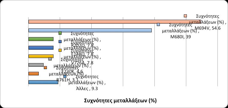
Results: The mean age at onset was 3.54±3.08 years and the cumulative follow-up time 9.88 years/patient. 75.7% of patients had a typical phenotype at onset, most frequently periodic febrile episodes (100%), abdominal pain (84.3%), rheumatic manifestations (arthritis/arthralgias 44.3%) and chest pain (40%). 64/67 patients carried ≥1 mutations (homozygotes 16, monozygotes 23, compound heterozygotes 25); the commonest mutations were M694V (57.1%) and M680I (37%), while the commonest compound heterozygocity, M694V/M680I (26.5%). M694V was significantly detected in patients with rheumatic manifestations (p=0.008). A complete or partial colchicine response was recorded in 44.2% and 47% of patients respectively, and unresponsiveness in 8.6% of them. In one refractory patient, treatment with biologics (anti-IL1) tamed the disease activity. No cases of amyloidosis ever developed so far.
Conclusions: These findings indicate that children with FMF who resident in Greece, have a typical phenotype with a mild disease course and carry the expected for Caucasians mutations. Their systemic follow-up and management in Organized Centers hinders non-compliance and contributes to an excellent disease outcome.
Keywords: Familial Mediterranean Fever, phenotype, subclinical inflammation, genotyping, MEFV mutations
Maria Trachana
Vasiliki Sgouropoulou
Polyxeni Pratsidou-Gertsi
Evangelia Farmaki
Vasiliki Tzimouli
Grigoris Pardalos
Florentia KanakoudiTsakalidou
Pediatric Immunology and Rheumatology Referral Center, First Department of Pediatrics, Aristotle University Thessaloniki, Ippokration Hospital Thessaloniki, Greece
MEditerraneanFeVer),
ARMS (Amplification Refractory Mutation Systems),
NIRCA, (Non-Isoptopic RNAse Cleavage Assay)
“Statistical Package for the
την M694V/M680I που διαπιστώθηκε σε 16/64 (25%). Τέλος, σε 23/64
Εικόνα
*Άλλες
M694I, M694Q, E140Q, P369S
1. Berkun Y, Eisenstein EM. Diagnostic criteria of familial Mediterranean fever. Autoimmun Rev 2014; 13:388-390.
2. Ozen S, Bilginer Y. A clinical guide to autoinflammatory diseases: familial Mediterranean fever and next of kin. Nat Rheum Rev doi: 10.1038/ nrrheum 2013.174 2013.
3. Piram M, Koné-Paut I, Lachmann HJ, Frenkel J, OzenS, Kuemmerle-Deschner J. Validation of the auto-inflammatory diseases activity index (AIDAI) for hereditary recurrent fever syndromes. Ann Rheum Dis 2014;73: 2168-2173.
4. Rigante D, Lopalco G, Tarantino G, Compagnone A, Fastiggi M, Cantarini M. Non-canonical manifestations of familial Mediterranean fever: a changing paradigm. Clin Rheumatol 2015 Mar 13. [Epub ahead of print] doi 10.1007/s10067-015-2916-z.
5. Ozçakar ZB, Yalçinkaya F, Yüksel S, Acar B, Gökmen D, Ekim M. Possible effect of subclinical inflammation on daily life in familial Mediterranean fever. Clin Rheumatol 2006; 25: 149-152.
6. Bayram MT, Çankaya T, Bora E, Kavukçu S, Ülgenalp A, Soylu A, et al. Risk factors for subclinical inflammation in children with Familial Mediterranean fever. Rheumatol Int 2015 Feb 11. [Epub ahead of print] doi 10.1007/s00296-015-3227-z.
7. Kapoor S. Assessing disease severity and activity in patients with familial Mediterranean fever. Inflammation 2014; 37: 833-834.
8. Ben-Zvi I, Danilesko I, Yahalom G, Kukuy O, Rahamimov R, Livneh A, et al. Risk factors for amyloidosis and impact of kidney transplantation on the course of familial Mediterranean fever. IMAJ 2012; 14: 221-224.
9. Ben-Chetrit E, Touitou I. The impact of MEFV gene identification on FMF: an appraisal after 15 years. Clin Exp Rheumatol 2012; 30 (3 Suppl 72): S3-6.
10. Soriano A, Pras E. Familial Mediterranean Fever: Genetic Update. IMAJ 2014; 16: 274–276.
11. Eisenberg S, Aksentijevich I, Deng Z, Kastner DL, Matzner Y. Diagnosis of familial Mediterranean fever by a molecular genetics method. Ann Intern Med 1998; 129: 539-542.
12. Cantarini L, Lopalco G, Cattalini M, Vitale A, Galeazzi M,Rigante M. Interleukin-1 as Ariadne’s Thread in Autoinflammatory and Autoimmune Disorders. IMAJ 2015: 17: 93-97.
13. Milhavet F, Cuisset L, Hoffman HM, Slim R, El-Shanti H, AksentijevichI, et al. The infevers autoinflammatory mutation online registry: update with new genes and functions. Hum Mutat 2008; 29: 803-808.
14. Livneh A, Langevitz P, Zemer D, Zaks N, Kees S, Lidar T, et al. Criteria for the diagnosis of familial Mediterranean fever. Arthritis Rheum 1997; 40: 1879-1885.
15. Federici S, Sormani MP, Ozen S, Lachmann HJ, Amaryan G, Woo P. Evidence-based provisional clinical classification criteria for autoinflammatory periodic fevers. Ann Rheum Dis2015;
74: 799-805.
16. Gül A. Familial Mediterranean fever phenotype and MEFV variations. Clin Exp Rheumatol. 2014; 32: 12-13.
17. Giancane G, TerHaar NM, Wulffraat N, Vastert SJ, Barron K, Hentgen V. Evidencebased recommendations for genetic diagnosis of familial Mediterranean fever. Ann Rheum Dis. 2015;74: 635-641.
18. Sozeri B, Kasapcopur O. Biological agents in familial Mediterranean fever focusing on colchicine resistance and amyloidosis. Curr Med Chem 2015: Mar 11. [Epub ahead of print] doi 10.2174/0929867322666150311152300.
19. La Regina M, Ben-Chetrit E, Gasparyan AY, Livneh A, Ozdogan H, Manna R. Current trends in colchicine treatment in familial Mediterranean fever. Clin Exp Rheumatol 2013; 31:41-46.
20. Özçakar ZB, Elhan AH, Yalçınkaya F. Can colchicine response be predicted in familial Mediterranean fever patients? Rheumatology (Oxford) 2014 ; 53: 1767-1572.
21. Grattagliano I, Bonfrate L, Ruggiero V, Scaccianoce G, Palasciano G, Portincasa P. Novel therapeutics for the treatment of familial Mediterranean fever: from colchicine to biologics. Clin Pharmacol Ther 2014; 95:89-97.
22.
2008; 20:52-58.
24. Ritis K, Giaglis S, Spathari N, Micheli A, Zonios D, Tzoanopoulos D, et al. Non-isotopic RNase cleavage assay for mutation detection in MEFV, the gene responsible for familial Mediterranean fever, in a cohort of Greek patients. Ann Rheum Dis 2004; 63:438-443.
25. Ozturk C, Halıcıoğlu O, Coker I, Gulez N, Sutçuoğlu S, Karaca N, et al. Association of clinical and genetical features in FMF with focus on MEFV strip assay sensitivity in 452 children from western Anatolia, Turkey. Clin Rheumatol 2012;31: 493–501.
26. Kilim Y, Magal N, Shohat M. Expanding the panel of MEFV mutations for routine testing of patients with a clinical diagnosis of familial Mediterranean fever. IMAJ 2011;13: 206-208.
27. Schnekenberg RP, Németh AH. Next-generation sequencing in childhood disorders. Arch Dis Child 2014;9: 284-290.
28. Ozen S, Demirkaya E, Duzova A, Erdogan O, Erken E, Gul A. et al. FMF50: a score for assessing outcome in familial Mediterranean fever. Ann Rheum Dis 2014;73: 897-901.
29. Özer S, Yılmaz R, Sönmezgöz E, Karaaslan E, Taşkın S, Bütün I et al. Simple Markers for Subclinical Inflammation in Patients with Familial Mediterranean Fever. Med Sci Monit 2015; 21: 298–303.
30. Celikbilek M, Dogan S, Akyol L, Borekci E, Zararsiz G, Kozan M et al. Neutrophil-lymphocyte ratio in patients with familial Mediterranean fever. J Clin Lab Anal 2015;29: 80-83.
31. Uslu AU, Deveci K, Korkmaz S, Aydin B, Senel S, Sancakdar E, Sencan M. Is neutrophil/lymphocyte ratio associated with subclinical inflammation and amyloidosis in patients with familial Mediterranean fever? Biomed Res Int 2013: 185317. http://dx.doi.org/10.1155/2013/185317.
32. Hentgen V, Grateau G, Stankovic-Stojanovic K, Amselem S Jéru I. Familial Mediterranean Fever in Heterozygotes: Are We Able to Accurately Diagnose the Disease in Very Young Children? Arthritis Rheum 2013; 65:1654-1662.
33. Marek-Yagel D, Berkun Y, Padeh S, Abu A, Reznik-Wolf H, Livneh A,et al. Clinical disease among patients heterozygous for familial Mediterranean fever. Arthritis Rheum 2009; 60: 18621866.
34. Booty MG, Chae JJ, Masters SL, Chae JJ, Masters SL, Remmers EF, et al. Familial Mediterranean fever with a single MEFV mutation: where is the second hit? Arthritis Rheum 2009; 60: 1851-1861.
35. Soylemezoglu O, Arga M, Fidan K, Gonen S, Emeksiz HC, Hasanoglu E, et al. Unresponsiveness to colchicine therapy in patients with familial Mediterranean fever homozygous for the
M694V mutation. J Rheumatol2010; 37: 182-189.
36. Gül A. Treatment of familial Mediterranean fever: colchicine and beyond. Isr Med Assoc J. 2014;16:281-284.
37. Rossi-Semerano L, Fautrel B, Wendling, Hachulla E, Galeotti C, Semerano L, et al. Tolerance and efficacy of off-label anti-interleukin-1 treatments in France: a nationwide survey. Orphanet J Rare Dis 2015 15; 10:19. doi: 10.1186/s13023-015-0228-7.
38. Brik R, Butbul-Aviel Y, Lubin S, Ben Dayan E, Rachmilewitz-Minei T, Tseng L, et al. Canakinumab for the treatment of children with colchicine-resistant familial Mediterranean fever: a 6-month open-label, single-arm pilot study. Arthritis Rheumatol 2014; 66: 3241-3243.
39. Ozen S, Demirkaya E, Amaryan G, Koné-Paut I, Polat A, Woo P. Results from a multicentre international registry of familial Mediterranean fever: impact of environment on the expression of a monogenic disease in children. Ann Rheum Dis 2014;73:662-667.
Correspondence
Μaria Gogou
Dimitriou Nika 44, 60 100, Κaterini e-mail: mariaangogou@ gmail.com
Polysomnographic study of sleep in children with idiopathic epilepsy
Maria Gogou, Katerina Haidopoulou, Maria Eboriadou, Evangelos Pavlou
Abstract
Introduction: Sleep disorders consist a frequent problem in children with epilepsy, while some studies correlate them with poor seizure control. The aim of our study is to investigate through polysomnography the prevalence of sleep disorders in children with idiopathic epilepsy in comparison to healthy children.
Methods: Totally 40 children with idiopathic epilepsy and 27 healthy children underwent overnight polysomnography. Respiratory and sleep architecture parameters were collected and analyzed. Statistical analysis was performed with SPSS software and level of significance was set at 0.05.
Results: The epilepsy and control group were similar in age, gender, Body Mass Index, and in frequency of hypertrophic adenoids or tonsils. The mean value of the Obstructive Apnea (-Hypopnea) Index and the prevalence of the Obstructive Sleep Apnea Syndrome were significantly higher in children with epilepsy in comparison to healthy children. Besides, children with epilepsy had a significantly lower sleep efficiency and a trend for shorter REM duration. Epileptic children with poor seizure control presented significantly longer apneic events compared to children with good seizure control. On the contrary, no significant differences were detectedin respiratory and sleep architecture parameters between generalized and focal epilepsy.
Conclusion: Our findings show that sleep breathing disorders are significantly more frequent in children with epilepsy than in healthy children. Sleep efficiency and REM stage duration are also disturbed and this can have a negative impact on quality of life, as well as on memory and learning. Children with epilepsy should be systematically screened for sleep problems during their follow-up.
Keywords: sleep disorders, epilepsy, children, sleep apneas, polysomnography
Maria Gogou
Evangelos Pavlou
2nd Department of Pediatrics, School of Medicine, Aristotle University of Thessaloniki, University General Hospital of Thessaloniki AHEPA
Katerina Haidopoulou
Maria Eboriadou
4th Department of Pediatrics, School of Medicine, Aristotle University of Thessaloniki, General Hospital of Thessaloniki Papageorgiou
SWS: SlowWaveSleep Care Excellence
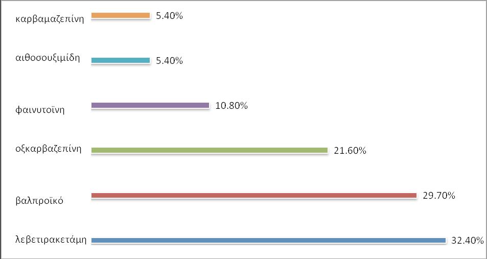
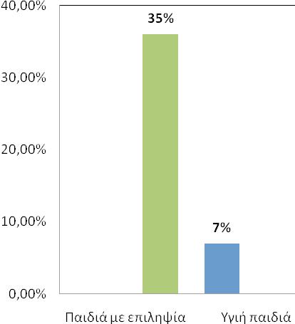
1,55±0,96 0,46±0,43 96,7±1 1,14±1,09 40,21±3,79
18,85±6,29
6,7±0,76 27,34±20,35 80±12 50,4±9,14 32±10,48 18,63±5,88 1,06±1,01
1,2 0,8 0,75 9,9% (2/22) 5,5% (1/18
(1-4, 7-16).
1. ChanB, CheongEY, NgSF, ChanYC, LeeQU, ChanKY. Evaluation of sleep disturbances in children with epilepsy: a questionnaire-based case-control study. Epilepsy Behav 2011;21:43740
2. Ong LC, Yang WW, Wong SW, alSiddiq F, Khu YS. Sleep habits and disturbances in Malaysian children with epilepsy. J Paediatr Child Health 2010; 46:80-4
3. Samaitienė R, Norkūnienė J, Tumienė B, Grikinienė J. Sleep and behavioral problems in rolandic epilepsy. Pediatr Neurol 2013; 48:115-22
4. Becker DA, Fennell EB, Carney PR. Daytime behavior and sleep disturbance in childhood epilepsy. Epilepsy Behav 2004; 5:708-15
5. Iber C, Ancoli-Israel S, Chesson AL Jr, Quan SF. The AASM Manual for the Scoring of Sleep and Associated Events: Rules, Terminology and Technical Specifications. American Academy of Sleep Medicine, 2007, pp. 47-8
6. Katz ES, Marcus CL. Diagnosis of obstructive sleep apnea. In: Sheldon SH, Ferber R, Kryger MH, Gozal D, editors. Principles and practice of pediatric sleep medicine. 2nd edition, Elsevier Saunders: New York, 2014, pp. 221-30
7. Maganti R, Hausman N, Koehn M, Sandok E, Glurich I, Mukesh BN. Excessive daytime sleepiness and sleep complaints among children with epilepsy. Epilepsy Behav 2006; 8:272-7
8. Larson AM, Ryther RC, Jennesson M, Geffrey AL, Bruno PL, Anagnos CJ, et al. Impact of pediatric epilepsy on sleep patterns and behaviors in children and parents. Epilepsia 2012; 53:1162-9
9. Wirrell E, Blackman M, Barlow K, Mah J, Hamiwka L. Sleep disturbances in children with epilepsy compared with their nearest-aged siblings. Dev Med Child Neurol 2005; 47:754-9
10. Tang SS, Clarke T, Owens J, Pal DK. Sleep behavior disturbances in rolandic epilepsy. J Child Neurol 2011; 26:239-43
11. Jain SV, Simakajornboon S, Shapiro SM, Morton LD, Leszczyszyn DJ, Simakajornboon N. Obstructive sleep apnea in children with epilepsy: prospective pilot trial. Acta Neurol Scand 2012; 125:3-6
12. Jain SV, Horn PS, Simakajornboon N, Glauser TA. Obstructive sleep apnea and primary snoring in children with epilepsy. J Child Neurol 2013; 28:77-82
13. Becker DA, Fennell EB, Carney PR. Sleep disturbance in children with epilepsy. Epilepsy Behav 2003; 4:651-8
14. Bruni O, Novelli L, Luchetti A, Zarowski M, Meloni M, Cecili M, et al. Reduced NREM sleep instability in benign childhood epilepsy with centro-temporal spikes. Clin Neurophysiol 2010; 121:665-71
15. Kaleyias J, Cruz M, Goraya JS, Valencia I, Khurana DS, Legido A, et al. Spectrum of polysomnographic abnormalities in children with epilepsy. Pediatr Neurol 2008;39:170-6
16. Pereira AM, Bruni O, Ferri R, Palmini A, Nunes ML. The impact of epilepsy on sleep architecture during childhood. Epilepsia 2012; 53:1519-25
17. Carroll JL, Donnelly DF. Respiratory physiology and pathophysiology during sleep. In: Sheldon SH, Ferber R, Kryger MH, Gozal D, editors. Principles and practice of pediatric sleep medicine. 2nd edition, Elsevier Saunders: New York, 2014, pp. 179-94
18. McKay LC, Atalla A, Morrell MJ. Physiology and neural control of breathing during sleep. Eur Respir Monogr 2010;50: 1–16
27. Ramelli GP, Donati F, Bianchetti M, Vassella F. Apnoeic attacks as an isolated manifestation of epileptic seizures in infants. Eur J Paediatr Neurol 1998;2:187-91
19. Hoellinger P, Khatami R, Gugger M, Hess CW, Bassetti CL. Epilepsy and obstructive sleep apnea. Eur Neurol 2006; 55:74-9
20. Kanemura H, Sano F, Maeda Y, Sugita K, Aihara M. Valproate sodium enhances body weight gain in patients with childhood epilepsy: a pathogenic mechanisms and open-label clinical trial of behavior therapy. Seizure 2012; 21:496-500
21. Verrotti A, Laus M, Scardapane A, Franzoni E, Chiarelli F. Thyroid hormones in children with epilepsy during long-term administration of carbamazepine and valproate. Eur J Endocrinol 2009; 160:81-6
22. Jain SV, Glauser TA. Effects of epilepsy treatments on sleep architecture and daytime sleepiness: an evidence-based review of objective sleep metrics. Epilepsia 2014; 55:26-37
23. Liu DB, Zhou LF, Zhong JW, Wang J. Changes of sleep architecture in children with obstructive sleep apnea syndrome. Zhonghua Er Ke Za Zhi 2004;42:284-6
24. Bourke R, Anderson V, Yang JS, Jackman AR, Killedar A, Nixon GM, et al. Cognitive and academic functions are impaired in children with all severities of sleep-disordered breathing. Sleep Med 2011;12:489-96
25. Kwon S, Seo HE, Hwang SK. Cognitive and other neuropsychological profiles in children with newly diagnosed benign rolandic epilepsy. Korean J Pediatr 2012; 55:383-7
26. Braakman HM, Ijff DM, Vaessen MJ, Debeij-van Hall MH, Hofman PA, Backes WH, et al. Cognitive and behavioural findings in children with frontal lobe epilepsy. Eur J Paediatr Neurol 2012; 16:707-15
27. Segal E, Vendrame M, Gregas M, Loddenkemper T, Kothare SV. Effect of treatment of obstructive sleep apnea on seizure outcomes in children with epilepsy. Pediatr Neurol 2012; 46:359-62
28. Bruni O, Novelli L, Luchetti A, Zarowski M, Meloni M, Cecili M, et al. Reduced NREM sleep instability in benign childhood epilepsy with centro-temporal spikes. Clin Neurophysiol 2010; 121:665-71
29. Nunes ML, Ferri R, Arzimanoglou A, Curzi L, Appel CC, Costa da Costa J. Sleep organization in children with partial refractory epilepsy. J Child Neurol 2003; 18:763-6
30. Eisensehr I, Parrino L, Noachtar S, Smerieri A, Terzano MG. Sleep in Lennox-Gastaut syndrome: the role of the cyclic alternating pattern (CAP) in the gate control of clinical seizures and generalized polyspikes. Epilepsy Res 2001; 46:241-50
31. Derry CP, Duncan S. Sleep and epilepsy. Epilepsy Behav 2013; 26:394-404
e-mail: chrisskaretsos@ hotmail.com
Correspondence
Karetsos Chris
Ierou Lochou 18 Amarousion, Athens
T. 6948596864
e-mail: chrisskaretsos@ hotmail.com
A decade of experience with gastros-
tomy placement in infants and children: A critical evaluation of indications, techniques and results
Karetsos Chris, Antoniou Dimitris, Mavridis Georgios
Abstract
Purpose: The presentation of our experience in gastrostomy placement in the last 10 years in our institute.
Materials and Methods: From 2005-2015, 40 gastrostomies placed in patients with neurological and metabolic syndromes. The age of children’s was ranged from 11 months to 14 years. In 10 patients the gastrostomy was placed with the open technique, in 25 with the percutaneous endoscopic technique and in 5 children with the laparoscopic assisted peg technique. The choice of technique was based on surgeon’s experience and the general condition of the patient.
Results: All the procedures were perfomed uneventfully and without major complications. The minor complications were 1 wound infection in the open group, 1 minor hemorrhage and 5 granulomas in the peg group due to eversion of mucosa of the stomach through the small incision. The progressive feeding in the peg group started in the first postoperative day while in the other groups in the fourth postoperative day. The duration of hospitalization was shorter in the peg and lap+peg group about 4 days in comparison with the open group. The postoperative pain was negligible.
Conclusions: The percutaneous endoscopic gastrostomy is fast and safe technique. The postoperative complications and the pain are minimum. The experience of the surgeon and the knowledge of indications and contraindications are the criteria in which must be based the selection of the technique.
Keywords: Percutaneous endoscopic gastrostomy, gastrostomy, complications
Karetsos Chris
Antoniou Dimitris
Mavridis Georgios
2nd Department of Pediatric Surgery, Aghia Sophia Children’s Hospital, Athens, Greece
Gauderer (3).
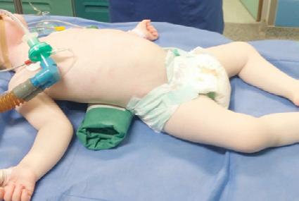
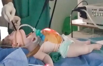
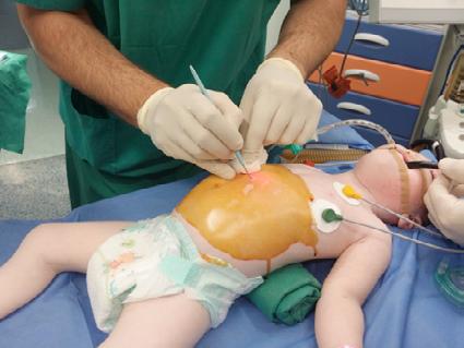
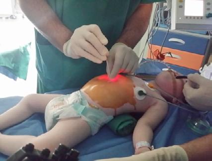
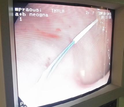
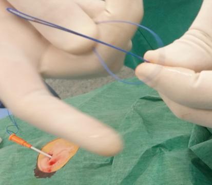
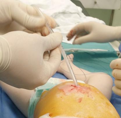
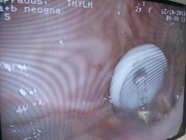
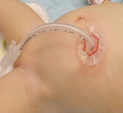
(1). Laura Baker, Alana L. Beres, Robert Baird: A systematic review and meta-analysis of gastrostomy insertion. Journal of Pediatric Surgery 50 (2015) 718–725.
(2). Stamm M. Gastrostomy by a new method. Med Newsl 1984;65.
(3). Gauderer M, Ponsky J, Izant RJ. Gastrostomy without laparotomy: a percutaneous endoscopic technique. J Pediatr Surg 1980;15(6):872–5.
(4). Zamakhshary M, Jamal M, Blair G, et al. Laparoscopic vs percutaneous endoscopic gastrostomy tube insertion: a new pediatric gold standard? J Pediatr Surg 2005;40(5):859–62
(5). Georgeson K. Laparoscopic fundoplication and gastrostomy. Semin Laparosc Surg1998;5:25–30.
(6). Khattak IU, Kimber C, Kiely EM, et al. Percutaneous endoscopic gastrostomy in paediatric practice: complications and outcome .J Pediatr Surg 1998;33:67-72
(7). Begum Akay, Tony R. Capizzani, Alice M. Lee, Robert A. Drongowski, James D. Geiger, Ronald B. Hirschl, George B. Mychaliska.Gastrostomy tube placement in infants and children: is there a preferred technique? Journal of Pediatric Surgery (2010) 45, 1147–1152
(8). Jessica Gonzalez-Hernandez, YahyaDaoud, Anne C. Fischer, Bradley Barth, Hannah G. Piper. Endoscopic button gastrostomy: Comparing a sutured endoscopic approach to the current techniques. Journal of Pediatric Surgery (2015) article in press.
Correspondence
Georgia Zisimopoulou
Thivon & Papadiamantopoulou, Goudi, 115 27, Greece
Τ. +306939613133
e-mail: dr_zis@yahoo.gr
Benign pancreatic hyperenzymemia or Gullo’s syndrome: a case report
Georgia Zisimopoulou, Nina Manolaki
Abstract
Benign pancreatic hyperenzymemia or Gullo’s syndrome is characterized by chronic elevation usually of all pancreatic enzymes in serum, in the absence of pancreatic or other disease. We report the case of a 2-year old asymptomatic girl with elevated serum levels of amylase and lipase. The physical examination was unremarkable. Possible causes of pancreatic hyperenzymemia were excluded by laboratory investigations and USS and MRCP imaging. Pancreatic hyperenzymemia, found in a 5-year old sibling of the patient, confirmed the diagnosis of familial benign pancreatic hyperenzymemia.
Keywords: benign pancreatic hyperenzymemia, Gullo’s syndrome, amylase, lipase, magnetic cholangiopancreatography
Georgia Zisimopoulou
Nina Manolaki
B’ Department of Pediatrics, “Agia Sophia” Children΄s Hospital
1. Gullo L.Chronic nonpathological hyperamylasemia of pancreatic origin Gastroenterology 1996;110(6):1905-1908.
2. Lionetti E, Francavilla R, Leonardi S, Tomarchio S, Gennaro A, Franzonello C, et al. Two rare cases of benign hyperlipasemia in children.WJCC 2014; 2(1):16–19.
3. Frulloni L, Patrizi F, Bernardoni L,Cavallini G. Pancreatic hyperenzymemia: Clinical significance and diagnostic approach JOP 2005; 6(6):536-551.
4. Gullo L. Day-to-day variations of serum pancreatic enzymes in benign pancreatic hyperenzymemia.ClinGastroenterolHepatol2007;5(1):70-74.
5. Gullo L,Ventrucci M, Barakat B,Migliori M, Tomassetti P,Pezzilli R. Effect of secretin on serum pancreatic enzymes and on the Wirsung duct in chronic nonpathological pancreatic hyperenzymemia.Pancreatology 2003; 3(3):191–194.
6. Mortele KJ,Wiesner W, Zou KH, Ros PR, Silverman SG.Asymptomatic nonspecific serum hyperamylasemia and hyperlipasemia: spectrum of MRCP findings and clinical implications. Abdom ImagingJ 2004;29(1):109-114.
7. Testoni PA, Mariani A,Curioni S, Giussani A,Masci E.Pancreatic ductal abnormalities documented by secretin-enhanced MRCP in asymptomatic subjects with chronic pancreatic hyperenzymemia. Amer J Gastroenterol 2009;104:1780-1786.
8. Pezzilli R, Morselli-Labate AM, Casadei R, Campana D, Rega D, Santini D, et al. Chronic asymptomatic hyperenzymemia is a benign condition in only half of the cases: a prospective study. Scand J Gastroenterol 2009;44:888-893.
9. Gullo L,Mantovani V, Manca M,Migliori M, Bastagli L,Pezzilli R. Mutations of the CFTR gene in idiopathic pancreatic hyperenzymemia. Pancreas 2005; 31(4):350-352.
10. Gullo L, Laghi L, Migliori M, Lucrezio L, Bianchi P, Randolf AE, et al.SPINK1 and PRSS1 mutations in benign pancreatic hyperenzymemia.Pancreas 2008; 37(1):31-35.
6936493425
F. 2310993514
e-mail: anastagab@yahoo. gr
Correspondence
Gkampeta Αnastasia
St. kiriakidi 1 54636, Thessaloniki
Τ. +302310994815
+306936493425
F. 2310993514
e-mail: anastagab@yahoo.
gr
Gkampeta Αnastasia
Pavlidou Εfterpi
Pavlou Εvangelos
2nd Pediatric Department, Aristotle University of Thessaloniki, AHEPA Hospital
Evangeliou Αthanasios
4th Pediatric Department, Aristotle University of Thessaloniki, Papageorgiou Hospital
Ketosis and electroencephalographical improvement following viral gastroenteritis in patient with West syndrome
Gkampeta Αnastasia, Pavlidou Εfterpi, Evangeliou Αthanasios, Pavlou Εvangelos
Abstract
Background: Epileptic seizures generally become more serious following infections. However, in rare cases epileptic seizures, mostly in patients with West syndrome, disappear or decrease in severity after viral infection.
Methods-results: We report an 11 months old female infant diagnosed with West syndrome whose clinical presentation and electroencephalogram findings improved following viral gastroenteritis.
Conclusion: The exact mechanism by which viral infection affects pathophysiology of epileptic activity is still unknown. Possible hypotheses need to be based on experimental and laboratory data for better understanding of the mechanism by which a viral infection may lead to spontaneous resolution of intractable epileptic seizures.
Keywords: gastroenteritis, West syndrome, electroencephalogram, ketosis.
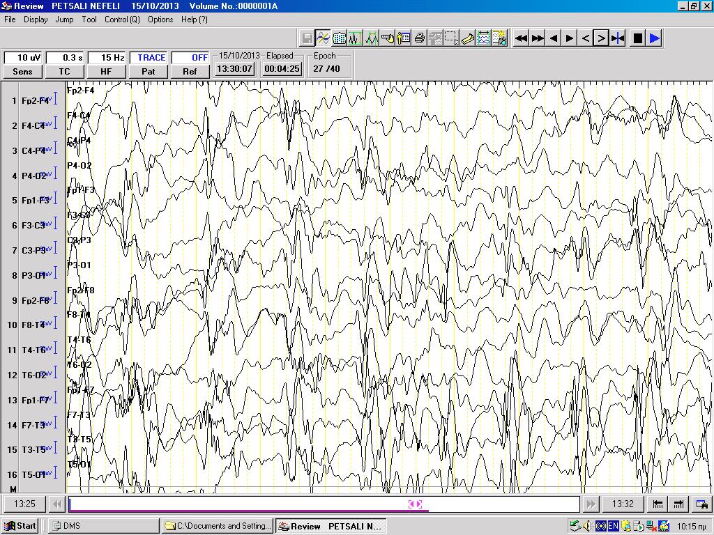
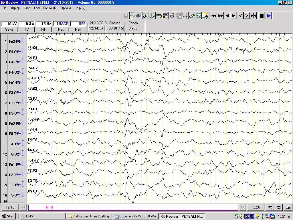
1. Yamamoto H, Yamano T, Niijima S, Kohyama J, Yamanouchi H. Spontaneous improvement of intractable epileptic seizures following acute viral infections. Brain Dev. 2004;26:377-379.
2. Yamamoto H, Kamiyama N, Murakami H, Miyamoto Y, Fukuda M. Spontaneous resolution of intractable epileptic seizures following HHV-7 infection. Brain Dev. 2007;29:185-188.
3. Hrachovy RA, Glaze DG, Frost JD Jr. A retrospective study of spontaneous remission and long-term outcome in patients with infantile spasms. Epilepsia. 1991;32:212-214.
4. West WJ. On a peculiar form of infantile convulsions. Lancet 1841;1:724-725.
5. Kurata S, Sakai A, Komiyama A. Infantile spasms and exanthema subitum. No To Hattatsu 1970;2:315-319.
6. Terada M, Kudo H, Abe T, Naito H, Nihei K. Unexpected improvement of intractable epilepsy following infections. Brain Dev 1984;6:422.
7. Fujita Y, Hiyoshi K, Wakasugi N, Sakuta R, Yanagida Y, Fuchigami T, et al. Transient improvement of the West syndrome in two cases following rotavirus colitis No To Hattatsu. 1988;20:59-63.
8. Hattori H. Spontaneous remission of spasms in West syndrome--implications of viral infection. Brain Dev. 2001;23:705-707.
9. Iwasaki T, Nonoda Y, Ishii M. Remission associated with human herpesvirus infection in west syndrome. J Child Neurol. 2006;21:886-890.
10. Fujita Y, Imai Y, Ishii W, Endo A, Arakawa C, Kohira R, et al. Improvement of intractable childhood epilepsy following acute viral infection. Brain Dev. 2011;33:62-68.
Correspondence
Koufopoulou Eleni
Thivon and Michalakopoulou St., 11527 Athens, T. +302132013416
e-mail: lena_kfpl@yahoo.gr
Panayiotopoulos syndrome. Case report and review of the literature
Koufopoulou Eleni, Athanassopoulou Panagiota, Dokou Aikaterini, Theodorou Virginia, Manolaki Nina
Abstract
Panayiotopoulos syndrome is benign autonomic epilepsy. A typical episode starts with vomiting, usually during sleep, followed by deviation of the eyes. Seizures frequently last longer than 30 minutes. Vomiting may be the most common symptom, but all functions of the autonomic system can be affected. The clinical manifestations of the syndrome often are attributed to gastroenteritis, migraine, cyclic vomiting, sleep disorders, syncope, encephalitis, intoxication. Diagnosis is based on clinical manifestations and ECG findings present in 90% of the cases. The prognosis is excellent as the majority of patients (75%) have only one or few episodes of seizures and all are symptom-free by the age of 16 years. The risk of epilepsy in adult life is similar to that in the general population. Due to the benign course of the syndrome, treatment is often withheld and, when deemed necessary, carbamazepine and levetiracetam seem to be the antiepileptic drugs of choice. Recently the syndrome has been related to SCN1A gene mutations. We describe the case of a 7-year old girl with Panayiotopoulos syndrome who remained undiagnosed for many years as the manifestations of the syndrome had been attributed to cyclic vomiting. Current literature is reviewed.
Keywords: Panayiotopoulos syndrome, autonomic seizures, occipital spikes.
Koufopoulou Eleni
Dokou Aikaterini
Manolaki Nina
2nd Department of Pediatrics, Aghia Sophia Children’s Hospital, Athens
Athanassopoulou Panagiota
Theodorou Virginia Neurology Department,
Aghia Sophia Children’s Hospital, Athens
Συντομογραφίες ΣΠ:
ILAE: International League Against Epilepsy
SLES: Syncope like epileptic seizures
SUDEP: Sudden unexpected death in epilepsy
NICE: National Institute of Clinical Excellence
1. Ferrie C, Caraballo R, Covanis A, Demirbilek V, Dervent A, Kivity S, et al. Panayiotopoulos syndrome: a consensus view. Dev Med Child Neurol 2006;48(3): 236-240.
2. Panayiotopoulos CP. Vomiting as an ictal manifestation of epileptic seizures and syndromes. J Neurol Neurosurg Psychiatr 1988; 51: 1448-1451.
3. Engel J Jr; International League Against Epilepsy (ILAE). A proposed diagnostic scheme for people with epileptic seizures and with epilepsy: report of the ILAE Task Force on Classification and Terminology. Epilepsia 2001; 42(6):796-803.
4. Berg AT, Berkovic SF, Brodie MJ, Buchhalter J, Cross JH, van Emde Boas W, et al. Revised terminology and concepts for organization of seizures and epilepsies: report of the ILAE Commission on Classification and Terminology, 2005-2009. Epilepsia 2010; 51(4): 676-685.
5. Ferrie CD, Caraballo R, Covanis A, Demirbilek V, Dervent A, Fejerman N, et al. Autonomic status epilepticus in Panayiotopoulos syndrome and other childhood and adult epilepsies: A consensus view. Epilepsia 2007; 48(6): 1165–1172.
6. Covanis A. Panayiotopoulos syndrome: a benign childhood autonomic epilepsy frequently imitating encephalitis, syncope, migraine, sleep disorder, or gastroenteritis. Pediatrics 2006; 118(4): e1237-e1243.
7. Lada C, Skiadas K, Theodorou V, Loli N, Covanis A. A study of 43 Patients with Panayiotopoulos syndrome, a common and benign childhood seizure susceptibility. Epilepsia 2003; 44(1): 81–88.
8. Panayiotopoulos CP, Michael M, Sanders S, Valeta T, Koutroumanidis M. Benign childhood focal epilepsies: assessment of established and newly recognized syndromes. Brain 2008; 131: 2264-2286.
9. Specchio N, Trivisano M, Di Ciommo V, Cappelletti S, Masciarelli G, Volkov J, et al. Panayiotopoulos syndrome: A clinical, EEG, and neuropsychological study of 93 consecutive patients. Epilepsia 2010; 51(10): 2098–2107.
10. Dirani M, Yamak W, Beydoun A. Panayiotopoulos syndrome presenting with respiratory arrest: A case report and literature review. Epilepsy Behav Case Rep 2015; 3: 12–14.
11. Gambardella A, Marini C. Clinical spectrum of SCN1A mutations. Epilepsia 2009; 50 (Suppl 5): 20-23.
12. Grosso S, Orrico A, Galli L, Di Bartolo R, Sorrentino V, Balestri P. SCN1A mutation associated with atypical Panayiotopoulos syndrome. Neurology 2007; 69(6): 609-611.
13. Livingston JH, Cross JH, Mclellan A, Birch R, Zuberi SM. A novel inherited mutation in the voltage sensor region of SCN1A is associated with Panayiotopoulos syndrome in siblings and generalized epilepsy with febrile seizures plus. J Child Neurol 2009; 24(4): 503-508.
14. Passamonti C, Petrelli C, Mei D, Foschi N, Guerrini R, Provinciali L, et al. A novel inherited SCN1A mutation associated with different neuropsychological phenotypes: is there a common core deficit? Epilepsy Behav 2015; 43: 89-92.
15. Mellish LC, Dunkley C, Ferrie CD, Pal DK. Antiepileptic drug treatment of rolandic epilepsy and Panayiotopoulos syndrome: Clinical practice survey and clinical trial feasibility. Arch Dis Child 2015; 100(1): 62-67.
16. Parisi P, Villa MP, Pelliccia A, Rollo VC, Chiarelli F, Verrotti A. Panayiotopoulos syndrome: diagnosis and management. Neurol Sci 2007; 28: 72–79.
17. Lacroix L, Fluss J, Gervaix A, Korff CM. Benzodiazepines in the acute management of seizures with autonomic manifestations: Anticipate complications! Epilepsia 2011; 52(10): e156–e159.
w w w.e-child .gr