

Paediatriki
154
158
166
176
188
202
220
Υποβολή εργασιών e-mail: grammateia@e-child.gr
Οδηγίες
http://e-child.gr/publications/ instructions-to-authors
Iδιοκτήτης
e-mail: grammateia@e-child.gr
246
256
Paediatriki
Volume 78 | Number 2 | April - May - June 2015
Trimonthly publication of the Greek Paediatric Society
154 SHORT ABSTRACTS
158
STATE OF THE ART - REVIEW ARTICLE
Revision of the jones criteria for the diagnosis of acute rheumatic fever. The contribution of the widespread use of echocardiography
Maria Gogou, Anastasia Keivanidou, Andreas Giannopoulos
166
REVIEW ARTICLES
Genetic predisposition and personalized nutritional intervention for childhood obesity
Kalliopi Gkouskou, Anastasia Markaki, Theodosios Theodosiou, Aristides Eliopoulos
176
Sexualization of children and adolescents
A. Tsitsika, V. Dimitrakopoulou
188 Growing pains
Maria N. Vasilopoulou, Maria Tsolia
202
Ciliopathies: The central role of primary cilia in a wide disease spectrum
Stavroula Psoni, Helena Fryssira
220
ORIGINAL ARTICLES
Prevalence of simple and abdominal obesity among 15 year old adolescents in Greece: results from the ADONUT study
Maria Grammatikopoulou, Dimitrios Poulimeneas, Konstantina Gerothanasi, Efstratios Kiranas, Maria Tsigga & ADONUT Study Group
President
A. Constantopoulos
Editorial board
Director
G. S. Varlamis
Members
S. Andronikou
E. Galanakis
A. Evangeliou
L. Thomaidou
M. Kanariou
A. Kapogiannis
S. Kitsiou-Tzeli
E. Mantadakis
P. Panagiotopoulou-Gartagani
A. Papadopoulou
V. Papaevagelou
A. Papathanassiou
A. Siamopoulou-Mavridou
A. Syrigou-Papavasiliou
Manuscript submission
e-mail: grammateia@e-child.gr
Instructions to authors: http://e-child.gr/publications/ instructions-to-authors
Owner Greek Paediatric Society 15, Mpakopoulou st. GR - 15451, Ν. Psychiko Tel.: +302107771140
e-mail: grammateia@e-child.gr
Annual subscription All foreign countries: US$50
234
Early changes of amino acids in critical ill children dependent on disease severity
Theonymfi Tavladaki, Anna Maria Spanaki, Helen Dimitriou, EvaggelosPapakonstantinou, George Briassoulis
246
Networks of primary care and welfare for deaf primary school children. Parents’ satisfaction
Bredaki Maria, Ktena Danai, Barbouni Anastasia, Kornarou Eleni
256
Νο Relapse And Effective Prevention Of Complications of Secondary Hemosiderosis with Iron Chelation With Deferasirox In Childhood Malignancies
Μicaela Nicolaou, Yiouli P. Ktena, Archontis Zampogiannis, Anastasia Athanasiadou, Spiros Vlahopoulos, George Lambrou, Maria Adamaki, Maria Moschovi
266
Α prospective evaluation new scoring system for diagnosis of acute appendicitis in children
Adelais Tzortzopoulou, Panagiota Giamarelou, Aikaterini Michail-Strantzia, Alexandros Passalidis
ΒΡΑΧΕΙΕΣ
SHORT ABSTRACTS

Παραπομπές:
1. Eliot L. What’s Going On in There? How the Brain and Mind Develop in the First Five Years of Life. New York, NY: Bantam Books; 1999. 2. UNICEF/World Health Organization. Integrating Early Childhood Development (ECD) Activities Into Nutrition Programmes in Emergencies. Why, What and How [guidance note]. 2014:1-16. 3. Hernandez-Reif M, Diego M, Field T. Preterm infants show reduced stress behaviors and activity after 5 days of massage therapy. Infant Behav Dev. 2007;30(4):557-561. 4. White-Traut RC, Schwertz D, McFarlin B, Kogan J. Salivary cortisol and behavioral state responses of healthy newborn infants to tactile-only and multisensory interventions. J Obstet Gynecol Neonatal Nurs. 2009;38:22-34. 5. Mindell JA, Telofski LS, Wiegand B, Kurtz ES. A nightly bedtime routine: impact on sleep in young children and maternal mood. Sleep. 2009;32(5):599-606. 6. White-Traut RC, Nelson MN, Silvestri JM, et al. Effect of auditory, tactile, visual, and vestibular intervention on length of stay, alertness, and feeding progression in preterm infants. Dev Med Child Neurol. 2002;44:91-97. 7. Field T, Cullen C, Largie S, Diego M, Schanberg S, Kuhn C. Lavender bath oil reduces stress and crying and enhances sleep in very young infants. Early Hum Dev. 2008;84(6):399-401. 8. Farroni T, Csibra G, Simion F, Johnson MH. Eye contact detection in humans from birth. Proc Natl Acad Sci U S A. 2002;99(14):9602-9605. 9. Herz RS. A naturalistic analysis of autobiographical memories triggered by olfactory visual and auditory stimuli. Chem Senses. 2004;29(3):217-224. 10. Sullivan RM, Toubas P. Clinical usefulness of maternal odor in newborns: soothing and feeding preparatory responses. Biol Neonate. 1998;74(6):402-408.
Correspondence
Μaria Gogou
Dimitriou Nika 44, 60100, Κaterini
e-mail: mariaangogou@ gmail.com
Revision of the jones criteria for the diagnosis of acute rheumatic fever. The contribution of the widespread use of echocardiography
Maria Gogou, Anastasia Keivanidou, Andreas Giannopoulos
Abstract
Although rheumatic fever has declined in incidence over the past decades, this disease remains an important cause of acquired cardiovascular morbidity in childhood and presents a very disproportionate geographic distribution. Besides, the increasing use of cardiac ultrasound has revealed subclinical carditis in many cases of children diagnosed with rheumatic fever without clinical findings of carditis. On this basis, in 2015 the American Heart Association’s Council on Cardiovascular Disease in the Young and its Rheumatic Fever, Endocarditis and Kawasaki Disease Committee revised the classical Jones criteria for the diagnosis of Rheumatic Fever defining populations as low-risk or moderate-/high-risk and including Doppler echocardiography findings of carditis in major criteria.
Keywords: rheumatic fever, revised criteria, subclinical carditis
Maria Gogou
Anastasia Keivanidou
Andreas Giannopoulos
Unit for Pediatric Cardiology, 2nd Department of Pediatrics, Aristotle University of Thessaloniki, University General Hospital of Thessaloniki AHEPA
Πίνακας
2.
3.
1. Seckeler MD, Hoke TR. The worldwide epidemiology of acute rheumatic fever and rheumatic heart disease. Clin Epidemiol 2011, 3:67-84
2. Ferrieri P; Jones Criteria Working Group. Proceedings of the Jones Criteria workshop. Circulation 2002, 106:2521-3
3. Dajani AS, Ayoub E, Bierman FZ, Bisno AL, Denny FW, Durack DT, et al. Special Writing Group of the Committee on Rheumatic Fever, Endocarditis, and Kawasaki Disease of the Council on Cardiovascular Disease in the Young of the American Heart Association. Guidelines for the diagnosis of rheumatic fever: Jones criteria, 1992 update [published correction appears in JAMA. 1993 ,269:476]. JAMA. 1992, 268:2069–73
4. Burke RJ, Chang C. Diagnostic criteria of acute rheumatic fever. Autoimmun Rev 2014, 13:503-7
5. Markowitz M. The decline of rheumatic fever: role of medical intervention: Lewis W. Wannamaker Memorial Lecture. J Pediatr 1985, 106:545–50
6. Milne RJ, Lennon DR, Stewart JM, Vander Hoorn S, Scuffham PA. Incidence of acute rheumatic fever in New Zealand children and youth. J Paediatr Child Health 2012,48:685–69145
7. Parnaby MG, Carapetis JR. Rheumatic fever in indigenous Australian children. J Paediatr Child Health 2010, 46:527–33
8. Atatoa-Carr P, Lennon D, Wilson N; New Zealand Rheumatic Fever Guidelines Writing Group. Rheumatic fever diagnosis, management, and secondary prevention: a New Zealand guideline. N Z Med J 2008, 121:59–69
9. Carapetis JR, Currie BJ. Rheumatic fever in a high incidence population: the importance of monoarthritis and low grade fever. Arch Dis Child 2001, 85:223–7
10. Merino Muñoz R, Viota Losada F, Sancho Madrid B, Castro Gussoni C, García-Consuegra Molina J. Rheumatic fever and post-streptococcal arthritis: clinical review. An Esp Pediatr 1991, 35:239–42
11.Koçak G, Imamoğlu A, Tutar HE, Atalay S, Türkay S. Poststreptococcal reactive arthritis: clinical course and outcome in 15 patients. Turk J Pediatr 2000, 42:101–4
12. Gewitz MH, Baltimore RS, Tani LY, Sable CA, Shulman ST, Carapetis J, et al. American Heart Association Committee on Rheumatic Fever, Endocarditis, and Kawasaki Disease of the Council on Cardiovascular Disease in the Young. Revision of the jones criteria for the diagnosis of acute rheumatic Fever in the era of Doppler echocardiography: a scientific statement from the
american heart association. Circulation 2015, 131:1806-18
13. Reményi B, Wilson N, Steer A, Ferreira B, Kado J, Kumar K, et al. World Heart Federation criteria for echocardiographic diagnosis of rheumatic heart disease: an evidence-based guideline. Nat Rev Cardiol 2012, 9:297–309
14. Vijayalakshmi IB, Vishnuprabhu RO, Chitra N, Rajasri R, Anuradha TV. The efficacy of echocardiographic criterions for the diagnosis of carditis in acute rheumatic fever. Cardiol Young 2008, 18:586–92
15. Ozdemir O, Işik S, Abaci A, Hizli S, Akelma AZ, Kişlal FM, et al. Silent enemy in acute rheumatic fever: subclinical carditis. Turk Kardiyol Dern Ars 2011, 39:41–6 16. Cann MP, Sive AA, Norton RE, McBride WJ, Ketheesan N. Clinical presentation of rheumatic fever in an endemic area. Arch Dis Child 2010, 95:455–7
17. Beg A, Sadiq M. Subclinical valvulitis in children with acute rheumatic fever. Pediatr Cardiol 2008, 29:619–23
18. Caldas AM, Terreri MT, Moises VA, Silva CM, Len CA, Carvalho AC, et al. What is the true frequency of carditis in acute rheumatic fever? A PROSPECTIVE clinical and Doppler blind study of 56 children with up to 60 months of follow-up evaluation. Pediatr Cardiol 2008, 29:1048–53
19. Rayamajhi A, Sharma D, Shakya U. Clinical, laboratory and echocardiographic profile of acute rheumatic fever in Nepali children. Ann Trop Paediatr 2007, 27:169–77
20. Ozer S, Hallioğlu O, Ozkutlu S, Celiker A, Alehan D, Karagöz T. Childhood acute rheumatic fever in Ankara, Turkey. Turk J Pediatr 2005, 47:120–4
21.Panamonta M, Chaikitpinyo A, Kaplan EL, Pantongwiriyakul A, Tassniyom S, Sutra S. The relationship of carditis to the initial attack of Sydenham’s chorea. Int J Cardiol 2004, 94:241–8
22. Ozkutlu S, Hallioglu O, Ayabakan C. Evaluation of subclinical valvar disease in patients with rheumatic fever. Cardiol Young 2003, 13:495–9
23. Karaaslan S, Demirören S, Oran B, Baysal T, Başpinar O, Uçar C. Criteria for judging the improvement in subclinical rheumatic valvitis. Cardiol Young 2003, 13:500–5
24. Khriesat I, Najada A, Al-Hakim F, Abu-Haweleh A. Acute rheumatic fever in Jordanian children. East Mediterr Health J 2003, 9:981–7
25. Lanna CC, Tonelli E, Barros MV, Goulart EM, Mota CC. Subclinical rheumatic valvitis: a long-term follow-up. Cardiol Young 2003,13:431–8
26. Figueroa FE, Fernández MS, Valdés P, Wilson C, Lanas F, Carrión F, et al. Prospective comparison of clinical and echocardiographic diagnosis of rheumatic carditis: long term follow up of patients with subclinical disease. Heart 2001, 85:407–10
27. Hilário MO, Andrade JL, Gasparian AB, Carvalho AC, Andrade CT, Len CA. The value of echocardiography in the diagnosis and followup of rheumatic carditis in children and adolescents: a 2 year prospective study. J Rheumatol 2000, 27:1082–6
28. Elevli M, Celebi A, Tombul T, Gökalp AS. Cardiac involvement in Sydenham’s chorea: clinical and Doppler echocardiographic findings. Acta Paediatr 1999, 88:1074–7
29.da Silva CH; Pediatric Committee, Sao Paulo Pediatric Rheumatology Society. Rheumatic fever: a multicenter study in the state of Sao Paulo. Rev Hosp Clin Fac Med Sao Paolo 1999, 54:85–90
30. Minich LL, Tani LY, Pagotto LT, Shaddy RE, Veasy LG. Doppler echocardiography distinguishes between physiologic and pathologic “silent” mitral regurgitation in patients with rheumatic fever. Clin Cardiol 1997, 20:924–6
31. Hoffman TM, Rhodes LA, Pyles LA, Balian AA, Neal WA, Einzig S. Childhood acute rheumatic fever: a comparison of recent resurgence areas to cases in West Virginia. W V Med J 1997, 93:260–3
32. Vasan RS, Shrivastava S, Vijayakumar M, Narang R, Lister BC, Narula J. Echocardiographic evaluation of patients with acute rheumatic fever and rheumatic carditis. Circulation 1996, 94:73–82
33. Maheu B, Costes P, Lionet P, Kamblock J, Papouin G, Mansourati J, et al. Contribution of Doppler echocardiography to the diagnosis of the first attack of acute rheumatic fever. Arch Mal Coeur Vaiss 1995, 88:1833–9
34. Abernethy M, Bass N, Sharpe N, Grant C, Neutze J, Clarkson P, et al. Doppler echocar-
diography and the early diagnosis of carditis in acute rheumatic fever. Aust N Z J Med 1994, 24:530–5
35. Veasy LG, Tani LY, Hill HR. Persistence of acute rheumatic fever in the intermountain area of the United States. J Pediatr 1994, 124:9–16
36. Wilson NJ, Voss L, Morreau J, Stewart JM, Lennon D. New Zealand guidelines for the diagnosis of acute rheumatic fever: small increase in the incidence of definite cases compared to the America Heart Association Jones criteria. N Z Med J 2013, 126:50–9
37. Caldas AM, Terreri MT, Moises VA, Silva CM, Carvalho AC, Hilário MO. The case for utilizing more strict quantitative Doppler echocardiographic criterions for diagnosis of subclinical rheumatic carditis. Cardiol Young 2007, 17:42–7
e-mail: gkouskoukal@ gmail.com
Correspondence
Kalliopi Gkouskou, Konstantinou Papadaki 4, Heraklion Crete
e-mail: gkouskoukal@ gmail.com Τ. +306938055379
Genetic predisposition and personalized nutritional intervention for childhood obesity
Kalliopi Gkouskou, Anastasia Markaki, Theodosios Theodosiou, Aristides Eliopoulos
Abstract
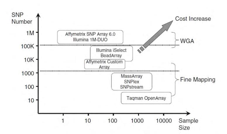
Introduction: Childhood obesity is a major public health issue especially in Greece where its prevalence is among the higher in Europe. This review aims to present genetic factors that permit a personalized nutritional intervention as well as the most cost effective technology for the analysis of these factors.
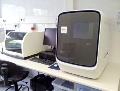
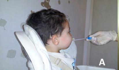
Methods: MeSHy program (Statnous, Greece) was utilized for a systematic review. Single nucleotide polymorphisms (SNPs), copy number variants (CNVs) and insertions/deletions that are related to childhood obesity and can determine a specific nutritional intervention were identified. In addition, a “market research” permitted the evaluation of the most cost-effective method for the analysis of these genetic factors. Finally, a pilot study of 24 children was performed for the initial evaluation of Οpen-array Real time PCR technology.
Results: Fifty genes (68 genetic factors) allow for the formation of a personalized nutritional intervention plan that aims to prevent or to treat childhood obesity. Open-array Real time PCR technology is bloodless, convenient, affordable and reliable for the evaluation of a great number of genetic factors in a large scale.
Conclusions: A personalized nutritional intervention based on genetic analyses for the prevention or treatment of childhood obesity is feasible with current technology.
Keywords: childhood obesity, genetic factors, personalized interventions
Kalliopi Gkouskou
Aristides Eliopoulos
Embiodiagnostics, Genetic Predisposition Research Company
Kalliopi Gkouskou
Aristides Eliopoulos
Medical School University of Crete, Molecular Biology Department
Kalliopi Gkouskou
Anastasia Markaki Department of Nutrition and Dietetics, TEI of Crete
Theodosios Theodosiou
Statnous, Statistical Consultancy
1.
2.
4.
Συντομογραφίες
rtPCR: Real time Polymerase Chain Reaction
CNV: copy number
variant
SNP: single nucleotide polymorphism
(n =870)
(n = 4329)
ΑΜΥ1 (copy number variation)
FTO (2 polymorhisms/epigenetic changes)
TMEM18
GNPDA2
MC4R
BDNF
TFAP2B
SEC16B
KCTD15
MTCH2
NEGR
POMC
NRXN3
FAIM2
GPRC5B
SH2B1
FANCL
LRRN6
TNNI3K
MAP2K5
HOXB5
CADM2
PTBP2
NUDT3
FLJ35779
ZNF608
TMEM160
RPL27A
OLFM4
APOA5
FABP2
GIPR
IRS1
MCM6(2 polymorphisms)
IL6
PPARGC1A
TAS2R38 (3 polymorphisms)
ACEI/D
SHBG*
PPMIK*
CLOCK*
15q26.1**
APOA5
APOE(2 polymorphisms)
CETP
LPL
NOS3
TCF7L2
Thombophilia panel (10 polymorphisms)
Vitamin D deficiency panel (4 polymorphisms)
Χαρακτηριστικά


1. Dietz WH. Overweight in childhood and adolescence. N Engl J Med. 2004 Feb 26;350(9):8557.
2. http://wwwoecdorg/els/health-systems/Obesity-Update-2014pdf.
3. Theodosiou T, Vizirianakis IS, Angelis L, Tsaftaris A, Darzentas N. MeSHy: Mining unanticipated PubMed information using frequencies of occurrences and concurrences of MeSH terms. J Biomed Inform. 2011 Dec;44(6):919-26.
4. Garver WS, Newman SB, Gonzales-Pacheco DM, Castillo JJ, Jelinek D, Heidenreich RA, et al. The genetics of childhood obesity and interaction with dietary macronutrients. Genes Nutr. 2013 May;8(3):271-87.
5. Heath V. Obesity: Genes and a hearty appetite conspire to increase childhood obesity risk. Nat Rev Endocrinol. 2014 Apr;10(4):187.
6. Young RP, Hopkins RJ. Genetic risk in childhood obesity: implications for clinical practice. JAMA Pediatr. 2013 Feb;167(2):196-7.
7. Qi Q, Chu AY, Kang JH, Jensen MK, Curhan GC, Pasquale LR, et al. Sugar-sweetened beverages and genetic risk of obesity. N Engl J Med. 2012 Oct 11;367(15):1387-96.
8. Gonzalez JR, Estevez MN, Giralt PS, Caceres A, Perez LM, Gonzalez-Carpio M, et al. Genetic risk profiles for a childhood with severely overweight. Pediatr Obes. 2014 Aug;9(4):272-80.
9. Domingue BW, Belsky DW, Harris KM, Smolen A, McQueen MB, Boardman JD. Polygenic risk predicts obesity in both white and black young adults. PLoS One. 2014;9(7):e101596.
10. Ciftdogan DY, Coskun S, Ulman C, Tikiz H. The association of apolipoprotein E polymorphism and lipid levels in children with a family history of premature coronary artery disease. J Clin Lipidol. 2012 Jan-Feb;6(1):81-7.
11. Roth CL, Hinney A, Reinehr T, Schreiner F, Nguyen TT, Muller T, et al. TCF7L2 polymorphism rs7903146 and predisposition for type 2 diabetes mellitus in obese children. Horm Metab Res. 2008 Oct;40(10):713-7.
12. Korner A, Berndt J, Stumvoll M, Kiess W, Kovacs P. TCF7L2 gene polymorphisms confer an increased risk for early impairment of glucose metabolism and increased height in obese children. J Clin Endocrinol Metab. 2007 May;92(5):1956-60.
13. Smart MC, Dedoussis G, Louizou E, Yannakoulia M, Drenos F, Papoutsakis C, et al. APOE, CETP and LPL genes show strong association with lipid levels in Greek children. Nutr Metab Cardiovasc Dis. 2010 Jan;20(1):26-33.
14. Kinik ST, Ozbek N, Yuce M, Yazici AC, Verdi H, Atac FB. PAI-1 gene 4G/5G polymorphism, cytokine levels and their relations with metabolic parameters in obese children. Thromb Haemost. 2008 Feb;99(2):352-6.
15. Micali N, Field AE, Treasure JL, Evans DM. Are obesity risk genes associated with binge eating in adolescence? Obesity (Silver Spring). 2015 Aug;23(8):1729-36.
16. Zhang D, Li Z, Wang H, Yang M, Liang L, Fu J, et al. Interactions between obesityrelated copy number variants and dietary behaviors in childhood obesity. Nutrients. 2015 Apr;7(4):3054-66.
17. Lv D, Zhang DD, Wang H, Zhang Y, Liang L, Fu JF, et al. Genetic variations in SEC16B, MC4R, MAP2K5 and KCTD15 were associated with childhood obesity and interacted with dietary behaviors in Chinese school-age population. Gene. 2015 Apr 15;560(2):149-55.
18. Llewellyn CH, Trzaskowski M, van Jaarsveld CH, Plomin R, Wardle J. Satiety mechanisms in genetic risk of obesity. JAMA Pediatr. 2014 Apr;168(4):338-44.
19. Wardle J, Carnell S, Haworth CM, Farooqi IS, O’Rahilly S, Plomin R. Obesity associated genetic variation in FTO is associated with diminished satiety. J Clin Endocrinol Metab. 2008 Sep;93(9):3640-3.
20. Baum JI, Gray M, Binns A. Breakfasts Higher in Protein Increase Postprandial Energy Expenditure, Increase Fat Oxidation, and Reduce Hunger in Overweight Children from 8 to 12 Years of Age. J Nutr. 2015 Aug 12.
21. Jonsson A, Franks PW. Obesity, FTO gene variant, and energy intake in children. N Engl J Med. 2009 Apr 9;360(15):1571-2; author reply 2.
22. Cecil JE, Tavendale R, Watt P, Hetherington MM, Palmer CN. An obesity-associated FTO gene variant and increased energy intake in children. N Engl J Med. 2008 Dec 11;359(24):255866.
23. Stutzmann F, Cauchi S, Durand E, Calvacanti-Proenca C, Pigeyre M, Hartikainen AL, et al. Common genetic variation near MC4R is associated with eating behaviour patterns in European populations. Int J Obes (Lond). 2009 Mar;33(3):373-8.
24. Scott RA, Bailey ME, Moran CN, Wilson RH, Fuku N, Tanaka M, et al. FTO genotype and adiposity in children: physical activity levels influence the effect of the risk genotype in adolescent males. Eur J Hum Genet. 2010 Dec;18(12):1339-43.
25. Viljakainen H, Andersson-Assarsson JC, Armenio M, Pekkinen M, Pettersson M, Valta H, et al. Low Copy Number of the AMY1 Locus Is Associated with Early-Onset Female Obesity in Finland. PLoS One. 2015;10(7):e0131883.
26. Greenhill C. Obesity. Copy number variants in AMY1 connected with obesity via carbohydrate metabolism. Nat Rev Endocrinol. 2014 Jun;10(6):312.
27. Falchi M, El-Sayed Moustafa JS, Takousis P, Pesce F, Bonnefond A, Andersson-Assarsson JC, et al. Low copy number of the salivary amylase gene predisposes to obesity. Nat Genet. 2014 May;46(5):492-7.
28. Mejia-Benitez MA, Bonnefond A, Yengo L, Huyvaert M, Dechaume A, Peralta-Romero J, et al. Beneficial effect of a high number of copies of salivary amylase AMY1 gene on obesity risk in Mexican children. Diabetologia. 2015 Feb;58(2):290-4.
29. Astrup A, Raben A, Geiker N. The role of higher protein diets in weight control and obesityrelated comorbidities. Int J Obes (Lond). 2015 May;39(5):721-6.
30. Kral TV, Whiteford LM, Heo M, Faith MS. Effects of eating breakfast compared with skipping breakfast on ratings of appetite and intake at subsequent meals in 8- to 10-y-old children. Am J Clin Nutr. 2011 Feb;93(2):284-91.
31. Reinehr T, Wolters B, Roth CL, Hinney A. FTO gene: association to weight regain after lifestyle intervention in overweight children. Horm Res Paediatr. 2014;81(6):391-6.
32. Lourenco BH, Qi L, Willett WC, Cardoso MA. FTO genotype, vitamin D status, and weight gain during childhood. Diabetes. 2014 Feb;63(2):808-14.
33. Newby PK. Plant foods and plant-based diets: protective against childhood obesity? Am J Clin Nutr. 2009 May;89(5):1572S-87S.
34.https://www3appliedbiosystemscom/cms/groups/global_marketing_group/documents/generaldocuments/cms_098456pdf.


Παιδο-δερματολογία
Παιδο-καρδιολογία
Παιδο-λοιμωξιολογία
Παιδο-νευρολογία
Παιδο-νεφρολογία
Παιδο-πνευμονολογία
Παιδο-ρευματολογία
Σακχαρώδης διαβήτης


Γενετική
Παιδική & εφηβική γυναικολογία
Παιδο-ακτινολογία
Παιδο-νευροχειρουργική
Παιδο-οδοντιατρική (Παιδοδοντική)
Παιδο-ορθοπεδική
Παιδο-οφθαλμολογία
Παιδο-ουρολογία
Παιδο-χειρουργική
Παιδο-ψυχιατρική Παιδο-ωτορινολαρυγγολογία



Τ./F.
e-mail:
Correspondence
Artemis Tsitsika
24 Mesogeion Av, Athens, 11527
e-mail: info@youth-health. gr T./F. +302107710824
Sexualization of children and adolescents
A. Tsitsika, V. Dimitrakopoulou
Abstract
Background: Sexualization is a relatively new issue. Τhe observation of the phenomenon and its impact on children and adolescents seem to interest the contemporary literature, constituting a modern age danger to individual sexual development, for both girls and boys. Part of the youth sexualization process is the repeated exposure to gender stereotypical ideas and images, which contributes to sexist attitudes and beliefs, as well as stereotyped perceptions of behavior towards men and women. Psychological researchers report that girls learn to think of and treat their own bodies as objects of others’ desires. Sexist attitudes and behaviors are well commercialized by the market and repetitively performed in popular culture. Girls are major consumers of media and receive and engage with these messages every day. According to bibliography, sexualization is a phenomenon present in most European countries. It occurs across cultures and social classes, although the channels may vary. Review evidence suggest that sexualization links to a variety of negative and harmful consequences which affect youth’s optimal emotional, psychosocial and sexual development. Current culture, parents, schools, and peers also sometimes contribute to the sexualization of girls. Research in Europe is limited and deficient in terms of intervention approaches towards the phenomenon. Adolescents should gain knowledge and life-coaching skills on areas that are not easy to deal with, such as self respect and filtering media and internet information.
Conclusions: Gaining more knowledge about sexualization and defining the phenomenon and its consequences to youth, would firstly facilitate the identification of the young victims, leading to efficient intervention approaches.
Keywords: sexualization, sexual objectification, adolescence, child abuse, sexism
A. Tsitsika
V. Dimitrakopoulou Adolescent Health Unit (Α.Η.U.), Second Department of Pediatrics, “P. & A. Kyriakou” Children’s Hospital, University of Athens, Greece
(30).
Dill, Gentile and Richter
Βιβλιογραφία
1. Task Force on the Sexualization of Girls ( 2007 ). Report of the APA Task Force on the Sexualization of Girls. Washington, DC: American Psychological Association. Available at www. apa.org/pi/wpo/sexualization.html
2. American Psychological Association, Report of the American Psychological Association Task Force on the Sexualisation of Girls (2010), Washington, DC, American Psychological Association (APA). Available at http://www.apa.org/pi/women/programs/girls/report-full.pdf
3. Fredrickson, B.L., Roberts, T.A. (1997). Objectification theory:Toward understanding women’s lived experience and mental health risks. Psychology of Women Quarterly, 21;173206.
4. McKinley, N.M., Hyde, J.S. (1996).The Objectified Body Consciousness Scale. Psychology of Women Quarterly, 20;181-215.
5. Strasburger, V.C. (2009). Children, adolescents and the media: what we know, what we don’t know and what we need to find out (quickly!).Arch Dis Child, 94(9);655-7.
6. Fine, M., McClelland,S.I. (2006). Sexuality Education and Desire: Still Missing after All These Years.Harvard Educational Review, 76(3);297-338.
7. Kaiser Family Foundation. (2003). New study finds children age zero to six spend as much time with TV, computers and video games as playing outside [Press release]. Available at www. kff.org/entmedia/entmedia102803nr.cfm
8. Roberts, D., Foehr, U., Rideout, V. (2005). Generation M: Media in the lives of 8-18 year olds. Menlo Park, CA: Kaiser Family Foundation.
9. Tsitsika, Α., Critselis, Ε., Kormas, G., Konstantoulaki, E., Constantopoulos, A., Kafetzis, D. (2009). Adolescent Pornographic Internet Site Use: A multivariate regression analysis of the predictive factors of use and psychosocial implications. Cyberpsychology& Behavior, 12(5);54550.
10. Tsitsika, A., Janikian, M., Tzavela, E., Schoenmakers, T.M., Ólafsson, K., Halapi, E., et al. (2013). Internet use and Internet addictive behaviour among European adolescents: A crosssectional study. National and Kapodistrian University of Athens (N.K.U.A.), Athens: EU NET ADB. Available at www.eunetadb.eu
11. Andsager, J.L., Roe, K. (1999). Country music video in country’s year of the woman. Journal of Communication, 49;69-82.
12. Seidman, S.A. (1992).An investigation of sex-role stereotyping in music videos. Journal of Broadcasting & Electronic Media, 36;209-216.
13. Sommers-Flanagan, R., Sommers-Flanagan, J., Davis, B. (1993).What’s happening on music television? A gender role content analysis. Sex Roles, 28;745-753.
14. Arnett, J.J. (2002).The sounds of sex: Sex in teens’ music and music videos. In Brown,J. Walsh-Childers, K. SteeleJ. (Eds.), Sexual teens, sexual media (253-264). Hillsdale, NJ: Erlbaum.
15. Gow, J. (1996). Reconsidering gender roles on MTV: Depictions in the most popular music
videos of the early 1990s. Communication Reports, 9;151-161.
16. McMahon, K. (1990).The Cosmopolitan ideology and the management of desire. Journal of Sex Research, 27;381-396.
17. Duffy, M., Gotcher, J.M. (1996). Crucial advice on how to get the guy:The rhetorical vision of power and seduction in the teen magazine YM. Journal of Communication Inquiry, 20;3248.
18. Liebau, C.P. (2007). Prude: How the sex obsessed culture damages girls. Boston, MA: Center Street Publishing.
19. Atlas, J.G., Smith, G.T., Hohlstein, L.A., McCarthy, D.M., Kroll, L. (2002). Similarities and differences between Caucasian and African American women on eating disorder risk factors and symptoms. International Journal of Eating Disorders, 32(3);326–334.
20. Barry, D.T., Grilo, C.M. (2002). Eating and body image disturbances in adolescent psychiatric inpatients: Gender and ethnicity patterns. International Journal of Eating Disorders, 32(3);335–343.
21. Goodman, J.R. (2002). Flabless is fabulous: How Latina and Anglo women read and incorporate the excessively thin body ideal into everyday experience. Journalism and Mass Communication Quarterly, 79(3);712–728.
22. Hesse-Biber, S., Leavy, P., Quinn, C.E., Zoino, J. (2006). The mass marketing of disordered eating and eating disorders: The social psychology of women, thinness and culture. Women’s Studies International Forum, 29;208–224.
23. Buckingham, D., Bragg, S. (2004). Young people, sex and the media: the facts of life? London, UK: Pelgrave.
24. Thornburgh, D., Lin, H. (2002). Youth, Pornography and the Internet. Washington, DC: National Academy Press.
25. Wolke, D., Woods, S., Stanford, K., Schulz, H. (2001). Bullying and victimization of primary school children in England and Germany: Prevalence and school factors. British Journal of Psychology, 92(4);673–696.
26. Brown, L.M. (2003). Girlfighting: Betrayal and rejection among girls. New York: New York University Press.
27. Petersen, J.L, Hyde, J.S. (2009). A longitudinal investigation of peer sexual harassment victimization in adolescence. J Adolesc, 32(5);1173-88.
28. Sidor, A., Baba, C.O., Marton-Vasarhelyi, E., Chereches, R.M. (2015). Gender differences in the magnitude of the associations between eating disorders symptoms and depression and anxiety symptoms. Results from a community sample of adolescents. J Ment Health, 19;1-5.
29. Vardar, E., Erzengin, M. (2011). The prevalence of eating disorders (EDs) and comorbid psychiatric disorders in adolescents: a two-stage community-based study. Turk PsikiyatriDerg, 22(4);205-12.
30. Beasley, B., Standley,T.C. (2002). Shirts vs. skins: Clothing as an indicator of gender stereotyping in video games. Mass Communication and Society, 5;279-293.
31. Dill, K.E., Gentile, D.A., Richter, W.A., Dill, J.C. (2005).Violence, sex, race, and age in popular video games:A content analysis. In Cole, E. Henderson-Daniel, J. (Eds.), Featuring females: Feminist analyses of media (115-130).Washington, DC:American Psychological Association.
32. Ward, L.M., Rivadeneyra, R. (2002). Dancing, strutting, and bouncing in cars:The women of music videos. Paper presented at the annual meeting of the American Psychological Association, Chicago.
33. O’Donohue, W., Gold, S.R., McKay, J.S. (1997). Children as sexual objects: Historical and gender trends in magazines. Sexual Abuse: Journal of Research & Treatment, 9;291-301.
34. Liss, M., Erchull, M.J., Ramsey, L.R.(2011).Empowering or oppressing? Development and exploration of the Enjoyment of Sexualization Scale.PersSocPsychol Bull, 37(1);55-68.
35. American Society of Plastic Surgeons. (2006a). 2005 cosmetic surgery age distributions 18 or younger.Available at www.plasticsurgery.org/public_education/ loader.cfm?url=/commonspot/ security/ getfile.cfm&PageID=17849
36. Slater, A., Tiggemann, M. (2002).A test of objectification theory in adolescent girls. Sex Roles, 46;343-349.
37. Groesz, L.M., Levine, M.P., Murnen, S.K. (2002).The effect of experimental presentation of thin media images on body satisfaction:A meta-analytic review. International Journal of Eating Disorders, 31;1-16.
38. Holmstrom, A.J. (2004).The effects of the media on body image:A meta-analysis.Journal of Broadcasting & Electronic Media, 48;196-217.
39. Stice, E., Shaw, H. (2003). Prospective relations of body image, eating, and affective disturbances to smoking onset in adolescent girls: How Virginia slims. Journal of Consulting and Clinical Psychology, 71;129-135.
40. Mitchell, K.J., Jones, L.M., Finkelhor, D., Wolak, J. (2011). Internet-facilitated commercial sexual exploitation of children: Findings from a nationally representative sample of law enforcement agencies in the U.S. Sexual Abuse: A Journal of Research and Treatment, 23(1);43-71.
41. Lindberg, S.M., Grabe, S., Hyde, J.S. (2007). Gender, pubertal development and peer sexual harassment predict objectified body consciousness in early adolescents. Journal of Research on Adolescence, 17;723–742.
42. Boyer, D., Chapman, L., Marshall, B.K. (1993). Survival sex in King County: Helping women out [Report to the King County Women’s Advisory Board]. Seattle,WA: Northwest Resource Associates.
43. Τσίτσικα, Α.,
et al (2010).
73:206-212.
44. Ward, L,.M., Vandenbosch, L., Eggermont, S. (2015). The impact of men’s magazines on adolescent boys’ objectification and courtship beliefs.J Adolesc, 39;49-58.
45. Near, C.E. (2013). Selling Gender: Associations of Box Art Representation of Female Characters With Sales for Teen- and Mature-rated Video Games. Sex Roles,1;68(3-4);252-269.
46. Bandura,A. (1986). Social foundations of thought and action:A social cognitive theory. Englewood Cliffs, NJ: Prentice-Hall.
47. Bandura,A. (1994). Social cognitive theory of mass communication. In Bryant, J.,Zillman, D. (Eds.), Media effects:Advances in theory and research (61-90). Hillsdale, NJ: Erlbaum.
48. Bussey, K., Bandura,A. (1984). Influence of gender constancy and social power on sex-linked modeling. Journal of Personality and Social Psychology, 47;1292-1302.
49. Bussey, K., Bandura,A. (1992). Self-regulatory mechanisms governing gender development. Child Development, 63;1236-1250.
50. Lips, H. (1989). Gender role socialization: Lessons in femininity. In Freeman, J. (Ed.), Women:A feminist perspective (197-216). Mountain View, CA: Mayfield.
51. Huston, A.C., Wright, J.C. (1998). Mass media and children’s development. In Damon, W., Sigel,I.E.,Renninger, K.A. (Eds.), Handbook of child psychology (Vol 4). Child psychology in practice (999-1058). New York:Wiley.
52. Tiggemann, M, Slater, A. (2014). Contemporary girlhood: maternal reports on sexualized behavior and appearance concern in 4-10 year-old girls.Body Image, 11(4);396-403.
53. Levine, M.P., Smolak, L., Hayden, H. (1994).The relation of sociocultural factors to eating attitudes and behaviors among middle school girls. Journal of Early Adolescence, 14;471-490.
54. Nichter, M. (2000). Fat talk:What girls and their parents say about dieting. Cambridge, MA: Harvard University Press.
55. Thorne, B. (1993). Gender play: Girls and boys in school. New Brunswick, NJ: Rutgers University Press.
56. Potter,W.J. (2004).Argument for the need for a cognitive theory of media literacy. American Behavioral Scientist, 48;266-272.
57. Abernathy, T.J., Webster, G., Vermeulen, M. (2002). Relationship between poverty and health among adolescents. Adolescence, 37;55-67.
58. Bonhauser, M., Fernandez, G., Püschel, K.,Yañez, F., Montero, J., Thompson, B., et al (2005). Improving physical fitness and emotional well-being in adolescents of low socioeconomic status in Chile: Results of a school-based controlled trial. Health Promotion International, 20;113-122.
59. Crews, D., Lochbaum, M.R., Landers, D.M. (2004). Aerobic physical activity effects on psychological well-being in low-income Hispanic children. Perceptual and Motor Skills, 98;319-
324.
60. McAuley, E. (1994). Physical activity and psychosocial outcomes. In Bouchard, C., Shephard,R.J., Stephens, T. (Eds.), Physical activity, fitness and health (551-568). Champaign, IL: Human Kinetics Publishers.
61. Gerber, J. (2008).Treatment of sexually compulsive adolescents. PsychiatrClin North Am,31(4);657-69.
Correspondence
Maria N. Vasilopoulou
8 Ippokratous str, Penteli, 15232
e-mail: mariza.vass@gmail. com Τ. +306944794442
Growing pains
Maria N. Vasilopoulou, Maria Tsolia
Abstract
“Growing pains” (GP) is the most common musculoskeletal complaint in childhood. The term refers to recurrent lower limb pain, affecting children aged 4-12 years of age. Its frequency in schoolchildren is estimated about 10-20%. It is a benign clinical entity. The diagnosis of GP can be made clinically, based on history and physical examination, by exclusion of more serious pathologic conditions.
This review article presents data concerning the epidemiology, the etiology/pathogenesis, the clinical characteristics, the differential diagnosis and the treatment of GP. It also reports all the available publications that studied: a) GP’s prevalence in different age groups of the general children’s population, b) the association between GP’s pathogenesis and several factors (growth, pain threshold, genetics, mild anatomic abnormalities, psychological distur- bance, restless legs syndrome, local overuse). Although many of these theories have not been sufficiently investigated, the lower pain threshold in children with GP, the absence of any evidence of inflammation, along with the familial nature of the disorder, classify GP in the group off unctional pain syndromes. These syndromes occur spontaneously or are easily provoked and they are characterized by genetic susceptibility, absence of any or mild somatic pathology and comorbid inter-relationships with other idiopathic pain syndromes. They are attributed to disordered somatosensory processing and they are influenced by psychological factors.
Keywords: Childhood, lower limb pains, growing pains, musculoskeletal pain syndromes.
Maria N. Vasilopoulou
Dr, Pediatrician-Intensivist, PICU Penteli’s Children Hospital
Maria Tsolia
Professor, 2nd Department of Pediatrics, School of Medicine, National and Kapodistrian University of Athens, Greece
1.
2.
3.
5.
6.
7.
Hawksley(6)
Naish and Apley(7)
Brenning(8)
Cullen and MacDonald
Oster and Nielsen (9),(11)
Abu-Arafeh and Russell(14)
Mikkelssonet al. (13)
Oberklaidetal.(14)
Evans A.(10)
VanDijkΑ. etal.(16)
Golding J, Northstone K (Avon Longitudinal study of Parents and Children (17)
4. Διάρκεια επεισοδίου <72 ωρών.
5. Πλήρης ύφεση των συμπτωμάτων στο διαστημα μεταξύ των επεισοδίων.
6. Εντόπιση στα κάτω άκρα (και σε αρθρώσεις)
1. Διάχυτα άλγη κάτω άκρων.
2. Τουλάχιστον 1 φορά/εβδομάδα το τελευταίο 3μηνο.
3. Μη τραυματικής αιτιολογίας.
Άλγος σε βραχίονες, κνήμες ή αρθρώσεις κατά τη διάρκεια του προηγούμενου
Bushnell
Ν.Gaucher
Leg Pain. In: Tunnessen WW, Roberts KB. Signs and Symptoms in Pediatrics, 3rd edition, Lippincott, Williams, and Wilkins, Philadelphia, 1999.p.633Atar D, Lehman WB, Grant AD. Growingpains.OrthopRev 1991; 20:133
1.
2.
3.
4.
1.
2.
3.
4.
5.
6.
7.
8.
9.
10.
Βιβλιογραφία
1. Peterson H. Growing pains, PediatrClin North Am 1986 ;33:1365-1372.
2. Wolraic ML, Felice ME, Drotar D. The classification of child and adolescent mental diagnosis in primary care. In: Diagnostic and Statistical Manual for Primary Care/DSM-PC Child and Adolescent Versions. Elk Grove Village, IL: American Academy of Pediatrics 1996.
3. Silber JT. Somatization Disorders: Diagnosis,Treatment and Prognosis. Pediatr Rev2011;32:56-64.
4. Duchamp M. Maladies de la croissance. In: Memoires de Medecine Practique, Levrault FG(Ed), Jean Frederic Lobstein,Paris,1823.
5. Editorial: Growing pains.Br Med J 1972; 3:365.
6. Hawksley JC. Race, rheumatism, and growing pains. Arch DisChild 1931;6:303
7. Naish JM, Apley J. “Growing pain”:a clinical study of non-arthritic limb pains in children. Arch Dis Child 1951;26:134.
8. Brenning R. Growing pains. ActaSoc Med Upsala 1960;65:185.
9. Oster J. Reccurent abdominal pain, headache and limb pains in children and adolescents. Pediatrics1972;50:429.
10. Evans AM, Scutter SD. Prevalence of “growing pains”in young children. J Pediatr 2004;145 :255.
11. Oster J, Nielsen A. Growing pains. A clinical investigation of a school population. Acta PaediatrScand 1972;61:329.
12. Brady M, Grey M. Growing pains: a myth or a reality. J Pediatr Health Care1989;3: 219
13. Mikkelsson M, Salminen JJ, Kautiainen H. “Non-specific musculoskeletal pain in preadolescents. Prevalence and 1-year persistence. Pain 1997;73:29-35
14. Abu-ArafehI, Russell G. Recurrent limb pain in school children. Arch Dis Child 1996;74:336
15. Oberklaid F, Amos D, Liu C, Jarman F, Sanson A, Prior M. “Growing pains” : clinical and behavioral correlates in a community sample. J Dev Behav Pediatr 1997;18:102-6.
16. Van Dijk A, McGrath PA, Pickett W, VanDen Kerkhof EG. Pain prevalence in nine- to 13-year-old school children. Pain Res Manage 2006;11:234-240.
17. Bishop L J, Northstone K, Emmett MP, Golding J. Parental accounts of the prevalence, causes and treatments of limb pain in children aged 5 to 13 years: a longitudinal cohort study. Arch Dis Child 2012;97:52-53.
18. Evans AM. Relationship between “growing pains” and foot posture in children: single-case experimental designs in clinical practice. J Am Podiatr Med Assoc 2003;93:111-117.
19. Baxter MP and Dulberg C. “Growing pains” in childhood. A proposal for treatment. J PediatrOrthop 1988;8:402.
20. Evans AM, Scutter SD. Are foot posture and functional health different in children with growing pains? PediatrInt 2007;49:991-6.
21. Kaspiris A, Zafiropoulou C. Growing pains in children: Epidemiological analysis in a Mediterranean population. Joint Bone Spine. 2009;76:486-90.
22. Annan JD, Abu-Rajab RB, Young D, Bennet G C. Growing pains in children are associated with joint hypermobility. J Bone Joint Surg Br 2010 vol.92-Bno.SUPP III 372
23. Viswanathan V, Khubchandani R P. Joint hypermobility and growing pains in school children. Clinical & Experimental Rheumatology, 2008; 26.5: 962.
24. Mikkelsson M, Salminen JJ, Kautiainen H. Joint hypermobility is not a contributing factor to musculoskeletal pain in pre-adolescents. JRheumatol. 1996 ; 23:1963-7
25. Leone V, Tornese G, Zerial M, Locatelli, C, Ciambra, R, Bensa, M, et al. Joint hypermobility and its relationship to musculoskeletal pain in schoolchildren: a cross-sectional study. Arch. Dis. Child. 2009;94:8 627-632.
26. Lampl M. In: Lampl M, ed. Saltation and Stasis in Human Growth and Development: Evidence, Methods and Theory. London: Smith Gordon,1999.
27. Noonan KJ, Farnum CE, Leiferman EM, Lampl M, Markel MD, Wilsman NJ. Growing pains: Are they due to increased growth during recumbency as documented in a lamb model? J Pediatr Orthop 2004; 24:726-731.
28. Hashkes PJ, Friedland O, Jaber L, Cohen HA, Wolach B, Uziel Y. Decreased pain threshold in children with growing pains. J Rheumatol 2004;31:610-3.
29. Uziel Y, Chapnick G, JaberL,Nemet Dan, Hashkes PJ. Five-year outcome of children with “growing pains”: correlations with pain threshold. The Journal of Pediatrics 2010;156: 838-840.
30. Pathirama S, Champion D, Jaaniste T, YeeA, Chapman C. Somatosensory test responses in children with growing pains. Journal of Pain Research 2011;4:393-400.
31. Friedland O, Hashkes PJ, Jaber L, Cohen H. A., Eliakim A., Wolach B., et al. Decreased bone speed of sound in children with growing pains measured by quantitative ultrasound. J Rheumatol 2005;32:1354-1357.
32. Foldes AJ, Rimon A, Keiman DD, Popovetzer MM. Quantitative ultrasound of the tibia:a novel approach for assessment of bone status. Bone 1995;17: 363-7.
33.
34. Lowe MR, Hashkes PJ. Growing pains: a noninflammatory pain syndrome of early childhood. Nature Clinical Practice Rheumatology 2008 ;4:542-549.
35. The International Classification of Sleep Disorders, 2ndedn.2005. American Academy of Sleep Medicine. Westchester.
36. Kotagal S, Silber MH. Childhood onset restless legs syndrome. Am Neurol 2004;56:803-7
37. Picchietti D, Allen RP, Walters AS., Davidson JE, Myers A, Ferini-Strambi L. Restless legs
syndrome: prevalence and impact in children and adolescents- the Peds REST study (see comment).Pediatrics 2007;120:253-266.
38. Walters AS. Is there a subpopulation of children with growing pains who really have restless legs syndrome? A review of the literature. Sleep Med 2002;3: 93-8
39. Rajaram SS, Walters AS, England SJ, Mehta D and Nizam F. Some children with growing pains may actually have restless legs syndrome.Sleep2004;27: 767-773.
40. Wong M W, Williamson B D, Qiu, W, Champion D, & Teng A. Growing pains and periodic limb movements of sleep in children. Journal of paediatrics and child health, 2014;50:455460.
41. Champion D, Flynn C, Taylor A, Jaaniste T. Growing pains shares genetic determinants with the restless legs syndrome. Twin Research and Human Genetics 2010; 13:250.39.
42. Evans AM, Scutter SD, Lang MG, Dansie BR. “Growing pains” in young children: a study of the profile, experiences and quality of life issues for four to six year old children with reccurent leg pain. The Foot 2006;16:120-124.
43. Wong MW. Growing pains and its associations with restless legs syndrome and other functional pain syndromes: A twin family case-control study.ΜFAC 4503 Independent Learning Project .Μarch 2011, University of New South Wales, Sidney, Australia.
44. Champion D, Pathirama S, Flynn C, Taylor A, Hopper JL, Berkovic SF, Jaaniste T ,Qiu W. Growing pains: twin family studyevidence for genetic susceptibility and a genetic relationship with restless legs syndrome. Eur J Pain 2012;16:1224-31.
45. Makay B. Is there a role of melatonin in the development of growing pains? Medical Hypotheses2009;72:225
46. Sherry DD. An overview of amplified musculoskeletal pain syndromes. J Rheumatol Suppl2000;58:44-48
47. Aromaa M, Sillanpaa M, Rautava P, Helenius H. Pain experience of children with headache and their families: A controlled study. Pediatrics 2000;106:270-275.
48. Roth-Isiqkeit A, Thyen U, Stoven H, Schwarzenberger J, Schmucker P. Pain among children and adolescents: restrictions in daily living and triggering factors. Pediatrics 2005;115:152-62.
49. Hashkes PJ, Gorenberg M, Oren V, Friedland O and Uziel Y. “Growing pains” in children are not associated with changes in vascular perfusion patterns in painful regions. Clin Rheumatol 2005;24:342-345.
50. Mayer EA, Bushnell MC. Functional Pain Disorders: Time for a Paradigm Shift? In E.A. Mayer & M.C. Bushnell (Eds.), Functional Pain Syndromes: Presentation and Pathophysiology (pp.431-565). Seattle: IASP Press.2009
51. Diatchenko L, Nackley AG, Slade GD, Fillingim RΒ, Maixner W. Idiopathic paindisorderspathways of vulnerability. Pain 2006;123:226-230.
52. Halliwell P, Monsell F.Growing pains: a diagnosis of exclusion. Practitioner 2001;245:620.
53. Pavone V, Lionetti E, Gargano V, Evola F, Costarella L, Sessa G. Growing Pains: A study of 30 cases and a review of literature. J Pediatr Orthop. 2011; 31:606-9.
54. Macarthur C, Wright JG, Srivastava R, Rosser W. Variability in physicians’ reported ordering and perceived reassurance value of diagnostic tests in children with “growing pains”. Arch Pediatr Adolesc Med 1996;150:1072.
55. Kocher MS, Bishop JA, Weed B, Hresko MT, Millis MB, Kim YJ, Kasser JR Delay in diagnosis of slipped capital femoral epiphysis. Pediatrics. 2004;113(4):e322
56. Hägglund G, Hansson LI, Ordeberg G, Sandström S. Βilaterality in slipped upper femoral epiphysis. J Bone Joint Surg Br. 1988;70(2):179
57. Springfield DS, Gebhardt MC. Bone and soft tissue tumors. In: Lovell and Winter’s Pediatric Orthopaedics, 6th, Morrissy RT, Weinstein SL (Eds), Lippincott Williams & Wilkins, Philadelphia 2006. p.493.
58. Widhe B, Widhe T. Initial symptoms and clinical features in osteosarcoma and Ewing sarcoma. J Bone Joint Surg Am. 2000;82: 667
59. Coady CM, Micheli L J. Stress fractures in the pediatric athlete. Clin Sports Med. 1997;16 (2):22
60. Wenger DR, Ward WT, Herring JA. Legg-Calvé-Perthes disease. J Bone Joint Surg Am. 1991;73(5):778.
61. Uno A, Hattori T, Noritake K, Suda H. Legg-Calvé-Perthes disease in the evolutionary period: comparison of magnetic resonance imaging with bone scintigraphy. J Pediatr Orthop. 1995;15(3):362.
62. Leirisalo-Repo M. Reactive arthritis. Scand J Rheumatol 2005 ;34:251.
63. Woolf CJ. Central sensitization: implications for the diagnosis and treatment of pain. Pain 2011;152:S2
64. Yunis MB, Masi AT. Juvenile primary fibromyalgia syndrome. A clinical study of thirtythree patients and matched normal controls. Arthritis Rheum 1985; 28:138.
Correspondence
Stavroula Psoni
Thivon and Levadias St., 11527, Athens
e-mail:psonistavroula@ gmail.com
T. / F. +302107795553
Ciliopathies: The central role of primary cilia in a wide disease spectrum
Stavroula Psoni, Helena Fryssira
Abstract
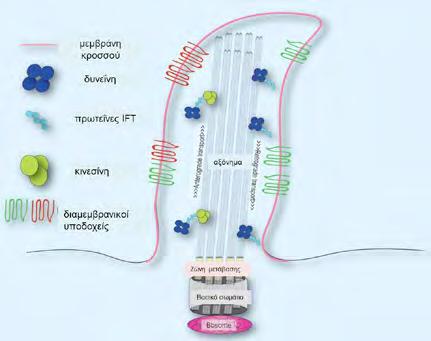
The ciliopathies comprise a heterogeneous group of disorders which are associated with mutations at genes coding for proteins for the formation or functionality of the primary or immotile cilia. Since these cilia are components of practically all cells, the ciliopathies present mainly with retinal degeneration, renal disease and brain anomalies and secondarily with hepatic fibrocystic disease, diabetes mellitus, obesity and skeletal dysplasias. More than 80 genes correspond to more than 20 distinct disorders, while many diseases sharing some of the above features may be classified as ciliopathies in the near future, when the cilia molecular mechanisms will be further clarified. The cilia mainly induce cell signals and thus facilitate the cellular paracrine activity. They also play major role in cell division and orientation through the participation in several developmental pathways. The study of ciliopathies aids the comprehension of the mechanisms of tumorigenesis, cysteogenesis, mental retardation and diabetes mellitus. The new diagnostic technologies such as Next Generation Sequencing have optimized the genetic research through the simultaneous study of several ciliopathetic genes. The dynamic development of Medical Genetics in this field provides a powerful tool in genetic diagnosis, counseling and management of the patients and their families.
Keywords: immotile cilia, retinal degeneration, renal disease, CNS malformations, heterogeneity
Stavroula Psoni
Helena Fryssira Medical Genetics National and Kapodistrian University of Athens, School of Medicine, “Aghia Sophia” Children’s Hospital, Athens
(Microtubule-Organizing-Center, MTOC) (13).
Συντομογραφίες
NGS: Next Generation
Sequencing
PCD: Primary Ciliary Dyskinesia
IFT: Intraflagellar Transport
MTOC: MicrotubuleOrganizing-Center
PCP: PlanarCellPolarity
Shh: Sonic-Hedgehog
Smo: Smoothened
PTCH1: Patched
Homologue 1
mTOR: mammalianTarget of Rapamycin
PC1: polycystin-1
BBS: Bardet-Biedl Syndrome
ADPKD: Autosomal
Dominant Polycystic Kidney Disease
ARPKD: Autosomal
Recessive Polycystic Kidney Disease
PC2: polycystin-2
ESRD: End-Stage Renal Disease
PKHD 1: Polycystic Kidney and Hepatic Disease 1
PCLD: Polycystic Liver Disease
vHL: von HippelLindau
TSC: Tuberous Sclerosis
NPH: nephronophthisis
MCKD: Medullary
Cystic Kidney Disease
JS: Joubert Syndrome
MTS: MolarTooth Sign
JSRD: Joubert Syndrome Related Disorders
OFD: Orofaciodigital Syndrome
MGS: Meckel-Gruber Syndrome
JATD: Jeune Asphyxiating Thoracic Dystrophy
EVC: Ellis-van Creveld Syndrome
SRPS: Short-Rib Polydactyly Syndrome
1. Μη κανονική
Wnt - Planar Cell Polarity (Non-Canonical Wnt-PCP):
(Planar Cell Polarity, PCP)
(22).
2. Κανονική οδός Wnt (Canonical Wnt pathway):
3. Οδός Sonic-Hedgehog (Shh)
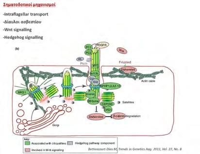
Gli (Gli1, Gli2, Gli3)(17) (Εικόνα 2).
4.
θηλαστικών (mammalian Target of Rapamycin)
Polycystic Kidney Disease, ADPKD
Autosomal Recessive Polycystic Kidney Disease,
CED: Cranioectodermal
Dysplasia
DDR: DNA Damage Response
PKHD1 (6p12.3p12.2)
PKD1 (16p13.3)
PKD2 (4q22.1)
PRKCSH (19p13.2) SEC (6q21)
VHL (3p25.3)
Polyductin/ fibrocystin
TSC1 (9q34.13)
TSC2 (16p13.3)
Polycystin-1 Polycystin-2
Hepatocystin SEC63
(Tuberous Sclerosis, TSC) (40). H
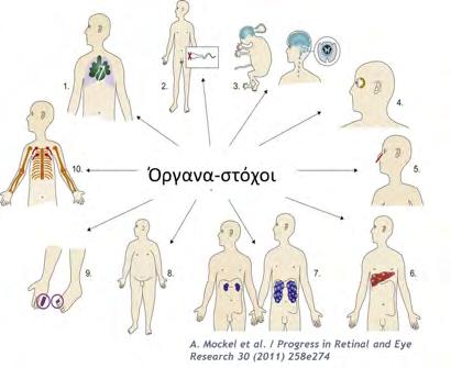
(NPH)
Συν. Senior-Løken (SLSN)
Ivemark) (45).
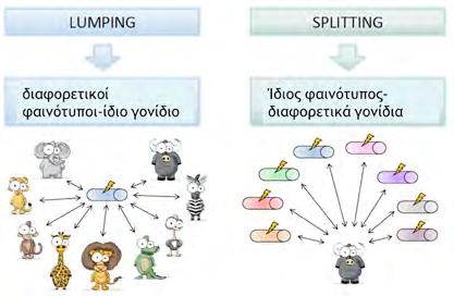
“lumping” και “splitting”. Από Enza Maria Valente, 5th Eur. Course in Clinical Dysmorphology, Rome 2013)
NPHP1 (2q13)
NPHP2/INVS (9q31.1)
NPHP3 (3q22.1)
NPHP4 (1p36.31)
NPHP5/IQCB1 (3q13.33)
NPHP6/CEP290 (12q21.2)
NPHP7/GLIS2 (16p13.3)
NPHP8/RPGRIP1L (16q12.2)
NPHP9/NEK8 (17q11.2)
NPHP10/SDCCAG8 (1q43)
NPHP11/TMEM67 (8q22.1)
NPHP12/TTC21B (2q24.3)
XPNPEP3/NPHPL1 (22q13.2)
NPHP1, INVS (NPHP2), NPHP3, NPHP4, IQCB1 (NPHP5), CEP290 (NPHP6), SDCAAG8 (NPHP10)
UMOD (16p12.3)
Nephrocystin-1 Inversin
Nephrocystin-3
Nephrocystin-4
IQCB1
CEP290 GLIS2
RPGRIP1L NEK8
SDCCAG8
Meckelin IFT139
XPNPEP3
Uromodulin/ Tamm-Horsfall protein
(Medullary Cystic Kidney Disease, MCKD)
συνεργιστικά (47).
Syndrome Related Disorders, JSRD) (40,51).
Συν. Bardet-Biedl (ΒΒS)
Συν. Alström
Συν. Ivemark
BBS1 (11q13.2)
BBS2 (16q12.2)
BBS3/ARL6 (3q11.2)
BBS4 (15q24.1)
BBS5 (2q31.1)
BBS6 /MKKS (20p12.2)
BBS7 (4q27)
BBS8/TTC8 (14q31.3)
BBS9 (7q14.3)
BBS10 (12q21.2)
BBS11/TRIM32 (9q33.1)
BBS12 (4q27)
BBS13/MKS1 (17q22)
BBS14/CEP290 (12q21.32)
BBS15/WDPCP (2p15)
BBS16/SDCCAG8 (1q43)
ALMS1 (2p13.1)
NPHP3 (3q22.1)
Nephrocystin-3
Συν. Joubert (JBTS)
(JSRD)
Συν. Meckel (MKS)
INPP5E/JBTS1 (9q34.3)
TMEM216/JBTS2 (11q12.2)
AHI1/JBTS3(6q23.3)
NPHP1/JBTS4 (2q13)
CEP290/JBTS5 (12q21.32)
TMEM67/JBTS6 (8q22.1)
RPGRIP1L/JBTS7 (16q12.2)
ARL13B/JBTS8 (3q11.1)
CC2D2A/JBTS9 (4p15.32)
OFD1/JBTS10 (Xp22.2)
KIF7 (15q26.1)
TCTN2 (12q24.31)
ATXN10 (22q13.31)
MKS1 (17q22)
TMEM216 /MKS2 (11q13)
TMEM67/MKS3 (8q22.1)
CEP290/MKS4 (12q21.32)
RPGRIP1L/MKS5 (16q12.2)
CC2D2A/MKS6 (4p15.32)
NPHP3(3q22.1)
TCTN2/MKS8 (12q24.31)
B9D1 (17p11.2)
INPP5E
TMEM216 Jouberin
Nephrocystin-1 CEP290 Meckelin
RPGRIP1L ARL13B CC2D2A OFD1 KIF7 Tectonic 2 Ataxin 10
MKS1
TMEM216 Meckelin CEP290 RPGRIP1L CC2D2A Nephrocystin-3 Tectonic 2 B9D1
Στοματοπροσωποδακτυλικό σύνδρομο 1 (Orofaciodigitals., OFD1)
Συν. βραχέων πλευρώνπολυδακτυλίας (shortribpolydactylys.) + Συν. Jeune/asphyxiating thoracic dystrophy, ATD)
Συν. Ellis-van Creveld (EVC)
Β9D2 (19q13.2)
OFD1 (Xp22.2)
IFT80 (3q25.33)
DYNC2H1 (11q22.3)
NEK1 (4q33)
TTC21B (2q24.3)
WDR35 (2p24.1)
IFT80/WDR56
DYNC2H1
NEK1
IFT1 39/THM1 IFT121/ WDR35
EVC (4p16.2)
EVC2 (4p16.2)
Kρανιοεκτοδερμική
(CED/Sensenbrenners.)
IFT122 (3q21.3)
WDR35 (2p24.1)
C14orf179 (14q24.3)
IFT122/ WDR10 IFT121/ WDR35 IFT43
δύο truncating “ισχυρών” μεταλλάξεων
γονίδια ΝPHP3, NPHP6/CEP290, NPHP8, NPHP11/ MKS3 προκαλούν MGS λόγω απώλειας πρωτεϊνικής λειτουργίας,
4.
1. Badano JL, Mitsuma N, Beales PL, Katsanis N. The ciliopathies: an emerging class of human genetic disorders. Annu Rev Genomics Hum Genet 2006;7:125-148
2. Waters AM, Beales PL. Ciliopathies: an expanding disease spectrum. Pediatr Nephrol 2011;26:1731-1737
3. Rosenbaum JL, Witman GB. Intraflagellar transport. Nat Rev Mol Cell Biol 2002;3:813-825
4. Salathe M. Regulation of mammalian ciliary beating. Annu Rev Physiol 2007;69:401-422
5. Knowles MR, Daniels LA, Davis SD, Zariwala MA, and Leigh MW. Primary ciliary dyskinesia: Recent advances in diagnostics, genetics, and characterizationof clinical disease. Am J Respir Crit Care Med 2013;188:913–922
6. Bettencourt-Dias M, Hildebrandt F, Pellman D, Woods G, Godinho SA. Centrosomes and
cilia in human disease. Trends Genet 2011; 27:307-315
7. Goetz SC, Anderson KV. The primary cilium: a signalling center during vertebrate development. Nat Rev Genet 2010;11:331-344
8. Hildenbrant F, Benzing T, Katsanis N. Ciliopathies. N Engl J Med 2011; 364: 1533-1543
9. Nigg EA, Ralf JW. Centrioles, centrosomes and cilia in health and disease. Cell 2009;139:663678
10. Ishikawa H, Marshall WF. Ciliogenesis: building the cell’s antenna. Nat Rev Mol Cell Biol 2011; 12:222-234
11. Carroll TJ, Yu J. The kidney and planar cell polarity. Curr Top Dev Biol 2012; 101:185-212
12. Pedersen LB, Rosenbaum JL. Intraflagellar transport (IFT) role in ciliary assembly, resorption and signalling. Curr Top Dev Biol 2008; 85:23-61
13. Kobayashi T, Dynlacht BD. Regulating the transition from centriole to basal body. J Cell Biol 2011; 193:435-444
14. Rosenbaum JL, Witman GB. Intraflagellar transport. Nat Rev Mol Cell Biol 2002; 3:813825
15. Singla V, Reiter JF. The primary cilium as the cell’s antenna; signaling at a sensory organelle. Science 2006; 313; 629-633
16. Ross AJ, May-Simera H, Eichers ER, Kai M, Hill J, Jagger DJ et al. Disruption of Bardet-Biedl syndrome ciliary proteins perturbs planar cell polarity in vertebrates. Nat Genet 2005;37;1135-1140
17. Ingham PW, McMahon AP. Hedgehog signaling in animal development: paradigms and principles.Genes Dev. 2001; 15:3059–3087
18. Louvi A, Artavanis-Tsakonas S. Notch signalling in vertebrate neural development. Nat Rev Neurosci 2006 Feb;7:93-102
19. Li WX. Canonical and non-canonical JAK-STAT signaling. Trends Cell Biol 2008 Nov;18:545-551
20. Hassan B, Akcakanat A, Holder AM, Meric-Bernstam F. Targeting the PI3-kinase/Akt/ mTOR signaling pathway. Surg Oncol Clin N Am 2013 Oct;22:641-664
21. Wang Z, Ahmad A, Li Y, Kong D, Azmi AS, Banerjee S et al. Emerging roles of PDGFD signaling pathway in tumor development and progression. Biochim Biophys Acta 2010 Aug;1806:122-130
22. Goggolidou P. Wnt and planar cell polarity signaling in cystic renal disease. Organogenesis 2014 Jan 1;10:86-95
23. Wei Q, Zhang Y, Li Y, Zhang Q, Ling K, Hu J. The BBSome controls IFT assembly and turnaround in cilia. Nat Cell Biol 2012 Sep;14(9):950-957
24. Milenkovic L, Scott MP, Rohatdi R. Lateral transport of Smoothened from the plasma membrane to the membrane of the cilium. J Cell Biol; 187:365-374
25. Huber TB, Walz G, Kuehn EW. mTOR and rapamycin in the kidney: signaling and therapeutic implications beyond immunosuppression. Kidney Int 2011 Mar;79:502-11
26. Beales PL, Elcioglu N, Woolf AS, Parker D, Flinter FA. New criteria for improved diagnosis of Bardet-Biedl syndrome: results of a population survey. J Med Genet 1999;6: 437-446
27. Xing DJ, Zhang HX, Huang N, Wu KC, Huang XF, Huang F et al. Comprehensive molecular diagnosis of Bardet-Biedl syndrome by high-throughput targeted exome sequencing. PLoS One 2014 Mar 7;9:e90599
28. Zaki MS, Sattar S, Massoudi RA, Gleeson JG.. Co-occurrence of distinct ciliopathy diseases in single families suggests genetic modifiers. Am J Med Genet Part A 2011;155:3042–3049
29. Baker K, Beales PL. Making sense of cilia in disease: the human ciliopathies. Am J Med Genet Part C (Seminars in Medical Genetics) 2009;151C:281-295
30. Gascue C, Katsanis N, Badano JL. Cystic diseases of the kidney: ciliary dysfunction and cystogenic mechanisms. Pediatr Nephrol 2011;26: 1181-1195
31. Steinman TI. Polycystic kidney disease: a 2011 update. Curr Opin Nephrol Hypertens. 2012;21:189-194
32. Bae KT, Zhu F, Chapman AB, Torres VE, Grantham JJ, Guay-Woodford LM et al. Magnetic resonance imaging evaluation of hepatic cysts in early autosomal-dominant polycystic kidney disease: the Consortium for Radiologic Imaging Studies of Polycystic Kidney Disease cohort.
Clin J Am Soc Nephrol 2006;1:64-69
33. Kang YR, Ahn JH, Kim KH, Choi YM, Choi J, Park JR. Multiple cardiovascular manifestations in a patient with autosomal dominant polycystic kidney disease. J Cardiovasc Ultrasound 2014;22:144-147
34. Pirson Y. Extrarenal manifestations of autosomal dominant polycystic kidney disease. Adv Chronic Kidney Dis 2010;17:173-180
35. Torres VE, Harris PC. Autosomal dominant polycystic kidney disease: the last 3 years. Kidney Int 2009;76:149-168
36. Harris PC, Bae KT, Rosetti S, Torres VE, Grantham JJ, Chapman AB et al. Cyst number but not the rate of cystic growth is associated with the mutated gene in autosomal dominant polycystic kidney disease. J Am Soc Nephrol 2006;17:3013-3019
37. Ward CJ, Hogan MC, Rossetti S, Walker D, Sneddon T, Wang X et al. The gene mutated in autosomal recessive polycystic kidney disease encodes a large, receptor-like protein. Nat Genet 2002;30:259-269
38. Turkbey B, Ocak I, Daryanani K, Font-Montgomery E, Lukose L, Bryant J et al. Autosomal recessive polycystic kidney disease and congenital hepatic fibrosis (ARPKD/CHF). Pediatr Radiol 2009 ;39:100-111
39. Wang S, Luo Y, Wilson PD, Witman GB, Zhou J. The autosomal recessive polycystic kidney disease protein is localized to primary cilia, with concentration in the basal body area. J Am Soc Nephrol 2004 Mar;15:592-602
40. Bergmann C. Educational paper: Ciliopathies. Eur J Pediatr 2012;171:1285-1300
41. Cnossen WR, Drenth JP. Polycystic liver disease: an overview of pathogenesis, clinical manifestations and management. Orphanet J Rare Dis 2014;9:69
42. Chou A, Toon C, Pickett J, Gill AJ. von Hippel-Lindau syndrome. Front Horm Res 2013;41:30-49
43. Crino PB, Nathanson KL, Henske EP. The tuberous sclerosis complex. N Engl J Med 2006;355:1345-1356
44. Hildenbradt F, Zhou W. Nephronopthisis-associated ciliopathies. J Am Soc Nephrol 2007;18:1855-1871
45. Simons M, Walz G. Polycystic kidney disease: cell division without a c(l)ue? Kidney Int 2006;70:854-864
46. Zaghloul NA, Katsanis N. Mechanistic insights into Bardet-Biedl syndrome, a model ciliopathy. J Clin Invest 2009;119: 428-437
47. Leitch C, Zaghloul NA, Davis EE, Stoetzel C, Diaz-Font A, Rix S et al. Hypomorphic mutations in syndromic encephalocele genes are associated with Bardet-Biedl syndrome. Nat Genet 2008;40:443-448
48. Marshall JD, Beck S, Maffei P, Naggert JK. Alström syndrome. Eur J Hum Genet 2007;15:1193-1202
49. Girard D1, Petrovsky N. Alström syndrome: insights into the pathogenesis of metabolic disorders. Nat Rev Endocrinol 2011;7:77-88
50. Parisi MA. Clinical and molecular features of Joubert Syndrome and related disorders. Am J Med Genet C Semin Med Genet 2009;151C:326-340
51. Brancati F, Dallapiccola B, Valente EM. Joubert Syndrome and related disorders. Orphanet J Rare Dis 2010;5:20
52. Coppieters F, Lefever S, Leroy BP, De Baere E. CEP290, a gene with many faces: mutation overview and presentation of CEP290base. Hum Mutat 2010;31:1097-1108
53. Bergmann C, Fliegauf M, Brüchle NO, Frank V, Olbrich H, Kirschner J et al. Loss of nephrocystin-3 function can cause embryonic lethality, Meckel-Gruber-like syndrome, situs inversus, and renal-hepatic-pancreatic dysplasia. Am J Hum Genet 2008 Apr;82:959-970
54. Beales PL, Bland E, Tobin JL, Baccelli C, Tuysuz B, Hill J et al. IFT80 which encodes a consrved intraflagellar transport protein, is mutated in Jeune asphyxiating thoracic dystrophy. Nat Genet 2007;39:727-729
55. Brugmann SA, Cordero DR, Helms JA. Craniofacial ciliopathies: A new classification for craniofacial disorders. Am J Med Genet Part A 2010;152A:2995-3006
56. Galdzicka M, Patnala S, Hirshman MG, Cai JF, Nitowsky H, Egeland JA et al. A new gene
EVC2, is mutated in Ellis-van-Creveld syndrome. Mol Genet Metab 2002;77:291-295
57. Elcioglu NH, Hall CM. Diagnostic dilemmas in the short-rib-polydactyly syndrome group. Am J Med Genet 2002;111:392-400
58. Konstantinidou AE, Fryssira H, Sifakis S, Karadimas C, Kaminopetros P, Agrogiannis G et al. Cranioectodermal dysplasia: A probable ciliopathy. Am J Med Genet Part A 2009;149A:22062211
59. Hoffer JL, Fryssira H, Konstantinidou AE, Ropers HH, Tzschach A. Novel WDR35 mutations in patients with cranioectodermal dysplasia (Sensenbrenner syndrome). Clin Genet 2013;83:92-95
60. Fry AM, Leaper MJ, Bayliss R. The primary cilium: guardian of organ development and homeostasis. Organogenesis 2014;10:62-68
61. Wahl PR, Serra AL, Le Hir M, Molle KD, Hall MN, Wüthrich RP. Inhibition of mTOR with sirolimus slows disease progression in Han:SPRD rats with autosomal dominant polycystic kidney disease (ADPKD). Nephrol Dial Transplant 2006 Mar;21:598-604
62. Bukanov NO, Moreno SE, Natoli TA, Rogers KA, Smith LA, Ledbetter SR et al. CDK inhibitors R-roscovitine and S-CR8 effectively block renal and hepatic cystogenesis in an orthologous model of ADPKD. Cell Cycle 2012;11:4040-4046
63. McIntyre JC, Williams CL, Martens JR. Smelling the roses and seeing the light: gene therapy for ciliopathies. Trends Biotechnol 2013;31:355-363
Correspondence
Maria Tsigga
Sindos, GR57400, Thessaloniki
e-mail: mtsigga@nutr. teithe.gr
Prevalence of simple and abdominal obesity among 15 year old adolescents in Greece: results from the
ADONUT
study
Maria Grammatikopoulou, Dimitrios Poulimeneas, Konstantina Gerothanasi, Efstratios Kiranas, Maria Tsigga & ADONUT Study Group
ADONUT Study Group: Calliope Aberiadou, EfrosyniAlexopoulou, DemetraApostolou, IoannaBozouri, Stella Chortiatinou, Olympia-Brenda Christodoulou, Maria Dalakoglou, PelagiaDamianou, Anastasia Daviti, Leonidas Dimitrakopoulos, KyriakiDimitriou-Kouspou, NikolaosDrougkas, AikateriniEfstathiadi, KonstantinosFamisis, Irene Foukou, Hermione Hatziaggelousi, DimitriosHatzipetrou, ArgyroHatzivasileiou, DiamantoulaHoremi, Thessalia-VaiaHroni, IoannisIdreos, Sofia Thanasogeorgou, Irene Theodoridou, Konstantina Theohari, IoannisKafantaris, AimiliaKakali, Marina Kalli, MeropiKalli, KonstantinosKalpakis, Christina Karabaliou, PanagiotaKarabela, AikateriniKaragkiozaki, Maria Karaventza, Georgia Kazasi, Maria Kiokaki, PolykseniKokka, Nikolaos Kokkinos, StamatiaKonstanta, EleniKotseridi, AhmetKourou, FotiniKsenaki, Anna Kyrgiou, Smaro-Maria Lazoglou, EleniMakantasi, ElianaMameka, Olga Maridaki, AikateriniMouratidou, Artemis Niarchou, Kyriaki Papa, Sofia Papagiannidou, KonstantinosPapathanasiou, AikateriniPappa, Charoula Pavli, AidonitsaPavlidi, Magdalene Pipeki, Athena Pourlioti, CharaPsachoula, Maria Psalidakou, Vera Psomopoulou, EleniRizou, EleniRomba, GeorgiosSbyrakis, SpyridoulaSoulaidopoulou, Maria Spanoudaki, AikateriniTriantafyllidou, VasilikiTriantafyllopoulou, Anastasia Troumpouki, KonstantiniaTsami, Georgia Tserepi, MarilenaTsivgouli, DespoinaValaora, Maria Verykokkou, Anna Vlahou, Anastasia Voulgaridou and Maria Zymara
Abstract
Background: Adolescent health is of cardinal importance, as a plethora of adulthood diseases are actually consolidated during adolescence. The aim of this study is to assess the prevalence of simple and abdominal obesity among 15-year-old Greek adolescents.
Methods: A nationally representative sample of 7028 adolescents, aged 12-19 years old, was recruited from schools throughout the country during 2010-2012. Body weight, stature and waist circumference were measured. The prevalence of each weight category was defined according to the International Obesity Task Force criteria and abdominal obesity was diagnosed according to the International Diabetes Federation.
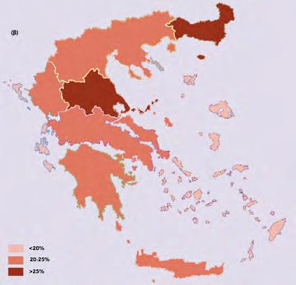
Results: The majority of the participants were normoweight (66.1%), 22.1% were overweight, 7.3% obese, and the remaining 4.6% were underweight. Overweight including obesity reached 35% among boys and 23.6% among girls, with increased prevalence in Thrace and Thessaly. High prevalence of underweight was observed in Epirus. In the total sample 9.5% was diagnosed with abdominal obesity, including 10.1% of the girls and 9.0% of the boys. Central obesity reached Ό of the population among girls in Thrace and Thessaly.
Maria Grammatikopoulou
Dimitrios Poulimeneas
Konstantina Gerothanasi
Efstratios Kiranas
Maria Tsigga
Department of Nutrition & Dietetics, Alexander
Technological Educational Institute, Thessaloniki
Conclusions: Differences are observed in the weight status tiers between different geographical regions of the country, among 15-year-old adolescents. Spatial analysis of the data provide a better approach in highlighting areas in need of intervention.
Key words: Overweight, obese, waist circumference, Greek, adolescents
GmbH
(14)
με το International Diabetes Federation (IDF) (15).
PASW Statistics 18 (SPSS Inc., HongKong).
λιποβαρή (3.8%) (PR:0.7, CI:0.6-0.9,p≤0.001) ή
(6.1%) (ΡR:0.9, CI:0.80.9,p≤0.001)
(26.2%) (PR:1.5, CI:1.4-1.6, p≤0.001) ή
(8.8%) (PR:1.5, CI:1.3-1.8, p≤0.001) συγκριτικά
(PR:0.8, CI:0.6-0.9, p≤0.002),
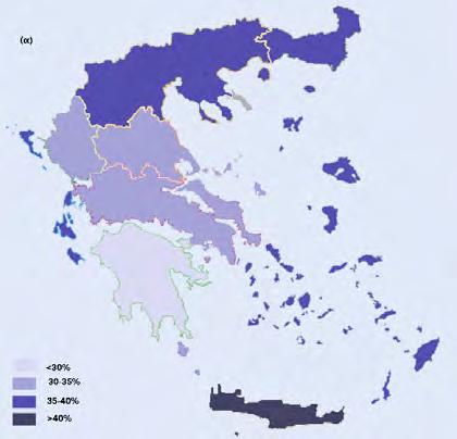
CI: Confidence Intervals
Βουλγαρία Ιρλανδία
Κύπρος
Νορβηγία
1. Patton GC, Coffey C, Cappa C, Currie D, Riley L, Gore F, et al. Health of the world’s adolescents: a synthesis of internationally comparable data. Lancet 2012;379:1665-1675.
2. Bibbins-Domingo K, Coxson P, Pletcher MJ, Lightwood J, Goldman L. Adolescent Overweight and Future Adult Coronary Heart Disease. NEngl J Med 2007;357:2371-2379.
3. Dietz DW. Health Consequences of Obesity in Youth: Childhood Predictors of Adult Disease. Pediatrics 1998;101(S2):518-525.
4. Tzotzas T, Kapantais E, Tziomalos K, Ioannidis I, Mortoglou A, Bakatselos S, et al. Epidemiological survey for the prevalence of overweight and abdominal obesity in Greek adolescents. Obesity (Silver Spring) 2008;16:1718–1722.
5. Grammatikopoulou MG, Poulimeneas D, Gounitsioti IS, et al. Prevalence of Simple and Abdominal Obesity in Greek Adolescents: The ADONUT Study. ClinObes 2014;4:303-308.
6. Lissau I, Overpeck MD, Ruan WJ, Due P, Holstein B, Hediger M, et al. Body mass index and overweight in adolescents in 13 European countries, Israel and the United States. Arch PediatrAdolesc Med 2004;158:27–33.
7. Velasquez-Mieyer P, Perez-Faustinelli S, Cowan PA. Identifying Children at Risk for Obesity, Type 2 Diabetes, and Cardiovascular Disease. Diabetes Spectr 2005;18:213-220.
8. Field AE, Cook NR, Gillman MW. Weight status in childhood as a predictor of becoming overweight or hypertensive in early adulthood. Obes Res 2005;13:163-169.
9. Hulens M, Beuven G, Claessens AL, Lefevre J, Thomis M, Philippaerts R, et al. Trends in BMI among Belgian children, adolescents and adults from 1969 to 1996. Int J Obes 2001;25:395399.
10. Cole TJ, Bellizzi MC, Flegal KM, Dietz WH. Establishing a standard definition for child overweight and obesity worldwide: international survey. BMJ2000;320:1240–1243.
11. Cole TJ, Flegal KM, Nicholls D, Jackson AA. Body mass index cut offs to define thinness in children and adolescents: international survey. BMJ2007;335:194.
12. Lien N, Henriksen HB, Nymoen LL, Wind M, Klepp KI. Availability of data assessing the prevalence and trends of overweight and obesity among European adolescents. Public Health Nutr2010;13:1680–1687.
13. National Heart, Lung, and Blood Institute’s (NHLBI) Obesity Education Initiative. The Practical Guide. Λ 1 Identification, Evaluation, and Treatment of Overweight and Obesity in Adults. North American Association for the Study of Obesity. NIH, 2000. [WWW document]. URL http://www.nhlbi.nih.gov/guidelines/obesity/prctgd_c.pdf (accessed July 16, 2015).
14. Fernandez JR, Redden D, Pietrobelli A,Allison DB. Waist circumference percentiles in na-
tionally representative samples of African-American, European-American, and Mexican-American children and adolescents. J Pediatr2004;145:439–444.
15. International Diabetes Federation. IDF consensus definition of metabolic syndrome in children and adolescents. International Diabetes Federation: Brussels, 2007.
16. McNutt L-A, Wu C, Xue X, Hafner. Estimating the Relative Risk in Cohort Studies and Clinical Trials of Common Outcomes. Am J Epidemiol 2003;157:940-943.
17. Santos CA, Fiaccone RL, Oliveira NF, Cunha S, Barreto ML, Carmo MB, et al. Estimating adjusted prevalence ratio in clustered cross-sectional epidemiological data. BMC Med Res Methodol 2008;8:80.
18. Karayiannis D, Yannakoulia Μ, Terzidou Μ, Sidossis LS, Kokkevi A. Prevalence of Overweight and Obesity in Greek School-Aged Children and Adolescents. Eur J ClinNutr 2003;57:1189-1192.
19. Kosti RI, Panagiotakos DB, Mihas CC, Alevizos A, Zampelas A, Mariolis A, et al. Dietary Habits. Physical Activity and Prevalence of Overweight/Obesity among Adolescents in Greece: The Vyronas Study. Med SciMonit 2007;13:CR437-444.
20. Ahluwalia N, Dalmasso P, Rasmussen M, Lipsky L, Currie C, Haug E, et al. Trends in Overweight Prevalence among 11-, 13- and 15-Year-Olds in 25 Countries in Europe, Canada and USA from 2002 to 2010. Eur J Public Health 2015;25(S2):28-32.
21. Tsartsali PK, Thompson JL, Jago R. Increased knowledge predicts greater adherence to the Mediterranean diet in Greek adolescents. Public Health Nutr 2009;12:208-213.
22. Mladenova S, Andreenko E. Prevalence of underweight, overweight, general and central obesity among 8-15- years old Bulgarian children and adolescents (Smolyan region, 2012-2014). NutrHosp 2015;31:2419-2427.
23. Whelton H, Harrington J, Crowley E, Kelleher V, Cronin M, Perry IJ. Prevalence of overweight and obesity on the island of Ireland: results from the North South Survey of Children’s Height, Weight and Body Mass Index, 2002. BMC Public Health2007;7:187.
24. Artero EG, Espaρa-Romero V, Ortega FB, Jiménez-Pavón D, Ruiz JR, Vicente-Rodríguez G, et al. Health-related fitness in adolescents: underweight, and not only overweight, as an influencing factor. The AVENA study. Scand J Med Sci Sports2010;20:418-427.
25. Vieno A, Santinello M, Martini MC. Epidemiology of overweight and obesity among Italian early adolescents: relation with physical activity and sedentary behaviour. EpidemiolPsichiatrSoc 2005;14:100-7.
26. Lazzeri G, Rossi S, Pammolli A, Pilato V, Pozzi T, Giacchi MV. Underweight and overweight among children and adolescents in Tuscany (Italy). Prevalence and short-term trends. J Prev Med Hyg 2008;49:13-21.
27. Savva SC, Kourides Y, Tornaritis M, Epiphaniou-Savva M, Chadjigeorgiou C, Kafatos A. Obesity in children and adolescents in Cyprus. Prevalence and predisposing factors. Int J Obes 2002;26:1036–1045.
28. Λαβίθη
2007.
29. Jϊlνusson PB, Eide GE, Roelants M, Waaler PE, Hauspie R, Bjerknes R. Overweight and obesity in Norwegian children: prevalence and socio-demographic risk factors.Acta Paediatr 2010;99:900-905.
30. Van den HurkΚ, Van DommelenP, Van Buuren S, Verkerk PH, HiraSing RA. Prevalence of overweight and obesity in the Netherlands in 2003 compared to 1980 and 1997. Arch Dis Child 2007;92:992–995.
31. Schønbeck Y, Talma H, van Dommelen P, Bakker B, Buitendijk S, HiraSing RA, et al. Increase in prevalence of overweight in Dutch children and adolescents: a comparison of nationwide growth studies in 1980, 1997 and 2009.PLoS One 2011;6:e27608.
32. Kantanista A, Osinμski W. Underweight in 14 to 16 year-old girls and boys: prevalence and associations with physical activity and sedentary activities. Ann Agric Environ Med 2014;21:114–119.
33. Jodkowska M, Oblacinska A, Tabak I. Overweight and obesity among adolescents in Poland: gender and regional differences. Public Health Nutr 2010;3:1688–1692.
34. Sardinha LB, Santos R, Vale S, Silva AM, Ferreira JP, Raimundo AM, et al. Prevalence of overweight and obesity among Portuguese youth: A study in a representative sample of 10–18-year-old children and adolescents. Int J PediatrObes2011;6:e124-128.
35. Santos R, Aires L, Santos P, Ribeiro JC, Mota J.Prevalence of overweight and obesity in a Portuguese sample of adults: results from the Azorean Physical Activity and Health Study. Am J Hum Biol 2008;20:78-85.
36. Öner N, Vatansever Ü, Sari A, Ekuklu G, Gόzel A, Karasalihoglu S, Boris NW. Prevalence of underweight, overweight and obesity in Turkish adolescents. Swiss Med Wkly 2004;134:529–533.
37. Ojala K, Tynjälä J, Välimaa R, Villberg J, Kannas L. Overweight Adolescents’ Self-Perceived Weight and Weight Control Behaviour: HBSC Study in Finland 1994–2010. J Obes. 2012; 2012:180176.
38. Baratta R, Degano C, Leonardi D, Vigneri R, Frittitta L. High prevalence of overweight and obesity in 11e15-year-old children from Sicily. NutrMetabCardiovasc Dis 2006;16:249-255.
39. Currie C, Zanotti C, Morgan A, Currie D, de Looze M, Roberts C, et al, eds. Social determinants of health and well-being among young people: Health Behaviour in School-aged Children (HBSC) study: international report from the 2009/2010 survey. Copenhagen, WHO Regional Office for Europe, 2012 (Health Policy for Children and Adolescents, No. 6) (http://www.euro. who.int/__data/assets/pdf_file/0003/163857/Social-determinants-of-health-and-well-beingamong-young-people.pdf, accessed 21 September 2015).
40. Vaezghasemi M, Lindkvist M, Ivarsson A,Eurenius E. Overweight and lifestyle among 13−15 year olds: A cross-sectional study in northern Sweden. Scand J Public Health2012;40:221–228.
41. Tanner JM. Growth at Adolescence, 2nd ed. Oxford, Blackwell Scientific Publishers, 1962. 42. de Gouw L, Klepp K-I, Vignerovα J, Lien N, Steenhuis IH, Wind M, et al. Associations between diet and (in)activity behaviours with overweight and obesity among 10 to 18 year old Czech Republic adolescents. Public Health Nutr2010;13:1701–1707.
43. de Moraes AC, Fadoni RP, Ricardi LM, Souza TC, Rosaneli CF, Nakashima AT, et al. Prevalence of abdominal obesity in adolescents: a systematic review. ObesRev 2011;12:69–77.
44. Ekelund U, Anderssen S, Andersen LB, Riddoch CJ, Sardinha LB, Luan J, et al. Prevalence and correlates of the metabolic syndrome in a population-based sample of European youth. Am J Clin Nutr 2009;89:90-96.
45. Hakanen M, Lagstrøm H, Pahkala K, Sillanmäki L, Saarinen M, Niinikoski H, et al. Dietary and lifestyle counseling reduces the clustering of overweight- related cardiometabolic risk factors in adolescents. Acta Paediatr 2010;99:888–895.
(PosthocanalysesSSvs. H, SIRSvs. H: p<0.001).
(PosthocanalysesSSvs. H, SIRSvs. H: p<0.004).
e-mail: efi.tavladaki@ gmail.com Τ.
Correspondence
Theonymfi Tavladaki
Anapafseos 32, Agios Nikolaos, Crete, ΤΚ 72100 e-mail: efi.tavladaki@ gmail.com Τ. +306945553102
Early changes of amino acids in critical ill children dependent on disease severity
Theonymfi Tavladaki, Anna Maria Spanaki, Helen Dimitriou, EvaggelosPapakonstantinou, George Briassoulis
Abstract
Background: Since the metabolism of amino acids in critically ill patients remains subject of extensive research over the last decade, 22 amino acid profile investigated in children with severe sepsis(SS) or severe trauma with systemic inflammatory response syndrome (SIRS)during the early phase of stress compared with their corresponding levels in normal children serum. Also we tried to consider the direction of this mobility (upward, downward) and to correlate with clinical parameters, as expressed by the severity of disease or inflammatory markers (CRP).
Materials-Methods: The study included thirty-five critically ill patients aged ≤ 18years, of which 15 met the criteria for severe sepsis and 20 criteria for SIRS.These patients were compared with 27 healthy children served as controls. Serum samples from patients were collected in the first 24-hour introduction to the intensive care unit
Results: The metabolic spectrum of amino acids significantly altered in patients with sepsis. Methionine levels in patients’ plasma were significantly lower than in healthy children (Post hoc analyses SS vs. H, SIRS vs. H: p <0.001). Taurine was significantly higher in PICU patients than in healthy children (Post hoc analyses SS vs. H, SIRS vs. H: p <0.004). In SS or SIRS patients, negative correlations were recorded between amino acid levels and severity of disease markers (p <0,05); Glutamine was negatively correlated with not only APACHE II but also with CRP (p <0,05).
Conclusions: Αmino acids show significant variations depending on the severity of the disease and the inflammatory response to stress. Acute stress dietary interventions with mixtures of amino acids in ICU patients cannot be correctly predicted and can negatively influence metabolism.
Theonymfi Tavladaki
Anna Maria Spanaki
George Briassoulis
Paediatric Intensive Care Unit (PICU) ,University Hospital of Crete
Helen Dimitriou
Hematology and Oncology Laboratory, University of Crete, Medical School
EvaggelosPapakonstantinou Neo Lab , Αthens
159,38±56,53
139,11±65,09
125,61±31,38
48,34±24,42 11,00±10,66
45,57±20,11
152,42±59,98
35,53±16,67
62,50±31,83
65,69±12,45
189,95±95,67
19,31±9,14
11,05±4,71
244,95±112,48
90,07±21,00
34,32±7,40
125,77±50,95
70,43±22,66
15,03±4,44
36,69±11,35
49,73±24,74
236,73±55,35
136,89±49,59
138,84±48,65
118,73±34,89
73,52±20,68
7,78±5,96
50,31±16,87
131,47±47,30
37,23±14,30
54,94±22,17
68,68±17,63
96,73±78,73
16,31±7,23
9,02±4,50
132,07±84,72
86,17±41,37
33,58±16,38
154,40±52,55
48,54±11,37
9,24±3,65
30,87±10,70
34,18±16,90
264,18±43,46
168,14±72,13
120,76±55,69
113,60±41,25
62,88±33,69 10±3,34
54,00±24,74
112,80±40,29
32,78±14,76
54,93±14,45
76,65±23,04
157,77±99,50
16,82±8,26
8,30±2,84
169,08±101,39
83,29±26,14
32,59±9,77
144,25±86,87
54,26±23,61
11,48±4,00
42,02±9,25
42,69±22,37
227,73±37,07 0,292 0,672
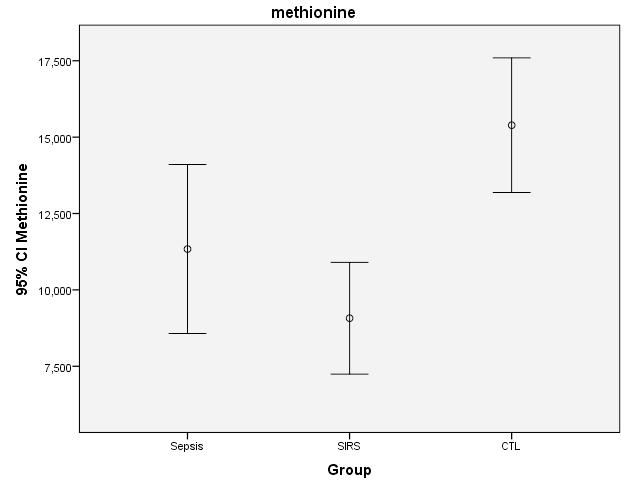
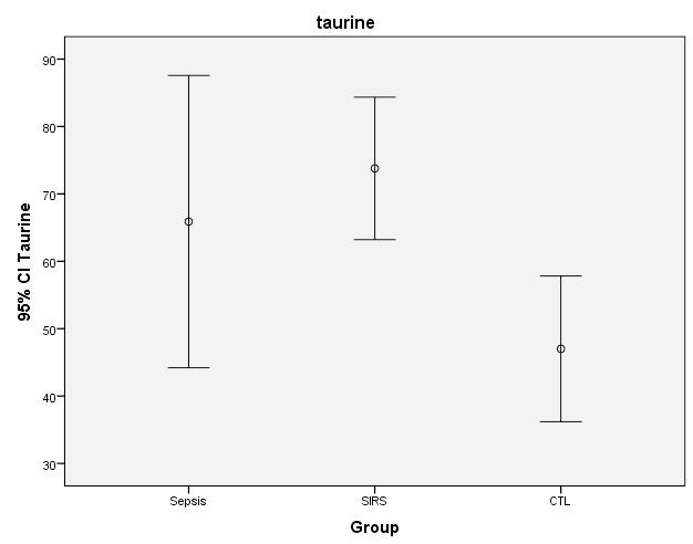
Chiarla
Mechteld A. R. Vermeulen et al (18)
Tomoya Hirose et al (9)
Βιβλιογραφία
1. Heyland DK, Novak F, Drover JW, Jain M, Su X, Suchner U. Should immunonutrition become routine in critically ill patients? A systematic review of the evidence. JAMA. 2001 Aug 22;286(8):944–53.
2. Dhaliwal R, Heyland DK. Nutrition and infection in the intensive care unit: what does the evidence show? Curr Opin Crit Care. 2005 Oct;11(5):461–7.
3. Casaer MP, Mesotten D, Hermans G, Wouters PJ, Schetz M, Meyfroidt G, et al. Early versus late parenteral nutrition in critically ill adults. N Engl J Med. 2011 Aug 11;365(6):506–17.
4. Heidegger CP, Berger MM, Graf S, Zingg W, Darmon P, Costanza MC, et al. Optimisation of energy provision with supplemental parenteral nutrition in critically ill patients: a randomised controlled clinical trial. Lancet. 2013 Feb 2;381(9864):385–93.
5. Weijs PJM, Wischmeyer PE. Optimizing energy and protein balance in the ICU. Curr Opin Clin Nutr Metab Care. 2013 Mar;16(2):194–201.
6. Askanazi J, Furst P, Michelsen CB, Elwyn DH, Vinnars E, Gump FE, et al. Muscle and plasma amino acids after injury: hypocaloric glucose vs. amino acid infusion. Ann Surg. 1980 Apr;191(4):465–72.
7. Chiarla C, Giovannini I, Siegel JH, Boldrini G, Castagneto M. The relationship between plasma taurine and other amino acid levels in human sepsis. J Nutr. 2000 Sep;130(9):2222–7.
8. Vente JP, von Meyenfeldt MF, van Eijk HM, van Berlo CL, Gouma DJ, van der Linden CJ, et al. Plasma-amino acid profiles in sepsis and stress. Ann Surg. 1989 Jan;209(1):57–62.
9. Hirose T, Shimizu K, Ogura H, Tasaki O, Hamasaki T, Yamano S, et al. Altered balance of the aminogram in patients with sepsis - the relation to mortality. Clin Nutr Edinb Scotl. 2014 Feb;33(1):179–82.
10. Druml W, Heinzel G, Kleinberger G. Amino acid kinetics in patients with sepsis. Am J Clin Nutr. 2001 May;73(5):908–13.
11. De Bandt J-P, Cynober L. Therapeutic use of branched-chain amino acids in burn, trauma, and sepsis. J Nutr. 2006 Jan;136(1 Suppl):308S – 13S.
12. Wang H, McNeil YR, Yeo TW, Anstey NM. Simultaneous determination of multiple amino acids in plasma in critical illness by high performance liquid chromatography with ultraviolet and
fluorescence detection. J Chromatogr B Analyt Technol Biomed Life Sci. 2013 Dec 1;940:53–8.
13. Tomiyasu M, Aida N, Watanabe Y, Mori K, Endo K, Kusakiri K, et al. Monitoring the brain metabolites of children with acute encephalopathy caused by the H1N1 virus responsible for the 2009 influenza pandemic: a quantitative in vivo 1H MR spectroscopy study. Magn Reson Imaging. 2012 Dec;30(10):1527–33.
14. Schaffer SW, Jong CJ, Ito T, Azuma J. Effect of taurine on ischemia-reperfusion injury. Amino Acids. 2014 Jan;46(1):21–30.
15. Marcinkiewicz J, Kontny E. Taurine and inflammatory diseases. Amino Acids. 2014 Jan;46(1):7–20.
16. Paauw JD, Davis AT. Taurine concentrations in serum of critically injured patients and ageand sex-matched healthy control subjects. Am J Clin Nutr. 1990 Oct;52(4):657–60.
17. Boelens PG, Houdijk APJ, de Thouars HN, Teerlink T, van Engeland MIA, Haarman HJTM, et al. Plasma taurine concentrations increase after enteral glutamine supplementation in trauma patients and stressed rats. Am J Clin Nutr. 2003 Jan;77(1):250–6.
18. Vermeulen MAR, van Stijn MFM, Visser M, Lemmens SMP, Houdijk APJ, van Leeuwen PAM, et al. Taurine Concentrations Decrease in Critically Ill Patients With Shock Given Enteral Nutrition. JPEN J Parenter Enteral Nutr. 2015 Jan 13;
19. Angus DC, Linde-Zwirble WT, Lidicker J, Clermont G, Carcillo J, Pinsky MR. Epidemiology of severe sepsis in the United States: analysis of incidence, outcome, and associated costs of care. Crit Care Med. 2001 Jul;29(7):1303–10.
20. Watson RS, Carcillo JA, Linde-Zwirble WT, Clermont G, Lidicker J, Angus DC. The epidemiology of severe sepsis in children in the United States. Am J Respir Crit Care Med. 2003 Mar 1;167(5):695–701.
21. Wheeler DS, Wong HR, Zingarelli B. Pediatric Sepsis - Part I: “Children are not small adults!.” Open Inflamm J. 2011 Oct 7;4:4–15.
22. Su L, Li H, Xie A, Liu D, Rao W, Lan L, et al. Dynamic changes in amino Acid concentration profiles in patients with sepsis. PloS One. 2015;10(4):e0121933.
23. Sax HC, Hasselgren PO, Talamini MA, Edwards LL, Fischer JE. Amino acid uptake in isolated, perfused liver: effect of trauma and sepsis. J Surg Res. 1988 Jul;45(1):50–5.
24. Semmler A, Smulders Y, Struys E, Smith D, Moskau S, Blom H, et al. Methionine metabolism in an animal model of sepsis. Clin Chem Lab Med CCLM FESCC. 2008;46(10):1398–402.
25. Dever JT, Elfarra AA. The biochemical and toxicological significance of hypermethionemia: new insights and clinical relevance. Expert Opin Drug Metab Toxicol. 2010 Nov;6(11):1333–46.
26. Zhang Q, Raoof M, Chen Y, Sumi Y, Sursal T, Junger W, et al. Circulating mitochondrial DAMPs cause inflammatory responses to injury. Nature. 2010 Mar 4;464(7285):104–7.
27. Bone RC, Balk RA, Cerra FB, Dellinger RP, Fein AM, Knaus WA, et al. Definitions for sepsis and organ failure and guidelines for the use of innovative therapies in sepsis. The ACCP/ SCCM Consensus Conference Committee. American College of Chest Physicians/Society of Critical Care Medicine. 1992. Chest. 2009 Nov;136(5 Suppl):e28.
28. Fujiwara M, Ishida Y, Nimura N, Toyama A, Kinoshita T. Postcolumn fluorometric detection system for liquid chromatographic analysis of amino and imino acids using o-phthalaldehyde/Nacetyl-L-cysteine reagent. Anal Biochem. 1987 Oct;166(1):72–8.
29. Wijnands KAP, Hoeksema MA, Meesters DM, van den Akker NMS, Molin DGM, Briedé JJ, et al. Arginase-1 deficiency regulates arginine concentrations and NOS2-mediated NO production during endotoxemia. PloS One. 2014;9(1):e86135.
30. Wijnands KAP, Castermans TMR, Hommen MPJ, Meesters DM, Poeze M. Arginine and citrulline and the immune response in sepsis. Nutrients. 2015 Mar;7(3):1426–63.
31. De Jonge WJ, Hallemeesch MM, Kwikkers KL, Ruijter JM, de Gier-de Vries C, van Roon MA, et al. Overexpression of arginase I in enterocytes of transgenic mice elicits a selective arginine deficiency and affects skin, muscle, and lymphoid development. Am J Clin Nutr. 2002 Jul;76(1):128–40.
32. Barbul A, Lazarou SA, Efron DT, Wasserkrug HL, Efron G. Arginine enhances wound healing and lymphocyte immune responses in humans. Surgery. 1990 Aug;108(2):331–6; discussion 336–7.
33. Bertolini G, Iapichino G, Radrizzani D, Facchini R, Simini B, Bruzzone P, et al. Early enteral immunonutrition in patients with severe sepsis: results of an interim analysis of a randomized multicentre clinical trial. Intensive Care Med. 2003 May;29(5):834–40.
34. Heyland DK, Dhaliwal R, Drover JW, Gramlich L, Dodek P, Canadian Critical Care Clinical Practice Guidelines Committee. Canadian clinical practice guidelines for nutrition support in mechanically ventilated, critically ill adult patients. JPEN J Parenter Enteral Nutr. 2003 Oct;27(5):355–73.
35. Dhaliwal R, Cahill N, Lemieux M, Heyland DK. The Canadian critical care nutrition guidelines in 2013: an update on current recommendations and implementation strategies. Nutr Clin Pract Off Publ Am Soc Parenter Enter Nutr. 2014 Feb;29(1):29–43.
36. Jordan I, Balaguer M, Esteban ME, Cambra FJ, Felipe A, Hernández L, et al. Glutamine effects on heat shock protein 70 and interleukines 6 and 10: Randomized trial of glutamine supplementation versus standard parenteral nutrition in critically ill children. Clin Nutr Edinb Scotl. 2015 Feb 7;
37. Briassoulis G. Are early parenteral nutrition and intensive insulin therapy what critically ill children need?*. Pediatr Crit Care Med J Soc Crit Care Med World Fed Pediatr Intensive Crit Care Soc. 2014 May;15(4):371–2.
Aθήνα, 11521 e-mail: ekornarou@esdy. edu.gr Τ. 2132010383, 2132010385
Correspondence
Kornarou Eleni
Αlexandras Av. 196, Athens, 11521
e-mail: ekornarou@esdy. edu.gr
Τ. +302132010383, +302132010385
Networks of primary care and welfare for deaf primary school children. Parents’ satisfaction
Bredaki Maria, Ktena Danai, Barbouni Anastasia, Kornarou Eleni
Abstract
Background: Deafness is a particularly difficult situation for children, as it affects their whole life.
Objective: Our objective was to assess the effectiveness of care and welfare networks, that are currently available to primary school children facing hearing problems and to evaluate the parents’ satisfaction from the use of these networks.
Material and Methods: We conducted our research, using anonymous questionnaires addressed to 96 parents of children with severe hearing problems or deafness. The children were chosen from two primary schools in Attica, while attending there (5/2013- 12/2013). SPSS was used for data analysis.
Results: 70 parents took part in the research (72.9%). 77.9% were mothers with mean age 40 years. 60% of the children with hearing problems were boys, with a mean age of 9 years. 61.4% of the children used hearing aid and 90% attended special courses. Parents were satisfied from informational campaignes, financial aid and emotional support from school, their other children/their partner, their family and deaf adults. School was found to be the most supporting formal care network (78.6%). Parents’ satisfaction from social services and insurance networks was ‘’inadequate’’.
Conclusions: The parents’ degree of satisfaction is directly dependent from the age when the child’s hearing problem was tracked and from the parents’ educational level; regardless of whether the children attended special courses (except for speech therapy) or not. More effective social support from non-official care networks and immediate vocational rehabilitation during adulthood from official care networks were considered necessary.
Keywords: deaf children, care-welfare networks
Bredaki Maria
Ktena Danai
Kornarou Eleni
National School of Public Health, Department of Epidemiology and Medical Statistics
Barbouni Anastasia
National School of Public Health, Department of Public Health
Διερευνήθηκαν
1.
2.
3.
4.
5.
6.
1.
Γ. (1998)
2. Bat-ChavaY (2000) Diversity of deaf identities. American Annals of the Deaf 145:420-428.
3. Northern J. L., & Downs, M. P. (1978). Hearing in children (2nd Ed.). Baltimore: Williams & Wilkins.
4. Clymer E. (1995). The psychology of deafness: Enhancing self – concept in the deaf and hearing impraired. Family Therapy, 22, 113-120.
5. Jambor E., Elliott M. (2005). Self-esteem and Coping Strategies among Deaf Students, journal of Deaf Studiesand Deaf Education, 10, 63-81.
6. Triantis H.,C. (1996). The psychological measurement of cultural syndromes. American Psychologist, 51, 407-413.
7. Schlesinger H.S. (2000). A developmental model applied to problems of deafness. Journal of Deaf Studies and Deaf Education, 5 (4), 349-361.
8. Emerton R. G. (1996). Marginality, biculturalism, and social identity of deaf people. In. I. Parasnis (Ed)., Cultural and language diversity and the deaf experience (pp. 136-145). Cambridge: CambridgeUniversityPress.
9. Καμπουράκος Κ. (2003), Εκπαίδευση
(2013).
http://prosvasi.uoa.gr
Correspondence
Moschovi Maria “Aghia Sofia” Children’s Hospital HematologyOncology Unit, First Department of Pediatrics, University of Athens, “Aghia Sofia” Children’s Hospital, Athens
e-mail: mmoschov@med. uoa.gr
Τ. +302107452132
Μicaela Nicolaou
Yiouli P. Ktena
Archontis Zampogiannis
Anastasia Athanasiadou
Spiros Vlahopoulos
George Lambrou
Maria Adamaki
Maria Moschovi Hematology-Oncology Unit, First Department of Pediatrics, University of Athens, “Aghia Sofia” Children’s Hospital
Νο relapse and effective prevention of complications of secondary hemosiderosis with iron chelation with deferasirox in childhood malignancies
Μicaela Nicolaou, Yiouli P. Ktena, Archontis Zampogiannis, Anastasia Athanasiadou, Spiros Vlahopoulos, George Lambrou, Maria Adamaki, Maria Moschovi
Summary
Introduction: Secondary iron overload often occurs in childhood malignancies, due to the multiple blood transfusions during therapy. Iron overload causes adverse effects but the iron mobilization induced during chelation may cause a recurrence of neoplastic desease, as it is used to tumor cell proliferation.
Aim of this study is to present the effectiveness and safety of deferasirox, in this group of patients. Method: 53 out of 252 children with malignancy in remission and secondary hemosiderosis received Deferasirox. MRI with special software was performed for the evaluation of liver and cardiac iron overload, as well as opthalmological-audiological testing prior to the initiation of deferasirox. Monitoring of ferritin and biochemical markers for kidney and liver function was performed before initiation of deferasirox and during therapy. Deferasirox was administered for an average of 6 months (SD:4.6 range: 0.4 to19.2).
Results: There was no recurrence of the neoplastic disease. The average monthly metabolic rate of ferritin levels was -10.8 mg/L before the initiation of treatment (p=0.02), and -92.8 mg/L during therapy with deferasirox (p<0.001). The difference in the monthly metabolic rate of ferritin levels before and after the initiation of therapy was -82 mg/L (p<0.001). Two children presented with skin rash, one with gastrointestinal disorders and one with fully reversible acute renal failure. Patients continued to have follow-up for three more years.
Conclusion: 1) There was no recurrence of the neoplastic disease. 2) Deferasirox was effective in reducing the iron load. 3) Adverse events were mild and reversible.
Keywοrds: secondary hemosiderosis, iron chelation, Deferasirox, complications, relapse, childhood malignancy
Acknowledgements
The authors express their acknowledgements to the children and their parents who agreed to participate in this study.
There was not any funding for the use of Deferasirox in this study.
(SD=98.
U/L (SD=61.
133-411),
U/L (SD=98.
0,2 - 1,2).
3-369).
-82 mg/L (95% CI, 111,6 έως 53.9 Ρ <0,001).
1. Ruccione, K.S., Midambi K, Sposto R et al., Association of projected transfusional iron burden with treatment intensity in childhood cancer survivors. Pediatr Blood Cancer, 2012. 59(4): p. 697-702.
2. Buss, J.L., F.M. Torti, and S.V. Torti, The role of iron chelation in cancer therapy. Curr Med Chem, 2003. 10(12): p. 1021-34.
3. Chueh, H.W., Sung KW, Lee SH, Yoo KH, Koo HH, Kim JY, et al., Iron chelation treatment with deferasirox prior to high-dose chemotherapy and autologous stem cell transplantation may reduce the risk of hepatic veno-occlusive disease in children with high-risk solid tumors. Pediatr Blood Cancer, 2012. 58(3): p. 441-7.
4. Leung, A.W., Chu WC, Lam WW, Lee V, Li CK. Magnetic resonance imaging assessment of cardiac and liver iron load in transfusion dependent patients. Pediatr Blood Cancer, 2009. 53(6): p. 1054-9.
5. Brissot, E., B.N. Savani, and M. Mohty, Management of high ferritin in long-term survivors after hematopoietic stem cell transplantation. Semin Hematol, 2012. 49(1): p. 35-42.
6. Gamberini, M.R., V. De Sanctis, and G. Gilli, Hypogonadism, diabetes mellitus, hypothyroidism, hypoparathyroidism: incidence and prevalence related to iron overload and chelation therapy in patients with thalassaemia major followed from 1980 to 2007 in the Ferrara Centre. Pediatr Endocrinol Rev, 2008. 6 Suppl 1: p. 158-69.
7. Wood, J.C., Noetzl L, Hyderi A, Joukar M, Coates T, Mittelman S. Predicting pituitary iron and endocrine dysfunction. Ann N Y Acad Sci, 2010. 1202: p. 123-8.
8. Albini, A., Pennesi G, Donatelli F, Cammarota R, De FS, Noonan DM. Cardiotoxicity of anticancer drugs: the need for cardio-oncology and cardio-oncological prevention. J Natl Cancer Inst, 2010. 102(1): p. 14-25.
9. Armstrong, G.T., M. Stovall, and L.L. Robison, Long-term effects of radiation exposure among adult survivors of childhood cancer: results from the childhood cancer survivor study. Radiat Res, 2010. 174(6): p. 840-50.
10. Ness, K.K., Saro H Armenian, Nina Kadan-Lottick, James G Gurney. Adverse effects of treatment in childhood acute lymphoblastic leukemia: general overview and implications for long-term cardiac health. Expert Rev Hematol, 2011. 4(2): p. 185-97.
11. Termuhlen, A.M., Tersak JM, Liu Q, Yasui Y, Stovall M, Weathers A, et al., Twenty-five year follow-up of childhood Wilms tumor: a report from the Childhood Cancer Survivor Study. Pediatr Blood Cancer, 2011. 57(7): p. 1210-6.
12. van der Pal H.J., van Dalen EC, Hauptmann M, Kok WE, Caron HN, van den Bos C, et al., Cardiac function in 5-year survivors of childhood cancer: a long-term follow-up study. Arch Intern Med, 2010. 170(14): p. 1247-55.
13. Delea, T.E., Sofrygin O, Thomas SK, et al., Cost effectiveness of once-daily oral chelation therapy with deferasirox versus infusional deferoxamine in transfusion-dependent thalassaemia patients: US healthcare system perspective. Pharmacoeconomics, 2007. 25(4): p. 329-42.
14. Buss, J.L., Greene BT, Turner J, Torti FM, Torti SV. Iron chelators in cancer chemotherapy. Curr Top Med Chem, 2004. 4(15): p. 1623-35.
15. Hann, H.W., M.W. Stahlhut, and C.L. Hann, Effect of iron and desferoxamine on cell growth and in vitro ferritin synthesis in human hepatoma cell lines. Hepatology, 1990. 11(4): p. 566-9.
16. Ohyashiki, J.H., Kobayashi C, Hamamura R, Okabe S, Tauchi T, Ohyashiki K. The oral iron chelator deferasirox represses signaling through the mTOR in myeloid leukemia cells by enhancing expression of REDD1. Cancer Sci, 2009. 100(5): p. 970-7.
17. Jeon, S.R., Lee JW, Jang PS, Chung NG, Cho B, Jeong DC. Anti-leukemic properties of deferasirox via apoptosis in murine leukemia cell lines. Blood Res, 2015. 50(1): p. 33-9.
e-mail: alpassal@yahoo.gr; alpassal@gmail.com Τ. 2132009221
Correspondence
Alexandros Passalidis
Thivon & Livadias,11527
e-mail: alpassal@yahoo.gr; alpassal@gmail.com Τ. +302132009221
Α prospective evaluation new scoring system for diagnosis of acute appendicitis in children
Adelais Tzortzopoulou, Panagiota Giamarelou, Aikaterini Michail-Strantzia, Alexandros Passalidis
Abstract
Οbjective: To evaluate a new scoring system for diagnosis of acute appendicitis in children with acute pain in the right iliac fossa, who are classified in the gray area in Paediatric appendicitis score (PAS 4-7).
Methods: Prospective, observational study of 39 children aged 4-15 years with right iliac fossa pain and classified in the gray zone in PAS, conducted at the pediatric emergency department during a nine months period (May 2014 - February 2015). These children evaluated with the new scoring system, which includes nine clinical and laboratory parameters, some of which are not included in PAS. The ultrasound findings were disregarded. The score of each patient according to the new score system was compared with the histological findings of his/her appendix. Results: Of the 39 patients enrolled 35 (89,7%) had inflammatory and necrotic appendicitis, according to the histological findings. The new scoring system was found to have sensitivity 97,1% (P<0,05), specificity of 25% (P<0,05), positive predictive value 91.8% (P<0,05) and negative predictive value of 50% (P<0,05).
Conclusions: Our new scoring system may contribute to the best decision of treatment in pediatric patients with acute appendicitis, that initially appear with unclear clinical symptoms and are classified in the gray area of PAS.
Keywords: Appendicitis, clinical pathway, biomarkers, Pediatric Appendicitis Score (PAS)
Adelais Tzortzopoulou
Alexandros Passalidis
Second Department of Pediatric Surgery, “Panagiotis & Aglaia Kyriakou” Children’s Hospital, Athens
Panagiota Giamarelou
Aikaterini MichailStrantzia
Department of Pathology, “Panagiotis & Aglaia Kyriakou” Children’s Hospital, Athens
Πίνακας 1: Pediatric Appendicitis Score (PAS)
Sign/Symptom
Cough/percussion/heel tapping tenderness at Right Lower Quadrant
Anorexia
Low-grade fever >38
Nausea/emesis
Right Lower Quadrant tenderness on light palpation
Leucocytosis (>10000/mm3)
Left shift (>75% neutrophilia) Ουδετεροφιλία >75%
Migration of pain to Right Lower Quadrant
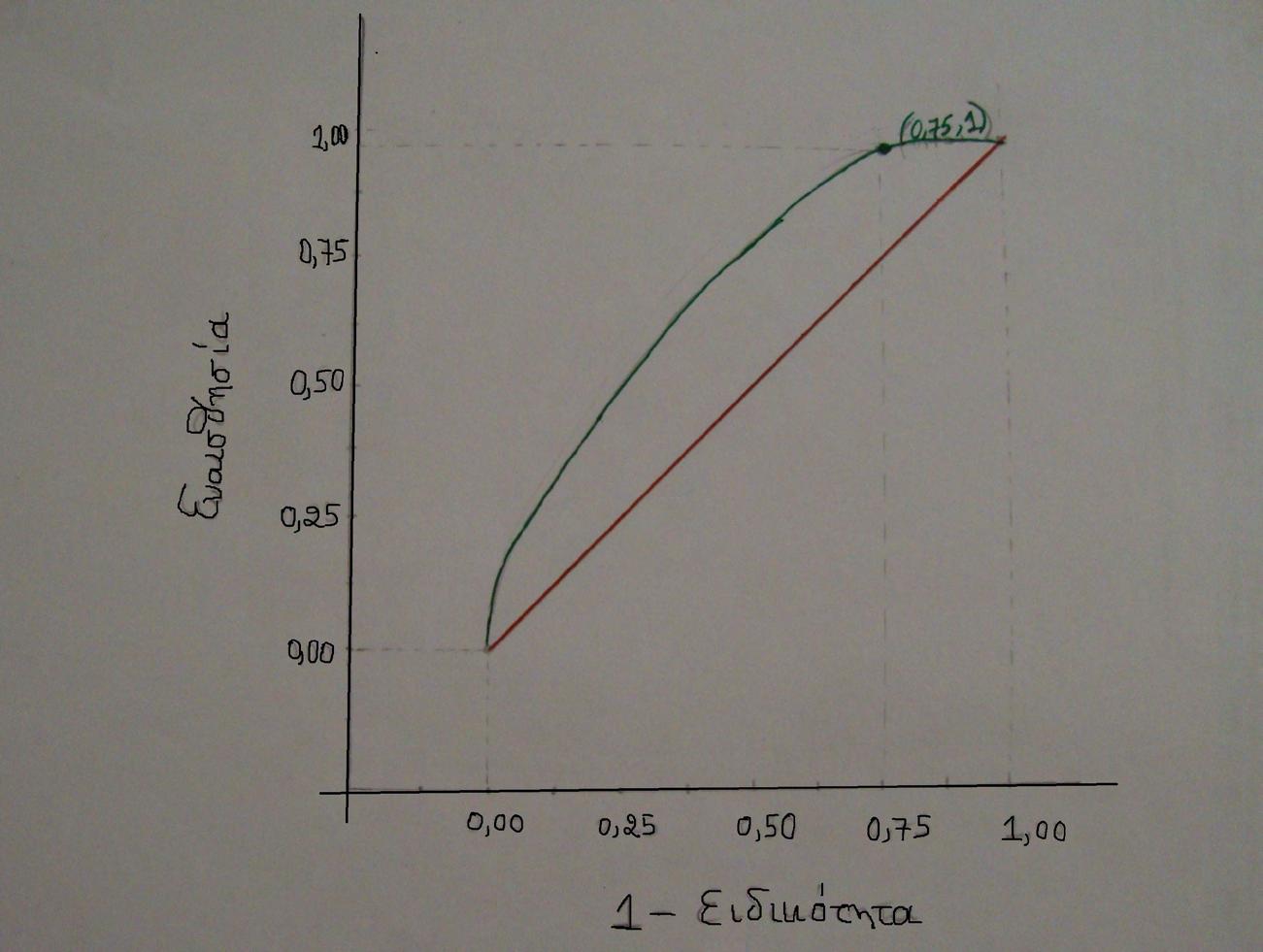
(9,28,31).
Βιβλιογραφία
1. Kelly- Quon Li, Tseng CH, Jen HC, Lee SL, Shew SB, Hospital type as a metric for racial disparities in pediatric appendicitis. J Am Coll Surg. 2013;216(1):74-82
2. Thompson GC. Variations in the diagnosis and management of acute appendicitis at Canadian pediatric emergency departments. American Academy of Pediatrics, Pediatr Emerg Care. 2012;28(10): 1099-1100
3. Joon Sung Kim, Acute Abdominal Pain in Children, Pediatr Gastroenterol Hepatol Nutr 2013, 16(4):219-224
4. Mandeville K, Pottker T, Bulloch B, Liu J, Using appendicitis scores in the pediatric ED. Am J Emerg Med. 2011Nov;29(9):972-7
5. Escriba A , Gamell AM, Fernandez Y, Quintilla JM, Cubells CL, Prospective validation of two systems of classification for the diagnosis of acute appendicitis, Emerg Care 2011Mar;27(3):1659
6. Samuel M, Pediatric Appendicitis Score, J Pediatric Surgery, 2002 Jun;37(6):877-81
7. Saucier A, Huang EY, Emeremni CA, Pershad J, Prospective evaluation of a clinical pathway for suspected appendicitis, 2014 Jan;133(1):e88-95
8. Neff LP , Ladd MR, Becher RD, Jordanhazy RA, Gallaher JR, Pranikoff T, Computerized tomography utilization in children with appendicitis-differences in reffering and children’s hospitals. Am Surg. 2011;77(8):1061-1065
9. Kulik DM, Uleryk EM, Maquire JL, Does this child have appendicitis? A systematic review of clinical prediction rules for children with acute abdominal pain. J Clin Epidemiol 2013 Jan;66(1):95-104
10. Bhangu A, Nepoqodiev D, Taylor C, Durkin N, Patel R, Accuracy of clinical coding from 1210 appendicectomies in a British district general hospital, International Journal of surgery 10(2012) 144-147
11. McCartan DP, Fleming FJ, Grace PA The management of right iliac fossa pain- is timing everything? Surgeon 2010;8(4):211-7
12. Guller U, Rosella L, McCall J, Brugger LE, Candinas D, Negative appendectomy and perforation rates in patients undergoing laparoscopic surgery for suspected appendicitis, Br J Surg 2011 Apr;98(4):589-95
13. Tomlinson D, von Baeyer CL, Stinson JN, Sung L, A Systematic Review of Faces Scales for the Self-report of Pain Intensity in Children, Pediatrics 2010 Nov;126(5):e1168-98
14. Salo M, Friman G, Stenstrom P, Ohlsson B, Arnbjornsson E, Appendicitis in children: Evaluation of the Pediatric Appendicitis Score in Younger and Older Childrenn, Surg Res Pract. 2014;2014:438076
15. Zakaria O, Sultan TA, Khalil TH, Wahba T, Role of clinical judgment and tissue harmonic imaging ultrasonography in diagnosis of paediatric acute appendicitis, World J Emerg Surg 2011 Nov16;6(1):39
16. Bundy DG, Byerley JS, Liles EA, Perrin EM, Katznelson J, Rice HE, Does this child have appendicitis?, JAMA 2007 Jul25;298(4):438-51
17. Sack U, Biereder B, Elouahidi T, Bauer K, Keller T, Trobs RB, Diagnostic value of blood inflammatory markers for detection of acute appendicitis in children, BMC Surg. 2006;6:15
18. Kwan KY, Nager AL, Diagnosing pediatric appendicitis: usefulness of laboratory markers, Am J Emerg Med. 2010 Nov;28(9):1009-15
19. Kharbanda AB, Cosme Y, Liu K, Spitalnik SL, Dayan PS, Discriminative Accuracy of Novel and Tranditional Biomarkers in Children with suspected appendicitis adjusted for duration of abdominal pain, Acad Emerg Med. 2011 Jun;18(6):567-574
20. C.-W. Yu, L.-I. Juan, Wu MH, Shen CJ, Wu JY, Lee CC, Systematic review and meta- analysis of the diagnostic accuracy of procalcitonin, C-reactive protein and white blood cell count for suspected acute appendicitis, British Journal of Surgery 2013;100:322-329
21. Γ.Ι. Αραπάκη,
2006
22. Lynn S. Bickley, BATE’S Guide to Physical Examination and History Taking, Tenth Edition 2009, Lippincott Wiliams & Wilkins
23. Otake S, Suzuki N, Takahashi A, Toki F, Nishi A, Yamamoto H et al, Histological analysis of appendices remoned during internal appendectomy after conservative management of pediatric patients with acute appendicitis with an inflammatory mass or abscess, Surg Today, 2014 Aug;44(8):1400-5
24. Lobo-Machin I, Delgado- Plasencia L, Hernandez- Gonzalez I, Brito-Garcia A, Burillo-Putze G, Bravo-Gutierrez A et al, Appendiceal diverticulitis and acute appendicitis: differences and similarities, Rev Esp Enferm Dig. 2014 Aug;106(7):452-8
25. Hornby ST, Shahtahmassebi G, Lynch S, Ladwa N, Stell DA, Delay to surgery does not influence the pathological outcome of acute appendicitis, Scand J Surg. 2014 Mar;103(1):5-11
26. Schellekens DH, Hulsewe KW, van Acker BA, van Bijnen AA, de Jaegere TM, Sastrowijoto SH et al, Evaluation of the diagnostic accuracy of plasma markers for early diagnosis in patients with suspected acute appendicitis, Acad Emerg Med. 2013 Jul;20(7):703-10
27. Tsioplis C, Brockschmidt C, Sander S, Henne-Bruns D, Kornmann M, Factors influencing the course of acute appendicitis in adults and children, Langenbecks Arch Surg. 2013 Aug;398(6):857-67
28. Sadot E, Wasserberg N, Shapiro R, Keidar A, Oberman B, Sadetzki S, Acute appendicitis in the twenty- first century: should we modify the management protocol?, J Gastrointest Surg. 2013 Aug;17(8):1462-70
29. Farach SM, Danielson PD, Walford NE, Harmel RP Jr, Chandler NM, Operative findings Are a Better Predictor of Resource Utilization in Pediatric Appendicitis, J Pediatr Surg. 2015 Sep;50(9):1574-8
30. F.Bruder Stapleton, Robert C. Tasker, Fluids and electrolytes. In: F.Bruder Stapleton, Robert C. Tasker et al, Oxford American Handbook of Pediatrics, New York: Oxford University Press 2009
31. Schuh S, Man C, Cheng A, Murphy A, Mohanta A, Moineddin R et al, Predictors of non- diagnostic ultrasound scanning in children with suspected appendicitis, J. Pediatr. 2011;158(1):112-118
w w w.e-child .gr