Paediatriki
Contents
vi LETTER FROM THE PRESIDEnT vii EDITORIAL
SpeciAl Article
63 Traffic accidents and posttraumatic stress among children and adolescents
G. Kolaitis, G. Giannakopoulos
reVieW ArticleS
69 Chlamydia trachomatis and Chlamydia pneumonia as respiratory system pathogens in infants and children
I. N. Mammas, K.N. Priftis, C. Koutsaftiki, N. Myriokefalitakis
75 The foetal / neonatal environment and the epigenome
T. Siahanidou
81 Thrombosis and thrombophilia in childhood
H. Platokouki-Komitopoulou
87 Congenital club foot. Questions about its treatment N. Markeas
OrigiNAl Article
95 Breastfeeding in northern Greece at early 21rst century
K. Papadopoulou-Legbelou, E. Papadopoulou-Alataki, M. Kavga, S. Karagianni, G. Varlamis
prActicAl iSSueS
102 Strabismus: What the paediatrician should know
A.G. Damanakis
107 The ethics of withdrawal of care and limitation of care in children – a statement of the principles that should guide decisions. Joint Statement of the Ethics group of the European Academy of Paediatrics
R.I. Ross Russell
cASe repOrtS
110 “nutcracker” syndrome: A pediatric case presentation
Attilakos, E. Alexopoulou, A. Papapetrou, A. Fretzayas, P. Nikolaidou Karpathiou, C. Liapis
114 A case of neonatal small left colon syndrome
E. Anastasiadou-Karageorgopoulou, A. Martinopoulou, M. Mpadouraki, A. Andreou
cliNicAl QuiZ
117 CLInICAL QUIZ
N. Karathanasis, P. Emmanouil, G. Simeoglou, C. Koutsaftiki, N. Miriokefalitakis
ΚλινιΚο QUIZ 11.
Traffic accidents and posttraumatic stress among children and adolescents
G. Kolaitis, G. Giannakopoulos
Abstract: Over the past decades, traffic accidents have become an epidemic in Greece. Children and youths are commonly victims posing a vast social and economic burden. The psychological impact of traffic accidents on children and adolescents has been poorly studied so far; nevertheless, traffic accidents are associated with depression, journey and generalized anxiety, acute stress disorder as well as high incidence of posttraumatic stress disorder (PTSD). PTSD is a psychopathological entity which can result in psychiatric co-morbidity and functional impairment, thus it is necessary to be recognised by parents and hospital staff, to be assessed and managed therapeutically. Trauma care should include procedures that could identify and prevent stress reactions in order to minimize the risk of associated psychological consequences. Psycho-education of children victims and their parents is recognized as a key component of effective interventions.
Key words: Psychological impact, acute stress disorder, posttraumatic stress disorder (PTSD), children, adolescents, traffic accidents.
Department of Child Psychiatry, University of Athens Medical School, "Aghia Sophia" Children's Hospital, Athens, Greece
Correspondence:
Gerasimos Kolaitis gkolaitis@med.uoa.gr Department of Child Psychiatry, University of Athens Medical School, "Aghia Sophia" Children's Hospital, Thivon & M. Asias, 11527 Athens, Greece
Bryant et al. (5), 2004 86 (59%), 5-16 ετών Post-traumatic stress disorder 25%
3
Ηνωμένο Βασίλειο (μέση τιμή 12.2), reaction index (RI), Impact of 18% στους 6 μήνες 55% αγόρια, 21% νοσηλευόμενα, event scale children’s version μέση τιμή ISSα 2.4 (IES-8)
Di Gallo et al. (9), 1997 53 (72%), 5-18 ετών (70% ≤ 12), Child post-traumatic stress 14% στους 3 μήνες Ηνωμένο Βασίλειο 67% αγόρια, μέση τιμή ISS 5.8 disorder reaction index (PTSD-RI)
Kassam-Adams & 177 (44%), 8–17 ετών
Clinician-administered PTSD 6% στους 3-12 μήνες Winston (10), 2004 ΗΠΑ scale for children and adolescents (CAPS-CA)
Keppel-Benson et al. (1), 50 (57%), 7-16 ετών
Child/adolescent revised version 14% στους 2-18 μήνες 2002 ΗΠΑ (μέση τιμή 11.6), 58% αγόρια, of the diagnostic interview for 22% ελαφρύς τραυματισμός, children and adolescents 72% μέτριος τραυματισμός, (DICA-R-C) 6% σοβαρός τραυματισμός
McDermott & 26 (34%), 8-13 ετών
Post-traumatic stress disorder 23% στους 3 μήνες Cvitanovich (6), (μέση τιμή 10.2), 50% αγόρια, reaction index for children 2000 Αυστραλία 72% νοσηλευόμενα (PTSD-RI)
Mirza et al., 119 (76%), 8-16 ετών (12% ≤ 11), The Fredrick’s reaction index 29% στις 4 εβδομάδες 1998 Ηνωμένο Βασίλειο 68% αγόρια, μέση τιμή ISS 2.3 (FRI) 13% στους 6 μήνες
Pervanidou et al. (12), 56, 7-18 ετών (μέση τιμή 10.7), Kiddie Schedule for Affective 37.7% στις 4 εβδομάδες
2007 Ελλάδα 66.7% αγόρια, 100% νοσηλευόμενα, Disorders and Schizophrenia - 18% στους 6 μήνες 36.8% ελαφρύς τραυματισμός, Lifetime version (K-SADS-PL) 35.1 % μέτριος τραυματισμός, 28.1% σοβαρός τραυματισμός
Schafer et al. (8), 72, 8-18 ετών
Impact of event scale - Revised 25% στους τρεις μήνες 2006 Germany
Stallard et al. (2), 158 (42%), 7-18 ετών
Clinician-administered PTSD 29% στις 4 εβδομάδες 2004 Ηνωμένο Βασίλειο (μέση τιμή 14.9), 48% αγόρια, scale for children (CAPS-C), 13% νοσηλευόμενα, μέση τιμή Impact of event scale (IES) Manchester triage scaleβ 2.7
Sturms et al. (7), 2005 51 (35.7%), 8-15 ετών Impact of event scale 12% στους 3 μήνες Ολλανδία (μέση τιμή 12.2), 53% αγόρια, 12% στους 6 μήνες 29% νοσηλευόμενα, 26% σοβαρός τραυματισμός
1. Keppel-Benson JM, Ollendick TH, Benson MJ. Posttraumatic stress in children following motor vehicle accidents. J Child Psychol Psychiatry 2002;43:203-212.
2. Stallard P, Salter E, Velleman R. Posttraumatic stress disorder following road traffic accidents - a second prospective study. Eur Child Adolesc Psychiatry 2004;13: 172-178.
3. Nikolaidis G, Zavras D, Bonikos D, Kyriopoulos J. Trends of mortality rates during the last thirty years in Greece. J Med Syst 2004;28:607-616.
4. Jones-Alexander J, Blanchard EB, Hickling EJ. Psychophysiological assessment of youthful motor vehicle accident survivors. Appl Psychophysiol Biofeedback 2005; 30:115-123.
5. Bryant B, Mayou R, Wiggs L, Ehlers A, Stores G. Psychological consequences of road traffic accidents for children and their mothers. Psychol Med 2004;34:335-346.
6. McDermott BM, Palmer LJ. Postdisaster emotional distress, depression and event-related variables: findings across child and adolescent developmental stages. Aust N Z J Psychiatry 2002;36:754-761.
7. Sturms LM, van der Sluis CK, Stewart RE, Groothoff JW, ten Duis HJ, Eisma WH. A prospective study on paediatric traffic injuries: health-related quality of life and posttraumatic stress. Clin Rehabil 2005;19:312-322.
8. Schafer I, Barkmann C, Riedesser P, Schulte-Markwort M. Posttraumatic syndromes in children and adolescents after road traffic accidents--a prospective cohort study. Psychopathology 2006;39:159-164.
9. Di Gallo A, Barton J, Parry-Jones WL. Road traffic accidents: early psychological consequences in children and adolescents. British Journal of Psychiatry 1997;170:358-362.
10. Kassam-Adams N, Winston FK. Predicting child PTSD: the relationship between acute stress disorder and PTSD in injured children. Journal of the American Academy of Child and Adolescent Psychiatry 2004;43:403-411.
11. Pervanidou P, Kolaitis G, Charitaki S, Margeli A, Ferentinos S, Bakoula C, et al. Elevated morning serum interleukin (IL)-6 or evening salivary cortisol concentrations predict posttraumatic stress disorder in children and adolescents six months after a motor vehicle accident. Psychoneuroendocrinology 2007;32:991-999.
12. Pervanidou P, Kolaitis G, Charitaki S, Lazaropoulou C, Papassotiriou I, Hindmarsh P, et al. The natural history of neuroendocrine changes in pediatric posttraumatic stress
disorder (PTSD) after motor vehicle accidents: progressive divergence of noradrenaline and cortisol concentrations over time. Biol Psychiatry 2007;62:1095-1102.
13. Landolt MA, Vollrath M, Timm K, Gnehm HE, Sennhauser FH. Predicting posttraumatic stress symptoms in children after road traffic accidents. J Am Acad Child Adolesc Psychiatry 2005;44:1276-1283.
14. Ellis A, Stores G, Mayou R. Psychological consequences of road traffic accidents in children. Eur Child Adolesc Psychiatry 1998;7:61-68.
15. de Vries AP, Kassam-Adams N, Cnaan A, Sherman-Slate E, Gallagher PR, Winston FK. Looking beyond the physical injury: posttraumatic stress disorder in children and parents after pediatric traffic injury. Pediatrics 1999;104:1293-1299.
16. Winston FK, Kassam-Adams N, Vivarelli-O'Neill C, Ford J, Newman E, Baxt C, et al. Acute stress disorder symptoms in children and their parents after pediatric traffic injury. Pediatrics 2002;109:e90.
17. Vasa RA, Grados M, Slomine B, Herskovits EH, Thompson RE, Salorio C, et al. Neuroimaging correlates of anxiety after pediatric traumatic brain injury. Biol Psychiatry 2004;55:208-216.
18. Daviss WB, Mooney D, Racusin R, Ford JD, Fleischer A, McHugo GJ. Predicting posttraumatic stress after hospitalization for pediatric injury. J Am Acad Child Adolesc Psychiatry 2000;39:576-583.
19. Meiser-Stedman RA, Yule W, Dalgleish T, Smith P, Glucksman E. The role of the family in child and adolescent posttraumatic stress following attendance at an emergency department. J Pediatr Psychol 2006;31:397-402.
20. Daviss WB, Racusin R, Fleischer A, Mooney D, Ford JD, McHugo GJ. Acute stress disorder symptomatology during hospitalization for pediatric injury. J Am Acad Child Adolesc Psychiatry 2000;39:569-575.
21. Kassam-Adams N, Winston FK. Predicting child PTSD: the relationship between acute stress disorder and PTSD in injured children. J Am Acad Child Adolesc Psychiatry 2004;43:403-411.
22. Landolt MA, Vollrath M, Laimbacher J, Gnehm HE, Sennhauser FH. Prospective study of posttraumatic stress disorder in parents of children with newly diagnosed type 1 diabetes. J Am Acad Child Adolesc Psychiatry 2005; 44:682-689.
23. Langeland W, Olff M. Psychobiology of posttraumatic stress disorder in pediatric injury patients: a review of the literature. Neuroscience and Biobehavioral Reviews 2008;32:161-174.
24. Nugent NR, Ostrowski S, Christopher NC, Delahanty DL. Parental posttraumatic stress symptoms as a moderator of child's acute biological response and subsequent posttraumatic stress symptoms in pediatric injury patients. Journal of Pediatric Psychology 2007;32:309-318.
25. Melnyk BM, Alpert-Gillis L, Feinstein NF, Crean HF, Johnson J, Fairbanks E, et al. Creating opportunities for parent empowerment: program effects on the mental health/coping outcomes of critically ill young children and their mothers. Pediatrics 2004;113:e597-607.
26. Ziegler MF, Greenwald MH, DeGuzman MA, Simon HK. Posttraumatic stress responses in children: awareness and practice among a sample of pediatric emergency care providers. Pediatrics 2005;115:1261-1267.
27. Saigh PA, Mroueh M, Bremner JD. Scholastic impairments among traumatized adolescents. Behav Res Ther 1997;35:429-436.
28. Kar N. Psychological impact of disasters on children: review of assessment and interventions. World J Pediatr 2009;5:5-11.
29. Veenema TG, Schroeder-Bruce K. The aftermath of violence: children, disaster, and posttraumatic stress disorder. J Pediatr Health Care 2002;16:235-244.
30. Chrousos GP, Gold PW. A healthy body in a healthy mind-and vice versa--the damaging power of "uncontrollable" stress. J Clin Endocrinol Metab 1998;83:1842-1845.
31. Gill JM, Saligan L, Woods S, Page G. PTSD is associated with an excess of inflammatory immune activities. Perspect Psychiatr Care 2009;45:262-277.
32. Langeland W, Olff M. Psychobiology of posttraumatic stress disorder in pediatric injury patients: a review of the literature. Neurosci Biobehav Rev 2008;32:161-174.
33. Perrin S, Smith P, Yule W. The assessment and treatment of Post-traumatic Stress Disorder in children and adolescents. J Child Psychol Psychiatry 2000;41:277-289.
34. American Psychiatric Association. Diagnostic and statistical manual of mental disorders- DSM-IV. Washington, DC: APA, 1994.
35. Miller JL. Post-traumatic stress disorder in primary care practice. J Am Acad Nurse Pract 2000;12:475-482; quiz 483-475.
36. Butler DJ, Moffic HS, Turkal NW. Post-traumatic stress reactions following motor vehicle accidents. Am Fam Physician 1999;60:524-531.
37. Meyer C, Steil R. [Post-traumatic stress disorder after traffic accidents]. Unfallchirurg 1998;101:878-893.
38. Gold JI, Kant AJ, Kim SH. The impact of unintentional pediatric trauma: a review of pain, acute stress, and posttraumatic stress. J Pediatr Nurs 2008;23:81-91.
39. Carr A. Interventions for post-traumatic stress disorder in children and adolescents. Pediatr Rehabil 2004;7:231-244.
Chlamydia trachomatis and Chlamydia pneumoniae as respiratory system pathogens in infants and children.
I. Ν. Mammas1, K. Ν. Priftis2, C. Koutsaftiki1, N. Myriokefalitakis1
Abstract: Chlamydia trachomatis and Chlamydia pneumoniae are two of the most common members of the Chlamydiaceae family that infect humans. Both species are small Gram-negative organisms the invasion of which into the host-cells is a prerequisite for their multiplication. Vertical transmission of Chlamydia trachomatis infection may result in the development of neonatal pneumonia. Chlamydia pneumoniae has been recognized as a common causative agent for community-acquired pneumonia in school-age children. Infection with Chlamydia pneumoniae has also been associated with asthma exacerbations. Laboratory detection of both species can be performed via isolation of the organism from nasopharyngeal cell culture, polymerase chain reaction (PCR) amplification, and serological studies. Recently, both species have been detected in bronchoalveolar lavage (BAL) fluid samples of children. This review presents data from the recent literature regarding the epidemiology and the clinical features of infection with Chlamydia trachomatis and Chlamydia pneumoniae in infants and children. We also discuss their role in the pathogenesis of asthma in childhood.
Key words: Chlamydia trachomatis, Chlamydia pneumoniae, respiratory pathogens, infants, asthma, children.
1 1st Department of Paediatrics, General Children’s Hospital of Penteli, Palaia Penteli, Athens, Greece
2 Department of Paediatric Pneumonology, General Children’s Hospital of Penteli, Palaia Penteli, Athens, Greece
Correspondence:
Ioannis N. Mammas mammasjo@hotmail.com
1st Department of Paediatrics, General Children’s Hospital of Penteli, 152 36 Palaia Penteli, Athens, Greece
( Chamydia psittaci
(Chlamydia pecorum
1.
1999:244–254.
2. Saikku P, Wang SP, Kleemola M, Brander R, Rusanen E, Grayston JT: An epidemic of mild pneumonia due to an unusual strain of Chlamydia psittaci. J Infect Dis 1985;151: 832–839.
3. Grayston JT, Kuo CC, Campbell LA, Wang SP: Chlamydia pneumoniae sp. nov. for Chlamydia sp. strain TWAR. Int J Syst Bacteriol 1989;39:88–90.
4. Darville T. Chlamydia trachomatis infections in neonates and young children. Semin Pediatr Infect Dis 2005;16:235–244.
5. Wu S, Shen L, Liu G. Study on vertical transmission of Chlamydia trachomatis using PCR and DNA sequencing. Chin Med J (Engl) 1999;112:396–399.
6. Βell TA, Stamm WE, Wang SP, Kuo CC, Holmes KK, Grayston JT. Chronic Chlamydia trachomatis infections in infants. JAMA 1992; 267: 400–402.
7. McIntosh K. Community-acquired pneumonia in children. N Engl J Med 2002;346:429–437.
8. Herieka E, Dhar J. Acute neonatal respiratory failure and Chlamydia trachomatis. Sex Transm Infect 2001;77:135–136.
9. Bavastrelli M, Midulla M, Rossi D, Salzano M. Chlamydia trachomatis infection in children with wheezing simulating asthma. Lancet 1992;339:1174.
10. Webley WC, Tilahun Y, Lay K, Patel K, Stuart ES, Andrzejewski C, Salva PS. Occurrence of Chlamydia trachomatis and Chamydia pneumoniae in paediatric respiratory infections. Eur Respir J 2009;33:360–367.
11. Majeroni BA, Ukkadam S. Screening and treatment for sexually transmited infections in pregnancy. Am Fam Physician 2007;76:265–270.
12. Σκουτέλης
Focus on Health Ltd,
2007:101–115.
13. Hammerschlag MR. The role of Chlamydia in upper respiratory tract infections. Curr Infect Dis Rep 2000;2: 115–120.
14. Tsai MH, Huang YC, Chen CJ, et al. Chlamydial pneumonia in children requiring hospitalization: effect of mixed infection on clinical outcome. J Microbiol Immunol Infect 2005;38:117–122.
15. Teig N, Anders A, Schmidt C, Rieger C, Gatermann S. Chlamydophila pneumoniae and Mycoplasma pneumoniae in respiratory specimens of children with chronic lung diseases. Thorax 2005;60:962–966.
16. Cunningham AF, Johnston SL, Julious SA, Lampe FC, Ward ME. Chronic Chlamydia pneumoniae infection and asthma exacerbations in children. Eur Respir J 1998;11:345–349.
17. Esposito S, Principi N. Asthma in children: are Chlamydia or Mycoplasma involved? Paediatr Drugs 2001;3:159–168.
18. Oba Y, Salzman G. Chlamydial pneumonias. Overview of infection with the three main chlamydial species that cause respiratory disease in humans. eMedicine, February 2007.
http://www.emedicine.com/med/topic341.htm.
19. Triga MG, Anthracopoulos MB, Saikku P, Syrogiannopoulos GA. Chlamydia pneumoniae infection among healthy children and children hospitalised with pneumonia in Greece. Eur J Clin Microbiol Infect Dis 2002;21:300–303.
20. Kumar S, Hammerschlag MR. Acute respiratory infection due to Chlamydia pneumoniae: Current status of diagnostic methods. Clin Infect Dis 2007;44:568–576.
21. Wubbel L, Muniz L, Ahmed A, Trujillo M, Carubelli C, McCoig C, Abramo T, Leinonen M, McCracken GH Jr. Etiology and treatment of community-acquired pneumonia in ambulatory children. Pediatr Infect Dis J 1999;18:98–104.
22. Michelow IC, Olen K, Lozano J, Rollins NK, Duffy LB, Ziegler T, Kauppila J, Leinonen M, McCracken GH Jr. Epidemiology and clinical characteristics of communityacquired pneumonia in hospitalized children. Pediatrics 2004;113:701–707.
23. Kurz H, Gopfrich H, Wabnegger L, Apfalter P. Role of Chlamydia pneumoniae in children hospitalised for community-acquired pneumonia in Vienna, Austria. Pediatr Pulmonol 2009;44:873–876.
24. Heiskanen-Kosma T, Korppi M, Laurila A, Jokinen C, Kleemola M, Saikku P. Chlamydia pneumoniae is an important cause of community-acquired pneumonia in school-aged children: serological results of a prospective, population-based study. Scand J Infect Dis 1999;31:255–259.
25. Kese D, Cizman M, Marin J, Zupanc TA. Chlamydia pneumoniae infections in patients with communityacquired pneumonia in Slovenia. Scand J Infect Dis 2003;34:172–176.
26. Jain R, Jain A, Agarwal J, Awasthi S. Chlamydia sp. in hospitalised children with community acquired pneumonia. Indian Pediatr 2007;44: 216–218.
27. Somer A, Salman N, Yalcin I, Agacfidan A. Role of M. pneumoniae and C. pneumoniae in children with community acquired pneumonia in Istanbul, Turkey. J Trop Pediatr 2006;52:173–178.
28. Lassmann B, Poetschke M, Ninteretse B, Issifou S, Winkler S, Kremsner PG, Graninger W, Apfalter P. Community-
acquired pneumonia in children in Lambarene, Gabon. Am J Trop Med Hyg 2008;79:109–114.
29. Webley WC, Salva PS, Andrzejewski C, et al. The bronchial lavage of pediatric patients with asthma contains infectious Chlamydia. Am J Respir Crit Care Med 2005;171:1083–1088.
30. Block S, Hedrick J, Hammerschlag MR, Cassell GH, Craft JC. Mycoplasma pneumoniae and Chlamydia pneumoniae in pediatric community-acquired pneumonia: comparative efficacy and safety of clarithromycin vs. erythromycin ethylsuccinate. Pediatr Infect Dis J 1995;14:471–477.
31. Esposito S, Bosis S, Faelli N, Begliatti E, Droghetti R, Tremolati E, Porta A, Blasi F, Principi N. Role of atypical bacteria and azithromycin therapy for children with recurrent respiratory tract infections. Pediatr Infect Dis J 2005;24:438–444.
32. Wark PA, Johnston SL, Simpson JL, Hensley MJ, Gibson PG. Chlamydia pneumoniae immunoglobulin A reactivation and airway inflammation in acute asthma. Eur Respir J. Eur Respir J 2002;20:834–840.
33. Allegra L, Blasi F, Centanni S, Cosentini R, Denti F, Raccanelli R, Tarsia P, Valenti V. Acute exacerbations of asthma in adults: role of Chlamydia pneumoniae infection. Eur Respir J 1994;7:2165–2168.
34. Lieberman D, Lieberman D, Printz S, Ben-Yaakov M, Lazarovich Z, Ohana B, Friedman MG, Dvoskin B, Leinonen M, Boldur I. Atypical pathogen infection in adults with acute exacerbation of bronchial asthma. Am J Respir Crit Care Med 2003;167:406–410.
35. Cunningham AF, Johnston SL, Julious SA, Lampe FC, Ward ME. Chronic Chlamydia pneumoniae infection and asthma exacerbations in children. Eur Respir J 1998; 11:345–349.
36. Esposito S, Blasi F, Arosio C, Fioravanti L, Fagetti L, Droghetti R, Tarsia P, Allegra L, Principi N. Importance of acute Mycoplasma pneumoniae and Chlamydia pneumoniae infections in children with wheezing. Eur Respir J 2000;16:1142–1146.
37. Biscardi S, Lorrot M, Marc E, Moulin F, Boutonnat-Faucher B, Heilbronner C, Iniguez JL, Chaussain M, Nicand E, Raymond J, Gendrel D. Mycoplasma pneumoniae and asthma in children. Clin Infect Dis 2004;38:1341–1346.
38. Gern JE, Lemanske RF Jr. Infectious triggers of pediatric asthma. Pediatr Clin North Am 2003;50:555–575.
39. Montalbano MM, Lemanske RF Jr. Infections and asthma in children. Curr Opin Pediatr 2002;14:334–337.
40. Emre U, Roblin PM, Gelling M, Dumornay W, Rao M, Hammerschlag MR, Schachter J. The association of Chlamydia pneumoniae infection and reactive airway disease in children. Arch Pediatr Adolesc Med 1994;148: 727–732.
41. ten Brinke A, van Dissel JT, Sterk PJ, Zwinderman AH, Rabe KF, Bel EH. Persistent airflow limitation in adultonset nonatopic asthma is associated with serologic evidence of Chlamydia pneumoniae infection. J Allergy Clin Immunol 2001 Mar;107:449–454.
42. Paldanius M, Juvonen R, Leinonen M, Bloigu A, Silvennoinen-Kassinen S, Saikku P. Asthmatic persons are prone to the persistence of Chlamydia pneumoniae antibodies. Diagn Microbiol Infect Dis 2007;59:117–122.
43. Martin RJ, Kraft M, Chu HW, Berns EA, Cassell GH. A link between chronic asthma and chronic infection. J Allergy Clin Immunol 2001;107:595–601.
44. Gencay M, Roth M. Chlamydia pneumoniae infections in asthma: clinical implications. Am J Respir Med 2003;2: 31–38.
45. Biscione GL, Corne J, Chauhan AJ, Johnston SL. Increased frequency of detection of Chlamydophila pneumoniae in asthma. Eur Respir J 2004;24:745–749.
46. Black PN, Scicchitano R, Jenkins CR, Blasi F, Allegra L, Wlodarczyk J, Cooper BC. Serological evidence of infection with Chlamydia pneumoniae is related to the severity of asthma. Eur Respir J 2000;15:254–259.
47. Zaitsu M. Does Chlamydia pneumoniae infection trigger to development of asthma in wheezy infants? J Asthma 2009;46:967–968.
48. Cook PJ, Davies P, Tunnicliffe W, Ayres JG, Honeybourne D, Wise R. Chlamydia pneumoniae and asthma. Thorax 1998;53:254–259.
49. Mills GD, Lindeman JA, Fawcett JP, Herbison GP, Sears MR. Chlamydia pneumoniae serological status is not associated with asthma in children or young adults. Int J Epidemiol 2000;29:280–284.
50. Normann E, Gnarpe J, Wettergren B, Janson C, Wickman M, Nordvall L. Association between Chlamydia pneumoniae antibodies and wheezing in young children and the influence of sex. Thorax 2006;61:1054–1058.
51. Richeldi L, Ferrara G, Fabbri LM, Lasserson TJ, Gibson PG. Macrolides for chronic asthma. Cochrane Database Syst Rev 2005:CD002997.51.
52. Normann E, Gnarpe J, Gnarpe H, Wettergren B. Chlamydia pneumoniae infection predicts a reduced risk for subsequent atopic disease. Acta Paediatr 2005;94:705–710.
53. Schmidt SM, Müller CE, Wiersbitzky SK. Inverse association between Chlamydia pneumoniae respiratory tract infection and initiation of asthma or allergic rhinitis in children. Pediatr Allergy Immunol 2005;16:137–144.
54. Malinverni R, Kuo CC, Campbell LA, Grayston JT. Reactivation of Chlamydia pneumoniae lung infection in mice by cortisone. J Infect Dis 1995;172: 593–594.
55. Cook PJ, Honeybourne D, Wise R, Davies P. Chlamydia pneumoniae antibody titres are significantly associated with the use of steroid medication in respiratory disease. Eur Respir J 1996; 9:5s.
56. von Hertzen LC. Role of persistent infection in the control and severity of asthma: focus on Chlamydia pneumoniae. Eur Respir J 2002;19:546–556.
57. Black PN, Scicchitano R, Jenkins CR, et al. Serological evidence of infection with Chlamydia pneumoniae is related to the severity of asthma. Eur Respir J 2000;15:254–259.
58. Hahn DL, Bukstein D, Luskin A, Zeitz H. Evidence for Chlamydia pneumoniae infection in steroid-dependent asthma. Ann Allergy Asthma Immunol 1998;80:45–49.
59. Kraft M, Cassell GH, Pak J, Martin RJ. Mycoplasma pneumoniae and Chlamydia pneumoniae in asthma: effect of clarithromycin. Chest 2002;121:1782–1788.
60. Black PN, Blasi F, Jenkins CR, Scicchitano R, Mills GD, Rubinfeld AR, Ruffin RE, Mullins PR, Dangain J, Cooper BC, David DB, Allegra L. Trial of roxithromycin in subjects with asthma and serological evidence of infection with Chlamydia pneumoniae. Am J Respir Crit Care Med 2001;164:536–541.
The foetal/neonatal enviroment and the epigenome
T. Siahanidou
Abstract: Several environmental factors, including stress, glucocorticoid therapy, maternal care and diet, may have an epigenetic impact on the foetal or neonatal genome and cause changes in chromatin (i.e., DNA methylation, histone acetylation) leading to modification of gene expression, and thus to alteration of the genetically determined phenotype, without any change in the DNA sequence (genotype). Epigenetic modifications of the fetal/neonatal genome are considered to be the main mechanisms by which environmental factors have a “programming” effect leading to permanent changes in the morphology and/or function of tissues and organs and to the development of various disturbances (neuroendocrine, metabolic, etc) and of degenerative disorders in later life. For example, in animals, foetal or neonatal exposure to increased glucocorticoid concentrations, either in response to stress or following administration of pharmacological doses of glucocorticoids, or as a result of insufficient maternal care during the first days of life, resulted in decreased glucocorticoid receptor expression and decreased numbers of glucocorticoid receptors in the hippocampus, impaired learning and memory capacity and increased vulnerability to stress in adult life. The epigenetic alterations can be transmitted across generations. Although they are relatively stable, they are potentially reversible, even in adulthood, according to animal studies. The prevention or repair of adverse epigenetic alterations in humans is a future goal.
Key words: Epigenetics, genome, foetus, neonate, programming, glucocorticoids
Neonatal Unit, 1st University Paediatric Clinic, “Aghia Sophia” Children’s Hospital, Athens
Correspondence: Tania Siahanidou siahan@med.uoa.gr
(phosphoenolpyruvate carboxykinase,
ΒιΒλιοΓραΦια
1. Seckl JR, Meaney MJ. Glucocorticoid programming. Ann NY Acad Sci 2004; 1032: 63-84.
2. Szyf M. The early life environment and the epigenome. Biochim Biophys Acta 2009; 1790: 878-885.
3. Weaver ICG. Epigenetic effects of glucocorticoids. Semin Fetal Neonat Med 2009; 14: 143-150.
4. Simmons RA. Developmental origins of adult disease. Pediatr Clin N Am 2009; 56: 449-466.
5. McGowan PO, Meaney MJ, Szyf M. Diet and the epigenetic (re)programming of phenotypic differences in behavior. Brain Res 2008; 1237: 12-24.
6. Langley-Evans SC. Developmental programming of health and disease. Proc Nutr Soc 2006; 65: 97-105.
7. van Bel F, Heijnen CJ. Perinatal programming and reprogramming by glucocorticoid therapy and perinatal stress. Semin Fetal Neonat Med 2009; 14: 127-129.
8. Mesquita AR, Wegerich Y, Patchev AV, Oliveira M, Leao P, Sousa N, Almeida OFX. Glucocorticoids and neuro- and behavioural development. Semin Fetal Neonat Med 2009; 14: 130-135.
9. Meaney MJ, Szyf M, Seckl JR. Epigenetic mechanisms of perinatal programming of hypothalamic-pituitary-adrenal function and health. Trends Mol Med 2007; 13: 269-277.
10. Kapoor A, Petropoulos S, Matthews SG. Fetal programming of hypothalamic-pituitary-adrenal (HPA) axis function and behavior by synthetic glucocorticoids. Brain Res Rev 2008; 57: 586-595.
11. Kapoor A, Dunn E, Kostaki A, Andrews MH, Matthews SG. Fetal programming of hypothalamic-pituitary-adrenal function: prenatal stress and glucocorticoids. J Physiol 2006; 572: 31-44.
12. Kapoor A, Leen J, Matthews SG. Molecular regulation of the hypothalamic-pituitary-adrenal axis in adult male guinea pigs after prenatal stress at different stages of gestation. J Physiol 2008; 586: 4317-4326.
13. French NP, Hagan R, Evans SF, Mullan A, Newnham JP. Repeated antenatal corticosteroids: effects on cerebral palsy and childhood behavior. Am J Obstet Gynecol 2004; 190: 588-595.
14. Trautman PD, Meyer-Bahlburg HF, Postelnek J, New MI. Effects of early prenatal dexamethasone on the cognitive and behavioral development of young children: results of a pilot study. Psychoneuroendocrinology 1995; 20: 439-449.
15. Van Den Bergh BR, Van Calster B, Smits T, Van Huffel S, Lagae L. Antenatal maternal anxiety is related to HPA-axis dysregulation and self-reported depressive symptoms in adolescence: A prospective study on the fetal origins of depressed mood. Neuropsychopharmacology 2007; 33: 536-545.
16. Nyirenda MJ, Lindsay RS, Kenyon CJ, Burchell A, Seckl JR. Glucocorticoid exposure in late gestation permanently programmes rat hepatic phosphoenolpyruvate carboxykinase and glucocorticoid receptor expression and causes glucose intolerance in adult offspring. J Clin Invest 1998; 101:2174-2181.
17. Shen CN, Seckl JR, Slack JM, Tosh D. Glucocorticoids suppress beta cell development and induce hepatic metaplasia in embryonic pancreas. Biochem J 2003; 375: 41-50.
18. Dodic M, Moritz K, Koikoulas I, Wintour EM. Programmed hypertension: kidney, brain or both? Trends Endocrinol Metab 2002; 13: 403-408.
19. Bogdarina IG, King PJ, Clark AJL. Characterization of the angiotensin (AT1b) receptor promoter and its regulation by glucocorticoids. J Mol Endocrinol 2009; 43: 73-80.
20. McMullen S, Langley-Evans SC. Maternal low-protein diet in rat pregnancy programs blood pressure through sexspecific mechanisms. Am J Physiol 2005; 288: R85-R90.
21. O’Connor DM, Blache D, Hoggard N, Brookes E, Wooding FB, Fowden AL, Forhead AJ. Developmental control of plasma leptin and adipose leptin messenger ribonucleic acid in the ovine fetus during late gestation: role of glucocorticoids and thyroid hormones. Endocrinology 2007; 148: 3750-3757.
22. Langdown ML, Holness MJ, Sugden MC. Effects of prenatal glucocorticoid exposure on cardiac calreticulin and calsequestrin protein expression during early development and in adulthood. Biochem J 2003; 371: 61-69.
23. Fowden AL, Forhead AJ. Hormones as epigenetic signals in developmental programming. Exp Physiol 2009; 94: 607-625.
24. Liu D, Diorio J, Tannenbaum B, Caldji C, Francis D,
Freedman A, et al. Maternal care, hippocampal glucocorticoid receptors, and hypothalamic-pituitary-adrenal responses to stress. Science 1997;277:1659-1662.
25. Champagne FA, Curley JP. Epigenetic mechanisms mediating the long-term effects of maternal care on development. Neurosci Biobehav Rev 2009;33:593-600.
26. McGowan PO, Sasaki A, Huang TCT, Unterberger A, Suderman M, Ernst C, et al. Promoter-wide hypermethylation of the ribosomal RNA gene promoter on the suicide brain. PloS One 2008; 3:e2085.
27. McGowan PO, Sasaki A, D’Alessio AC, Dyrnov S, Labonte B, Szyf M, et al. Epigenetic regulation of the glucocorticoid receptor in human brain associates with childhood abuse. Nat Neurosci 2009; 12: 342-358.
28. Champagne FA, Weaver ICG, Diorio J, Dymov S, Szyf M, Meaney MJ. Maternal care associated with methylation of the estrogen receptor-a1b promoter and estrogen receptor-a expression in the medial preoptic area of female offspring. Endocrinology 2006;147:2909-2915.
29. Champagne FA. Epigenetic mechanisms and the transgenerational effects of maternal care. Front Neuroendocrinol 2008;29:386-397.
30. MacLennan NK, James SJ, Melnyk S, Piroozi A, Jernigan S, Hsu JL, et al. Uteroplacental insufficiency alters DNA methylation, one-carbon metabolism, and histone acetylation in IUGR rats. Physiol Genomics 2004; 18: 43-50.
31. Ke X, Lei Q, James SJ, Kelleher SL, Melnyk S, Jernigan S, et al. Uteroplacental insufficiency affects epigenetic determinants of chromatin structure in brains of neonatal and juvenile IUGR rats. Physiol Genomics 2006;25:16-28
32. Heijmans BT, Tobi EW, Stein AD, Putter H, Blauw GJ, Susser ES, et al. Persistent epigenetic differences associated with exposure to famine in humans. PNAS 2008;105:17046-17049.
33. Sinclair KD, Allegrucci C, Singh R, Gardner DS, Sebastian S, Bispham J, et al. DNA methylation, insulin resistance, and blood pressure in offspring determined by maternal periconceptional B vitamin and methionine status. PNAS 2007;104:19351-19356.
34. Aagaard-Tillery KM, Grove K, Bishop J, Ke X, Fu Q, McKnight R, Lane RH. Developmental origins of disease and determinants of chromatin structure: maternal diet modifies the primate fetal epigenome. J Mol Endocrinol 2008;41:91-102.
35. Drake AJ, Walker BR, Seckl JR. Intergenerational consequences of fetal programming by in utero exposure to glucocorticoids in rats. Am J Physiol Regul Integr Comp Physiol 2005;288:R34-R38.
36. Francis D, Diorio J, Liu D, Meaney MJ. Nongenomic transmission across generations of maternal behavior and stress responses in the rats. Science 1999; 286: 1155-1158
37. Champagne FA, Weaver ICG, Diorio J, Sharma S, Meaney MJ. Natural variations in maternal care are associated with estrogen receptor alpha expression and estrogen sensitivity in the medial preoptic area. Endocrinology 2003;144:47204724.
38. Chapman D, Scott K. The impact of maternal intergenerational risk factors on adverse developmental outcomes. Developmental Review 2001; 21: 305-325.
39. Weaver ICG, Meaney MJ, Szyf M. Maternal care effects on the hippocampal transcriptome and anxiety-mediated behaviors in the offspring that are reversible in adulthood. PNAS 2006; 103: 3480-3485.
40. Waterland RA, Jirtle RL. Transportable elements: Targets for early nutritional effects on epigenetic gene regulation. Mol Cell Biol 2003; 23: 5293-5300.
41. Dolinoy DC, Weidman JR, Waterland RA, Jirtle RL. Maternal genistein alters coat color and protects Avy mouse offspring from obesity by modifying the fetal epigenome. Environ Health Perspect 2006; 114: 567-572.
42. Weaver ICG, Champagne FA, Brown SE, Dymov S, Sharma S, Meaney MJ, Szyf M. Reversal of maternal programming of stress responses in adult offspring through methyl supplementation: Altering epigenetic marking later in life. J Neurosci 2005; 25: 11045-11054.
Thrombosis and thrombophilia in childhood
H. Platokouki-Komitopoulou
Abstract: Thrombosis is a rare event during childhood. Its incidence may increase in the years to come, as children with underlying diseases have longer overall survival, intervention techniques predisposing to thrombosis are common practice in intensive care units, advanced diagnostic imaging methods are currently available and paediatricians are better informed. In addition to the environmental risk factors for thrombosis, many polymorphisms in genes encoding almost all the haemostatic proteins have been found. The role of these defects in the pathogenesis of thrombosis has not yet been fully elucidated. Thrombophilia encompasses both the clinical predisposition (i.e., spontaneous, extended, recurrent thrombotic episodes or thrombosis at a young age) and the laboratory identification of congenital risk factors for thrombosis. In recent years, paediatric haematologists are frequently being asked to evaluate children for thrombophilia, although the cost of testing has increased dramatically and the clinical utility of laboratory findings is increasingly debated. Children most likely to benefit from thrombophilia testing include patients with acute thrombosis (to aid decision for the duration of anticoagulation therapy) or individuals at high risk for thrombosis, with a known positive family history. Abnormal laboratory findings on thrombophilia testing are not necessarily or causatively associated with thrombosis in asymptomatic children. Conversely, normal test results may provide false reassurance. Future research may lead to the precise estimation of the role and the contribution to thrombosis risk of each thrombophilic factor.
Key words: Τhrombosis, thrombophilia, laboratory testing, children.
Haemostasis/Haemophilia Unit
“Aghia Sophia” Children’s Hospital, Athens
Correspondence: Helen PlatokoukiKomitopoulou eplatokouki@paidonagiasofia.gr
“Aghia Sophia” Children’ Hospital
Haemophilia/Haemostasis Unit 11527 Athens
C (1981) (3)
V (FV Leiden)
S (1984)
C (Activated Protein C Resistance - APCR) (5),
(FII 20210A) (6),
ΑΡΑ αναφέρονται: αναστολή ινωδόλυσης, αναστολή συστήματος PC/PS, αναστολή έκκρισης
προστακυκλίνης, anti-β2GPI ή annexin V και ενίσχυση ενεργοποίησης αιμοπεταλίων.
δυνητικοί θρομβοφιλικοί παράγοντες αναφέρονται τα υψηλά επίπεδα των παραγόντων VIII, IX,
FXII,
-
ση (MTHFR),
για τη ζωή. Πρώιμη
αναγνώριση της ανεπάρκειας επιβάλλει
πλάσμα (FFP) (8).
επίπεδα PC, ΡS ή AT ενδέχεται να βρίσκονται παροδικώς χαμηλότερα των φυσιολογικών στην
Βιβλιογραφία
1. Jordan FL, Nandorff A. The familial tendency in thromboembolic disease. Acta Med Scand 1956;156:267-275.
2. Egeberg O. Inherited antithrombin deficiency causing thrombophilia. Thromb Diath Haemorrh 1965;13:516-530.
3. Griffin JH, Evatt B, Zimmerman TS, Kleiss AJ, Wideman C. Deficiency of protein C in congenital thrombotic disease. J Clin Invest 1981;68:1370-1373.
4. Comp PC, Esmon CT. Recurrent venous thromboembolism in patients with partial deficiency of protein S. N Engl J Med 1984;311:1525-1528.
5. Bertina RM, Koeleman BP, Koster T, Rosendaal FR, Driven RJ, de Ronde H et al. Mutation in blood coagulation FV associated with resistance to activated protein C. Nature 1994;369:64-67.
6. Poort SR, Rosendaal FR, Reitsma PH, Bertina RM. A common genetic variation in the 3’-untranslated region of the prothrombin gene is associated with elevated plasma prothrombin levels and an increase in venous thrombosis. Blood 1996;88:3698-3703.
7. Reitsma PH, Rosendaal FR. Past and future of genetic research in thrombosis. J Thromb Haemost 2007;5(1):264-269.
8. Raffini L. Thrombophilia in children: Who to test, How, When, and Why? Hematology (American Society of Hematology/ Edeucation Program Book) 2008;228-235.
9. Rosendaal FR. Thrombosis in the young: epidemiology and risk factors. A focus on venous thrombosis. Thromb Haemost 1997;18:1-6.
10. Andrew M. David M. Adams M, Ali K, Anderson R, Barnard D et al.Venous thromboembolic complications in children:first analyses of the Canadian Registry of VTE. Blood 1994;83:1251-1257.
11. Chan AK, deVeber G, Monagle P, Brooker LA, Massicotte PM. Venous thrombosis in children. J Thromb Haemost 2003;1:1443-1455.
12. Turebylu R, Salis R, Erbe R, Martin D, Lakshminrushima S, Ryan RM. Genetic prothrombotic mutations are common in neonates but are not associated with umbilical catheterassociated thrombosis. J Perinatol 2007;27:490-495
13. van Omen CH, Heijboer H, Buller HR, Heijmans HS, Peters M. Venous thromboembolism in childhood: a prospective two-year registry in The Netherlands. J Paediatr 2001;139:676-681.
14. Mitchell LG, Andrew M, Hanna K, Abshire T, Halton J, Anderson et al. A prospective cohort study determining the prevalence of thrombotic events in children with acute lymphoblastic leukemia and a central venous line who are treated with L-asparaginase: results of the Prophylactic Antithrombin Replacement in Kids with ALL treated with L-asparaginase (PARKAA) Study. Cancer 2003;97:508-516.
15. Komitopoulou A, Platokouki H, Kapsimali Z, Pergantou H, Adamtziki E, Aronis S. Mutations and polymorphisms in genes affecting hemostasis proteins and homocysteine metabolism in children with arterial ischemic stroke. Cerebrovasc Dis 2006;22:13-20.
16. Komitopoulou A, Platokouki H, Kapsimali Z, Moschovi M, Kattamis A, Pergantou H, Aronis S. Mutations and polymorphisms in genes affecting haemostasis components in children with thromboembolic disease. Pathophysiol Haemost Thromb 2006;35:392-397.
17. Revel-Vilk SK. Thrombophilia in children with venous disease. Thromb Res 2006;118:59-60.
18. Ehrenforth S, Junker R, Koch HG. Kreuz W, Münchow N, Scharrer I, et al. Multicentre evaluation of combined prothrombotic defects associated with thrombophilia in childhood. Childhood Thrombophilia Study Group. Eur J Paediatr 1999;158(3): S97-104.
19. Strater R, Becker S, von Eckardstein A, Heinecke A, Gutsche S, Junker R, et al. Prospective assessment of risk factors for recurrent stroke during childhood- a 5-year follow up study. Lancet 2002;360:1540-1545.
20. Ganesan V, Prengler M, Wade A, Kirkham FJ. Clinical and radiological recurrence after childhood arterial ischemic stroke. Circulation 2006;114:2170-2177.
21. Mackay MT, Monagle P. Perinatal and early childhood stroke and thrombophilia. Pathology 2008;40:116-123.
22. Lane DA, Grant PJ. Role of hemostatic polymorphisms in venous and arterial thrombotic disease. Blood 2009;95:1517-1532.
23. Franco RI, Reitsma PH. Gene polymorphisms of the haemostatic system and the risk for arterial thrombotic disease. Br J Haematol 2001;115:491-506.
24. Khan S. Hereditary thrombophilia. A review. Thrombosis J 2006;4:15-23.
25.
2007.
26. Lim W, Crower MA, Eikelboom JW. Management of antiphospholipid antibody syndrome: a systematic review.
JAMA 2006;295(9):1050-1057
27. Cochrane Database of Systematic Reviews 2009;issue 1.
28. Middeldorp L. Thrombophilia: an update. Semin Thromb Haemost 2007;33(6)
29. Nowak-Gottl U, Junker R, Kreuz W, von Eckardstein A, Kosch A, Nohe N et al. Risk of recurrent venous thrombosis with combined risk factors. Blood 2001;97:858-862.
30. Manco-Johnson MJ, Grabowski EF, Hellgreen M, Kemahli AS, Massicotte MP, Muntean W, et al. Laboratory testing for thrombophilia in pediatric patients. On behalf of the Subcommittee for Perinatal and Pediatric Thrombosis of the Scientific and Standardization Committee of the International Society of Thrombosis and Haemostasis (ISTH). Thromb Haemost 2002;88:155-156.
31. Monagle P, Chalmers E, Chan A, deVeber G, Kirkham F, Massicote P, et al. Antithrombotic therapy in neonates and children: American College of Chest Physicians (ACCP) Evidence-Based Clinical Practice Guidelines (8th Edition). Chest 2008;133:887-968.
32. Manco-Johnson MJ. How I treat venous thrombosis in children. Blood 2006;107:21-29.
33. Martinelli I. Pros and cons of thrombophilia testing: pros. J Thromb Haemost 2003;1:410-411.
34. Ho WK, Hankey GJ, Quinlan DJ, Eikelboom JW. Risk of recurrent venous thromboembolism in patients with common thrombophilia: a systematic review. Arch Intern Med 2006;166:729-736.
35. Young G, Albisetti M, Bonduel M, Brandao L, Chan A, Friedriechs F et al. Impact of inherited thrombophilia on venous thromboembolism in children: a systematic review and meta-analysis of observational studies. Circulation 2008;118(13):1373-1382.
36. Michaels LA. A response to: laboratory testing for thrombophilia in pediatric patients. Thromb Haemost 2003;89:204-205.
37. Thornburg CD, Dixon N, Paulyson-Nunez K, Ortel T. Thrombophilia screening in asymptomatic children. Thromb Res 2008;121: 597-604.
38. Walker ID, Greaves M, Preston FE. Investigation and management of heritable thrombophilia. Br J Haematol 2001;14:512-528.
39. Tormene D, Simioni P, Prandoni P, Franz F, Zerbinati P, Tognin G, et al. The incidence of venous thromboembolism in thrombophilic children: a prospective cohort study. Blood 2002;100:2403-2405.
40. Tormene D, Covasso S, Rossetto V, Simioni P. Thrombosis and thrombophilia in children: A systematic review. Semin Thromb Hemost 2006;3297);724-728.
41. Rosendaal FR, Holmerhorst FM, Vandenbroucke JP. Oral contraceptives, hormone replacement therapy and thrombosis. Thromb Haemost 2001;86:112-123.
42. Homocysteine: The Rubik’s cube of cardiovascular risk factors [Editorial]. Mayo Clin Proc 2008;83(11):1200-1202.
43. Bazzano LA, Reynolds K, Holder KN, He J. Effect of folic acid supplementation on risk of cardiovascular diseases: a meta-analysis of randomized controlled trials. JAMA 2006;296(22):2720-2726.
44. Antoniades C, Antonopoulos AS, Tousoulis K, Marinou K, Stefanadis C. Homocysteine and coronary atherosclerosis: from folate fortification to the recent clinical trials.
45. Moat SJ. Plasma total homocysteine: instigator or indicator of cardiovascular disease? Ann Clin Biochem 2008;45(4): 345-348.
Congenital club foot. Questions about its treatment
N. Markeas
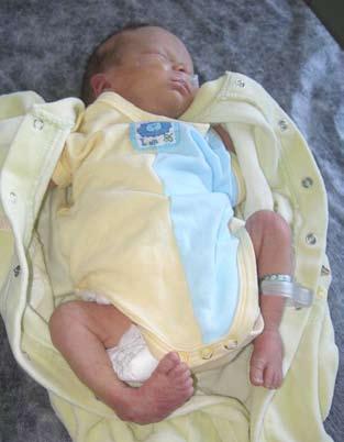
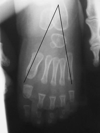
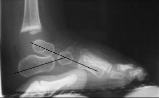
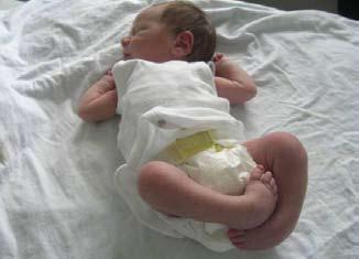
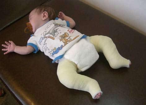
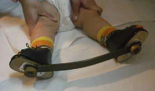
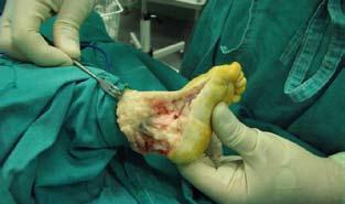
Abstract: Talipes equinovarus is probably the most common congenital deformity of the developing skeleton requiring intensive treatment by paediatricians and orthopedic surgeons. Its frequency ranges between 1 and 2 cases per 1,000 live births. Numerous theories have been proposed on the aetiology of congenital club foot, reflecting the difficulty of authors to agree on the subject, as well as on the various clinical signs of deformity used for its diagnosis. Dysmorphism of the talus appears to be the main reason for the typical deformity, which includes contractures of the periarticular soft tissues (sheaths, ligaments, tendons). Evaluation of the severity of the deformity is required for monitoring the progress of treatment and for prognostis. This review evaluates clinical criteria of classification and certain radiological indices that appear to be useful for estimation of the outcome of treatment. The initial treatment should be nonoperative, and the earlier the treatment begins, the more likely it is to be successful. The most popular nonoperative methods of treatment for club foot are the French (functional) and the Ponseti, which are based on weekly manipulation and cast application to hold the correction in place. The short-term results of the use of the conservative methods are successful but the long-term results are not always satisfactory. Certain factors can cause failure, including delayed onset of treatment, mistaken manipulations, social and financial issues, etc. The main indications for surgical release are when, on the one hand the initial deformity persists, despite the application of conservative measures, and on the other hand the radiological indices remain pathological. Surgical procedure should be limited to percutaneous lengthening of Achilles tendon or release of the posterior soft tissues. In more severe cases, wide ligament and capsular divisions, sheath openings, and tendons lengthenings may be justified.
Key words: Congenital club foot, criteria of choice, classification systems, Ponseti method, surgical treatment
markeasn@otenet.gr
2nd Orthopaedic Department, Children’s Hospital “P. & A. Kyriakou”
Correspondence: N. Markeas markeasn@otenet.gr
2nd Orthopaedic Department, Children’s Hospital “P. & A. Kyriakou” Athens, Greece
(Goldner and Fitch, Carroll and colleagues, Pirani and associates κ.λπ.) που χρησιμο-
(14).
(22,25,30,31).
Denis Browne.
4.
5.
Ευχαριστίες
(MA, University of Reading, UK)
Βιβλιογραφία
1. Bazopoulou - Kyrkanidou E. What causes the lameness of Hephaestus? Paediatriki 2008;71:283.
2. Kawashima T, Uhthoff HK: Development of the foot in prenatal life in relation to idiopathic club foot. J Pediatr Orthop 1990;10:232.
3. Fukuhara K, Schollmeier G, Uhthoff HK: The pathogenesis of clubfoot. A histomorphometric and immunohistochemical study of fetuses. J Bone Joint Surg Br 1994; 76:450.
4. Irani RN, Sherman MS: The pathological anatomy of idiopathic club foot. J Bone Joint Surg Am 1963;45:45.
5. Loren GJ, Karpinski NC, Mubarak SJ: Clinical implications of clubfoot histopathology. J Pediatr Orthop 1998;18:765.
6. Dobbs MB, Gordon JE, Schoenecker PL: Absent posterior tibial artery associated with idiopathic clubfoot. A report of two cases. J Bone Joint Surg Am 2004;86:599.
7. Dietz F: The genetics of idiopathic clubfoot. Clin Orthop Relat Res 2002;401:39.
8. Tachdjian MO: Disorders of the foot. In John Anthony Herring’s (ed): Pediatric Orthopaedics, 4th edition. Philadelphia, WB Saunders, 2008, p 1070.
9. Cahuzac JP, Navascues J, Baunin C, et al: Assessment of the position of the navicular by three dimensional magnetic resonance imaging in infant foot deformities. J Pediatr Orthop B 2002;11:134.
10. Scarpa Antonio: Commentarius de Penitiori Ossium Structura, 1799
11. Dobbs MB, Gurnett CA: Update on clubfoot: etiology and treatment. Clin Orthop Relat Res. 2009; 467(5):1146-53.
12. Ippolito E: Update on pathologic anatomy of clubfoot. J Pediatr Orthop B. 1995;4:17.
13. Ippolito E, Ponseti IV: Congenital club foot in the human fetus. A histological study. J Bone Joint Surg Am 1980;62:8.
14. Flynn JM, Donohoe M, Mackenzie WG: An independent assessment of two clubfoot-classification systems. J Pediatr Orthop 1998;18:323.
15. Laliotis N: Clinical evaluation of clubfoot. Acta Orthop Trauma Hell. 2006;57(3):162.
16. Dimeglio A, Bensahel H, Souchet P, et al: Classification of clubfoot. J Pediatr Orthop B 1995;4:129.
17. van Mulken JM, Bulstra SK, Hoefnagels NH: Evaluation of the treatment of clubfeet with the Dimeglio score. J Pediatr Orthop 2001;21:642.
18. Beaty JH: Congenital Anomalies of lower extremity. In: Campbell’s Operative Orthopaedics, S.T.Canale (editor), 10th edition, Mosby 2003.
19. Farsetti P, De Maio F, Russolillo L, Ippolito E: CT study on the effect of different treatment protocols for clubfoot pathology. Clin Orthop Relat Res. 2009;467(5):1243.
20. Pous JG, Dimeglio A: Neonatal surgery in clubfoot. Orthop Clin North Am 1978;9:233.
21. Macnicol MF: Congenital talipes equinovarus (clubfoot). In Hetal B, Rowley D, Cracciolo A, (eds) Surgery of Disorders of the Foot and Ankle. London, Martin Dunitz 1996, p 154.
22. Ponseti IV: Congenital Clubfoot: Fundamentals of Treatment. Oxford, Oxford University Press, 1996.
23. Turco VJ: Surgical correction of the resistant clubfoot. One-stage posteromedial release with internal fixation: A preliminary report. J Bone Joint Surg Am 1971;53:477.
24. Kite J: The Clubfoot. New York, Grune and Stratton, 1964.
25. Herzenberg JE, Radler C, Bor N: Ponseti versus traditional methods of casting for idiopathic clubfoot. J Pediatr Orthop 2002;22:517.
26. Noonan KJ, Richards BS: Nonsurgical management of idiopathic clubfoot. J Am Acad Orthop Surg 2003;11:392.
27. Richards BS, Johnston CE, Wilson H: Nonoperative clubfoot treatment using the French physical therapy method. J Pediatr Orthop 2005;25:98.
28. Bensahel H: The intimacy of clubfoot: The ways of functional treatment. J Pediatr Orthop B 1994;3:155.
29. Bensahel H: The early physiotherapic treatment in clubfoot: Results at 10 years follow up. Paper presented at the annual meeting of the Pediatric Orthopedic Society of North America, 2000, Vancouver, British Columbia.
30. Ponseti IV: Common errors in the treatment of congenital clubfoot. Int Orthop 1997;21:137.
31. Colburn M, Williams M: Evaluation of the treatment of idiopathic clubfoot by using the Ponseti method. J Foot Ankle Surg 2003;42:259.
32. Cummings RJ: The effectiveness of botulinum A toxin as an adjunct to the treatment of clubfeet by the Ponseti method: a randomized, double blind, placebo controlled study. J Pediatr Orthop. 2009;29(6):564.
33. Abbas M, Qureshi OA, Jeelani LZ, Azam Q, Khan AQ, Sabir AB: Management of congenital talipes equinovarus by Ponseti technique: a clinical study. J Foot Ankle Surg. 2008;47(6):541.
34. Sanghvi AV, Mittal VK: Conservative management of idiopathic clubfoot: Kite versus Ponseti method. J Orthop Surg (Hong Kong). 2009;17(1):67.
35. Halanski MA, Davison JE, Huang JC, Walker CG, Walsh SJ, Crawford HA: Ponseti method compared with surgical treatment of clubfoot: a prospective comparison. J Bone Joint Surg Am 2010;92(2):270-8.
36. Evans AM, Van Thanh D: A review of the Ponseti method and development of an infant clubfoot program in Vietnam. J Am Podiatr Med Assoc. 2009;99(4):306.
37. Hegazy M, Nasef NM, Abdel-Ghani H: Results of treatment of idiopathic clubfoot in older infants using the Ponseti method: a preliminary report. J Pediatr Orthop B 2009;18(2):76.
38. Wang YZ, Wang XW, Zhang P, Wang XS: Application of Ponseti method in patients older than 6 months with congenital talipes equinovarus. Beijing Da Xue Xue Bao. 2009;41(4):452.
39. Sanghvi AV, Mittal VK: Conservative management of idiopathic clubfoot: Kite versus Ponseti method. J Orthop Surg (Hong Kong). 2009;17(1):67.
40. Volz R, Paulsen M, Morcuende J: Distal tibia/fibula fractures following clubfoot casting. report of four cases. Iowa Orthop J. 2009;29:117.
41. Willis RB, Al-Hunaishel M, Guerra L, Kontio K: What proportion of patients need extensive surgery after failure of the Ponseti technique for clubfoot? Clin Orthop Relat Res. 2009;467(5):1294.
42. Kessler JI: A new flexible brace used in the Ponseti treatment of talipes equinovarus. J Pediatr Orthop B. 2008;17(5):247.
43. Avilucea FR, Szalay EA, Bosch PP, Sweet KR, Schwend RM: Effect of cultural factors on outcome of Ponseti treatment of clubfeet in rural America. J Bone Joint Surg Am. 2009;91(3):530.
44. Janicki JA, Narayanan UG, Harvey B, Roy A, Ramseier LE, Wright JG: Treatment of neuromuscular and syndromeassociated (no idiopathic) clubfeet using the Ponseti method. J Pediatr Orthop. 2009;29(4):393.
45. Kowalczyk B, Lejman T: Short-term experience with Ponseti casting and the Achilles tenotomy method for clubfeet treatment in arthrogryposis multiplex congenita. J Child Orthop. 2008;2(5):365.
46. Bradford EG: Treatment of clubfoot. J Bone Joint Surg. 1889;s1-1:89.
47. Park SS, Kim SW, Jung BS, Lee HS, Kim JS:Selective softtissue release for recurrent or residual deformity after conservative treatment of idiopathic clubfoot. J Bone Joint Surg Br. 2009;91(11):1526.
48. Carroll NC, Mc Martry R, Leete SF: The pathoanatomy of congenital clubfoot. Orthop Clin North Am 1978;9:225.
49. Carroll NC: Preoperative clinical assessment of clubfoot. In Simons GW (ed): The Clubfoot. New York, SpringerVerlag, 1993,p.77.
50. Goldner JL: Congenital talipes equinovarus-fifteen years of surgical treatment. Curr Pract Orthop Surg 1969;4:61.
51. Yngve DA, Gross RH, Sullivan JA: Clubfoot release without wide subtalar release. J Pediatr Orthop 1990;10:473.
52. Simons GW: Complete subtalar release in clubfeet. Part II-
comparison with less extensive procedures. J Bone Joint Surg Am 1985;67:1056.
53. McKay DW: New concept of and approach to clubfoot treatment. Section II-correction of the clubfoot. J Pediatr Orthop 1983;3:10.
Breastfeeding in northern Greece at early 21st century
Kyriaki Papadopoulou-Legbelou, Efimia Papadopoulou-Alataki, Maria Kavga, Stamatia Karagianni, George Varlamis.
Abstract
Background: This study aimed to determine the prevalence of breast-feeding and exclusive breast-feeding in the Northern Greece in the last decade, to compare data from this decade with data from the previous one and to correlate factors that can increase or decrease breast-feeding duration.
Methods: A questionnaire was used to define the demographics of the test group and collect information on delivery and breast-feeding. The data was then separated to 3 time periods: 1990-1994, 2000-2004, 2005-2009.
Results: Breast-feeding and exclusive breast-feeding rates were 91,1% and 19,6% respectively for the first period, 80,2% and 29,8% for the second, 81,6% and 29% for the third period. Highest exclusive breastfeeding rates were recorded among mothers who are immigrants, older (aged >35 years of age), mothers with a higher educational level, nonsmoking mothers, mothers who gave birth to full term neonates and among those who were assisted/informed by a midwife. Breast-feeding lasted for more than 6 months among 11,2% of mothers at first period, while the percentage for the second and third period stood at 15,1% and 5,5% respectively. Inadequate milk supply was cited as the most frequent cause of weaning, while, in smaller percentages, early maternal reemployment (especially in the third period) and various health reasons were also reported as factors of breast feeding cessation.
Conclusions: Breast-feeding and exclusive breast-feeding rates are very low in Northern Greece. Examining the reasons that lead to its termination as well as communicating its benefits can contribute to an increase of these rates in our country.
Key words: Breastfeeding, exclusive breastfeeding, supplementary feeding.
4th Dept. of Pediatrics “Papageοrgiou” Hospital N. Efkarpia, Thessaloniki, Greece
Correspondence: K. Papadopoulou-Legbelou kelipap@gmail.com
4th Dept. of Pediatrics “Papageοrgiou” Hospital N. Efkarpia, 56403 Thessaloniki, Greece
Ηλικία
Κάπνισμα
Σύνολο
(p=0,029),
Βιβλιογραφία
1. Gartner LM, Morton J, Lawrence RA, Naylor AJ, O’Hare D, Schanler RJ et al. American Academy of Pediatrics Section on Breastfeeding. Breastfeeding and the use of human milk. Pediatrics 2005; 115: 496-506.
2. Stuebe A. The risks of not breastfeeding for mothers and infants. Rev Obstet Gynecol 2009; 2: 222-231.
3. WHO. Infant and young child nutrition. World Health Organization 2001, 54th World Health Assembly, May 1422, WHA 54.2, Agenda item 13.1. Geneva: WHO, 2001.
4. Li R, Darling N, Maurice E, Barker L, Grummer-Strawn LM. Breastfeeding rates in the United States by characteristics of the child, mother, or family: The 2002 National Immunization Survey. Pediatrics 2005; 115:e31-7.
5. Cattaneo A, Yngve A, Koletzko B, Guzman LR. Protection, promotion and support of breast-feeding in Europe: current situation. Public Health Nutr 2005; 8: 39-46.
6. US Department of Health and Human Services. Healthy People 2010: Conference Edition. Vols I and II. Washington, DC: US Government Printing Office; 2000: 47-48.
7. Yngve A, Sjӧstrӧm M. Breastfeeding in countries of the European Union and EFTA: current and proposed recommendations, rationale, prevalence, duration and trends. Public Health Nutr 2001; 4(2B): 631-645.
8. Ryan AS, Wenjun Z, Acosta A. Breastfeeding continues to increase into the new millennium. Pediatrics 2002;110:11031109.
9. Antoniou E, Daglas M, Iatrakis G, Kouronis G, Greatsas G. Factors associated with initiation and duration of breastfeeding in Greece. Clin Exp Obstet Gynecol 2005; 32: 37-40.
10. Theofilogiannakou M, Skouroliakou M, Gounaris A, Panagiotakos D, Markantonis SL. Breast-feeding in Athens, Greece: Factors Associated With Its Initiation and Duration. J Pediatr Gastroenterol Nutr 2006; 43: 379-384.
11. Ladomenou F, Kafatos A, Galanakis E. Risk factors related to intention to breastfeed, early weaning and suboptimal duration of breastfeeding. Acta Paediatrica 2007; 96: 14411444.
12. Taylor LK, Lim K, Neville SE. Newborn feeding practices at the time of discharge from hospital in NSW in 2007: a descriptive study. N S W Public Health Bull 2009; 20: 177181.
13. Scott JA, Landers MC, Hughes RM, Binns CW. Factors associated with breastfeeding at discharge and duration of breastfeeding. J Paediatr Child Health 2001; 37: 254-261.
14. Ludvigsson JF, Ludvigsson J. Socio-economic determinants, maternal smoking and coffee consumption and exclusive breastfeeding in 10205 children. Acta Paediatr 2005; 94: 1310-1319.
15. Ντουρντούφη A,
2006; 18: 347-352.
16. Weiser TM, Lin M, Garikapaty V, Feyerharm RW, Bensyl DM, Zhu BP. Association of maternal smoking status with breastfeeding practices: Missouri 2005. Pediatrics 2009; 124: 1603-1610.
17. Αδάμ Ε, Νάκου Σ, Πούλη Θ. Απόψεις, προθέσεις και πρακτική
1997; 44: 189-196.
σε πληθυσμούς της Αθήνας. Δελτ. Α΄ Παιδιατρ Κλιν Πανεπιστημίου
18. Sheehan D, Krueger P, Watt S, Sword W, Bridle B. The Ontario Mother and Infant Survey: breastfeeding outcomes. J Hum Lact 2001; 17: 211-219.
19. Ertem IO, Votto N, Leventhal JM. The timing and predictors of the early termintion of breastfeeding. Pediatrics 2001; 107: 543-548.
20. Labarere J, Gelbert-Baudino N, Ayral AS, Duc C, Berchotteau M, Bouchon N et al. Efficacy of breastfeeding support provided by trained clinicians during an early, routine, preventive visit: a prospective, randomized, open trial of 226 mother-infant pairs. Pediatrics 2005; 115: e139-146.
21. Senarath U, Dibley MJ, Agho KE. Factors associated with nonexclusive breastfeeding in five East and Southeast Asian countries. A multilevel analysis. J Hum Lact 2010; Jan 28 (Epub ahead of Print)
22. Lu MC, Lange L, Slusser W, Hamilton J, Halfon N. Provider encouragement of breast-feeding: evidence from a national survey. Obstet Gynecol 2001; 97: 290-295.
23. Scott JA, Binns CW, Oddy WH, Graham KI. Predictors of breastfeeding duration: evidence from a cohort study. Pediatrics 2006; 117: e646-655.
24. Scott JA, Binns CW. Factors associated with the initiation and duration of breastfeeding: a review of the literature. Breastfeed Rev 1999; 7: 5-16.
Α.
Correspondence:
Α. G. Damanakis damanakis@ath.forthnet.gr ?????????????
Strabismus: What the Paediatrician should know
Α. G. Damanakis
Abstract: This is a concise review of the most common types of strabismus that are encountered in childhood. It is addressed to the pediatrician, and includes the necessary information on this subject that will help him consult, direct correctly and support the patient during the usually longlasting treatment of this problem.
Key words: Strabismus, esotropia, exotropia
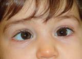
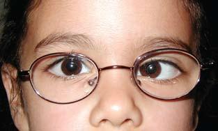
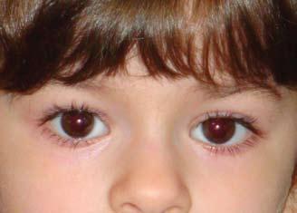
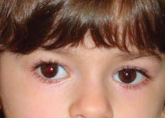
Εικόνα
Εικόνα 2. Επίκανθος.
1.
2004.
2. Noorden GK von. Burian – von Noorden’ s Binocular Vision and Ocular Motility. 4th ed. CV Mosby Co, St Louis, 1990.
3. Parks MM. Ocular motility and strabismus. Hagerstown, Harper and Row Publishers Inc; 1975.
4. Pediatric Eye Disease Investigator Group. The course of moderate amblyopia treated with patching in children: experience of the amblyopia treatment study. Am J Ophthalmol 2003;136:620-629.
5. Pediatric Eye Disease Investigator Group. The clinical spectrum of early-onset esotropia:experience of the Congenital Esotropia Observational Study. Am J Ophthalmol 2002;133:102-108.
6. Roth A, Speeg-Schatz C. Eye muscle surgery. SwetsZeitlinger Publishers; 2001.
The ethics of withdrawal of care and limitation of care in children - a statement of the principles that should guide decisions
Joint statement of the Ethics Group of the European Academy of Paediatrics
R I Ross
Russell on behalf of the Ethics Working Group of the European Academy of Paediatrics
Abstract: The decision to withhold or withdraw care from a child can be one of the most difficult, emotionally and ethically, made by a paediatrician. There is little literature available that can guide us through the considerations that need to be addressed, especially as our society becomes increasingly multicultural. The ethics group of the European Academy of Paediatrics comprises paediatricians from across Europe, many with years of experience in developing ethical frameworks in their own countries. This group has compiled a statement of principles that is intended to act as a guide to colleagues faced with these complex decisions.
Key words: limitation of care, ethics, end of life.
Introduction
In 2001 the ethics working group of the Confederation of the European Specialists of Paediatrics (CESP), itself the predecessor of the European Academy of Paediatrics (EAP), published a statement concerning the withholding or withdrawing of life support in children1. It is now almost 10 years since that work was undertaken, and November 2010 marked the 21st anniversary of the UN adoption of the ‘Convention on the Rights of the Child2’;it therefore seems an appropriate time to review the recommendations made.
The key purpose of this document is to acknowledge suffering in children, and recognise that such suffering may exceed any future prospects that can be envisaged for that child. In such a situation, the duty of those caring for the child is to consider whether palliative support should take precedence over therapeutic intervention. Sometimes that discussion may be very acute, and continuing care may be considered futile or unbearable. In others, the prognosis may be more long term and so much more difficult to predict. In such cases the balance of opinion may need to be more cautious. In many circumstances these decisions should be undertaken as part of a comprehensive discussion about end of life planning3
In recognising this the original statement emphasised the obligation of a paediatrician to protect the dignity of a dying child, and set out 12 specific guidelines for those involved in such situations. Since then there have been a number of publications looking at this issue in both adult and paediatric or neonatal practice4-9 .
These have all emphasised the importance of a multi-disciplinary approach to such discussions, and the involvement of families and patients in any
discussions, but they have also emphasised the differences in attitude that still exist between different communities. These differences vary from practical issues of how different professional and carers are actively involved9,10 to issues such as the active hastening of death in severely ill neonates11 There are also substantial legal and cultural differences between countries and communities and in the way in which they approach death, particularly when that involves a child. In such circumstances it would be inappropriate to set out detailed guidelines on how such situations should be handled. Rather, we believe that all paediatricians are able to (and should) advocate on behalf of their patients, and that the ethical foundations for the care of such children contain many commonly shared principles that can be made explicit.
In this position statement, we have set out those principles that underpin our approach to the setting of therapeutic limits.
Fundamental Principles9,12
Partnership of Care. Granted the compelling presumption in favour of life, there is a general duty of care with the primary intention of sustaining life and restoring patients to health. Whether or not a child can be restored to health, there is an absolute duty to comfort and to cherish that child and to prevent pain and suffering.
Healthcare professionals and parents therefore form a partnership, whose function is to serve the best interests of the child. This also involves respecting the wishes and views of the child in the light of their knowledge, understanding and experience. Children should participate as fully as possible in decision making, but the age at which maturity permits such involvement is very variable.
Respect for Children’s Rights. The United
Department of Paediatrics, Addenbrooke’s Hospital, United Kingdom
Correspondence:
R I Ross Russell robert.ross-russell@ addenbrookes.nhs.uk
Department of Paediatrics, Box 181, Addenbrooke’s Hospital, Hills Road, Cambridge CB2 0QQ, United Kingdom
Nations Convention on the Rights of the Child (1989)2 , sets out fundamental principles which govern how children should be treated. These affirm the right of a child to the highest obtainable standards of health (article 24), the right to receive and express information (article 13), to have those views given weight (article 12) and to have their ‘best interests’ as a primary consideration (article 3).
Axioms on which to base practice
From these fundamental principles flow a number of general axioms which may govern practice. These are:
The duty of care is not an absolute duty to preserve life by all means. There is no obligation to provide life sustaining treatment if the benefits of that treatment no longer outweigh the burden to the patient.
Optimal ethical decision-making concerning children requires open and timely communication between healthcare professionals and the child and family, respecting their values and beliefs.
The wishes of a child who has obtained sufficient understanding and experience in the evaluation of treatment options should be given substantial consideration in the decision making process. The antecedent wishes and preferences of the child, if known, should also carry considerable weight given that conditions at the time for action match those envisaged in advance.
A competent child is ethically able to refuse life sustaining although this would generally be expected to occur in the context of a wider agreement about that child’s ‘best interests’.
Parents may ethically decide on behalf of children who are unable, for whatever reason, to express preferences, unless they are clearly acting against the child’s best interest or are unable, unwilling or persistently unavailable to make decisions on behalf of their child.
In general, resolution of disagreement should be by discussion, consultation and consensus.
Changing the emphasis of care from life sustaining treatment to palliation can be entirely consistent with the best interests of a patient.
The withdrawal of procedures designed to alleviate pain or promote comfort should only ever be undertaken at the express and rational direction of the child.
It follows that use of medication and other treatments which may incidentally hasten death may be justified if their primary aim is to relieve suffering.
The importance of language
Discussions about limiting care frequently occur at a time of great stress for the family. The importance of introducing discussion about end of life issues needs to be balanced by an awareness of the effect of that discussion on families.
Limiting care should never imply lack of care. Language used by staff must emphasise the balance between ‘cure’ and ‘care’ for a patient. Phrases such as withdrawing or withholding care may be taken to imply that a patient will be left without any support.
All those involved in a decision on care limitation need to have a clear understanding of the details of that decision, which should be recorded in the notes and copied to the carers. In particular there should be clarity over what supportive care will be offered (eg oxygen, suction, analgesia). This recognises that every such decision is individual and specific to the patient concerned 13,14 . In some units, the phrase Do Not Resuscitate (DNR) has been considered too blunt and the use of alternative phrases (such as Allowing a Natural Death (AND)) have been proposed15
Decisions often vary with time, and may need to be rediscussed at intervals. Decisions made at a time of acute illness may be reconsidered after recovery, and the environment (eg hospital, respite care, home) may also influence decisions. These changes in opinion and plan are entirely acceptable and appropriate, but must be explicit and documented.
Training staff to handle end of life discussions is essential. At present such training is limited and mostly experiential16. New methods to teach ethical issues to healthcare professionals may need to be developed (R Ross Russell, personal communication).
In some institutions, clinical ethical committees exist who can offer advice. There is some data to support this approach17,18, but there is a lack of evidence about their effectiveness19 .
Dealing with conflict
Despite best intentions, it is inevitable in such emotional situations that disagreements can arise20. Such issues may involve those directly associated with the child, or at some distance, such as spiritual leaders or senior family members. It is important that clinicians have established a procedure to deal with such situations and that this is in place prior to discussions. On some occasions there may also be conflict between two carers. It is important that engagement with all such parties occurs, and that attempts are made at a consensus. Previous studies have shown that disagreement between clinicians and parents is the most common issue and usually relates to problems with spiritual beliefs or communication 21 , although within-team conflict (usually about the accuracy of the prognosis) may also occur18,21. They may be resolved by facilitated discussion, by external independent opinion or by time9,22. External opinion may be from independent professionals, from mediators or from clinical ethical committees 9 . It is important that every effort is made to avoid bias in the decision making, and that all areas of uncertainty are clearly and openly debated23. As a last resort legal opinion
may be necessary. However it is rarely important that decisions are made quickly, and a period of time that allows a family to reflect on the advice they have been given is frequently beneficial.
Bereavement support
There is some evidence to suggest that continued support beyond the point of death can be beneficial to families23. Where possible, opportunities for families to meet trained staff to discuss any issues that may still concern them should be arranged. Recommendations for the structure of such meetings have been published24
Summary
Dealing with major clinical decisions, when those decisions involve the possible death of a child, is an emotional and difficult time. It can be easy to allow one’s passion to obscure good decision making, but equally easy to lose one’s sensitivity to a situation in trying to reach objective decisions. We offer these principles that we believe are generic to all the major cultural and religious groups, and hope that they may guide clinicians and carers in this difficult area.
Acknowledgements
We are grateful for comments on this manuscript from a number of colleagues, including Dr D Korošec, Slovenian National Ethics Centre, and Dr V M Drnovšek, Ministry of Health, Slovenia.
References
1. Kurz R. Decision making in extreme situations involving children: withholding or withdrawal of life supporting treatment in paediatric care. Statement of the ethics working group of the Confederation of the European Specialists of Paediatrics (CESP). Eur J Pediatr 2001; 160(4):214-216.
2. United Nations. Convention on the rights of the child. 1989.
3. Fraser J, Harris N, berringer AJ, Prescott H, Finlay F. Advanced care planning in children with life-limiting conditions – the Wishes Document. Arch Dis Child 2010; 95: 79-82
4. Stark Z, Hynson J, Forrester M. Discussing withholding and withdrawing of life-sustaining medical treatment in paediatric inpatients: audit of current practice. J Paediatr Child Health 2008; 44(7-8):399-403.
5. Sprung CL, Cohen SL, Sjokvist P, Baras M, Bulow HH, Hovilehto S et al. Ethicus study group. End of life practices in European intensive care units: the Eticus study. JAMA 2003; 290: 790-797
6. Devictor DJ, Tissieres P, Gillis J, Truog R. Intercontinental
differences in end-of-life attitudes in the pediatric intensive care unit: results of a worldwide survey. Pediatr Crit Care Med 2008; 9(6):560-566
7. Lago PM, Piva J, Garcia PC, Troster E, Bousso A, Sarno MO et al. End-of-life practices in seven Brazilian pediatric intensive care units. Pediatr Crit Care Med 2008; 9(1):26-31.
8. Lantos JD, Berger AC, Zucker AR. Do-not-resuscitate orders in a children's hospital. Crit Care Med 1993; 21(1):52-55.
9. Nuffield Council on Bioethics 2006. Critical care decisions in fetal and neonatal medicine: ethical issues. http://www. nuffieldbioethics.org/neonatal-medicine
10. Moselli NM, Debernardi F, Piovani F. Forgoing life sustaining treatments: differences and similarities between North America and Europe. Acta Anaesthesiol Scand 2006; 50:1177-1186
11. Verhagen E, Sauer PJ. The Groningen protocol-euthanasia in severely ill newborns. N Engl J Med 2005; 352(10):959-962.
12. Witholding or Withdrawing Life Sustaining Treatment in Children: RCPCH May 2004
13. Hazebroek FW, Tibboel D, Mourik M, Bos AP, Molenaar JC. Withholding and withdrawal of life support from surgical neonates with life-threatening congenital anomalies. J Pediatr Surg 1993; 28(9):1093-1097.
14. O'Mara MS, Chapyak D, Greenhalgh DG, Palmieri TL. End of life in the pediatric burn patient. J Burn Care Res 2006;27(6):803-808.
15. Sokol D. The death of DNR. BMJ 2009; 339: b1723
16. Moore P, Kerridge I, Gillis J, Jacobe S, Isaacs D. Withdrawal and limitation of life-sustaining treatments in a paediatric intensive care unit and review of the literature. J Paediatr Child Health 2008;44(7-8):404-408.
17. Larcher VF, Last B, McCarthy JM. Paediatrics at the cutting edge: do we need clinical ethics committees? J Med Ethics 1997;23:245-249
18. Førde R, Vandvik IH. Clinical ethics, information and communication: a review of 31 cases from a clinical ethics committee. J Med Ethics 2005;31: 73-77
19. Williamson L, McLean S, Connell J. Clinical ethics committees in the United Kingdom: towards evaluation. Med Law Int 2007;8:221-238
20. McHaffie HS et al. Crucial Decisions at the beginning of Life. Radcliffe Medical Press (2001).
21. Verhagen AA, de Vos M, Dorscheidt JH, Engels B, Hubben JH, Sauer PJ. Conflicts about end-of-life decisions in NICUs in the Netherlands. Pediatrics. 2009 Jul;124(1):e112-9.
22. General Medical Council. Treatment and care towards the end of life. May 2010.
23. Schneiderman G, Winders P, Tallett S, Feldman W. Do child and/or parent bereavement programs work? Can J Psychiatry 1994; 39: 215-218
24. Cook P, White DK, Ross Russell RI. Bereavement support following sudden and unexpected death: guidelines for care. Arch Dis Child. 2002; 87: 36-8
1
2
1 Third Department of Pediatrics, «Attikon» University Hospital, Athens University Medical School
2 Second Department of Radiology, «Attikon» University Hospital, Athens University Medical School
3 Department of Vascular Surgery, «Attikon» University Hospital, Athens University Medical School
Correspondence: Achilleas Attilakos attilakos@hotmail.com
Third Department of Pediatrics, «Attikon» University Hospital, Athens University Medical School 30 Miltiadou, Glyfada, 16675, Athens, Greece
«nutcracker» syndrome: A pediatric case presentation
A. Attilakos1, E. Alexopoulou2, A. Papapetrou3, A. Fretzayas1, P. Nicolaidou-Karpathiou1, C. Liapis3
Abstract: Nutcracker syndrome is caused by the compression of the left renal vein between the superior mesenteric artery and the aorta (anterior syndrome), or rarely, between the aorta and the vertebral columns (posterior syndrome). The phenomenon results in left renal venous hypertension. The syndrome is manifested by left flank pain and macroscopic or microscopic, non-glomerular, hematuria. Also, in females, the syndrome may be manifested as “pelvic congestion syndrome”, characterized by symptoms of dysmenorrhea, lower abdominal pain, dysouria and pelvic, vulvar, gluteal or thigh varices. In males, nutcracker syndrome may appear as left varicocele or lower limb varices. Other manifestations of the syndrome include postural proteinouria and chronic fatigue syndrome. We present a case of a child with 2 episodes of macroscopic hematuria and flank pain, in which the diagnosis of nutcracker syndrome was proposed. Extensive laboratory evaluation excluded other causes of hematuria. Magnetic resonance imaging revealed the compression of the left renal vein between the superior mesenteric artery and the aorta. The child was treated conservatively, while macroscopic hematuria subsided gradually during a few days period. Nutcracker syndrome should be included in the differential diagnosis of a patient with macroscopic, non-glomerular, hematuria, especially when hematuria is combined with left flank pain.
Key words: Nutcracker syndrome, hematuria, child.
εξέταση ούρων των γονέων ήταν, επίσης, φυσιολογικά. Πρό-
σθετος έλεγχος στον ασθενή με αντιπυρηνικά και
αντι-DNA αντισώματα, συμπλήρωμα C4, ανοσφαι-
ευρήματα. Ο ασθενής εξήλθε από το νοσοκομείο με οδηγίες
1. El-Sadr AR, Mina E. Anatomical and surgical aspects in the operative management of varicocele. Urol Cutaneous Rev. 1950; 54:257-262.
2. Chait A, Matasar KW, Fabian CE, Mellins HZ. Vascular impressions on the ureters. Am J Roentgenol Radium Ther Nucl Med. 1971; 111:729-749.
3. De Schepper A.: Nutcracker phenomenon of the left renal vein pathology. J Belg Rad 1972; 55: 507-511.
4. Jang YB, Kang KP, Lee S, Kim W, Kwak HS, Park SK. Posterior nutcracker phenomenon. Nephrol Dial Transplant. 2005; 20:2573-2574.
5. Ahmed K, Sampath R, Khan MS. Current trends in the diagnosis and management of renal nutcracker syndrome: a review. Eur J Vasc Endovasc Surg. 2006; 31:410-416.
6. Rudloff U, Holmes RJ, Prem JT, Faust GR, Moldwin R, Siegel D. Mesoaortic compression of the left renal vein (nutcracker syndrome): case reports and review of the literature. Ann Vasc Surg. 2006; 20:120-129.
7. Shin JI, Lee JS, Kim MJ. The prevalence, physical characteristics and diagnosis of nutcracker syndrome. Eur J Vasc Endovasc Surg. 2006; 32:335-336.
8. Scultetus AH, Villavicencio JL, Gillespie DL. The nutcracker syndrome: its role in the pelvic venous disorders. J Vasc Surg. 2001; 34:812-819.
9. Pan CG. Evaluation of gross hematuria. Pediatr Clin North Am. 2006; 53:401-412.
10. Στεφανίδης
2006; 69:75,78-79.
11. Wang L, Yi L, Yang L, Liu Z, Rao J, Liu L, Yang J. Diagnosis and surgical treatment of nutcracker syndrome: a singlecenter experience. Urology. 2009; 73:871-876.
12. Okada M, Tsuzuki K, Ito S. Diagnosis of the nutcracker phenomenon using two-dimensional ultrasonography. Clin Nephrol. 1998; 49:35–40.
13. Kim SH, Cho SW, Kim HD, Chung JW, Park JH, Han MC. Nutcracker syndrome: diagnosis with Doppler US. Radiology. 1996; 198:93-97.
14. Takebayashi S, Ueki T, Ikeda N, Fujikawa A. Diagnosis of the nutcracker syndrome with color Doppler sonography: correlation with flow patterns on retrograde left renal venography. AJR Am J Roentgenol. 1999; 172:39-43.
15. Shin JI, Park JM, Lee JS, Kim MJ. Doppler ultrasonographic indices in diagnosing nutcracker syndrome in children. Pediatr Nephrol. 2007; 22:409-413.
16. Shin JI, Park JM, Lee JS, Kim MJ. Effect of renal Doppler ultrasound on the detection of nutcracker syndrome in children with hematuria. Eur J Pediatr. 2007; 166:399-404.
17. Cheon JE, Kim WS, Kim IO, Kim SH, Yeon KM, Ha IS, Cheong HI, Choi Y. Nutcracker syndrome in children with gross haematuria: Doppler sonographic evaluation of the left renal vein. Pediatr Radiol. 2006; 36:682-686.
18. Ragazzi M, Milani G, Edefonti A, Burdick L, Bianchetti MG, Fossali EF. Left renal vein entrapment: a frequent feature in children with postural proteinuria. Pediatr Nephrol. 2008; 23:1837-1839.
19. Shin JI, Park JM, Lee SM, Shin YH, Kim JH, Lee JS, Kim MJ. Factors affecting spontaneous resolution of hematuria in childhood nutcracker syndrome. Pediatr Nephrol. 2005; 20:609-613.
20. Shin JI, Park JM, Lee JS, Kim MJ. Morphologically improved nutcracker syndrome in an 11-year-old girl with hematuria. Pediatr Int. 2007; 49:677-679.
21. Wei SM, Chen ZD, Zhou M. Intravenous stent placement for treatment of the nutcracker syndrome. J Urol. 2003; 170:1934-1935.
1
1 Department of Neonatology, Hippokrateion General Hospital of Thessaloniki
2 Pediatric Radiology Department
Correspondence:
Efrosini Anastasiadou info@karageorgopoulos.gr Department of Neonatology, Hippokrateion General Hospital of Thessaloniki
A case of neonatal small left colon syndrome.
E.
Anastasiadou- Karageorgopoulou¹, A. Martinopoulou¹, M.Μpadouraki2, A.Andreou¹.
Abstract: Neonatal small left colon syndrome is a functional intestinal obstruction with a significant association with maternal diabetes. The case of neonatal small left colon syndrome of a male infant, who was born to a mother with diabetes mellitus type 1, is reported. The newborn infant had absent spontaneous passage of meconium, abdominal distention and bad peripheral circulation. A contrast enema showed a small left colon. Normal stooling pattern was established after multiple enemas for 3 days.
Key words: Neonatal small left colon syndrome.
σαφηνισθεί, όμως έχουν προταθεί πολλές υποθέ -
σεις, που περιλαμβάνουν νευρικούς χυμικούς και φαρμακο-προκαλούμενους μηχανισμούς. Οι Davis και συν. περιγράφουν
ύπαρξη
ανώριμου
πλέγματος ( 1 ). Όμως αυτό το εύρημα παρουσιάζεται μόνο στο
Hirschsprung,
Βιβλιογραφία
1. Davis WS, Allen RP, Favara, BESlovis TL. Neonatal small left colon syndrome. Am J Roentgenol Radium Ther Nucl Med. 1974;120:322-329.
2. Νixon, GW, Condon, VR, Stevart, DR Intestinal perforation as a complication of the neonatal small left syndrome. Presented at the seventeeth annual meeting of the Society for Pediatric Radiology. San Fransisco Calif., 1974.
3. Stewart DR, Nixon GW, Johnson DG and Condon VR Neonatal small left colon syndrome. Ann Surg 1977;186(6): 741-745.
4. Philipart, AI, Reed, JO and Georgeson, KE Neonatal Small Left Syndrome: Intramural Not Intraluminal Obstruction J Pediatr Surg 1975;10:733-739.
5. Βerdon WE, Slovis TL, Campell JB, Baker DH and Haler JO. Neonatal small left syndrome: its relationship to aganglionosis and meconium plug syndrome.Radiology, 1977; 125,457-462.
6. Aνδρέου
1982:24-26.
7. Hernanz-Schulman M. Imaging of neonatal gastrointestinal obstruction. Radiol Clin North Am.1999;37:1163-1186.
8. Bloom SR, Edwards AV, Vaughn NJA: The role of the autonomic nervous innervations in the control of glucagon release during hypoglycemia in the calf. J Physiol (Lond) 1974;236:611.
9. Skarsgard E, Blair G. Small Left Colon Syndrome Sep 30, 2008(Internet). Webpage:http://emedicine.medscape.com/ article/937183-print
10. Burge D, Drewett M. Meconium plug obstruction. Pediatr Surg Int. 2004;20(2):457-62.
11. Χατζησταματίου
k/mm3 (Π=20%, Λ=67%, M=8%), CRP=0,1 mg/dl, AST=44 IU/L, ALT=25 IU/L, Κ/Α
IgG.
κατά CMV (IgM και IgG),
Mycoplasma pneumoniae (IgM και IgG), τα anti-
Gianotti-Crosti
Henoch-Schönlein
Gianotti
Mycoplasma
Bartonella henselae, Borrelia burgdorferi
(6).
1. Gianotti F. [Report on a special case of toxic infection characterized by a desquamative erythematoinfiltrative eruption with lenticular foci and a selective localization at the extremities.] Soc Ital Dermatol Sifilogr Sezioni Interprov Soc Ital Dermatol Sifilogr. 1955 Nov-Dec;96(6):678-97.
2. Jindal T, Arora VK. Gianotti-crosti syndrome Indian Pediatr. 2000 Jun;37(6):683-4.
3. Toda G, Ishimaru Y, Mayumi M, Oda T. Infantile papular acrodermatitis (Gianotti's disease) and intrafamilial occurence of acute hepatitis B with jaundice: age dependency of clinical manifestations of hepatitis B virus infection. J Infect Dis. 1978 Aug;138(2):211-6.
4. Ishimaru Y, Ishimaru H, Toda G, Baba K, Mayumi M. An epidemic of infantile papular acrodermatitis (Gianotti's disease) in Japan associated with hepatitis-B surface antigen subtype ayw. Lancet 1976 Apr 3;1(7962):707-9.
5. Kanzaki S, Kanda S, Terada K, Nohno S, Kumano K, Narahara K et al. Detection of hepatitis B surface antigen subtype adr in an epidemic of papular acrodermatitis of childhood (Gianotti's disease). Acta Med Okayama. 1981 Dec;35(6):407-10.
6. Caputo R, Gelmetti C, Ermacora E, Gianni E, Silvestri A. Gianotti-Crosti syndrome: a retrospective analysis of 308 cases. J Am Acad Dermatol. 1992 Feb;26(2 Pt 1):207-10.
7. Cambiaghi S, Scarabelli G, Pistritto G, Gelmetti C. Gianotti-Crosti syndrome in an adult after influenza virus vaccination. Dermatology. 1995;191(4):340-1.
8. Andiran N, Sentürk GB, Bükülmez G. Combined vaccination by measles and hepatitis B vaccines: a new cause of Gianotti-Crosti syndrome. Dermatology. 2002;204(1):75-6.
9. Ricci G, Patrizi A, Neri J. Gianotti-Crosti syndrome and allergic background. Acta Derm Venereol 2003;83(3):202-5.
10. Brandt O, Abeck D, Gianotti R. Gianotti-Crosti syndrome. J Am Acad Dermatol. 2006 Jan;54(1):136-45.
19
24-27
27 νοεμβρίου 2011
www.sydnox2011.gr e-mail karpenisi@sydnox2011.gr
Stratos Vassilikos, Αθήνα
30 νοεμβρίου- Excellence in Paediatrics 2011 Istanbul, Turkey
3 δεκεμβρίου Website: www.excellence-in-paediatrics.org
30 νοεμβρίου- Excellence in Child Mental Health Istanbul, Turkey
3 δεκεμβρίου Website: www.excellence-in-child-mentalhealth.org
1-2 December 2011 1st PnAE Congress on Paediatric nursing Istanbul, Turkey Website: www.pnae-congress.org
3 δεκεμβρίου 2011 2ο
210 3634944, Fax: 210 3631690
Website: www.ppc.org.gr 10
INFO: GK advertising Τηλ.: 6945 597848
E-mail: info@gkad.gr
Website: www.belife.gr
INFO: GK advertising
Website: www.belife.gr
E-mail: info@gkad.gr
Tηλ.: 6945 597848, 210 9710130
•
•
•
•
•