Vascular complications after kidney transplantation in children
Stylianos Kykalos1, Georgios C. Sotiropoulos1, Birgitta Kranz2, Udo Vester2, Peter F. Hoyer2
Abstract: Vascular complications after kidney transplantation in children consist a complex problem.
Kinking of the graft due to lack of space, and secondary anastomotic stenosis due to diameter discrepancy of vessel lumen are pure surgical complications. Renal vascular thrombosis (Virchow’s Triad and immunological factors) is the main cause of graft loss. Vascular changes of small vessels after renal transplantation such as thrombotic microangiopathy and graft vasculopathy are further analysed.
Concluding, it comes out that although the risk factors for great vessels are well understood and can be prevented, the changes of small vessels remain an unresolved problem.
Key words: Paediatric kidney transplantation, paediatric surgery, vascular complications, rejection.
Introduction
Vascular complications or vascular problems after kidney transplantation in children are multidimensional. The localisation is oriented to anatomy.
The major vessels are mostly affected by operative and thrombotic complications. In the small vessels the thrombotic microangiopathy, the vascular and the chronic transplant rejections are predominantly responsible. The complications could be also as immunological and non-immunological classified. A classification in the context of kidney transplantation is quite difficult, since it is not always possible to differentiate the immunological from other trigger processes.
Operative complications
Surgical complications rarely happen nowadays, due to the modern state of surgical technique. They are usually caused by vascular characteristics of the transplanted kidney. Thus, if an aberrant vessel can not be anastomosed, it should be ligated with possible complication of a pole-, kidney pelvis- or ureteronecrosis.
The anatomical relationships in childish situs are often narrow. A possible kinking due to compression after abdominal closure, can lead to either renal artery stenosis plus reduction of organ’s perfusion or in venous stasis. Because of the vascular calibre discrepancy between the child's and adult’s graft vessels, secondary anastomotic stenosis, leading to renovascular hypertension, is possible.
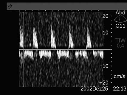
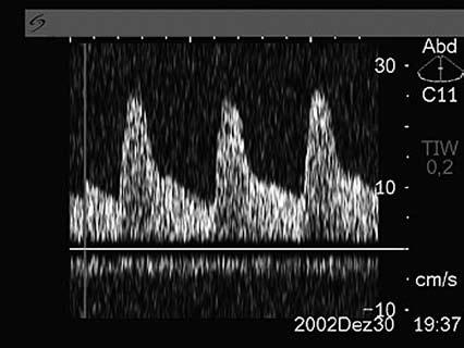
Surgical vascular complications should no longer result in graft loss, especially if early postoperative or even in the operating room a routine Doppler monitoring (Figure 1, 2) is
performed. The assessment of diastolic flow velocity and the calculation of resistance index are of crucial importance. A high vascular resistance is found at high parenchyma pressure, by external compression or by swelling of the kidney for example by an acute tubular necrosis. A negative diastolic flow should lead, without
1 Department of General, Visceral and Transplantation Surgery
University Hospital, Essen, Essen Germany
2 Department of Pediatrics II, Pediatric Nephrology, Endocrinology, Gastroenterology and Transplant Medicine, University Hospital, Essen, Essen Germany
Correspondence: Georgios C. Sotiropoulos georgios.sotiropoulos@ uni-due.de Department of General, Visceral and Transplantation Surgery
University Hospital Essen Hufelandstr. 55, 45122 Essen, Germany
Figure 1. Αcute outflow obstruction.
Figure 2. Αcute outflow obstruction after decompression.
delay, to an operational revision, in order to exclude a venous compression or renal vein thrombosis.
Thrombotic complications
30 years ago Lindström et al. had already defined [1] the thrombotic complications as the most common surgical cause of graft loss. Arbus et al. published in 1983 the experience of the paediatric transplantation program in Toronto [2] and concluded that thrombosis occurred more frequently in children less than 6 years who had received a large adult kidney. This phenomenon was particularly pronounced, when the natural kidneys were left with a high rest diuresis in situ. Broyer [3] in 1990 published the experience of the paediatric transplantation program in Paris. A renal vein thrombosis was responsible in 12 from 53 cases of graft loss. Young age of donor or recipient was identified as special risk factors. In contrast, protective effects were found regarding the occurrence of vascular rejection under the use of OKT3. A prospective study conducted in Paris proved that the prophylactic enoxaparin, did significantly reduce the thrombosis rate in the treated patients [4].
The cause of graft failure in about 2000 children was investigated in an analysis of the North American Paediatric Renal Transplantation Cooperative Study (NAPRTCS) [5,6]. Over a study period of 15 years, the chronic rejection with a rate of 33%, followed by acute rejection and renal vascular thrombosis with 15% and 12% respectively, proved to be the main causes of graft loss. The analysis of the last five years resulted in a change of the leading causes: renal vascular thrombosis was in the first place with 21%, followed by chronic and acute rejection with 19% and 9% respectively. The overall incidence of renal vascular thrombosis was 2.1%. The risk factors for the occurrence of renal vascular thrombosis were analyzed in a multivariate analysis (use of IL-2 receptor antibodies, deceased donors’ kidneys, previous transplantation, peritoneal dialysis before transplantation and more than 5 transfusions). Only the use of IL2 receptor antibodies seemed to have a significant effect on decreasing the incidence of renal vascular thrombosis. If this conclusion represents a statistical artifact of registry data, or whether immunological factors do really trigger the occurrence of renal vascular thrombosis can not be answered from this study.
Virchow's triad and thrombotic complications after renal transplantation
In a differentiated approach to the possible causes of vascular thrombosis after kidney transplantation, the classical Virchow's triad is particularly helpful,
since not a single factor, but a combination of them, is responsible for the occurrence of renal vascular thrombosis.
Stasis: risk factors for stasis consist all low flow situations in the transplanted kidney, especially in the transplantation of adult kidneys in an infant recipient, and predominantly in cases where the anastomosis is not between renal arteries and aorta [7], but between renal artery and iliac vessels of the young receiver. The large for size situation leads to a reduction of blood flow velocity. Simultaneous organ swelling with increase in the resistance may also reduce the blood flow velocity. Congestion and compression of the kidney transplant in a young child are additional risk factors that occur more frequently in the transplantation.
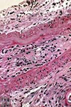
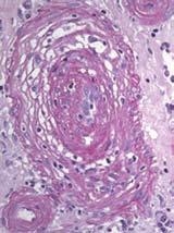
Endothelial lesions: reperfusion injury may provoke an endothelial swelling and an increased expression of adhesion molecules on endothelial cells, favoring the adhesion of platelets and leukocytes. Frequently, endothelial lesions do occur during a vascular rejection procedure (Figure3, 4). Calcineurin inhibitors, such as cyclosporine and tacrolimus, may lead to a vascular toxic vasoconstriction. For an older kidney, possible atherosclerosis represents an additional risk factor. Furthermore, the use of additional patches and a long warm ischemia time should be mentioned as surgical causes of endothelial lesions.
Increase of viscosity: this can occur in the framework of dehydration [8], particularly in cases of polyuria. Infants who receive an adult’s organ are, due to high fluid turnover, at particular risk for the development of dehydration. Changes in the coagulation factors, particularly in the direction of hypercoagulability [9] play a significant role. More frequent risk factors for the development of thrombosis are the APC-resistance (hetero-or homozygosity for mutations of factor V), hetero-or homozygosity for mutations in
Figure 3. Αcute vascular rejection.
the prothrombin gene, increased levels of homocysteine, elevated levels of factor VIII and the Lupus anticoagulant. Rarer causes of an increased risk of thrombosis are the degradation of proteins C, S activity, the decreased concentration of ATIII, and the presence of cardiolipin antibodies and lipoprotein a. Very rare causes are dysfibrinogenemia and increased activity of factor IX and XI.
A possible accumulation of the above mentioned risk factors, in the context of Virchow's triad, during kidney transplantation in childhood, sets this age group at high risk of thrombosis.
Strategies to reduce renal vascular thrombosis
In Essen a preoperative screening for coagulation disorders [9] is being routinely performed since 1998, as part of the paediatric kidney transplantations program. The determined factors include prothrombin time, partial thrombin time, fibrinogen, antithrombin III, protein C and S, activated protein C resistance (APC), factor V Leiden mutation (FVR506Q), prothrombin mutation (G20210A), antiphospholipid antibodies and MTHFR mutations.
In 90 studied children, there were found11 cases (12.2%) of increased risk of thrombosis. A factor V Leiden mutation was found in 7 cases, a fibrinogen mutation in two cases, antiphospholipid antibodies were detected in two cases (one child had a history of two graft losses as a result of renal vein thrombosis) and in 5 cases, thrombosis had already occurred in the background without any identifiable cause. The Essen-based therapeutic strategy for patients without risk of thrombophilia consists of a postoperative low dose heparin for 2 weeks. By patients with thrombophilia, the heparinisation begins already intraoperatively and continues for the two first postoperative weeks. All patients receive low-dose aspirin for 1 year.
In the study by Kranz et al. no case of thrombosis occurred, while bleeding complications occurred twice (in one patient without and in one with thrombophilia), but in both cases without any further adverse consequences. The first year Graft Survival rates in patients without and with thrombophilia were 93% and 100% respectively
Vascular changes of small vessels after renal transplantation
Vascular changes in the small vessels occur as a result of thrombotic microangiopathy, of vascular rejections or of the so-called chronic graft vasculopathy.
The thrombotic microangiopathy (Figure 5) is a histopathological description and not a unique entity. Fibrin deposits in the glomerular and pre-glomerular capillaries are found both in the hyperacute rejection as well as the hemolytic-uremic syndrome. In contrast, the vascular toxicity of calcineurin inhibitor is mainly in the arteriole, the so-called typical cyclosporine or tacrolimusassociated arteriolopathy with hyaline necrosis.
The hemolytic-uremic syndrome after kidney transplantation is a complication that has been described since the beginning of transplantation in the 70 years. Originally, it was equated with severe vascular rejection with insufficient immunosuppression (steroids, azathioprine). Furthermore, the hemolyticuremic syndrome after transplantation was thought to be induced by the calcineurin inhibitors- [10], as this phenomenon occurred in isolated cases of cyclosporine treatment, particularly at high but even at low doses. A switch to tacrolimus showed that this phenomenon may occur with tacrolimus as well. In all proposed theories, this phenomenon is still not properly understood. On the other hand, it is certain that the HUS is not only by calcineurin inhibitors triggered, as there are also many described cases after application of mTOR inhibitors [10].
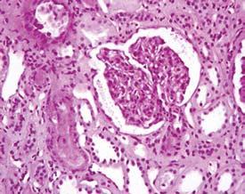
Figure 5. Τhrombotic microangiopathy (1st posttransplantation day)
Figure 4. Αcute chronic vascular rejection.
In cases where a hemolytic uremic syndrome was the cause of renal insufficiency, exists always the danger of disease recurrence in the graft [11,12]. A more detailed recent analysis shows that the risk of recurrence exists exclusively for the atypical HUS. In the so-called typical HUS, which is Verotoxin-induced, the recurrence probability is very low. In a retrospective analysis of patients with loss of the transplanted kidney due to recurrent HUS, the molecular genetic studies revealed frequent defects in the complement system, in particular factor H mutations [13]. Virtually all patients who showed a factor H mutation, have lost their graft in context of a recurrent HUS. In a special investigation in cooperation with Professor Zipfel, Jena, we did demonstrate an antibody against factor H, in a patient with an atypical hemolytic uremic syndrome.
The identification of these risk factors is of great importance, because without new concepts, transplantations in this special group of patients can not promise any success.
Chronic graft vasculopathy
Chronic graft vasculopathy is currently the most common cause of chronic graft dysfunction. The causes are multifactorial. Human immunological factors are likely involved. On the other hand, non-immunological causes, such as arterial hypertension, are considered as pacemaker of chronic graft vasculopathy. Especially in childhood and adolescence, the antihypertensive treatment is often inadequate.[14] Another contributing factor is calcineurin inhibitors. Whether calcineurin inhibitor-free immunosuppressive protocols would resolve this problem, is still open.
The question of endothelial repair processes in the childhood, particularly after an ischemia-reperfusion injury, is unclear. The role of circulating epithelial precursor cells in paediatric graft recipients has not been studied yet, but is eventually indicated by the presence of chimeric endothelial cells in the vascular endothelium of renal transplant (own observations).
Summary
In transplanted children, all vessel segments in the graft kidney could be affected by vascular problems. As risk factors of great vessels complications are easily identifiable, a cautious approach can reduce the problem. Regarding small vessels, many different pathogenetic mechanisms are implicated in thrombotic
microangiopathies, influencing the appropriate therapeutic decision. The problem of chronic graft vasculopathy, which is especially important for the longterm prognosis in children, remains unresolved. Prospective studies with sufficient long-term observation could eventually prove helpful in this direction.
references
1. Lindstrom BL, O. Lindfors (1977) Surgical complications in 500 kidney transplantations. Proc Eur Dial Transplant Assoc 14: 353-361
2.Arbus GS, B. E. Hardy (1983) Cadaveric renal transplants in children under 6 years of age. Kidney Int Suppl 15: S111-115
3.Broyer M, A. Mitsioni (1990) Early failures in kidney transplantation: A retrospective study of 53 cases. Ann Pediatr (Paris) 37: 87-90
4.Broyer M, Gagnadoux MF, Sierro A, Fischer AM, Revillon Y, Jan D, Beurton D, Niaudet P (1991) [Prevention of vascular thromboses after renal transplantation using low molecular weight heparin]. Ann Pediatr (Paris) 38: 397-9
5. Benfield MR (2003) Current status of kidney transplant: update 2003. Pediatr Clin North Am 50: 1301-34
6.McDonald RA, Smith JM, Stablein D, Harmon WE (2003) Pretransplant peritoneal dialysis and graft thrombosis following pediatric kidney transplantation: a NAPRTCS report. Pediatr Transplant 7: 204-8
7.Vester U, Kranz B, Testa G, Paul A, Broelsch CE, Hoyer PF (2004) Medical and surgical aspects of pediatric renal transplantation using living donors. Transplant Proc 36: 1308-10
8.Ismail H, Kalicinski P, Drewniak T, Smirska E, Kaminski A, Prokurat A, Grenda R, Szymczak M, Chrupek M, Markiewicz M (1997) Primary vascular thrombosis after renal transplantation in children. Pediatr Transplant 1: 43-7
9.Kranz B, Vester U, Nadalin S, Paul A, Broelsch CE, Hoyer PF (2006) Outcome after kidney transplantation in children with thrombotic risk factors. Pediatr Transplant 10: 788-93
10.Ponticelli C, Banfi G (2006) Thrombotic microangiopathy after kidney transplantation. Transpl Int 19: 789-94
11.Quan A, Sullivan EK, Alexander SR (2001) Recurrence of hemolytic uremic syndrome after renal transplantation in children: a report of the North American Pediatric Renal Transplant Cooperative Study. Transplantation 72: 742-5
12.Eijgenraam FJ, Donckerwolcke RA, Monnens LA, Proesmans W, Wolff ED, van Damme B (1990) Renal transplantation in 20 children with hemolytic-uremic syndrome. Clin Nephrol 33: 87-93
13.Zimmerhackl LB, Besbas N, Jungraithmayr T, van de Kar N, Karch H, Karpman D, Landau D, Loirat C, Proesmans W, Prufer F, Rizzoni G, Taylor MC (2006) Epidemiology, clinical presentation, and pathophysiology of atypical and recurrent hemolytic uremic syndrome. Semin Thromb Hemost 32: 113-120
14.Seeman T., Hypertension after renal transplantation.Pediatr Nephrol. 2009 May;24(5):959-972
Hypertension and obesity in children and adolescents
S. Stabouli1, V. Kotsis2
Abstract: Obesity is a chronic disease prevalent in both developed and developing countries, which affects children and adolescents as well as adults. Epidemiological studies have demonstrated a higher prevalence of hypertension in obese than in non-obese children and adolescents in a variety of ethnic and racial groups, using clinic blood pressure measurements. Τhe pathogenesis of obesity induced hypertension implicates adipose tissue derivatives (adipokines and cytokines), neurohumoral pathways, metabolic functions and modulation of pressor/depressor mechanisms. Evidence is accumulating of early target organ damage in obese young people, emphasizing the fact that the adverse effects of weight gain may be apparent even in childhood. There is compelling evidence that overweight status and elevated blood pressure are closely related and increase cardiovascular risk synergistically. The pathogenetic process of cardiovascular disease appears to begin in childhood, and obesity may accelerate the progression of the disease. Weight loss and exercise remain the cornerstone for the management of obese children and adolescents with hypertension. The presence of left ventricular hypertrophy may be an indication for more aggressive therapy aimed at inducing the regression of silent target organ damage before a clinical event develops. In addition to lifestyle modifications, antihypertensive medication should be considered for obese children with stage 2 hypertension and for those with left ventricular hypertrophy.
Key words: Obesity, hypertension, target organ, sympathetic nervous system
1 Paediatric Intensive Care Unit, Hippokration Hospital, Thessaloniki
2 Hypertension-ABPM centre, 3rd Department of Medicine, Papageorgiou Hospital, Thessaloniki, Greece
Correspondence: S. Stabouli sstaboul@med.uoa.gr Paediatric Intensive Care Unit, Hippokration Hospital, Thessaloniki 55132, Kalamaria
Regression
(Stabouli et al, unpublished data).
1. Lobstein T, Baur L, Uauy R; IASO International Obesity TaskForce. Obesity in children and young people: a crisis in public health. Obes Rev 2004;5 (Suppl 1):4-104.
2. Wang Y, Lobstein T. Worldwide trends in childhood overweight and obesity. Int J Pediatr Obes 2006;1:11-25.
3. Ogden CL, Flegal KM, Carroll MD, Johnson CL. Prevalence and trends in overweight among US children and adolescents, 1999-2000. JAMA 2002;288:1728-1732.
4. Jackson-Leach R, Lobstein T. Estimated burden of paediatric obesity and co-morbidities in Europe. Part 1. The increase in the prevalence of child obesity in Europe is itself increasing. Int J Pediatr Obes 2006;1:26-32.
5. International Obesity Task Force. EU childhood obesity “out of control”: IOTF Childhood Obesity Report May 2004.
6. Daniels SR, Arnett DK, Eckel RH, Gidding SS, Hayman LL, Kumanyika S, et al. Overweight in children and adolescents: pathophysiology, consequences, prevention, and treatment. Circulation 2005;111:1999-2012.
7. Lobstein T, Jackson-Leach R. Estimated burden of paediatric obesity and co-morbidities in Europe. Part 2. Numbers of children with indicators of obesity-related disease. Int J Pediatr Obes 2006;1:33-41.
8. World Health Organization. Obesity: Preventing and managing the global epidemic. Report of a WHO consultation. Geneva, Switzerland: WHO technical report series 894, 2000.
9. American Academy of Pediatrics, Committee on Nutrition. Prevention of pediatric overweight and obesity. Pediatrics 2003;112:424-430.
10. Cole TJ, Bellizzi MC, Flegal KM, Dietz WH. Establishing a
standard definition for child overweight and obesity worldwide: international survey. BMJ 2000; 320:1240-3.
11. Cole TJ, Freeman JV, Preece MA. British 1990 growth reference centiles for weight, height, body mass index and head circumference fitted by maximum penalized likelihood. Stat Med 1998;17:407-29.
12. Davy KP, Hall JE. Obesity and hypertension: two epidemics or one? Am J Physiol Regul Integr Comp Physiol 2004;286:R803-13.
13. Gutin B, Basch C, Shea S, Contento I, DeLozier M, Rips J, Irigoyen M, Zybert P. Blood pressure, fitness, and fatness in 5- and 6-year-old children. JAMA 1990;264:1123-7.
14. Clarke WR, Woolson RF, Lauer RM. Changes in ponderosity and blood pressure in childhood: the Muscatine Study. Am J Epidemiol 1986;124:195-206.
15. Sorof JM, Daniels S. Obesity hypertension in children: a problem of epidemic proportions. Hypertension 2002;40:441-447.
16. Robinson RF, Batisky DL, Hayes JR, Nahata MC, Mahan JD. Body mass index in primary and secondary pediatric hypertension. Pediatr Nephrol 2004;19:1379-84.
17. Macedo ME, Trigueiros D, de Freitas F. Prevalence of high blood pressure in children and adolescents. Influence of obesity. Rev Port Cardiol 1997;16:27-28.
18. Sorof JM, Poffenbarger T, Franco K, Bernard L, Portman RJ. Isolated systolic hypertension, obesity, and hyperkinetic hemodynamic states in children. J Pediatr 2002;140;660-666.
19. Sorof JM, Lai D,Turner G, Poffenbarger T, Portman RJ. Overweight, ethnicity, and the prevalence of hypertension in school-aged children. Pediatrics 2004;113:475-482.
20. Rosner B, Prineas R, Daniels SR, Loggie J. Blood pressure differences between blacks and whites in relation to body size among US children and adolescents. Am J Epidemiol 2000;151:1007-19.
21. Freedman DS, Dietz WH, Srinivasan SR, Berenson GS. The relation of overweight to cardiovascular risk factors among children and adolescents: the Bogalusa Heart Study. Pediatrics 1999;103:1175-82.
22. Sorof JM, Urbina EM, Cunningham RJ, Hogg RJ, MoxeyMims M, Eissa MA, et al. Ziac Pediatric Hypertension Study Group. Screening for eligibility in the study of antihypertensive medication in children: experience from the Ziac Pediatric Hypertension Study. Am J Hypertens 2001;14:783-7.
23. Stabouli S, Kotsis V, Papamichael C, Constantopoulos A, Zakopoulos N. Adolescent obesity is associated with high ambulatory blood pressure and increased carotid intimalmedial thickness. J Pediatr 2005;147: 651-6.
24. Grassi G, Seravalle G, Cattaneo BM, Bolla G, Lafranchi A, Colombo M, et al. Sympathetic activation in obese normotensive subjects. Hypertension 1995;25:560-563.
25. Rocchini AP, Yang IQ, Gokee A. Hypertension and insulin resistance are not directly related in obese dogs. Hypertension 2004; 43:1011-1016.
26. Wofford MR, Anderson DC Jr, Brown CA, Jones DW, Miller ME, Hall JE. Antihypertensive effect of alpha- and beta-adrenergic blockade in obese and lean hypertensive subjects. Am J Hypertens 2001; 14:694-8.
27. Voors AW, Webber LS, Berenson GS. Resting heart rate and pressure-rate product of children in a total biracial community. The Bogalusa Heart Study. Am J Epidemiol. 1982;116:276–286
28. Rocchini AP, Katch V, Schork A, Kelch RP. Insulin and blood pressure during weight loss in obese adolescents. Hypertension 1987;10:267–273.
29. Hall JE, Brands MW, Hildebrandt DA, Kuo J, Fitzgerald S. Role of sympathetic nervous system and neuropeptides in obesity hypertension. Brazilian J Med Biol Research 2000; 33:605-618.
30. Hall JE. Mechanisms of abnormal sodium handling in obesity hypertension. Am J Hypertens 1997; 10:S49-S55.
31. Ruano M, Silvestre V, Castro R, García-Lescún MC, Rodríguez A, Marco A, et al. Morbid obesity, hypertensive disease and the renin-angiotensin-aldosterone axis. Obes Surg 2005;15:670-6.
32. Kidambi S, Kotchen J, Grim C, Raff H, Mao J, Singh R, et al. Association of Adrenal Steroids With Hypertension and the Metabolic Syndrome in Blacks. Hypertension 2007;49;704-711.
33. Massièra F, Bloch-Faure M, Ceiler D, Murakami K, Fukamizu A, Gasc JM, et al. Adipose angiotensinogen is involved in adipose tissue growth and blood pressure regulation. The FASEB Journal 2001;15:2727-2729.
34. Hall JE, Brands MW, Dixon WN, Smith MJ. Obesityinduced hypertension. Renal function and systemic hemodynamics. Hypertension 1993; 22:292-299.
35. Landsberg L. Diet, obesity and hypertension: A hypothesis involving insulin, the sympathetic nervous system, and adaptive thermogenesis. Q J Med 1986;236:1081-1090.
36. Sakaguchi T, Bray GA. Intrahypothalamic injection of insulin decreases firing rate of sympathetic nerves. Proc Natl Acad Sci USA 1987;84:2012-2014.
37. Hall JE. Hyperinsulinemia: A link between obesity and hypertension? Kidney International 1993;43:1402-1417.
38. Hall JE, Brands MW, Zappe DH, Dixon WN, Mizelle L, Reinhart GA, Hildebrandt DA. Hemodynamic and renal responses to chronic hyperinsulinemia in obese, insulinresistant dogs. Hypertension 1995;25:994-1002.
39. DeCourten M, Zimmet P, Hodge A, Collins V, Nicolson M, Staten M, et al. Hyperleptinemia: the missing link in the metabolic syndrome? Diabetic Medicine 1997;14:200-208.
40. Wynne K, Stanley S, McGowan B, Bloom S. Appetite control. Journal of Endocrinology 2005;184:291-318.
41. Engeli S, Sharma A. Emerging concepts in the pathophysiology and treatment of obesity-associated hypertension. Curr Opin Cardiol 2002;17:355-359
42. Correia ML, Haynes WG, Rahmouni K, Morgan DA, Sivitz WI, Mark AL. The concept of selective leptin resistance: evidence from agouti yellow obese mice. Diabetes 2002;51:439-42.
43. Denis RGP, Bing C, Brocklehurst S, Harrold JA, Vernor RG, Williams G. Diurnal changes in hypothalamic neuropeptide and SOCS-3 expression: effects of lactation and relationship with serum leptin and food intake. J Endocr 2004;183:173-181.
44. Weyer C, Funahashi T, Tanaka S, Hotta K, Matsuzawa Y, Pratley RE, et al. Hypoadiponectinemia in obesity and type 2 diabetes: close association with insulin resistance and hyperinsulinemia. J Clin Endocr Metab 2001;86:19301935.
45. Pilz S, Horejsi R, Moller R, Almer G, Scharnagl H, Stojakovic T, et al. Early Atherosclerosis in Obese Juveniles Is Associated with Low Serum Levels of Adiponectin. J Clin Endocrinol Metab 2005;90: 4792–4796.
46. Sethna CB, Leonard MB, Gallagher PR, Meyers KE. Serum adiponectin levels and ambulatory blood pressure monitoring in pediatric renal transplant recipients. Transplantation. 2009;88:1030-7.
47. Rees A, Thomas N, Brophy S, Knox G, Williams R. Cross sectional study of childhood obesity and prevalence of risk factors for cardiovascular disease and diabetes in children aged 11-13.BMC Public Health. 2009;9:86.
48. Litwin M, Sladowska J, Syczewska M, Niemirska A, Daszkowska J, Antoniewicz J, et al. Different BMI cardiovascular risk thresholds as markers of organ damage and metabolic syndrome in primary hypertension.Pediatr Nephrol. 2008;23:787-96.
49. Kim F, Pham M, Maloney E, Rizzo NO, Morton GI, Wisse BE, et al. Vascular inflammation, insulin resistance and reduced nitric oxide production precede the onset of peripheral insulin resistance. Arteriosclerosis, Thrombosis, and Vascular Biology 2008;28:1982-1988.
50. Kapiotis S, Holzer G, Schaller G, Haumer M, Widhalm H, Weghuber D, et al. A Proinflammatory State Is Detectable in Obese Children and Is Accompanied by Functional and Morphological Vascular Changes. Arterioscler. Thromb. Vasc. Biol 2006;26;2541-2546.
51. Meyer A, Kundt G, Steiner M, Schuff-Werner P, Kienast W. Impaired Flow-Mediated Vasodilation, Carotid Artery Intima-Media Thickening, and Elevated Endothelial Plasma Markers in Obese Children: The Impact of Cardiovascular Risk Factors. Pediatrics 2006;117;15601567.
52. Geiss HC, Parhofer KG, Schwandt P. Parameters of childhood obesity and their relationship to cardiovascular risk factors in healthy prepubescent children. Int J Obes Relat Metab Disord 2001;25:830-837.
53. Wright CM, Parker L, Lamont D, Craft AW. Implications of childhood obesity for adult health: findings from thousand families’ cohort study. BJM 2001;323:1280-1284.
54. McGill HC Jr, McMahan CA, Zieske AW, Malcom GT, Tracy RE, Strong JP. Effects of nonlipid risk factors on atherosclerosis in youth with a favorable lipoprotein profile. Circulation 2001;103:1546 –1550.
55. Kortelainen ML. Adiposity, cardiac size and precursors of coronary atherosclerosis in children: a prospective study of 210 violent deaths. Int J Obes Relat Metab Disord 1997;21:691-697.
56. Toprak A, Wang H, Chen W, Paul T, Srinivasan S, Berenson G. Relation of childhood risk factors to left ventricular hypertrophy (eccentric or concentric) in relatively young adulthood (from the Bogalusa Heart Study). Am J Cardiol 2008;101: 1621-5.
57. Berenson GS, Srinivasan SR, Bao W, Newman WP 3rd, Tracy RE, Wattigney WA. Association between multiple cardiovascular risk factors and atherosclerosis in children and young adults. The Bogalusa Heart Study. N Engl J Med 1998;338: 1650-6.
58. Daniels SR, Kimball TR, Morrison JA, Khoury P, Witt S, Meyer RA. Effect of lean body mass, fat mass, blood pressure, and sexual maturation on left ventricular mass in children and adolescents. Statistical, biological, and clinical significance. Circulation.. 1995;92: 3249-54.
59. Stabouli S, Kotsis V, Zakopoulos N. Ambulatory blood pressure monitoring and target organ damage in pediatrics. J Hypertens 2007;25:1979-86.
60. Hanevold C, Waller J, Daniels S, Portman R, Sorof J. International Pediatric Hypertension Association. The effects of obesity, gender, and ethnic group on left ventricular hypertrophy and geometry in hypertensive children: a collaborative study of the International Pediatric Hypertension Association. Pediatrics.2004;113:328-33.
61. Daniels SR, Loggie JM, Khoury P, Kimball TR. Left ventricular geometry and severe left ventricular hypertrophy in children and adolescents with essential hypertension. Circulation 1998;97: 1907-11.
62. Tounian P, Aggoun Y, Dubern B, Varille V, Guy-Grand B, Sidi D, et al. Presence of increased stiffness of common carotid artery and endothelial dysfunction in severely obese children: a prospective study. Lancet 2001;358: 14001404.
63. Reinehr T, Kiess W, de Sousa G, Stoffel-Wagner B, Wunsch R. Intima media thickness in childhood obesity: relations to inflammatory marker, glucose metabolism, and blood pressure. Metabolism 2006;55:113-8.
64. Sorof JM, Alexandrov AV, Cardwell G, Portman RJ. Carotid artery intimal-medial thickness and left ventricular hypertrophy in children with elevated blood pressure. Pediatrics 2003;111:61-6.
65. Redon J, Ruilope LM. Microalbuminuria as an intermediate endpoint in essential hypertension: evidence is coming. J Hypertens 2004;22:1679–1681.
66. Nguyen S, McCulloch C, Brakeman P, Portale A, Hsu CY. Being overweight modifies the association between cardiovascular risk factors and microalbuminuria in adolescents. Pediatrics 2008;121:37-45.
67. Thompson DR, Obarzanek E, Franko DL, Barton BA, Morrison J, Biro FM, et al. Childhood overweight and cardiovascular disease risk factors: the National Heart, Lung, and Blood Institute Growth and Health Study. J Pediatr 2007;150:18-25.
68. Morrison JA, Sprecher DL, Barton BA, Waclawiw MA, Daniels SR. Overweight, fat patterning, and cardiovascular disease risk factors in black and white girls: The National Heart, Lung, and Blood Institute Growth and Health Study. J Pediatr 1999;135:458-64.
69. Morrison JA, Barton BA, Biro FM, Daniels SR, Sprecher DL. Overweight, fat patterning, and cardiovascular disease risk factors in black and white boys. J Pediatr 1999;135:451-7.
70. Reinehr T, de Sousa G, Toschke AM, Andler W. Long-term follow-up of cardiovascular disease risk factors in children after an obesity intervention. Am J Clin Nutr 2006;84:490-6.
71. Spear BA, Barlow SE, Ervin C, Ludwig DS, Saelens BE, Schetzina KE, et al. Recommendations for treatment of child and adolescent overweight and obesity. Pediatrics 2007;120 (Suppl 40):S254-88.
72. McGovern L, Johnson JN, Paulo R, Hettinger A, Singhal V, Kamath C, et al. Clinical review: treatment of pediatric obesity: a systematic review and meta-analysis of randomized trials. J Clin Endocrinol Metab 2008;93:4600-5.
73. Oude Luttikhuis H, Baur L, Jansen H, Shrewsbury VA, O'Malley C, Stolk RP, et al. Interventions for treating obesity in children. Cochrane Database Syst Rev 2009 ;1:CD001872.
74. European Medicines Agency. Press Release: Meeting Highlights from the Committee for Medicinal Products for Human Use (CHMP); 22 January 2010 EMA/ CHMP/32068/21010.
75. Inge TH, Krebs NF, Garcia VF, Skelton JA, Guice KS, Strauss RS, et al. Bariatric surgery for severely overweight adolescents: concerns and recommendations. Pediatrics 2004;114:217-23.
76. Lande MB, Flynn JT. Treatment of hypertension in children and adolescents. Pediatr Nephrol 2009;24:1939-49.
77. Mancia G, Grassi G, Zanchetti A. New-onset diabetes and antihypertensive drugs. J Hypertens 2006;24:3–10.
78. Lurbe E, Cifkova R, Cruickshank JK, Dillon MJ, Ferreira I, Invitti C,et al. Management of high blood pressure in children and adolescents: recommendations of the European Society of Hypertension.European Society of Hypertension. J Hypertens. 2009 ;27:1719-42.
79. Pischon T, Sharma AM. Use of beta-blockers in obesity hypertension: potential role of weight gain. Obes Rev 2001; 2:275–280.
80. Jacob S, Rett K, Henriksen EJ. Antihypertensive therapy and insulin sensitivity: do we have to redefine the role of beta-blocking agents? Am J Hypertens 1998;11:1258–1265.
Neonatal Intensive Care Unit (NICU), Child Health Department, University of Ioannina, Greece
Correspondence:
Vasileios Giapros vgiapros@cc.uoi.gr
Neonatal Intensive Care Unit, Child Health Department, University of Ioannina, Greece
Early growth disturbances and development of the metabolic syndrome
V. Giapros, S. Andronikou
Abstract: Individuals who are born either small (SGA) or large (LGA) for gestational age and those who experience early catch-up growth are characterized as having early growth disturbances. These conditions have been related with several parameters of the metabolic syndrome as insulin resistance (IR), diabetes mellitus (DM) type-2, obesity, elevated blood pressure and dislipidemia. Derangements in the production, secretion and action of insulin in SGA children have been observed as early as in the age of 3 years with deterioration with age. These individuals are prone to develop obesity and central fat distribution, which is linked with IR. Individuals with in-utero growth restriction may have a lower nephron endowment and their blood pressure may be elevated at preschool age. Postnatal catch-up growth in SGA children is related with abnormal serum lipids profile while childhood obesity in the same group of children is related with elevated triglycerides. Birth weight and any information about the type, cause, and severity of inutero growth restriction as well as postnatal catch-up growth patterns should be recorded in these children. There is inadequate evidence to recommend special interventions or screening test in individuals with early growth disturbances outside of normal clinical practice.
Key words: Low birth weight, metabolic syndrome, insulin resistance, blood pressure, in-utero growth restriction
Βιβλιογραφία
1. Barker DJP, Osmond C, Winder PD Margetts B, Simmonds SJ. Weight in infancy and death from ischaemic heart disease. Lancet 1989;2:577-580
2. Stein CE, Fall CHD, Kumaran K, Osmond C, Cox V, Barker DJ. Fetal growth and coronary heart disease in South India. Lancet 1996; 348: 1269-1273
3. Hales CN, Barker DJP, Clark PMS, Cox LJ, Fall C, Osmond C et al. Fetal and infant growth and impaired glucose tolerance at age 64. BMJ 1991;303: 1019-1022
4. Newsome CA, Shiell AW, Fall CHD, Phillips DI, Shier R, Law CM. Is birth weight related to later glucose and insulin metabolism? A systematic review. Diabetes Med 2003;20: 339-348
5. Curhan GC, Chertow GM, Willet WC. Birth weight and adult hypertension and obesity in women. Circulation 1996;94: 1310-1315
6. Barker D. Adult consequences of fetal growth restriction. Clin Obstetr Gynecol 2006; 49:270-283
7. Boubred F, Daniel L, Buffat C, Feuerstein JM, Tsimaratos M, Oliver C, Dignat-George F, Lelièvre-Pégorier M, Simeoni U. Early postnatal overfeeding induces early chronic renal dysfunction in adult male rats. Am J Physiol Renal Physiol. 2009;297:F943-51
8. Villar-Martini VC, Carvalho JJ, Neves MF, Aguila MB, Mandarim-de-Lacerda CA. Hypertension and kidney alterations in rat offspring from low protein pregnancies. J Hypertens 2009; 27:S47-51
9. Grundy SM, Brewer HB Jr, Cleeman JI, Smith SC Jr, Lenfant C; American Heart Association; National Heart, Lung, and
Blood Institute. Definition of metabolic syndrome: Report of the National Heart, Lung, and Blood Institute/American Heart Association conference on scientific issues related to definition. Circulation 2004 27; 109 :433-438
10. Στεφανοπούλου
2009; 72:274-282
11. Saenger P, Czernichow P, Hughes I, Reiter E. Small for gestational age: Short stature and beyond. Endocrin Rev 2007; 28: 219-251
12. Embleton ND, Pang N, Cooke RJ. Postnatal malnutrition and growth retardation : an inevitable consequence of current recommendations in preterm infants? Pediatrics 2001;107: 270–273
13. Monteiro P,Victora C. Rapid growth in infancy and childhood and obesity in later life-a systematic review. Obesity Rev 2005; 6: 143-154
14. Ibanez L, Lopez-Bermejo A, Suarez L, Marcos MV, Diaz M, de Zegher F. Visceral adiposity without overweight in children born small for gestational age. J Clin Endocrinol Metab 2008: 93; 2079-2083
15. Ibanez L, Lopez-Bermejo A, Diaz M, Marcos MV, Casano P, de Zegher F. Abdominal fat partitioning and high molecular weight adiponectin in short children born small for gestational age. J Clin Endocrinol Metab 2009;94: 10491052
16. Ong KK, Petry CJ, Emmett PM, Sandhu MS, Kiess W, Hales CN, et al. ALSPAC study team. Insulin sensitivity and secretion in normal children related to size at birth, postnatal growth, and plasma insulin-like growth factor-I levels. Diabetologia 2004;47:1064-1070
17. Reinehr T, Kleber M and Toschke AM. Small for gestational age status is associated with metabolic syndrome in overweight children. Eur J Endocrinol 2009; 160: 579–584
18. Sun M, Schutz Y, Grezzani A, Clementi M, Gaudino R, Maffeis C. Body size and early growth in appropriate- and large-for-gestational-age infants. Acta Paediatr 2003; 92: 1127-1132
19. Hediger M, Overpeck M, Maurer K, Kuczmarski RJ, McGlynn A, Davis WW. Growth of Infants and Young Children Born. Small or Large for Gestational Age. Arch Pediatr Ad Med 1998;152: 1225-1231
20. Boney CM, Verma A, Tucker R, Vohr BR. Metabolic syndrome in childhood: Association with birth weight, maternal obesity, and gestational diabetes mellitus. Pediatrics 2005;115:290-296
21. Evagelidou E, Kiortsis D, Bairaktari E, Giapros V, Cholevas V, Tzallas C et al. Lipid profile, glucose homeostasis, blood pressure, and obesity-anthropometric markers in macrosomic offspring of nondiabetic mothers. Diabetes Care 2006; 29: 1197-1201
22. Beltrand J, Levy-Marchal C. Pathophysiology of insulin resistance in subjects born small for gestational age. Best Pract Res Clin Endocrinol Metab 2008; 22 503-515
23. Mericq V, Ong KK, Bazaes R, Peña V, Avila A, Salazar T, et al. Longitudinal changes in insulin sensitivity and secretion from birth to age three years in small- and appropriate-forgestational-age children. Diabetologia 2005; 48:2609-2614
24. Evagelidou EN, Giapros VI, Challa AS, Kiortsis DN, Tsatsoulis AA, Andronikou SK. Serum adiponectin levels, insulin resistance, and lipid profile in children born small for gestational age are affected by the severity of growth retardation at birth. Eur J Endocrinol 2007;156:271-277
25. Jaquet D, Deghmoun S, Chevenne D, Collin D, Czernichow P, Lévy-Marchal C. Dynamic change in adiposity from fetal to postnatal life is involved in the metabolic syndrome associated with reduced fetal growth. Diabetologia 2005; 48: 849-855
26. Veening MA, Van Weissenbruch MM, Delemarre-Van De Waal HA. Glucose tolerance insulin sensitivity and insulin secretion in children born small for gestational age. J Clin Endocrinol Metab 2002; 87: 4657-4661
27. Fewtrell MS, Doherrty C, Cole TJ, Stafford M, Hales CN, Lucas A. Effects of size at birth, gestational age and early growth in preterm infants on glucose and insulin concentrations at 9-12 years. Diabetologia 2000; 43: 714717
28. Bazaes RA, Alegria A, Pittaluga E Avila A, Iniguez G, Mericq V. Determinants of insulin sensitivity and secretion in very-low-birth-weight children. J Clin Endocrinol Metab 2004: 89; 1267-1272
29. Rogers I and the EURO-BLCS study group. The influence of birthweight and intrauterine enviroment on adiposity and fat distribution in later life. Intern J Obes 2003; 27: 755-777
30. Elia M, Betts P, Jackson DM, Mulligan J. Fetal programming of body dimensions and percentage body fat measured in prepubertal children with a 4-component model of body composition, dual-energy X-ray absorptiometry, deuterium dilution, densitometry, and skinfold thicknesses. Am J Clin Nutr 2007;86:618-624
31. Matthes JW, Lewis PA, Davies DP, Bethel JA. Body size and subcutaneous fat patterning in adolescence. Arch Dis Child 1996; 75:521-523.
32. Meas T, Deghmoun S, Armoogum P, Alberti C, LevyMarchal. Consequences of being born small for gestational age on body composition: an 8-year follow-up study. J Clin Endocrinol Metab 2008; 93 :3804-3809
33. Hediger ML, Overpeck MD, Kuczmarski RJ, McGlynn A, Maurer KR, Davis WW. Muscularity and fatness of infants and young children born small- or large-for-gestationalage. Pediatrics 1998;02(5):E60
34. Ong KK. Catch-up growth in small for gestational age babies: good or bad? Curr Opin Endocrinol Diabetes Obes 2007;14: 30-34
35. Ekelund U, Ong K, Linné Y, Neovius M, Brage S, Dunger DB et al. Upward weight percentile crossing in infancy and early childhood independently predicts fat mass in young adults: the Stockholm Weight Development Study (SWEDES).Am J Clin Nutr 2006; 83:324-330
36. McCarthy A, Hughes R, Tilling K, Davies D, Smith GD, Ben-Shlomo Y. Birth weight; postnatal, infant, and childhood growth; and obesity in young adulthood: evidence from the Barry Caerphilly Growth Study. Am J Clin Nutr 2007; 86:907-913
37. Bol VV, Delattre AI, Reusens B, Raes M, Remacle C. Forced catch-up growth after fetal protein restriction alters the adipose tissue gene expression program leading to obesity in adult mice. Am J Physiol Regul Integr Comp Physiol 2009; 297 :R291-29
38. Guan H, Arany E, van Beek JP, Chamson-Reig A, Thyssen S, Hill DJ, Yang K. Adipose tissue gene expression profiling reveals distinct molecular pathways that define visceral adiposity in offspring of maternal protein-restricted rats. Am J Physiol Endocrinol Metab. 2005;288:E663-673
39. Giapros V, Evagelidou E, Challa A, Kiortsis D, Drougia A, Andronikou S. Serum adiponectin and leptin levels and insulin resistance in children born large-for-gestationalage are affected by the degree of overweight. Clin Endocrinol 2007; 66: 353-359
40. Vickers MH, Gluckman PD, Coveny AH, Hofman PL, Cutfield WS, Gertler A, et al. Neonatal leptin treatment reverses developmental programming. Endocrinology 2005;146:4211-4216
41. Singhal A, Fewtrell M, Cole TJ, Lucas A. Low nutrient intake and early growth for later insulin resistance in adolescents born preterm. Lancet 2003; 361:1089-1097
42. Huxley R, Owen CG, Whincup PH, Cook DG, Colman S, Collins R. Birth weight and subsequent cholesterol levels: exploration of the "fetal origins" hypothesis. JAMA 2004; 292:2755-2764
43. Tenhola S, Martikainen A, Rahiala E, Herrgârd E, Halonen P, Voutilainen R. Serum lipid concentrations and growth characteristics in 12-year-old children born small for gestational age. Pediatr Res 2000;48:623-628
44. Torre P, Ladaki C, Scire G, Spadoni GL, Cianfarani S. Catch-up growth in body mass index is associated neither with reduced insulin sensitivity nor with altered lipid profile in children born small for gestational age. J Endocrinol Invest 2008;31:760-764
45. Bavdekar A, Yajnik C, Fall C, Bapat S, Pandit A, Deshpande V et al. Insulin resistance syndrome in 8-year-old Indian children. Small at birth, big at 8 years, or both? Diabetes 1999; 48:2422-2429.
46. Rotteveel J, van Weissenbruch MM, Twisk JWR, DelemarreVan de Waal HA. Abnormal lipid profile and hyperinsulinaemia after a mixed meal: additional cardiovascular risk factors in young adults born preterm. Diabetologia 2008; 51:1269–1275
47. Merzouk H, Bouchenak M, Loukidi B, Madani S, Prost J, Belleville J: Fetal macrosomia related to maternal poorly controlled type 1 diabetes strongly impairs serum lipoprotein concentrations and composition. J Clin Pathol 2000; 53:917-923
48. Huxley RR, Shiell AW, Law CM. The role of size at birth and postnatal catch-up growth in determining systolic blood pressure: a systematic review of the literature. J Hypertens 2000;18:815-831
49. .Huxley R, Neil A, Collins R. Unraveling the fetal origins hypothesis: is there really an inverse association between birth weight and subsequent blood pressure? Lancet 2002; 360:659-665
50. Lackland DT, Egan BM, Syddal HE, Barker DJ. Associations between birthweight and antihypertensive medication in black and white Americans. Hypertension 2002; 39:179183
51. Schreuder MF, Nyengaard JR, Fodor M, van Wijk JA, Delemarre-van de Waal HA. Glomerular number and function are influenced by spontaneous and induced low birth weight in rats. J Am Soc Nephrol 2005; 16: 2913–2919
52. Manalich R, Reyes L, Herrera M, Melendi C, Fundora I. Relationship between weight at birth and the number and size of renal glomeruli in humans: a histomorphometric study. Kidney Int 2000; 58: 770–773
53. Hinchliffe SA, Lynch MR, Sargent PH, Howard CV, Van Velzen D. The effect of intrauterine growth retardation on
the development of renal nephrons. Br J Obstet Gynaecol 1992; 99:296–301
54. Brenner BM, Chertow GM. Congenital oligonephronia and the etiology of adult hypertension and progressive renal disease. Am J Kidney Dis 1994; 23: 171–175
55. Keller G, Zimmer G, Mall G, Ritz E, Amann K. Nephron number in patients with primary hypertension. N Engl J Med 2003; 348: 101–108
56. Woods LL, Weeks DA, Rasch R. Hypertension after neonatal uninephrectomy in rats precedes glomerular damage. Hypertension 2001; 38: 337–342
57. Rodriguez M, Gomez A, Abitbol, Chandar J, Duara S, Zilleruel G. Histomorphometric analysis of postnatalglomerulogenesis in extremely preterm infants. Pediatr Devel Pathol 2004; 7: 17–25
58. Giapros V, Papadimitriou P, Challa A, Andronikou S: The effect of intrauterine growth retardation on renal function in the first two months of life. Nephrol Dial Transplant 2007;22:96-103
59. Giapros V, Drougia A, Hotoura E, Papadopoulou F, Argyropoulou M, Andronikou S. Kidney growth in smallfor-gestational-age infants: Evidence of early accelerated renal growth. Nephrol Dial Transplant. 2006;21:3422-3427
60. Drougia A, Giapros V, Hotoura E, Papadopoulou F, Argyropoulou M, Andronikou S. The effects of gestational age and growth restriction on compensatory kidney growth. Nephrol Dial Transplant 2009;24:142-148
61. Basioti M, Giapros V, Kostoula A, Cholevas V, Andronikou S. Growth restriction at birth and kidney function during childhood. Am J Kidney Dis 2009;54:850-858
62. Franco MC, Christofalo DM, Sawaya AL, Ajzen SA, Sesso R. Effects of low birth weight in 8- to 13-year-old children: Implications in endothelial function and uric acid levels. Hypertension 2006; 48:45-50
63. Law CM, Shiell AW, Newsome CA, Syddall HE, Shinebourne EA, Fayers PM, et al .Fetal, infant, and childhood growth and adult blood pressure: a longitudinal study from birth to 22 years of age. Circulation 2002; 105:1088-1092
64. Keijzer-Veen MG, Schrevel M, Finken MJ, Dekker F, Nauta J, Hille E, et al, Dutch POPS-19 Collaborative Study Group: Microalbuminuria and lower glomerular filtration rate at young adult age in subjects born very premature and after intrauterine growth retardation. J Am Soc Nephrol 2005; 16: 2762-2768
65. Keijzer-Veen M, Finken M, Nauta J, Dekker F, Hille E, Frölich M, et al, on behalf of the Dutch POPS-19 Collaborative Study Group. Is Blood Pressure Increased 19 Years After Intrauterine Growth Restriction and Preterm Birth? A Prospective Follow-up Study in the Netherlands. Pediatrics 2005; 116: 725-731
66. Gluckman PD, Hanson MA, Cooper C, Thornburg KL. Effect of In Utero and Early-Life Conditions on Adult Health and Disease. N Engl J Med 2008;359:61-73
67. Fowden AL, Giussani DA, Forhead AJ. Endocrine and metabolic programming during intrauterine development. Early Hum Dev 2005; 81:723-734
68. Chong S, Whitelaw E. Epigenetic germline inheritance. Curr Opin Genet Dev.2004; 14:692-696
69. Eriksson JG, Lindi V, Uusitupa M, Forsén TJ, Laakso M, Osmond C et al. The effects of the Pro12Ala polymorphism of the peroxisome proliferator-activated receptor-gamma2 gene on insulin sensitivity and insulin metabolism interact with size at birth. Diabetes 2002; 51: 2321-2324
70. Belizán JM, Villar J, Bergel E, del Pino A, Di Fulvio S, Galliano SVet al. Long-term effect of calcium supplementation during pregnancy on the blood pressure of offspring: follow up of a randomized controlled trial. BMJ 1997; 315:281-285
71. Clayton PE, Cianfarani S, Czernichow P, Johannsson G, Rapaport R, Rogol A. Management of the child born small for gestational age through to adulthood: a consensus statement of the International Societies of Pediatric Endocrinology and the Growth Hormone Research Society. J Clin Endocrinol Metab. 2007; 92:804-810
4th Dept of Pediatrics Papageorgiou Hospital
Correspondence: Maria Kavga maroula_k4@yahoo.gr
4th Dept of Pediatrics Papageorgiou Hospital N. Efkarpia, 56403, Thessaloniki, Greece
The role of B type natriuretic peptide in congenital heart disease
Μ. Kavga, G. Varlamis
Abstract: B-type natriuretic peptide (BNP) is a cardiac hormone, secreted mainly by the ventricles in response to volume expansion and pressure load. In the last decades, BNP has been widely recognized as a reliable marker in adult cardiological evaluation, but BNP applications in children have remained limited, and for paediatric patients, age- and sex-related normal values have not yet been compiled.
The data from the few studies published to date suggest that the measurement of BNP may be a useful marker in managing i) children with heart failure of various origins, ii) children treated with antracyclines, and iii) children who have had a cardiac transplantation.
There is growing interest in research on BNP and pro-BNP in children with congenital heart disease. Serum BNP and pro-BNP levels appear to be of future value as effective biomarkers of systolic ventricular dysfunction in paediatric patients with congenital heart disease.
This literature review firstly describes the natriuretic peptide family and secondly summarizes the current state of knowledge regarding the use of BNP in both paediatrics and paediatric cardiology.
Key words: B-type natriuretic peptide, congenital heart disease, children.
είναι ένας ισχυρός, νατριουρητικός, διουρητικός παράγοντας
RAAS (Renin Angiotensin Aldosterone System-ρενίνης-αγγειοτενσίνης-
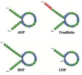
(5) (Εικόνα 2).
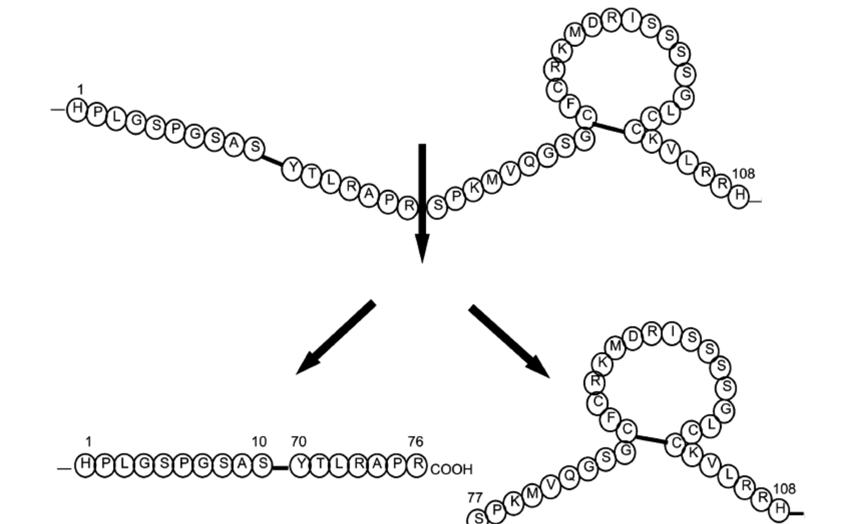
BNP (134
(108 αμινοξέα).
BNP (76
BNP (32
Giannakoulas et
-
επικοινωνία (υπερφόρτωση όγκου), στένωση ισθμού της αορτής (υπερφόρτωση πίεσης) και διατατική μυοκαρδιοπάθεια (συστολική δυσλειτουργία) και διαπίστωσαν
(ventricular septal defect - VSD),
(patent ductus arteriosus - PDA)
(atrial septal defect
1. Maeda K, Takayoshi T, Wada A, Hisanaga T, Kinoshita M. Plasma brain natriuretic peptide as a biochemical marker of high left ventricular end-diastolic pressure in patients with symptomatic left ventricular dysfunction. Am Heart 1998; 135: 825–832.
2. Maisel AS, Krishnaswamy P, Nowak RM, McCord J, Hollander JE, Duc P, et al, and the Breathing Not Properly Multinational Study Investigators. Rapid measurement of B-type natriuretic peptide in the emergency diagnosis of heart failure. N Engl J Med 2002; 347: 161–167.
3. Mavinkurve-Groothuis AM, Kapusta L, Nir A, Groot-Loonen J. The role of biomarkers in the early detection of anthracyclineinduced cardiotoxicity in children: a review of the literature. Pediatr. Hematol. Oncol 2008; 25(7): 655-64.
4. Favilli S, Frenos S, Lasagni D, Frenos F, Pollini I, Bernini G et al. The use of Btype natriuretic peptide in paediatric
patients: a review of literature. Journal of Cardiovascular Medicine 2009;10: 298-302.
5. Levin ER, Gardner DG, Samson WK. Natriuretic peptides. N Engl J Med 1998; 339: 321–8.
6. Cauliez Β, Berthe MC, Lavoinne A.Le BNP :aspects physiologiques, biologiques et cliniques. Ann Biol Clin 2005; 63(1): 15-25.
7. Baughman KL. B Type natriuretic peptide –a window to the heart. N Engl J Med. 2002; 347: 158-9.
8. Sudoh T, Kangawa K, Minamino N & Matsuo. A new natriuretic peptide in porcine brain. Nature (Lond) 1988; 332:78–81.
9. Davis GK, Bamforth F, Sarpal A, Dicke F, Rabi Y, Lyon M. B-type natriuretic peptide in pediatrics. Clinical Biochemistry 2006; 39: 600-605.
10. Hall C. Essential biochemistry and physiology of (NT-pro) BNP. The European Journal of Heart Failure.2004; 6: 257–260.
11. Doust JA, Pietrzak E, Dobson A, Glasziou P. How well does B-type natriuretic peptide predict death and cardiac events in patients with heart failure: systematic review. BMJ 2005; 330: 625-633.
12. Wu AH. B type Natriuretic peptide and its clinical utility in patients with heart failure, MLO Med Lab Obst 2001;33: 10-14.
13. Μaisel A, Mehra MR. Understanding Β type Natriuretic Peptide and its role in the diagnosis and monitoring congestive heart failure. Clin Comerstone 2005; l1: 7-17.
14. Dickstein K, Cohen-Solal A, Filippatos G, McMurray J, Ponikowski P, Poole-Wilson PA et al. ESC Guidelines for the diagnosis and treatment of acute and chronic heart failure 2008. European Heart Journal. 2008;29:2388–2442.
15. Koch A, Zink S, Singer H. B-type natriuretic peptide in paediatric patients with congenital heart disease. European Heart Journal. 2006; 27, 861–866.
16. Giannakoulas G, Dimopoulos K, Bolger AP, Tay EL, Inuzuka R, Bedard E et al. Usefulness of Natriuretic Peptide Levels to Predict Mortality in Adults With Congenital Heart Disease. Am J Cardiol. 2010; 105: 869-73.
17. Tan LH, Jefferies JL, Liang JF, Denfield SW, Dreyer WJ, Mott AR, et al. Concentrations of brain natriuretic peptide in the plasma predicts outcomes of treatment of children with decompensated heart failure admitted to the intensive care unit. Cardiol.Young 2007; 17: 397-406.
18. Price JF, Thomas AK, Grenier M, Eidem BW, O’ Brian Smith E, Denfield SW, et al. B type natriuretic peptide predicts adverse cardiovascular events in paediatric outpatients with chronic left ventricular systolic dysfunction. Circulation 2006; 114: 1063-1069.
19. Koulouri S, Acherman R, Wong P, Chan L, Lewis A. Utility of B-Type Natriuretic Peptide in Differentiating Congestive Heart Failure from Lung Disease in Pediatric Patients with Respiratory Distress. Pediatr. Cardiology. 2004; 25: 341-6.
20. Hayakawa H, Komada Y, Hirayama M, Hori H, Ito M, Sakurai M. Plasma levels of natriuretic peptides in relation to doxorubicin-induced cardiotoxicity and cardiac function in children with cancer. Med Pediatr Oncol 2001; 37:4-9.
21. Soker M, Kervancioglou M. Plasma concentrations of NTPro BNP and cardiac troponin –I in relation to doxorubicininduced cardiomyopathy and cardiac function in childhood malignancy. Saudi Med J 2005; 26: 1197-1202.
22. Ekstein S, Nir A, Rein AJ, Perles J, Bar-Oz B, Salpeter L, et al. N terminal pro Btype natriuretic peptide as a marker for acute anthracyclina cardiotoxicity in children. J Pediatr. Hematol Oncol 2007; 29: 440-444.
23. Germanakis I, Kalmanti M, Parthenakis F, Nikitovic D, Stiakaki E, Patrianakos A, et al. Correlation of plasma N-terminal pro-brain natriuretic peptide levels with left ventricle mass in children treated with anthracyclines. Int J Cardiol. 2006; 108(2): 212-5.
24. Bryant J, Picot J, Levitt G, Sullivan I, Baxter L, Clegg A. Cardioprotection against the toxic effects of anthracyclines given to children with cancer: a systematic review. Health Technol Assess. 2007; 11(27): 1-84.
25. Lan Y, Chang R, Alejos J, Burch C, Wetzel GT. B-type natriuretic peptide in children after cardiac transplantation. J.Heart Lung Transplant 2004; 23: 558-63.
26. Rossano JW, Denfield SW, Kim JJ, Price JF, Jefferies JL, Decker JA, et al. B-type natriuretic peptide is a sensitive screening test for acute rejection in pediatric heart transplant patients. J Heart Lung Transplant. 2008; 27(6): 649-654.
27. Kurotobi S, Kawakami N, Shimizu K, Aoki H, Nasuno S, Takahashi K, et al. Brain natriuretic peptide as a hormonal marker of ventricular diastolic dysfunction in children with Kawasaki disease. Pediatr Cardiol. 2005;26(4):425-30.
28. Oana S, Terai M, Tanabe M, Kohno Y, Ohnuma N. Plasma brain natriuretic peptides and renal hypertension. Pediatr Nephrol. 2000;14(8-9):813-5.
29. Khan K, Talwar S. Screening for familial hypertrophic cardiomyopathy using brain natriuretic peptide. Eur Heart J. 1999;20(7):550.
30. Kaditis AG, Alexopoulos EI, Hatzi F, Kostadima E, Kiaffas M, Zakynthinos E,et al. Overnight change in brain natriuretic peptide levels in children with sleep-disordered breathing. Chest. 2006;130(5):1377-84.
31. Koch A, Singer H. Normal values of B type natriuretic peptide in infants, children, and adolescents. Heart. 2003;89:875–8.
32. Cantinotti M, Clerico A, Murzi M, Vittorini S, Emdin M. Clinical relevance of measurement of brain natriuretic peptide and N-terminal pro-brain natriuretic peptide in pediatric cardiology. Clin Chim Acta. 2008;390(1-2):12-22.
33. Giuliani I, Rieunier F, Larue C, Delagneau JF, Granier C, Pau B,et al.Assay for Measurement of Intact B-Type Natriuretic Peptide Prohormone in Blood. Clinical Chemistry 2006; 52: 1054–1061.
34. Westerlind A, Wåhlander H, Lindstedt G, Lundberg P-A, Holmgren D. Clinical signs of heart failure are associated with increased levels of natriuretic peptide types B and A in children with congenital heart defects or cardiomyopathy. Acta Paediatr 2004; 93: 340-5.
35. Cowley CG, Bradley JD, Shaddy RE. B-type natriuretic peptide levels in congenital heart disease. Pediatr Cardiol 2004; 25: 336–340.
36. Hopkins EW, Chen Z, Fukagawa KN, Hall C, Knot HJ. Increased atrial and brain natriuretic peptides in adults with cyanotic congenital heart disease: enhanced understanding of the relationship between hypoxia and natriuretic peptide secretion. Circulation. 2004; 109; 28722877.
37. Trojnarska O, Gwizdala A, Katarzynski S, Katarzynska A, Oko-Sarnowska Z, Grajek S et al. The BNP concentrations and exercise capacity assessment with cardiopulmonary
stress test in cyanotic adult patients with congenital heart diseases. Int J Cardiol. 2008 Nov 28. [Epub ahead of print]
38. Ilkiw LR, Miller-Hance CW ,Nihill RM. The Pulmonary Circulation. In Garsin AJr, Bricker JT, McNamara GD. The science and practice of pediatric cardiology. Lea and Febiger. Philadelphia, London, 1990, pp371-373.
39. Nihill RM. Clinical management of patients with pulmonary hypertension. In: Moss and Adams: Heart disease in infants, children and adolescents, Fifth edition. Edited by Emmanouilides G., Allen H., Riemenschneider TA., Cutgesell PH., Williams and Wilkins, Baltimore, Philadelphia, Hong Kong, London, Munich, Sydney, Tokyo:1695-1699.
40. McLaughlin VV, McGoon MD. Pulmonary Arterial Hypertension. Circulation. 2006;114:1417-1431.
41. Galie N, Hoeper M, Humbert M et al. The Task Force for the Diagnosis and Treatment of Pulmonary Hypertension of the European Society of Cardiology (ESC) and the European Respiratory Society (ERS), endorsed by the International Society of Heart and Lung Transplantation (ISHLT). Guidelines for the diagnosis and treatment of pulmonary hypertension. Eur Heart J 2009, 20: 24932537.
42. Suda K, Matsumura M, Matsumoto M. Clinical implication of plasma natriuretic peptides in children with ventricular septal defect. Pediatr Int. 2003; 45: 249–254.
43. Kunii Y, Kamada M, Ohtsuki S, Araki T, Kataoka K, Kageyama M et al. Plasma brain natriuretic peptide and the evaluation of volume overload in infants and children with congenital heart disease. Acta Med Okayama. 2003;57:191–197.
44. Ozhan H, Albayrac S, Uzun H, Ordu S, Kaya A, Yazici M. Correlation of plasma -type natriuretic peptide with shunt severity in Patients with Atrial or Ventricular Septal Defect. Pediatr Cardiol .2007;28:272-275.
45. Nir A, Bar-Oz B, Perles Z, Korach A, Rein AJJT. N-terminal pro-B-type natriuretic peptide: reference plasma levels from birth to adolescence. Elevated levels at birth and in infants and children with heart diseases. Acta Paediatr 2004;93:603–607.
46. Sanjeev S, Pettersen M, Lua J, Thomas R, Shankaran S, L’Ecuyer T. Role of plasma B-type natriuretic peptide in screening for hemodynamically significant patent ductus arteriosus in preterm neonates. J.Perinatol 2005;25:709-713.
47. Choi BM, Lee KH, Eun BL, Yoo KH, Hong YS, Son CS, et al. Utility of rapid B-type natriuretic peptide assay for diagnosis of symptomatic patent ductus arteriosus in preterm infants. Pediatrics 2005;115:255-261.
48. Davlouros P, Karatza A, Xanthopoulou I, Dimittriou G, Georgiopoulou A, Mantagos S, et al. Diagnostic role of plasma BNP levels in neonates with signs of congenital heart disease.International Journal of Cardiology 2009;25:1-5.
49. Koch A, Kitzsteiner T, Zink S, Cesnjevar R, Singer H. Impact of cardiac surgery on plasma levels of B-type natriuretic peptide in children with congenital heart disease. Intern J Cardiol 2007; 114: 339–44.
50. Mir TS, Haun C, Lilje C, Laer S, Weil J. Utility of N-terminal brain natriuretic peptide plasma concentrations in comparison to lactate and troponin in children with congenital heart disease following open-heart surgery. Pediatr Cardiol 2006; 27: 209–16.
51. Ationu A, Singer DR, Smith A, Elliott M, Burch M, Carter ND. Studies of cardiopulmonary bypass in children: implications for the regulation of brain natriuretic peptide. Cardiovasc Res 1993; 27: 1538–41.
52. Shih CY, Sapru A, Oishi P, Azakie A, Karl TR, Harmon C, et al. Alterations in plasma B-type natriuretic peptide levels after repair of congenital heart defects: A potential perioperative marker. J Thorac Cardiovasc Surg 2006; 131: 632–638.
53. Berry JG, Askovich B, Shaddy RE, Hawkins JA, Cowley CG. Prognostic Value of B Type Natriuretic Peptide in Surgical Palliation of Children with Single-Ventricle Congenital Heart Disease. Pediatr Cardiol.2008; 29: 70–75.
54. Dodge-Khatami A, Büchel VE, Knirsch W, Kadner A, Rousson V, Dave H et al. Brain Natriuretic Peptide and Magnetic Resonance Imaging in Tetralogy With Right Ventricular Dilatation. Ann Thorac Surg 2006; 82: 983–8.
55. Koch A, Zink S, Glöckler M ,Seeliger T, Dittrich S Plasma levels of B-type natriuretic peptide in patients with tetralogy of Fallot after surgical repair. Int J Cardiol. 2009; 18. [Epub ahead of print]
56. Nir A , Nasser N. Clinical Value of NT-proBNP and BNP in Pediatric Cardiology. Journal of Cardiac Failure 2005;11:76-80
Γενικό Νοσοκομείο
«Ασκληπιείο Βούλας»,
Φιλίππου Όλγα filiolga@hotmail.com
Β. Παύλου 1, Βούλα, Τ.Κ. 16673
Paediatric Clinic, General Hospital “Asklipeio Voulas”, Voula, Attiki
Correspondence: Olga Filippou filiolga@hotmail.com
1 V. Pavlou St., Voula, TK 16673
The effect on atherogenic factors of a programme modifying dietary and physical exercise habits in overweight and obese children
O. Filippou, C. Vliora, M. Dolianiti, K. Κaranasiou, S. Papadakou-Lagogianni
Abstract
Introduction: This prospective study aimed to evaluate the short-term effects of a 6-month combined dietary and physical activity intervention programme on overweight and obese children and adolescents.
Methods: A total of 170 overweight and obese children (109 boys, 96 obese children) aged 3.5-14 years (mean: 8.9 ± 2.8 years) were included in the study. During their first visit to the outpatient obesity clinic, anthropometric indices, arterial blood pressure and eating and physical activity habits were recorded and their lipid profile was determined. Dietary and physical exercise instructions were given and the participants were invited to attend for follow-up after 3 and 6 months.
results: At the 6 month follow up examination 58 subjects (34.1%) returned for examination. In 19 (32.7%), a reduction in the body mass index (BMI) percentile was observed (mean reduction: -5.241 percentiles, p<0.001). In the measurements carried out before and after intervention, various changes were detected: a mean reduction in total cholesterol (T-C) by -10mg/dl (p=0.009), a mean reduction in LDL-C by –9.9 mg/dl (p=0.007), a mean increase in HDL-C by +2.3mg/dl (p=0.067) and a marginal decrease in blood pressure (p=0.085). A reduction in weekly high-calorie food consumption led to a decrease in levels of T-C and triglycerides (TGL) and in the appearance of normal LDL-C values (p=0.048, p=0.066 και p=0.042 respectively). Taking up systematic physical exercise was significantly associated with a decrease in levels of T-C and LDL-C (p=0.009 and p=0.057, respectively), and in the return of LDL-C to
normal levels (p=0.059). Increase in the frequency of systematic exercise resulted in a borderline drop of BMI (p=0.052).
Conclusions: A combined short-term intervention improved the BMI and atherogenic factors substantially in overweight and obese children.
Key words: Intervention, atherogenic risk factors, BMI, childhood obesity, follow-up.
SDS
αρτηριακή υπέρταση, δυσλιπι-
ΔΜΣ. Η
απώλεια ΒΣ ήταν 2,6 Ε.Θ. (p=0,009),
ΔΜΣ κατά 5,241 Ε.Θ. (p<0,001). Οι μεταβολές του ΒΣ και του ΔΜΣ ήταν ανεξάρτητες
παχυσαρκίας (Πίνακας 2).
Σε 47 παιδιά (27,64%) ελήφθη νέος βιοχημικός έλεγχος (Πίνακας 1). Διαπιστώθηκε σημαντική πτώ-
στις τιμές της Τ-C (μέση μείωση: -10,1mg/dl, p=0,009) και της LDL-C (μέση μείωση: -9,9mg/dl, p=0,007), μη στατιστικά σημαντική ελάτωση στις τιμές των TGL (μέση μείωση: -4,9mg/dl, p=0,470) καθώς και οριακή
της HDL-C (μέση αύξηση: +2,3mg/dl, p=0,067), πριν και μετά την παρέμβαση.
Σε 58 άτομα (34,1%) επανεξετάστηκε
μα μείωσαν οριακά τις τιμές της ΑΠ (p=0,085). Ειδικότερα, το 31,6% των συμμετεχόντων παρουσίασε ελάττωση της ΑΠ, ενώ
Αναψυκτικά 1,6 ± 2 0,6 ± 1 0,001 (φορές/εβδομάδα)
Έτοιμα
(φορές/εβδομάδα)
Μπισκότα
(φορές/εβδομάδα)
Χυμοί
(φορές/εβδομάδα)
Τσιπς
(φορές/εβδομάδα)
Tηλεθέαση
(ώρες/ημέρα)
Οργανωμένο
(ώρες/εβδομάδα)
με οργανωμένο άθλημα, είχε ως αποτέλεσμα την ελάττωση της τιμής της T-C και της LDL-C (p=0,009 και p=0,057, αντίστοιχα), ενώ σχετίστηκε με την επάνοδο παθολογικών τιμών LDL-C, σε φυσιολογικές τιμές (p=0,059). Τέλος, η αύξηση της εβδομαδιαίας συχνότητας
1. Eliakim A, Kaven G, Berger I, Friedland O, Wolach B, Nemet D. The effect of a combined intervention on body
mass index and fitness in obese children and adolescents – a clinical experience. Eur J Pediatr 2002;161:449-454.
2. Ciarke WR, Lauer RM. Does childhood obesity track into adulthood? Crit Rev Food Sci Nutr. 1993;33(4-5):423-30.
3. Viner RM, Cole Tj. Who changes body mass between adolescence and adulthood? Factors predicting change in BMI between 16 years and 30 years in the 1970 British birth cohort. Int J Obes 2006;30:1368-1374.
4. Berenson GS, Srinivasan SR, Bao W, Newman WP, Tracy RE, Wattigney WA. Association between multiple cardiovascular risk factors and atherosclerosis in children and young adults. The Bogalusa study. N. Engl J Med 1998;338:1650-1656.
5. Mahoney LT, Burns TL, Stanford W, Thompson BH, Witt JD, Rost CA, et al. Coronary risk factors measured in childhood and young adult life are associated with coronary artery calcification in young adults: The Muscatine study. J Am Coll Cardiol 1996;27:277-284.
6. Must A, Jacques PF, Dallal GE, Bajema CJ, Dietz WH. Long-term morbidity and mortality of overweight adolescents: a follow-up of the Harvard Growth Study of 1922-1935. N Engl J Med 1992;327:1350-1355.
7. Χιώτης Δ, Κρίκος
18
Δελτίο Α΄ Παιδ. Κλινικής 2004;51:139-154.
8. National High Blood Pressure Education Program Working Group on High Blood Pressure in Children and Adolescents. The Fourth Report on the Diagnosis, Evaluation and Treatment of High Blood Pressure in Children and Adolescents. Pediatrics 2004;114(2):555-576.
9. Daniels SR, Frank R, Greer FM and Τhe Committee on Nutrition. Lipid Screening and Cardiovascular Health in Childhood. Pediatrics 2008;122:198–208.
10. Φιλίππου
Τασιοπούλου Β, Ζήβα-Πετροπούλου Μ, Παπαδάκου-Λαγογιάννη
διά της Ν.Α. Αττικής. Επίδραση του σύγχρονου τρόπου
2008;71: 373-379.
11. Savoye M, Shaw M, Dziura J, Tamborlane W, Rose P, Guandalini C et al. Effects of a weight management program on body composition and metabolic parameters in overweight children: a randomized controlled trial. JAMA 2007;297:2697-2704.
12. Nemet D, Barkan S, Epstein Y, Friedland O, Kowen G, Eliakim A. Short- and long- term beneficial effects of a combined dietary-behavioral-physical activity intervention for the treatment of childhood obesity. Pediatrics 2005; 115:e443-e449.
13. Sung RYT, Yu CW, Chang SKY, Mo SW, Woo KS, Lam CWK. Effects of dietary intervention and strength training on blood lipid level in obese children. Arch Dis Child 2002;86:407-410.
14. Reinehr T, Kersting M, Alexy U, Andler W. Long term follow-up of overweight children: after training, after a single consultation session and without treatment. JPGN 2003;37:72-74.
15. Reinehr T, De Sousa G, Toschke AM, Andler W. Longterm follow-up of cardiovascular disease risk factors in children after an obesity intervention. Am J Clin Nutr 2006;84:490–6.
16. Nicklas TA, Baranowski T, Cullen KW, Berenson G. Eating patterns, dietary quality and obesity. J Am Coll Nutr 2001; 20:599-608.
17. McGill HC, McMahan CA, Herderick EE, Zieske AW, Malcom GT, Tracy RE, et al, for the Pathobiological Determinants of Atherosclerosis in Youth (PDAY) Research Group: Obesity accelerates the progression of coronary atherosclerosis in young men. Circulation 2002; 105:2712-2718.
18. Kiess W, Marcus C, Wabitsch M. Η
2008.
19. Kelley G, Kelley K. Aerobic exercise and lipids and lipoproteins in children and adolescents: A meta-analysis of randomized controlled trials. Atherosclerosis 2007; 191:447-453.
20. Maffeis C, Talamini G, Tato L. Influence of diet, physical activity and parents' obesity on children's adiposity: a four year longitudinal study. Int J Obes 1998;22:758-764.
21. Reinehr T, Andler W. Changes in the atherogenic risk factor profile according to degree of weight loss. Arch Dis Child 2004;89:419-422. 22.Λιναρδάκης
2007;70:24-36.
23. Hanson L, Hedner T, Lindholm L. The captopril prevention project (CAPPP) in hypertension-baseline data and current status. Blood Press 1997;6:365-367.
24. Wang JG, Staessen JA. Benefits of antihypertensive pharmacologic therapy and blood pressure reduction in outcome trials. J Clin Hypertens 2003;5:66-75.
25. Savoye M, Berry D, Dziura J, Shaw M, Serrecchia JB, Barbetta G, et al. Anthropometric and psychosocial changes in obese adolescents enrolled in a Weight Management Program. J Am Diet Assoc 2005;105(3):364-370.
26. Sothern M. Obesity prevention in children: physical activity and nutrition. Nutrition 2004;20:704-708.
27. Knip M, Nuutinen O. Long-term effects of weight reduction on serum lipids and plasma insulin in obese children. Am J Clin Nutr 1993;57:490-3.
28. Reinehr T, Brylak K, Alexy U, Kersting M, Andler W. Predictors to success in outpatient training in obese children and adolescents. Int J Obes Relat Metab Disord 2003:27:1087-1092.
29. Tershakovec AM, Kuppler K. Ethnicity, insurance type, and follow-up in a pediatric weight management program. Obes Res. 2003;11(1):17–20.
30.
The protective role of breastfeeding against pyelonephritis in infants with urinary tract infection
D. Doganis, D.
Delis, M. Mavrikou, G. Issaris, A. Martirosova, L. Stamoyiannou, K. Siafas
Abstract
Background: Much evidence shows that breastfeeding protects infants from infection and especially urinary tract infections, but the possible protective effect of breastfeeding against the development of lesions of acute pyelonephritis (APN) among infants with first time urinary tract infection (UTI) has not yet been sufficiently evaluated.
Methods: We studied 137 infants (males: 78, females: 59) aged 0.5-12 months (median 3.1) admitted to the hospital with their first time UTI and we evaluated the effect of breastfeeding on the findings of Technetium-99m-dimercaptosuccinic acid (DMSA) scintigraphy performed within 28 days of the diagnosis of UTI (median time: 5 days).
results: Findings of APN on DMSA scan were documented in 31/69 (45%) of currently breastfed infants, in 25/45 (56%) of infants who had been breastfed in the past for different periods of time and in 16/23 (70%) infants, who had never been breastfed. Among boys, APN was detected in 52%, 48% and 56% of infants with ongoing breastfeeding, prior breastfeeding and no breastfeeding respectively (p=ns) whereas among girls the corresponding percentages were 29%, 62.5% and 79% (p=0.008).
Conclusions: A protective role of breastfeeding against the development of acute pyelonephritis lesions on DMSA scan was noted only in girls with UTI.
Key words: Breastfeeding, infants, acute pyelonephritis.
1st and 3rd Paediatric Clinics, “P. & A. Kyriakou” Children’s Hospital, Athens Correspondence: Dimitrios Doganis doganisd@gmail.com 1st Paediatric Clinic, “P. & A. Kyriakou” Children’s Hospital, Athens, Greece
Ladomenou
Βιβλιογραφία
1. Hanson LA, Korotkova M, Håversen L, Mattsby-Baltzer I, Hahn-Zoric M, Silfverdal SA et al. Breast-feeding, a complex support system for the offspring. Pediatr Int. 2002;44:347–352.
2. Quigley MA, Kelly YJ, Sacker A. Breastfeeding and hospitalization for diarrheal and respiratory infection in the United Kingdom millennium cohort study. Pediatrics. 2007;119:e837–e842.
3. Wold AE, Adlerberth I. Breast feeding and the intestinal microflora of the infant implications for protection against infectious diseases. Adv Exp Med Biol. 2000;478:77-93.
4. Hanson LA. Protective effects of breastfeeding against urinary tract infection. Acta Paediatr. 2004;93:154-156.
5. Coppa GV, Gabrielli O, Giorgi P, Catassi C, Montanari MP, Varaldo PE et al. Preliminary study of breastfeeding and bacterial adhesion to uroepithelial cells. Lancet. 1990;335:569-571.
6. Jodal U. Does breast feeding protect against urinary tract infection in the first few months of life? Pediatr Nephrol. 1992;6:344.
7. Marild S, Hansson S, Jodal U, Oden A, Svedberg K. Protective effect of breastfeeding against urinary tract infection. Acta Paediatr. 2004;93:164-168.
8. Marild S, Jodal U, Mangelus L. Medical histories of children with acute pyelonephritis compared with controls. Pediatr Infect Dis J. 1989;8:511-515.
9. Pisacane A, Graziano L, Mazzarella G, Scarpellino B, Zona G. Breast-feeding and urinary tract infection. J Pediatr. 1992;120:87-89.
10. Mansour L, Mansour A. Breast feeding protects infants against urinary tract infection. New Egypt J Med. 1993;8:463-464.
11. Levy I, Comarsca J, Davidovits M, Klinger G, Sirota L, Linder N. Urinary tract infection in preterm infants: the protective role of breastfeeding. Pediatr Nephrol. 2009;24:527-531.
12. Rushton HG. The evaluation of acute pyelonephritis and renal scarring with technetium 99m-dimercaptosuccinic acid renal scintigraphy: evolving concepts and future directions. Pediatr Nephrol. 1997;11:108-120.
13. Majd M, Rushton HG. Renal cortical scintigraphy in the diagnosis of acute pyelonephritis. Semin Nucl Med. 1992;22:98-111.
14. Hoberman A, Charron M, Hickey RW, Baskin M, Kearney DH, Wald ER. Imaging Studies after a First Febrile Urinary Tract Infection in Young Children. N Engl J Med. 2003;348:195-202.
15. Gartner LM, Morton J, Lawrence RA, Naylor AJ, O’Hare D, Schanler RJ et al. Breastfeeding and the use of human milk. Pediatrics. 2005;115: 496–506.
16. Ladomenou F, Moschandreas J, Kafatos A, Tselentis Y, Galanakis E. Protective effect of exclusive breastfeeding against infections during infancy: a prospective study. Arch Dis Child. 2010;95:1004-1008.
17. Leventhal JM, Shapiro ED, Aten CB, Berg AT, Egerter SA. Does breast-feeding protect against infections in infants less than 3 months of age? Pediatrics. 1986;78:896-903.
18. van der Waaij LA, Limburg PC, Mesander G, van der Waaij D. In vivo IgA coating of anaerobic bacteria in human faeces. Gut 1996;38:348–354.
19. Winberg J, Bollgren I, Gothefors L, Herthelius M, Tullus K. The prepuce: a mistake of nature? Lancet 1989;1: 598–599.
20. Nuutinen M, Uhari M. Recurrence and follow-up after urinary tract infection under the age of 1 year. Pediatr Nephrol. 2001;16:69-72.
21. Ηowie PW, Forsyth JS, Ogston SA, Clark A, Florey CV. Protective effect of breast feeding against infection. Br Med J. 1990;300:11-16.
22. Gordon I, Barkovics M, Pindoria S, Cole TJ, Woolf AS. Primary vesicoureteric reflux as a predictor of renal damage in children hospitalized with urinary tract infection: a systematic review and meta-analysis. J Am Soc Nephrol. 2003;14:739-744.
Neonatal Intensive Care Unit, “P. and A. Kyriakou” Children’s Hospital, Athens, Greece
Correspondence: S. Mouskou stelli_m@yahoo.com 15 Efessou St., Papagou 15669, Athens
Hereditary spherocytosis in newborn infants. Case report
S. Mouskou,
A. Nika, N. Mastrantonaki, , G. Kourakis
Abstract: Hereditary spherocytosis is the most common haemolytic anaemia due to a red cell membrane defect. It is the result of abnormalities in the membrane proteins which leads to loss of the erythrocyte shape and their early destruction in the spleen. The disease is transmitted by autosomal dominant inheritance. A significant number of patients, however, represent de novo mutations. The clinical course is extremely variable; some children have severe haemolytic anaemia and jaundice, and need blood transfusions early in their life, while others are asymptomatic, with mild haemolysis which is detected incidentally. Splenectomy is the indicated treatment after the age of 5 years. The cases are presented of 2 neonates with jaundice and haemolytic anemia. Only one had positive family history for spherocytosis. The 2 infants had a different clinical course during their hospitalization.
Key words: Hereditary spherocytosis, haemolytic anaemia, jaundice, neonate.
Εξήλθε
3
Βιβλιογραφία
1. Bolton-Maggs PHB, Stevens RF, Dodd N.J, Lamont G, Tittensor P, King M-J. Guidelines for the diagnosis and management of hereditary spherocytosis. British Journal of Hematology 2004;126: 455-474
2. Oski Inherited abnormalities of the erythrocyte membrane. In Nathan and Oski .Hematology of Infancy and Childhood. vol 1. 5th ed. Philadelphia, PA: WB Saunders ; 1985 p.578-601
3. Oski FA Anemia in the neonatal period. In :Oski FA, Naiman JL. Hematologic problems in the newborn. 3rd ed. Philadelphia, PA:WB Saunders; 1982 p. 56
4. Gallagher P G. Red Cell Membrane Disorders. Hematology 2005;13-18
5. Asselin B L. Hemolytic Anemias. In: Gellis and Kagan’s Current Pediatric Therapy 16, WB Saunders; 1999 p. 685-688
6. Smedley JC, Bellingham AJ. Current problems in haematology 2: Hereditary spherocytosis. J Clin Pathol 1991;44:441-444
7. Perrota S, Gallagher GP, Mohandas N. Hereditary spherocytosis. Lancet 2008;372:1411-1426
8. Segel GB. Hereditary Spherocytosis. In: Nelson. Textbook of Pediatrics 18th ed PA:WB Saunders; 2007 p. 2020-2023
9. Berardi A, Lugli L, Ferrari F, Gargano G, D’Apolito M, Marrone A, Iolascon A. Kernicterus associated with hereditary spherocytosis and UGT!A! promoter polymorphism. Biol Neonate 2006;90:243-246
10. Schroter W, Kahsnitz E. Diagnosis of hereditary spherocytosis in newborn infants. J Pediatr 1983;103(3):460-63
11. Saada V, Cynober T, Brossard Y, Schischmanoff PO, Sendar A, Cohen H et al.Incidence of hereditary spherocytosis in a population of jaundiced neonates. Pediatr Hematol Oncol 2006;23:387-397
12. Alloisio N, Texier P, Vallier A, Ribeiro ML, Morle L, Bozan M et al. Modulation of clinical expression and band 3 deficiency in hereditary spherocytosis. Blood 2004;103: 3233-3240
13. Gallagher PG, Weed SA, Tse WT, Benoit L, Morrow JS, Marchesi SL et al. Recurrent fatal hydrops fetalis associated with a nucleotide substitution in the erythrocyte betaspectrin gene. J Clin Invest 1995;95:1174-1182
14. Delhommeau F, Cynober T, Schischmanoff PO, Rohrlich P, Delaunay J, Mohandas N, Tchermia G. Natural history of hereditary spherocytosis during the first year of life. Blood 2000;95:393-397
15. Shapiro CM, Josephson AM, Rozengvaig S, Kauffman A. Hereditary spherocytosis in the neonatal period. J Pediatr 1957;50:308-314,
16. Mentzer WC, Bertram HL, Hereditary spherocytosis: Clinical features, diagnosis, and treatment www.uptodate.com
17. King MJ, Smythe JS, Mushens, R. Eosin-5-maleimide
binding to band 3 and Rh-related proteins forms the basis of a screening test for hereditary spherocytosis. Br J Haematol 2004;124:106
18. Kedar PS, Colan RB, Kulkarnis S, Ghosh K, Mohanty D. Experience with eosin-5-maleimide as a diagnostic tool for red cell membrane cytoskeleton disorders. Clin Lab Haematol 2003; 24(6):373-376.
19. Tchernia G., Delhommeau F., Perrotta S. Recombinant erythropoietin therapy as an alternative to blood transfusions in infants with hereditary spherocytosis. J Hematol 2000;1:146-152.
20. Schiff Mr., Hays S, Sann L.. Use of erythropoietin in a newborn suffering from hereditary spherocytosis. Recombinant human erythropoietin (r-HuEPO) therapy in a newborn with hereditary spherocytosis. Arch Pediatr 2003;10: 333-336.
21. Hosono S., Hosono A, Mugishima H. Successful recombinant erythropoietin therapy for a developing anemic newborn with hereditary spherocytosis. Pediatr Int 2006; 48:178-180.
1st Paediatric Department, “P. & A. Kyriakou” Children’s Hospital
Correspondence: A. Papadopoulou papadop5@otenet.gr
1st Paediatric Department, “P. & A. Kyriakou” Children’s Hospital Athens, Greece
Topics of Pediatric Allergy in “Pediatriki”
P. Maggina
1rst Pediatric Clinic
Children’s Hospital
“P. & A. Kyriakou”
Correspondence:
Paraskevi Maggina vivian.maggina@hotmail.com
1rst Pediatric Clinic
Children’s Hospital
“P. & A. Kyriakou” 11527, Athens, Greece
2006; 69(3):167-168
http://www.e-child.gr/oldmagazines/Paediatriki_2006_69_167_168.pdf
69(3):193-198
http://www.e-child.gr/oldmagazines/Paediatriki_2006_69_193_198.pdf
Παιδιατρική 2005; 68(6):399-408
http://www.e-child.gr/oldmagazines/Pediatriki_2005_68_No6_P399_408.pdf
2006; 69(3):169-177
http://www.e-child.gr/oldmagazines/Paediatriki_2006_69_169_177.pdf
2006; 69(6): 417-424
http://www.e-child.gr/oldmagazines/Paediatriki_2006_69_p_417_424.pdf
Παιδιατρική 2007; 70(5):416-419
http://www.e-child.gr/oldmagazines/Paediatriki_2007_70_p_416_419.pdf
Αντιφλεγμονώδης
Παιδιατρική 2006; 69(6): 425-431
http://www.e-child.gr/oldmagazines/Paediatriki_2006_69_p_425_431.pdf
Νεφελοποιημένη
M.Β.
K.Ν.
Παιδιατρική 2008; 71(3):231-234
http://www.e-child.gr/oldmagazines/Paediatriki_2008_71_p_231_234.pdf
Παιδιατρική 2007; 70(4):330-335
http://www.e-child.gr/oldmagazines/Paediatriki_2007_70_p_330_335.pdf
http://www.e-child.gr/sites/default/files/Pediatriki_73_2_90_102.pdf
A. Σοφιανού-Κατσούλη, G. Du Toit, G. Lack
Παιδιατρική 2005; 68(5):337-341
http://www.e-child.gr/oldmagazines/Paediatriki_2005_68_No5_P337_341.pdf
http://www.e-child.gr/oldmagazines/Paediatriki_2008_71_p_475_481.pdf
http://www.e-child.gr/oldmagazines/Paediatriki_2008_71_p_486_492.pdf
Crohn
15. Costamagna G, Shah SK, Riccioni ME, Foschia F, Mutignani M, Perri V, Vecchioli A, Brizi MG, Picciocchi A, Marano P: A prospective trial comparing small bowel radiographs and video capsule endoscopy for suspected small bowel disease. Gastroenterology 2002, 123(4):999-1005.
16. Cheifetz AS, Kornbluth AA, Legnani P, Schmelkin I, Brown A, Lichtiger S, Lewis BS: The risk of retention of the capsule endoscope in patients with known or suspected Crohn's disease. The American journal of gastroenterology 2006, 101(10):2218-2222.
17. Herrerias JM, Caunedo A, Rodriguez-Tellez M, Pellicer F, Herrerias JM, Jr.: Capsule endoscopy in patients with suspected Crohn's disease and negative endoscopy. Endoscopy 2003, 35(7):564-568.
18. Marmo R, Rotondano G, Piscopo R, Bianco MA, Siani A, Catalano O, Cipolletta L: Capsule endoscopy
versus enteroclysis in the detection of small-bowel involvement in Crohn's disease: a prospective trial. Clin Gastroenterol Hepatol 2005, 3(8):772-776.
19. Attard TM, Horton KM, DeVito K, Darbari A, OlivaHemker M, Thompson R, Cuffari C: Pediatric jejunoileitis: a severe Crohn's disease phenotype that requires intensive nutritional management. Inflammatory bowel diseases 2004, 10(4):357-360.
20. Wagtmans MJ, Verspaget HW, Lamers CB, van Hogezand RA: Clinical aspects of Crohn's disease of the upper gastrointestinal tract: a comparison with distal Crohn's disease. The American journal of gastroenterology 1997, 92(9):1467-1471.
21. A Papadopoulou, MO Rawashdeh, GA Brown, AS Mc Neish, IW Booth Remission following an elemental diet or prednisolone in Crohn's disease. Acta Paediatrica 1995; 84:79-83.
ΥΠΟΥΡΓΕΙΟ ΥΓΕΙΑΣ ΚΑΙ
ΚΟΙΝΩΝΙΚΗΣ ΑΛΛΗΛΕΓΓΥΗΣ
ΔΙΟΙΚΗΣΗ 6η ΥΓΕΙΟΝΟΜΙΚΗΣ ΠΕΡΙΦΕΡΕΙΑΣ
ΠΕΛΟΠΟΝΝΗΣΟΥ- ΙΟΝΙΩΝ ΝΗΣΩΝ-ΗΠΕΙΡΟΥ
& ΔΥΤ. ΕΛΛΑΔΑΣ
ΑΓΙΟΣ ΔΙΟΝΥΣΙΟΣ
ΓΡΑΦΕΙΟ ΔΙΟΙΚΗΤΗ
ΠΛΗΡ.: Σ. Δρόσσου
Έχοντας υπόψη : 1. Τις
13 του Ν. 1965/91
άρθρου 84 του Ν.2071/92. της παρ.1 του άρθρου 5 του Ν. 2194/94.
39 του Ν.2737/99
του Ν.2716/99
του άρθρου 10 του του Ν.2955/01
του άρθρου 19 του Ν.3106/03
του Ν.3172/03
του άρθρου 3 του Ν.3527/07
το Π.Δ. 131/87 (ΦΕΚ73/Α/87)
της παρ.2 του άρθρου 43 του Ν.1759/88
Ν.2072/92.
του Ν.3204/03
του Ν.3754/2009 όπως τροποποιήθηκε
2. Την ΔΥ13α/39832/97 (ΦΕΚ1088/97 τ.Β’) απόφαση
αξιολόγησης υποψήφιων για
ΑΔΑ: 4Α194690ΒΞ-Μ
ΑΝΑΡΤΗΤΕΑ ΣΤΟ
ΖΑΚΥΝΘΟΣ 4-3-2011
Α.Π.: 1219
Σχετ. 1089/24-2-11, 219/14-1-11
3868/10
3. την ΔΥΙγ/οικ.41255/92 (ΦΕΚ 97/25-2-93 τ.Β’) Υπουργική Απόφαση όπως
με την ΔΥΙγ/οικ.25338/10-5-93 (ΦΕΚ 376/93 τ.Β’) όπως
ΔΥ13α/29804/15-9-97 (ΦΕΚ 859/26-9-97 τ. Β’).
4. Τον οργανισμό του Γ.Ν. Ζακύνθου.
5. Την υπ' αριθμ. 7465/3-11-2010 άγονη προκήρυξη
6. Την υπ' αριθμ. 7705/10-11-2010άγονη
8. Την υπ' αριθμ. Υ10α/162918/24-12-10 (Φ.Ε.Κ. 1250/31-12-2010) απόφαση της Υ.Υ.
9.
ΑΔΑ: 4Α194690ΒΞ-Μ
17-21 ιανουαρίου 2011 Pediatric Emergency Medicine: Sarasota, ΗΠΑ
An Evidence-Based Approach
Contact: Christy or Kathryn
Tel.: 1-866-267-4263
Fax: 1-941-365-7073
E-mail: mail@ams4cme.com
22
Makedonia Palace
Πληροφορίες: Global Events
Τηλ.: 2310 247743
Fax: 2310 247746
E-mail: info@globalevents.gr
17-20 Φεβρουαρίου 2011 the first Global congress for consensus in
Pediatrics and Child Health
Contact: CIP Sectretariat
Tel.: 41-0-22-533-0948
Fax: 41-0-22-580-2953
E-mail: cip@cipediatrics.org
13-17 Mαρτίου 2011 6th World Congress on Pediatric Critical Care
World Federation of Pediatric Intensive and Critical Care Societies (WFPICCS)
Phone: +61 2 9265 0700, Fax: +61 2 9267 5443
Email: pcc2011@arinex.com.au
17 Μαρτίου 2011
- «Μητώ» Τηλ: 210 68 69000 www.mitera.gr 7-10
2011 AlAPE Updates in Pediatrics Panama City, Contact: Astrid Sender
Tel.: 41-0-22-533-0948
Fax: 41-0-22-580-2953
E-mail: asender@paragon-conventions.com
22nd Annual Congress of ESPU
European Society for Paediatric Urology
E-mail: meeting@espu.org 4 - 6
2nd European Conference on Pediatric and ΜontreuxNeonatal Cardiac Intensive Care
Tel. (+41-22) 839 84 84. Fax (+41-22) 839 84 85
Email : info@epncic.com www.epncic.com
11-14 Μαΐου 2011 3rd International Congress on
lymphoma-leukemia-Myeloma
Contact: Ipek Durusu
Tel.: 90-312-490-9897
Fax: 90-312-490-9868
E-mail: admin@thd.org.tr
12-15
Μαΐου 2011
3-6 ιουνίου 2011
10th European Symposium on Pediatric Αθήνα Cochlear Implantation
Divani Apollon Palace
Πληροφορίες: GOLDAIR Congress
Τηλ.: 210 3274570
Φαξ: 210 3311021
E-mail: congress@goldair.gr
2nd Summer School of Pediatric dermatology Aegean Sea
European Society of Pediatric Dermatology & Hellenic Cruise, Greece
Society of Pediatric Dermatology
Πληροφορίες: Erasmus Conferences Tours & Travel S.A.
Τηλ: 210 7257693, 210 7257531
Ε-mail: info@espdsummerschool2011.org www.espdsummerschool2011.org
15-18 ιουνίου 2011 International Association of Paediatric dentistry Αθήνα
23rd Congress (IAPd 2011)
Πληροφορίες: Μαρία Ράντου
Τηλ.: 210 6889130
Φαξ: 210 6844777
E-mail: iapd2011@candc-group.com
23-26 ιουνίου 2011 5th Europaediatrics
European Paediatric Association (EPA/UNEPSA)
Tel.: +30 210 6889130, Fax: +30 210 6844777
E-mail: europaediatrics2011@candc-group.com
14-17 Οκτωβρίου 2011
Βιέννη, Αυστρία
52nd Annual Meeting of the European Society Newcastle, for Paediatric research Αγγλία
European Society for Paediatric Research
Tel: +41 22 908 0488, Fax: +41 22 906 9140
E-mail: espr@kenes.com