291Age-related serial plasma citrulline levels in preterm neonates
H.P. Ioannou, E. Diamanti, Z. Tsampoura, V. Drossou-Agakidou, P. Augoustides-Savvopoulou
296Immune status and immune recovery in children with lymphoma at the end of treatment (chemotherapy and/or radiotherapy) and on follow-up evaluation
S. Kosmidis, A. Pourtsidis, D. Bouhoutsou, M. Baka, M. Varvoutsi, D. Doganis, C. Kallergi, N. Douladiris, M. Synodinou, F. Saxoni-Papageorgiou, H. Vasilatou-Kosmidis
255Genetic studies in childhood acute lymphoblastic leukaemia
F. Tzifi, A. Kolialexi, K. Salavoura, S. Kitsiou, A. Mavrou
263Cytogenetic abnormalities in leukaemias of childhood
∞. Galla-Voumvouraki
271Non-random X inactivation: clinical consequences of the skewing effect
G. Lavranos, R. Angelopoulou
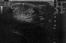
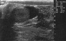
277Testicular microlithiasis
D. Thomas, ∂. Vlahopapadopoulou, V. Papadakis
ORIGINAL ARTICLES
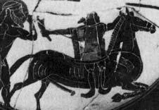
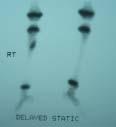
283What causes the lameness of Hephaestus?
∂. µazopoulou-∫yrkanidou
291Age-related serial plasma citrulline levels in preterm neonates
H.P. Ioannou, E. Diamanti, Z. Tsampoura, V. Drossou-Agakidou, P. Augoustides-Savvopoulou
296Immune status and immune recovery in children with lymphoma at the end of treatment (chemotherapy and/or radiotherapy) and on follow-up evaluation
S. Kosmidis, A. Pourtsidis, D. Bouhoutsou, M. Baka, M. Varvoutsi, D. Doganis, C. Kallergi, N. Douladiris, M. Synodinou, F. Saxoni-Papageorgiou, H. Vasilatou-Kosmidis
301Clinico-epidemiological data on children hospitalized with acute gastroenteritis due to rotavirus (1984-2007)
O. Tsiatsiou, √. Papagiannopoulou, Aik. Sarantopoulou, C. Alexandratos, D. Karabaxoglou, π. Kavaliotis
PRACTICAL ISSUE
304Sudden infant death syndrome: Risk factors and the latest guidelines
S. Stefanaki, A. Tsilimigkaki
CASE REPORTS
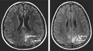
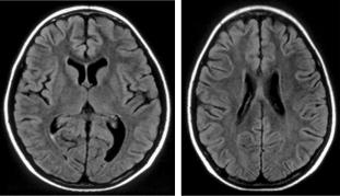
311Posterior reversible encephalopathy syndrome: a report of two cases and literature review
V. Askiti, ∞. Mitsioni, S. ªastroyianni, P. Gourtzelidis, π. Nikas, C. Stefanidis
317McCune-Albright syndrome. A case report
V. Aggelakou, K. Anyfantakis, K. Stamataki, H. Kokori, P. Papachileos, C. Hadjiathanassiou†
CLINICAL QUIZ
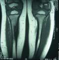
32316-month old boy with lameness N. Markeas, I. Papachristos
PAEDIATRIC NEWS IN BRIEF
324 Update on vaccination 2008 E. Galanakis
NEWS FROM THE INTERNET
327 The web page of the Royal College of Paediatrics and Child Health (RCPCH) C. Stefanidis
Thank you for your continuous interest in “Paediatriki” and the increasing number of submitted papers. We would like to inform you that Dr Michael Anthracopoulos (Pneumonology), Dr Vassiliki Papaevagelou (Infectious Diseases), Dr Antigone Syrigou-Papavasiliou (Neurology) and Dr Evangelia Charmandari (Endocrinology) are the new members of the Editorial Board. Dr Syrigou-Papavasiliou and Dr Anthracopoulos were formerly members of the Editorial Board of “Paediatriki”.
Dr Andriani Vazaiou-Gerasimidi, Dr Eustathia Katsarou-Pektasides, Dr Nicolaos Papadopoulos and Dr Marisa Tsolia withdrew from the Editorial Board. We would like to thank them for their longterm and important contribution.
On this occasion we would like to invite you to send us questions that arise from your every day practice. These questions will become the topics of the section: “Ask the expert”.
Genetic studies in childhood acute lymphoblastic leukaemia
F. Tzifi1, A. Kolialexi1, K. Salavoura2,
S. Kitsiou1, A. Mavrou1
Abstract: Research in the field of cancer genetics has given rise to a plethora of studies, which indicate that cancer is a genetic disorder of the somatic cells. The classification of childhood haematological malignancies combines cytologic, immunologic and cytogenetic characteristic features. Mutations of proto-oncogenes or tumour-suppressor genes play an important role in the pathogenesis of leukaemias. The application of molecular cytogenetics offers new potential for the laboratory investigation of haematological malignancies in children, through the identification of numerical or structural chromosomal abnormalities, which can determine the diagnosis, prognosis and treatment of the disease. Acute lymphoblastic leukaemia (ALL) is the most common leukaemia in childhood. In this review the molecular, chromosomal and immunophenotypic features of ALL in childhood are reported, which facilitate comprehension of the biology of ALL and its clinical evaluation and treatment.
Key words: ALL, children, oncogenes, chromosome abnormalities.
1 Laboratory of Medical Genetics, University of Athens, “Aghia Sophia” Children’s Hospital, Athens, Greece 2 Department of Immunology and Histocompatibility, “Aghia Sophia” Children’s Hospital, Athens, Greece
Correspondence: Ariadni Mavrou ariamav@hol.gr Laboratory of Medical Genetics, University of Athens, “Aghia Sophia” Children’s Hospital Thivon & Levadias St., 115 27, Athens, Greece
2. Kebriaei P, Anastasi J, Larson RA. Acute lymphoblastic leukaemia: diagnosis and classification. Best Pract Res Clin Haematol 2002;15:597-621.
3. McKenna RW. Multifaceted approach to the diagnosis and classification of acute leukemias. Clin Chem 2000;46:12521259.
4. Basso G, Buldini B, De Zen L, Orfao A. New methodologic approaches for immunophenotyping acute leukemias. Haematologica 2001;86:675-692.
5. Campana D, Coustan-Smith E. Advances in the immunological monitoring of childhood acute lymphoblastic leukaemia. Best Pract Res Clin Haematol 2002;15:1-19.
6. Weir EG, Borowitz MJ. Flow cytometry in the diagnosis of acute leukemia. Semin Hematol 2001;38:124-138.
7. Orfao A, Ortuno F, de Santiago M, Lopez A, San Miguel J. Immunophenotyping of acute leukemias and myelodysplastic syndromes. Cytometry A 2004;58:62-71.
8. Béné MC. Immunophenotyping of acute leukaemias. Immunol Lett 2005;98:9-21.
9. Riley RS, Massey D, Jackson-Cook C, Idowu M, Romagnoli G. Immunophenotypic analysis of acute lymphocytic leukemia. Hematol Oncol Clin North Am 2002;16:245-299, v.
10. Burmeister T, Thiel E. Molecular genetics in acute and chronic leukemias. J Cancer Res Clin Oncol 2001;127:80-90.
11. Heerema NA, Nachman JB, Sather HN, La MK, Hutchinson R, Lange BJ, et al. Deletion of 7p or monosomy 7 in pediatric acute lymphoblastic leukemia is an adverse prognostic factor: a report from the Children's Cancer Group. Leukemia 2004;18:939-47.
12. Sutcliffe MJ, Shuster JJ, Sather HN, Camitta BM, Pullen J, Schultz KR, et al. High concordance from independent studies by the Children's Cancer Group (CCG) and Pediatric Oncology Group (POG) associating favorable prognosis with combined trisomies 4, 10, and 17 in children with NCI Standard-Risk B-precursor Acute Lymphoblastic Leukemia: a Children's Oncology Group (COG) initiative. Leukemia 2005;19:734-740.
13. Zemanova Z, Michalova K, Sindelarova L, Smisek P, Brezinova J, Ransdorfova S, et al. Prognostic value of structural chromosomal rearrangements and small cell clones with high hyperdiploidy in children with acute lymphoblastic leukemia. Leuk Res 2005;29:273-281.
14. Raimondi SC, Zhou Y, Mathew S, Shurtleff SA, Sandlund JT, Rivera GK, et al. Reassessment of the prognostic significance of hypodiploidy in pediatric patients with acute lymphoblastic leukemia. Cancer 2003;98:2715-2722.
16. Romana SP, Poirel H, Leconiat M, Flexor MA, Mauchauffé M, Jonveaux P, et al. High frequency of t(12;21) in childhood B-lineage acute lymphoblastic leukemia. Blood 1995;86: 4263-4269.
17. Zelent A, Greaves M, Enver T. Role of the TEL-AML1 fusion gene in the molecular pathogenesis of childhood acute lymphoblastic leukaemia. Oncogene 2004;23:4275-4283.
18. Van Delft FW, Bellotti T, Luo Z, Jones LK, Patel N, Yiannikouris O, et al. Prospective gene expression analysis accurately subtypes acute leukaemia in children and establishes a commonality between hyperdiploidy and t(12;21) in acute lymphoblastic leukaemia. Br J Haematol. 2005;130:26-35.
19. Stams WA, den Boer ML, Beverloo HB, Meijerink JP, van Wering ER, Janka-Schaub GE, et al. Expression levels of TEL, AML1, and the fusion products TEL-AML1 and AML1-TEL versus drug sensitivity and clinical outcome in t(12;21)-positive pediatric acute lymphoblastic leukemia. Clin Cancer Res 2005;11:2974-2980.
20. Ferrando AA, Look AT. Clinical implications of recurring chromosomal and associated molecular abnormalities in acute lymphoblastic leukemia. Semin Hematol 2000;37: 381-395.
21. Hoelzer D, Gökbuget N, Ottmann O, Pui CH, Relling MV, Appelbaum FR, et al. Acute lymphoblastic leukemia. Hematology Am Soc Hematol Educ Program 2002;162-192.
22. Radich JP. Philadelphia chromosome-positive acute lymphocytic leukemia. Hematol Oncol Clin North Am 2001; 15:21-36.
24. Carè A, Cianetti L, Giampaolo A, Sposi NM, Zappavigna V, Mavilio F, et al. Translocation of c-myc into the immunoglobulin heavy-chain locus in human acute B-cell leukemia. A molecular analysis. EMBO J 1986;5:905-911.
25. Pais A, Amare Kadam P, Raje G, Sawant M, Kabre S, Jain H, et al. Identification of various MLL gene aberrations that lead to MLL gene mutation in patients with acute lymphoblastic leukemia (ALL) and infants with acute leukemia. Leuk Res 2005;29:517-526.
26. Attarbaschi A, Mann G, König M, Steiner M, Strehl S, Schreiberhuber A, et al. Mixed lineage leukemia-rearranged childhood pro-B and CD10-negative pre-B acute lymphoblastic leukemia constitute a distinct clinical entity. Clin Cancer Res 2006;12:2988-2994.
27. Pui CH, Gaynon PS, Boyett JM, Chessells JM, Baruchel A, Kamps W, et al. Outcome of treatment in childhood acute lymphoblastic leukaemia with rearrangements of the 11q23 chromosomal region. Lancet 2002;359:1909-1915.
28. Stam RW, den Boer ML, Passier MM, Janka-Schaub GE, Sallan SE, Armstrong SA, et al. Silencing of the tumor suppressor gene FHIT is highly characteristic for MLL gene rearranged infant acute lymphoblastic leukemia. Leukemia 2006;20:264-271.
29. Kode J, Dudhal N, Banavali S, Advani S, Chiplunkar S. Clonal T-cell receptor gamma and delta gene rearrangements in T-cell acute lymphoblastic leukemia at diagnosis: predictor of prognosis and response to chemotherapy. Leuk Lymphoma 2004;45:125-133.
30. Ferrando AA, Neuberg DS, Staunton J, Loh ML, Huard C, Raimondi SC, et al. Gene expression signatures define novel oncogenic pathways in T cell acute lymphoblastic leukemia. Cancer Cell 2002;1:75-87.
31. Berger R, Dastugue N, Busson M, Van Den Akker J, Pérot C, Ballerini P, et al. t(5;14)/HOX11L2-positive T-cell acute lymphoblastic leukemia. A collaborative study of the Groupe Français de Cytogénétique Hématologique (GFCH). Leukemia 2003;17:1851-1857.
32. Gottardo NG, Jacoby PA, Sather HN, Reaman GH, Baker DL, Kees UR. Significance of HOX11L2/TLX3 expression in children with T-cell acute lymphoblastic leukemia treated on Children's Cancer Group protocols. Leukemia 2005;19:1705-1708.
33. Watt PM, Hoffmann K, Greene WK, Brake RL, Ford J, Kees UR. Specific alternative HOX11 transcripts are expressed in paediatric neural tumours and T-cell acute lymphoblastic leukaemia. Gene 2003;323:89-99.
34. Iolascon A, Faienza MF, Coppola B, Moretti A, Basso G, Amaru R, et al. Frequent clonal loss of heterozygosity (LOH) in the chromosomal region 1p32 occurs in childhood T cell acute lymphoblastic leukemia (T-ALL) carrying rearrangements of the TAL1 gene. Leukemia 1997;11:359-363.
35. Anderegg B, Horstmann M, Ernst M, Kabisch H. Transcription of tal-1, a putative oncogene playing an important role in childhood T-ALL, can be shown in normal peripheral blood cells by a highly sensitive RT-PCR assay. Pediatr Hematol Oncol 1997;14:349-358.
36. Montpetit A, Larose J, Boily G, Langlois S, Trudel N, Sinnett D. Mutational and expression analysis of the chromosome 12p candidate tumor suppressor genes in pre-B acute lymphoblastic leukemia. Leukemia 2004;18:1499-1504.
38. Bertin R, Acquaviva C, Mirebeau D, Guidal-Giroux C, Vilmer E, Cavé H. CDKN2A, CDKN2B, and MTAP gene dosage permits precise characterization of mono- and biallelic 9p21 deletions in childhood acute lymphoblastic leukemia. Genes Chromosomes Cancer 2003;37:44-57.
39. Harrison CJ, Foroni L. Cytogenetics and molecular genetics of acute lymphoblastic leukemia. Rev Clin Exp Hematol 2002;6:91-113.
40. Papadhimitriou SI, Polychronopoulou S, Tsakiridou AA, Androutsos G, Paterakis GS, Athanassiadou F. p16 inactivation associated with aggressive clinical course and fatal outcome in TEL/AML1-positive acute lymphoblastic leukemia. J Pediatr Hematol Oncol 2005;27:675-677.
41. Kanerva J, Niini T, Vettenranta K, Riikonen P, Mäkipernaa A, Karhu R, et al. Loss at 12p detected by comparative genomic hybridization (CGH): association with TEL-AML1 fusion and favorable prognostic features in childhood acute lymphoblastic leukemia (ALL). A multi-institutional study. Med Pediatr Oncol 2001;37:419-425.
42. Haferlach T, Kern W, Schnittger S, Schoch C. Modern diagnostics in acute leukemias. Crit Rev Oncol Hematol 2005;56:223-234.
43. Kolialexi A, Tsangaris GT, Kitsiou S, Kanavakis E, Mavrou A. Impact of cytogenetic and molecular cytogenetic studies on hematologic malignancies. Anticancer Res 2005;25:29792983.
44. Harrison CJ, Moorman AV, Barber KE, Broadfield ZJ, Cheung KL, Harris RL, et al. Interphase molecular cytogenetic screening for chromosomal abnormalities of prognostic significance in childhood acute lymphoblastic leukaemia: a UK Cancer Cytogenetics Group Study. Br J Haematol 2005;129:520-530.
45. Carroll WL, Bhojwani D, Min DJ, Moskowitz N, Raetz EA. Childhood acute lymphoblastic leukemia in the age of genomics. Pediatr Blood Cancer 2006;46:570-578.
46. Holleman A, Cheok MH, den Boer ML, Yang W, Veerman AJ, Kazemier KM, et al. Gene-expression patterns in drugresistant acute lymphoblastic leukemia cells and response to treatment. N Engl J Med 2004;351:533-542.
47. Johansson B, Mertens F, Mitelman F. Clinical and biological importance of cytogenetic abnormalities in childhood and adult acute lymphoblastic leukemia. Ann Med 2004; 36:492-503.
Cytogenetic abnormalities in leukaemias of childhood
∞. Galla-Voumvouraki
Abstract: During the past two decades, the close association between specific chromosomal abnormalities and acute childhood leukaemias has been established and identification of clonal rearrangements of the involved genes has been made by molecular biology assays. It is suggested that detailed evaluation of the chromosomal rearrangements and the genes involved could contribute to the classification, prognosis and treatment of cancer of the haematopoetic system. This is a review of the most frequent chromosomal abnormalities associated with childhood leukaemias.
Department of Genetics, 2nd Department of Paediatrics, University of Athens, “P. & A. Kyriakou” Children’s Hospital, Athens, Greece
Correspondence: ∞ggeliki Galla-Voumvouraki Department of Genetics, 2nd Department of Paediatrics, University of Athens, “P. & A. Kyriakou” Children’s Hospital, Athens, Greece
1. Crist N, Pui CH. The Leukemias. In: Nelson W, Behrman R, Kliegman R, Arvin A, editors. Textbook of Paediatrics. 17th ed. Philadelphia: WB Saunders Company; 1996. p. 1694-1697.
2. Raimondi SC. Cytogenetics of Lymphoid Neoplasias. In: Mark HFL, editor. Medical Cytogenetics. New York: Marcel Dekker Inc.; 2000. p. 349-411.
3. Third International Workshop on Chromosomes in Leukaemia (1980): clinical significance of chromosomal abnormalities in acute lymphoblastic leukemia. Cancer Genet Cytogenet 1981;4:111-137.
4. Secker-Walker LM, Chessells JM, Stewart EL, Swansbury GJ, Richards S, Lawler SD. Chromosomes and other prognostic factors in acute lymphoblastic leukaemia: a longterm follow-up. Br J Haematol 1989;72:336-342.
5. Bloomfield CD, Secker-Walker LM, Goldman AI, Van Den Berghe H, de la Chapelle A, Ruutu T, et al. Six-year follow-
up of the clinical significance of karyotype in acute lymphoblastic leukemia. From the Sixth International Workshop on chromosomes in Leukemia 1987. Cancer Genet Cytogenet 1989;40:171-185.
6. Williams DL, Raimondi SC, Pui CH, Rivera GK. Evolving chromosome patterns and new cytogenetic concepts in childhood acute lymphoblastic leukemia. In: Gale RP, Hoelzer D, editors. Acute Lymphoblastic Leukemia: UCLA Symposia on Molecular and Cellular Biology. New Series, Vol. 108. New York: Wiley-Liss; 1990. p. 91-100.
7. Raimondi SC. Current status of cytogenetic research in childhood acute lymphoblastic leukemia. Blood 1993;81: 2237-2251.
8. Rivera GK, Raimondi SC, Hancock ML, Behm FG, Pui CH, Abromowitch M, et al. Improved outcome in childhood acute lymphoblastic leukaemia with reinforced early treatment and rotational combination chemotherapy. Lancet 1991;337:61-66.
9. Rubin CM, Le Beau MM, Mick R, Bitter MA, Nachman J, Rudinsky R, et al. Impact of chromosomal translocations on prognosis in childhood acute lymphoblastic leukemia. J Clin Oncol 1991;9:2183-2192.
10. Pui CH, Raimondi SC, Dodge RK, Rivera GK, Fuchs LA, Abromowitch M, et al. Prognostic importance of structural chromosomal abnormalities in children with hyperdiploid (greater than 50 chromosomes) acute lymphoblastic leukemia. Blood 1989;73:1963-1967.
11. Trueworthy R, Shuster J, Look T, Crist W, Borowitz M, Caroll A, et al. Ploidy of lymphoblasts is the strongest predictor of treatment outcome in B-progenitor cell acute lymphoblastic leukemia of childhood: a Pediatric Oncology Group study. J Clin Oncol 1992;10:606-613.
13. Martin PL, Look AT, Schnell S, Harris MB, Pullen J, Shuster JJ, et al. Comparison of fluorescence in situ hybridization, cytogenetic analysis, and DNA index analysis to detect chromosomes 4 and 10 aneuploidy in pediatric acute lymphoblastic leukemia: a Pediatric Oncology Group study. J Pediatr Hematol Oncol 1996;18:113-121.
14. Whitehead VM, Vuchich MJ, Carroll AJ, Lauer SJ, Mahoney D, Shuster JJ, et al. Accumulation of methotrexate polyglutamates (MTX PGS) in lymphoblasts, ploidy and trisomy of both chromosomes 4 and 10 in childhood Bprogenitor cell acute lymphoblastic leukemia (ALL): a Pediatric Oncology Group study. Blood 1994;84 (suppl 1): 515a.
15. White DM, Crolla JA, Ross FM. Detection of minimal residual disease in childhood acute lymphoblastic leukaemia using fluorescence in-situ hybridization. Br J Haematol 1995; 91:1019-1024.
16. Heinonen K, Mahlamäki E. Detection of numerical chromosome abnormalities by FISH in childhood acute lymphoblastic leukemia. Cancer Genet Cytogenet 1996;87: 123-126.
17. Rivera GK, Raimondi SC, Hancock ML, Behm FG, Pui CH, Abromowitch M, et al. Improved outcome in childhood acute lymphoblastic leukaemia with reinforced early treatment and rotational combination chemotherapy. Lancet 1991;337:61-66.
18. Watson MS, Carroll AJ, Shuster JJ, Steuber CP, Borowitz MJ, Behm FG, et al. Trisomy 21 in childhood acute lymphoblastic leukemia: a Pediatric Oncology Group study (8602). Blood 1993;82:3098-3102.
19. Pettenati MJ, Rao N, Wofford M, Shuster JJ, Pullen DJ, Ling MP, et al. Presenting characteristics of trisomy 8 as the primary cytogenetic abnormality associated with childhood acute lymphoblastic leukemia. A Pediatric Oncology Group (POG) Study (8600/8493). Cancer Genet Cytogenet 1994;75:6-10.
20. Pui CH, Caroll AJ, Head D, Raimondi SC, Shuster JJ, Crist WM, et al. Near-triploid and near-tetraploid acute lymphoblastic leukemia of childhood. Blood 1990;76:590-596.
21. Pui CH, Williams DL, Raimondi SC, Rivera GK, Look AT, Dodge RK, et al. Hypodiploidy is associated with a poor prognosis in childhood acute lymphoblastic leukemia. Blood 1987;70:247-253.
22. Rieder H, Schnittger S, Bodenstein H, Schwonzen M, Wörmann B, Berkovic D, et al. Dic(9;20): a new recurrent chromosome abnormality in adult acute lymphoblastic leukemia. Genes Chromosomes Cancer 1995;13:54-61.
23. Heerema NA, Maben KD, Bernstein J, Breitfeld PP, Neiman RS, Vance GH. Dicentric (9;20)(p11;q11) identified by fluorescence in situ hybridization in four pediatric acute lymphoblastic leukemia patients. Cancer Genet Cytogenet 1996;92:111-115.
24. Translocations involving 9p and/or 12p in acute lymphoblastic leukemia. United Kingdom Cancer Cytogenetics Group (UKCCG). Genes Chromosomes Cancer 1992;5: 255-259.
25. Behrendt H, Charrin C, Gibbons B, Harrison CJ, Hawkins JM, Heerema NA, et al. Dicentric (9;12) in acute lymphocytic leukemia and other hematological malignancies: report from a dic(9;12) study group. Leukemia 1995;9:102-106.
26. Raimondi SC, Shurtleff SA, Downing JR, Rubnitz J, Mathew S, Hancock M, et al. 12p abnormalities and the TEL gene (ETV6) in childhood acute lymphoblastic leukemia. Blood 1997;90:4559-4566.
27. Filatov LV, Saito M, Behm FG, Rivera GK, Raimondi SC. A subtle deletion of 12p by routine cytogenetics is found to be a translocation to 21q by fluorescence in situ hybridization: t(12;21)(p13;q22). Cancer Genet Cytogenet 1996;89: 136-140.
28. Pui CH, Raimondi SC, Hancock ML, Rivera GK, Ribeiro RC, Mahmoud HH, et al. Immunologic, cytogenetic, and clinical characterization of childhood acute lymphoblastic leukemia with the t(1;19)(q23;p13) or its derivative. J Clin Oncol 1994;12:2601-2606.
29. Troussard X, Rimokh R, Valensi F, Leboeuf C, Fenneteau O, Guitard AM, et al. Heterogeneity of t(1;19)(q23;p13) acute leukaemias. French Haematological Cytology Group. Br J Haematol 1995;89:516-526.
30. Hunger SP. Chromosomal translocations involving the E2A gene in acute lymphoblastic leukemia: clinical features and molecular pathogenesis. Blood 1996;87:1211-1224.
31. Human gene mapping 11. London Conference (1991). Eleventh International Workshop on Human Gene Mapping. London, UK, August 18-22, 1991. Cytogenet Cell Genet 1991;58(1-4):1-2156.
32. Chen CS, Sorensen PH, Domer PH, Reaman GH, Korsmeyer SJ, Heerema NA, et al. Molecular rearrangements on chromosome 11q23 predominate in infant acute
lymphoblastic leukemia and are associated with specific biologic variables and poor outcome. Blood 1993;81:23862393.
33. Behm FG, Raimondi SC, Frestedt JL, Liu Q, Crist WM, Downing JR, et al. Rearrangements of the MLL gene confers a poor prognosis in childhood acute lymphoblastic leukemia, regardless of presenting age. Blood 1996;87: 2870-2877.
34. Rubnitz JE, Link MP, Shuster JJ, Carroll AJ, Hakami N, Frankel LS, et al. Frequency and prognostic significance of HRX rearrangements in infant acute lymphoblastic leukemia: a Pediatric Oncology Group study. Blood 1994; 84:570-573.
35. Strout M, Caligiuri M, Bloomfield C. Recent advances in the Biology and management of primary acute myeloid leukemia with rearrangements of the MLL gene. MedGenMed 2000;2:1-8.
36. Schlieben S, Borkhardt A, Reinisch I, Ritterback J, Janssen JW, Ratei R, et al. Incidence and clinical outcome of children with BCR/ABL-positive acute lymphoblastic leukemia (ALL). A prospective RT-PCR study based on 673 patients enrolled in the German pediatric multicenter therapy trials ALL-BFM-90 and CoALL-05-92. Leukemia 1996;10: 957-963.
37. Herm ans A, Heisterkamp N, von Linden M, van Baal S, Meijer D, van der Plas D, et al. Unique fusion of bcr and cabl genes in Philadelphia chromosome positive acute lymphoblastic leukemia. Cell 1987;51:33-40.
38. Clark SS, McLaughlin J, Crist WM, Champlin R, Witte ON. Unique forms of the abl tyrosine kinase distinguish Ph1-positive CML from Ph1-positive ALL. Science 1987; 235:85-88.
39. Lugo TG, Pendergast AM, Muller AJ, Witte ON. Tyrosine kinase activity and transformation potency of bcr/abl oncongene products. Science 1990;247:1079-1082.
40. Voncken JW, Kaartinen V, Pattengale PK, Germeraad WT, Groffen J, Heisterkamp N. BCR/ABL P210 and P190 cause distinct leukemia in transgenic mice. Blood 1995;86: 46034611.
41. Hayashi Y, Raimondi SC, Look AT, Behm FG, Kitchingman GR, Pui CH, et al. Abnormalities of the long arm of chromosome 6 in childhood acute lymphoblastic leukemia. Blood 1990;76:1626-1630.
42. Murphy SB, Raimondi SC, Rivera GK, Crone M, Dodge RK, Behm FG, et al. Nonrandom abnormalities of chromosome 9p in childhood acute lymphoblastic leukemia: association with high-risk clinical features. Blood 1989:74: 409-415.
43. Okuda T, Shurtleff SA, Valentine MB, Raimondi SC, Head DR, Behm F, et al. Frequent deletion of p16INK4a/MTS1
and p15INK4b/MTS2 in pediatric acute lymphoblastic leukemia. Blood 1995;85:2321-2330.
44. Kees UR, Ranford PR, Hatzis M. Deletions of the p16 gene in pediatric leukemia and corresponding cell lines. Oncogene 1996;12:2235-2239.
45. Bowman WP, Shuster JJ, Cook B, Griffin T, Behm F, Pullen J, et al. Improved survival for children with B-cell acute lymphoblastic leukemia and stage IV small noncleaved-cell lymphoma: a pediatric oncology group study. J Clin Oncol 1996;14:1252-1261.
46. Blackwood EM, Eisenman RN. MAX: a helix-loop-helix zipper protein that forms a sequence-specific DNA-binding complex with Myc. Science 1991;251:1211-1217.
47. Ayer DE, Kretzner L, Eisenman RN. Mad: a heterodimeric partner for Max that antagonizes Myc transcriptional activity. Cell 1993;72:211-222.
48. Cytogenetic abnormalities in adult acute lymphoblastic leukemia: correlations with hematologic findings and outcome. A Collaborative Study of the Groupe Français de Cytogénétique Hématologique. Blood 1996;87:3135-3142.
50. Bianchi E, Rogge L. Dissecting oncogenes and tyrosine kinases in AML cells. MedGenMed 2003;5:10.
51. Boehm T, Foroni L, Kaneko Y, Perutz MF, Rabbitts TH. The rhombotin family of cysteine-rich LIM-domain oncogenes: distinct members are involved in T-cell translocations to human chromosomes 11p15 and 11p13. Proc Natl Acad Sci USA 1991;88:4367-4371.
52. Royer-Pokora B, Loos U, Ludwig WD. TTG-2, a new gene encoding a cysteine-rich protein with the LIM motif, is overexpressed in acute T-cell leukaemia with the t(11;14) (p13;q11). Oncogene 1991;6:1887-1893.
53. Xia Y, Brown L, Yang CY, Tsan JT, Siciliano MJ, Espinosa R III, et al. TAL2, a helix-loop-helix gene activated by the (7;9)(q34;q32) translocation in human T-cell leukemia. Proc Natl Acad Sci USA 1991;88:11416-11420.
54. Mellentin JD, Smith SD, Cleary ML. Lyl-1, a novel gene altered by chromosomal translocation in T cell leukemia, codes for a protein with a helix-loop-helix DNA binding motif. Cell 1989;58:77-83.
55. Arber DA, Stein AS, Carter NH, Ikle D, Forman SJ, Slovak ML. Prognostic impact of acute myeloid leukemia classification. Importance of detection of recurring cytogenetic abnormalities and multilineage dysplasia on survival. Am J Clin Pathol 2003;119:672-680.
56. Marcucci G, Caligiuri M, Bloomfield C. Core binding factor (CBF) primary acute myeloid leukemia: biology and clinical implications. MedGenMed 2000;2:1-13.
Non-random X inactivation: clinical consequences of the skewing effect
G. Lavranos, R. Angelopoulou
Abstract: X chromosome inactivation is a normal genetic phenomenon, causing the transcriptional silencing of almost every gene located on all X chromosomes but one in mammalian embryos, so that only a single gene copy remains active per cell. This procedure remains stable in all the diploid cells of the adult organism and applies to both normal females with karyotype 46,XX (1 inactive X) and individuals with numerical/quantitative chromosomal abnormalitites of the gonosomes. In humans, contrary to monotremes, marsupials and rodent mammals, the selection of the X chromosome to be inactivated is made in random order, so that a different chromosome is active in each cell deriving from a different blastomere of the embryo. Thus, affected individuals may be viewed as functional mosaics, which results both in their protection from the expression of pathological/mutated alleles and in an increase of biodiversity, in cases of differences in X chromosome alleles, with the creation of new, intermediate phenotypes due to co-dominance. In certain cases, however, for a yet unknown reason, X chromosome inactivation does not conform to the random pattern associated with favourable expression of a specific allele. Such incidents have been associated with a variety of clinical manifestations, including several genetic syndromes, recurrent spontaneous abortion, congenital immune deficiencies and autoimmune disorders. In this review, novel data on these alleged correlations are discussed, along with guidelines on their evaluation within the context of clinical genetics.
Key words: Genetic dosage compensation, X chromosome inactivation, clinical genetics.
Laboratory of HistologyEmbryology, Faculty of Medicine, National and Kapodistrian University of Athens
Correspondence: Roxani Angelopoulou rangelop@med.uoa.gr Laboratory of HistologyEmbryology, Faculty of Medicine, National and Kapodistrian University of Athens 115 27, Goudi, Athens, Greece
2. Manolakou P, Lavranos G, Angelopoulou R. Molecular patterns of sex determination in the animal kingdom: a comparative study of the biology of reproduction. Reprod Biol Endocrinol 2006;4:59.
3. Lyon MF. X-chromosome inactivation and human genetic disease. Acta Paediatr Suppl 2002;91:107-12.
4. Brown CJ, Ballabio A, Rupert JL, Lafreniere RG, Grompe M, Tonlorenzi R, et al. A gene from the region of the human X inactivation centre is expressed exclusively from the inactive X chromosome. Nature 1991;349:38-44.
5. Penny GD, Kay GF, Sheardown SA, Rastan S, Brockdorff N. Requirement for Xist in X chromosome inactivation. Nature 1996;379:131-137.
6. Lee JT, Jaenisch R. Long-range cis effects of ectopic X-inactivation centres on a mouse autosome. Nature 1997;386:275-279.
7. Sado T, Wang Z, Sasaki H, Li E. Regulation of imprinted X-chromosome inactivation in mice by Tsix. Development 2001;128:1275-1286.
8. Migeon BR, Chowdhury AK, Dunston JA, McIntosh I. Identification of TSIX, encoding an RNA antisense to human XIST, reveals differences from its murine counterpart: implications for X inactivation. Am J Hum Genet 2001;69:951-960.
9. Cohen DE, Davidow LS, Erwin JA, Xu N, Warshawsky D, Lee JT. The DXPas34 repeat regulates random and imprinted X inactivation. Dev Cell 2007;12:57-71.
10. Hornecker JL, Samollow PB, Robinson ES, Vandeberg JL, McCarrey JR. Meiotic sex chromosome inactivation in the marsupial Monodelphis domestica. Genesis 2007;45:696-708.
11. Angelopoulou R, Lavranos G, Manolakou P. Regulatory RNAs and chromatin modification in dosage compensation: a continuous path from flies to humans? Reprod Biol Endocrinol 2008;6:12.
12. Lau AW, Brown CJ, Penaherrera M, Langlois S, Kalousek DK, Robinson WP. Skewed X-chromosome inactivation is common in fetuses or newborns associated with confined placental mosaicism. Am J Hum Genet 1997;1353-1361.
13. Bouayed Abdelmoula N, Portnoi MF, Keskes L, Recan D, Bahloul A, Boudawara T, et al. Skewed X-chromosome inactivation pattern in SRY positive XX maleness: a case report and review of literature. Ann Genet 2003;46:11-18.
14. Manola KN, Stavropoulou C, Georgakakos VN, Zoi K, Fisfis M, Evmorfiadis I, et al. Switch in X-inactivation in a JAK2 V617F-negative case of polycythemia vera with two acquired X-autosome translocations. Leuk Res 2007;31:1009-1014.
16. Akabori S, Takano T, Fujito H, Takeuchi Y. West syndrome in a patient with balanced translocation t(X;18) (p22;p11.2). Pediatr Neurol 2007;37:64-66.
17. Marble M, Voeller KS, May MM, Stevenson RE, Schwartz CE, Simensen RJ. Pelizaeus-Merzbacher syndrome: neurocognitive function in a family with carrier manifestations. Am J Med Genet A 2007;143A:1442-1447.
18. Aral B, de Saint Basile G, Al-Garawi S, Kamoun P, Ceballos-Picot I. Novel nonsense mutation in the hypoxanthine guanine phosphoribosyltransferase gene and nonrandom
X-inactivation causing Lesch-Nyhan syndrome in a female patient. Hum Mutat 1996;7:52-58.
19. Rosenberg C, Wouters CH, Szuhai K, Dorland R, Pearson P, Poll-The BT, et al. A Rett syndrome patient with a ring X chromosome: further evidence for skewing of X inactivation and heterogeneity in the aetiology of the disease. Eur J Hum Genet 2001;9:171-177.
20. Tomkins DJ, McDonald HL, Farrell SA, Brown CJ. Lack of expression of XIST from a small ring X chromosome containing the XIST locus in a girl with short stature, facial dysmorphism and developmental delay. Eur J Hum Genet 2002;10:44-51.
21. Stankiewicz P, Thiele H, Giannakudis I, Schlicker M, Baldermann C, Krüger A, et al. Kabuki syndrome-like features associated with a small ring chromosome X and XIST gene expression. Am J Med Genet 2001;102:286-292.
22. Matsumoto N, Niikawa N. Kabuki make-up syndrome: a review. Am J Med Genet C Semin Med Genet 2003;117C:57-65.
23. Beever CL, Pen ~ aherrera MS, Langlois S, Robinson WR. X chromosome inactivation patterns in Russell-Silver syndrome patients and their mothers. Am J Med Genet A 2003;123A:231-235.
24. Hellenbroich Y, Grzeschik KH, Krapp M, Jarutat T, Lehrmann-Petersen C, Buiting K, et al. Reduced penetrance in a family with X-linked dominant chondrodysplasia punctata. Eur J Med Genet 2007;50:392-398.
25. Orstavik RE, Tommerup N, Eiklid K, Orstavik KH. Nonrandom X chromosome inactivation in an affected twin in a monozygotic twin pair discordant for Wiedemann-Beckwith syndrome. Am J Med Genet 1995;56:210-214.
26. Hansen RS, Stöger R, Wijmenga C, Stanek AM, Canfield TK, Luo P, et al. Escape from gene silencing in ICF syndrome: evidence for advanced replication time as a major determinant. Hum Mol Genet 2000;9:2575-2587.
27. Gartler SM, Hansen RS. ICF syndrome cells as a model system for studying X chromosome inactivation. Cytogenet Genome Res 2002;99(1-4):25-29.
28. Patra SK, Patra A, Dahiya R. Histone deacetylase and DNA methyltransferase in human prostate cancer. Biochem Biophys Res Commun 2001;287:705-713.
29. Yan L, Yang X, Davidson NE. Role of DNA methylation and histone acetylation in steroid receptor expression in breast cancer. J Mammary Gland Biol Neoplasia 2001;6:183-192.
30. Schneider R, Bannister AJ, Kouzarides T. Unsafe SETs: histone lysine methyltransferases and cancer. Trends Biochem Sci 2002;27:396-402.
31. Kristiansen M, Helland A, Kristensen GB, Olsen AO, Lo / nning PE, Bo / rresen-Dale AL, et al. X chromosome inactivation in cervical cancer patients. Cancer Genet Cytogenet 2003;146:73-76.
32. Novelli M, Cossu A, Oukrif D, Quaglia A, Lakhani S, Poulsom R, et al. X-inactivation patch size in human female tissue confounds the assessment of tumor clonality. Proc Natl Acad Sci USA 2003;100:3311-3314.
33. Kawakami T, Okamoto K, Sugihara H, Hattori T, Reeve AE, Ogawa O, et al. The roles of supernumerical X chromosomes and XIST expression in testicular germ cell tumors. J Urol 2003;169:1546-1552.
34. Kristiansen M, Langer A, Knudsen GP, Weber BL, Bo / rresen-Dale A-L, O / rstavik KH. X chromosome inactivation pattern in female patients with breast cancer. Breast Cancer Res 2000;2:P1.10.
35. Ganesan S, Silver DP, Greenberg RA, Avni D, Drapkin R, Miron A, et al. BRCA1 supports XIST RNA concentration on the inactive X chromosome. Cell 2002;111:393-405.
36. Huang KC, Rao PH, Lau CC, Heard E, Ng SK, Brown C, et al. Relationship of XIST expression and responses of ovarian cancer to chemotherapy. Mol Cancer Ther 2002;1:769-776.
37. Passarge E, Fries E. X chromosome inactivation in Xlinked hypohidrotic ectodermal dysplasia. Nature New Biol 1973;245:58-59.
38. O / rstavik KH, Knudsen GP, Nordgarden H, Ormerod E, Stro / mme P, Lazarou LP, et al. Severe hypohidrotic ectodermal dysplasia in a girl caused by a de novo 9;X insertion that includes XIST and disrupts the EDA gene. Am J Med Genet A 2007;143A:1510-1513.
39. Chitnis S, Monteiro J, Glass D, Apatoff B, Salmon J, Concannon P, Gregersen PK. The role of X-chromosome inactivation in female predisposition to autoimmunity. Arthritis Res 2000;2:399-406.
40. Hernaãndez-Molina G, Svyryd Y, Saãnchez-Guerrero J, Mutchinick OM. The role of the X chromosome in immunity and autoimmunity. Autoimmun Rev 2007;6:218-222.
41. Martin-Villa JM, Corell A, Ramos-Amador JT, Ruiz-Contreras J, Arnaiz-Villena A. Higher incidence of autoantibodies in X-linked chronic granulomatous disease carriers: random X-chromosome inactivation may be related to autoimmunity. Autoimmunity 1999;31:261-264.
42. Demura M, Takeda Y, Yoneda T, Furukawa K, Tachi A, Mabuchi H. Completely skewed X-inactivation in a mentally retarded young female with pseudohypoparathyroidism type IB and juvenile renin-dependent hypertension. J Clin Endocrinol Metab 2003;88:3043-3049.
43. Pegoraro E, Whitaker J, Mowery-Rushton P, Surti U, Lanasa M, Hoffman EP. Familial skewed X inactivation: a molecular trait associated with high spontaneous-abortion rate maps to Xq28. Am J Hum Genet 1997;61:160-170.
44. Beever CL, Stephenson MD, Pen~aherrera MS, Jiang RH, Kalousek DK, Hayden M, et al. Skewed X-chromosome inactivation is associated with trisomy in women ascertained on the basis of recurrent spontaneous abortion or chromosomally abnormal pregnancies. Am J Hum Genet 2003;72:399-407.
45. Sullivan AE, Lewis T, Stephenson M, Odem R, Schreiber J, Ober C, et al. Pregnancy outcome in recurrent miscarriage patients with skewed X chromosome inactivation. Obstet Gynecol 2003;101:1236-1242.
46. Bagislar S, Ustuner I, Cengiz B, Soylemez F, Akyerli CB, Ceylaner S, et al. Extremely skewed X-chromosome inactivation patterns in women with recurrent spontaneous abortion. Aust NZJ Obstet Gynaecol 2006;46:384-387.
47. Bretherick KL, Metzger DL, Chanoine JP, Panagiotopoulos C, Watson SK, Lam WL, et al. Skewed X-chromosome inactivation is associated with primary but not secondary ovarian failure. Am J Med Genet A 2007;143A:945-951.
48. Kline J, Kinney A, Levin B, Kelly A, Yu CY, Brown S, et al. X-chromosome inactivation and ovarian age during the reproductive years. Fertil Steril 2006;85:1488-1495.
49. Uz E, Dolen I, Al AR, Ozcelik T. Extremely skewed X-chromosome inactivation is increased in pre-eclampsia. Hum Genet 2007;121:101-105.
D. Thomas1,2, ∂. Vlahopapadopoulou2, V. Papadakis3
Abstract: Testicular microlithiasis (TM) is a relatively uncommon condition in the paediatric population, characterized by the presence of hyperechoic foci (microliths) within the testicular parenchyma, usually found incidentally in patients referred for scrotal ultrasound (U/S) for various reasons. It is encountered at a higher prevalence in children with genetic syndromes and has been associated with both benign and malignant lesions of the testes and other tissues. Although the true prevalence of TM in the population is still unknown, recent data on large series of patients who underwent scrotal U/S give ranges of 0.6 to 18.1% for the adult population and 1.1 to 1.9% for children. The natural history of TM cannot be predicted with precision. There have been several reports of testicular tumour development in patients with TM, indicating the possibility of it being a pre-malignant condition. ∆he reported rate of development of malignancy varies from 6-46% in adults and 0-12.5% in children. It has been estimated that almost 8% of subjects with TM will develop testicular tumour in the 6 years following identification. This probability is about the same for children and adults, but the long term risk of tumour development is not known at present. For the follow-up of children with isolated TM, annual U/S screening, regular careful self-examination and physical examination by a physician are highly recommended.
Key words: Microlithiasis, testes.
1 Department of Endocrinology, EANP “ªetaxa”, Piraeus
2 Department of Growth and Development, “P. & A. Kyriakou” Children’s Hospital, Athens, Greece
3 Department of Paediatric Haematology-Oncology, “Aghia Sophia” Children’s Hospital, Athens, Greece
Correspondence: Elpida Vlahopapadopoulou elpis.vl@gmail.com Department of Growth and Development, “P. & A. Kyriakou” Children’s Hospital 24, Mesogion St. 115 27, Athens, Greece
1. Miller FN, Sidhu PS. Does testicular microlithiasis matter? A review. Clin Radiol 2002;57:883-890.
2. Dell’Acqua A, Tomà P, Oddone M, Ciccone MA, Marsili E, Derchi LE. Testicular microlithiasis: US findings in six pediatric cases and literature review. Eur Radiol 1999;9:940-944.
3. Renshaw AA. Testicular calcifications: incidence, histology and proposed pathological criteria for testicular microlithiasis. J Urol 1998;160:1625-1628.
4. Weinberg AG, Currarino G, Stone IC Jr. Testicular microlithiasis. Arch Pathol 1973;95:312-314.
5. Drut R, Drut RM. Testicular microlithiasis: histologic and immunohistochemical findings in 11 pediatric cases. Pediatr Dev Pathol 2002;5:544-550.
6. Corut A, Senyigit A, Ugur SA, Altin S, Ozcelik U, Calisir H, et al. Mutations in SLC34A2 cause pulmonary alveolar microlithiasis and are possibly associated with testicular microlithiasis. Am J Hum Genet 2006;79:650-656.
7. Campagnoli MF, Pucci A, Garelli E, Carando A, Defilippi C, Lala R, et al. Familial tumoral calcinosis and testicular microlithiasis associated with a new mutation of GALNT3 in a white family. J Clin Pathol 2006;59:440-442.
8. Cast JE, Nelson WM, Early AS, Biyani S, Cooksey G, Warnock NG, et al. Testicular microlithiasis: prevalence and tumor risk in a population referred for scrotal sonography. AJR Am J Roentgenol 2000;175:1703-1706.
9. Otite U, Webb JA, Oliver RT, Badenoch DF, Nargund VH. Testicular microlithiasis: is it a benign condition with malignant potential? Eur Urol 2001;40:538-542.
10. Skyrme RJ, Fenn NJ, Jones AR, Bowsher WG. Testicular microlithiasis in a UK population: its incidence, associations and follow-up. BJU Int 2000;86:482-485.
11. Pourbagher MA, Kilinc F, Guvel S, Pourbagher A, Egilmez T, Ozkardes H. Follow-up of testicular microlithiasis for subsequent testicular cancer development. Urol Int 2005;74:108-112.
12. Derogee M, Bevers RF, Prins HJ, Jonges TG, Elbers FH, Boon TA. Testicular microlithiasis, a premalignant condition: prevalence, histopathologic findings, and relation to testicular tumor. Urology 2001;57:1133-1137.
13. Bach AM, Hann LE, Hadar O, Shi W, Yoo HH, Giess CS, et al. Testicular microlithiasis: what is its association with testicular cancer? Radiology 2001;220:70-75.
14. Sakamoto H, Shichizyou T, Saito K, Okumura T, Ogawa Y, Yoshida H, et al. Testicular microlithiasis identified ultrasonographically in Japanese adult patients: prevalence and associated conditions. Urology 2006;68:636-641.
15. Höbarth K, Susani M, Szabo N, Kratzik C. Incidence of testicular microlithiasis. Urology 1992;40:464-467.
16. Middleton WD, Teefey SA, Santillan CS. Testicular microlithiasis: prospective analysis of prevalence and associated tumor. Radiology 2002;224:425-428.
17. Aso C, Enr›quez G, Fité M, Toraãn N, Pirfi C, Piqueras J, et al. Gray-scale and color Doppler sonography of scrotal disorders in children: an update. Radiographics 2005;25:1197-1214.
18. Leenen AS, Riebel TW. Testicular microlithiasis in children: sonographic features and clinical implications. Pediatr Radiol 2002;32:575-579.
19. Dagash H, Mackinnon EA. Testicular microlithiasis: what does it mean clinically? BJU Int 2007;99:157-160.
20. Furness PD 3rd, Husmann DA, Brock JW 3rd, Steinhardt GF, Bukowski TP, Freedman AL, et al. Multi-institutional study of testicular microlithtiasis in childhood: a benign or premalignant condition? J Urol 1998;160:1151-1154.
21. McEniff N, Doherty F, Katz J, Schrager CA, Klauber G. Yolk sac tumor of the testis discovered on a routine annual sonogram in a boy with testicular microlithiasis. AJR Am J Roentgenol 1995;164:971-972.
22. Arrigo T, Messina MF, Valenzise M, Rosano M, Alaggio R, Cecchetto G, et al. Testicular microlithiasis heralding mixed germ cell tumor of the testis in a boy. J Endocrinol Invest 2006;29:82-85.
23. Kocaoglu M, Bozlar U, Bulakbasi N, Saglam M, Uçöz T, Somuncu I. Testicular microlithiasis in pediatric age group: ultrasonography findings and literature review. Diagn Interv Radiol 2005;11:60-65.
24. Bieger RC, Passarge E, McAdams AJ. Testicular intratubular bodies. J Clin Endocrinol Metab 1965;25:1340-1346.
25. Ganem JP, Workman KR, Shaban SF. Testicular microlithiasis is associated with testicular pathology. Urology 1999; 53:209-213.
26. Bennett HF, Middleton WD, Bullock AD, Teefey SA. Testicular microlithiasis: US follow-up. Radiology 2001;218: 359-363.
27. Bach AM, Hann LE, Shi W, Giess CS, Yoo HH, Sheinfeld J, et al. Is there an increased incidence of contralateral testicular cancer in patients with intratesticular microlithiasis? AJR Am J Roentgenol 2003;180:497-500.
28. Golash A, Parker J, Ennis O, Jenkins BJ. The interval of development of testicular carcinoma in a patient with previously demonstrated testicular microlithiasis. J Urol 2000; 163:239.
29. Salisz JA, Goldman KA. Testicular calcifications and neoplasia in patient treated for subfertility. Urology 1990;36:557560.
30. Frush DP, Kliewer MA, Madden JF. Testicular microlithiasis and subsequent development of metastatic germ cell tumor. AJR Am J Roentgenol 1996;167:889-890.
31. Howard RG, Roebuck DJ, Metreweli C. The association of mediastinal germ cell tumour and testicular microlithiasis. Pediatr Radiol 1998;28:998.
32. Quane LK, Kidney DD. Testicular microlithiasis in a patient with a mediastinal germ cell tumour. Clin Radiol 2000;55:642-644.
33. Sato K, Komatsu K, Maeda Y, Ueno S, Koshida K, Namiki M. Case of mediastinal seminoma with testicular microlithiasis. Int J Urol 2002;9:114-116.
34. Nishiyama T, Terunuma M, Iwashima A, Souma T, Hirahara H. Testicular microlithiasis with mediastinal choriocarcinoma: a case report. Int J Urol 1998;5:301-302.
35. Berthelsen JG, Skakkebaek NE. Value of testicular biopsy in diagnosing carcinoma in situ testis. Scand J Urol Nephrol 1981;15:165-168.
36. Von der Maase H, Ro/rth M, Walbom-Jo / rgensen S, So/rensen BL, Christophersen IS, Hald T, et al. Carcinoma in situ of contralateral testis in patients with testicular germ cell cancer: study of 27 cases in 500 patients. Br Med J (Clin Res Ed) 1986;293:1398-1401.
37. Vachon L, Fareau GE, Wilson MG, Chan LS. Testicular microlithiasis in patients with Down syndrome. J Pediatr 2006;149:233-236.
38. Wasniewska M, Matarazzo P, Weber G, Russo G, Zampolli M, Salzano G, et al. Clinical presentation of McCune-Albright syndrome in males. J Pediatr Endocrinol Metab 2006;19:619-622.
39. Zugor V, Zenker M, Schrott KM, Schott GE. [Frasier syndrome: a rare syndrome with WT1 gene mutation in pediatric urology] [Article in German]. Aktuelle Urol 2006;37: 64-66.
40. Aizenstein RI, Hibbeln JF, Sagireddy B, Wilbur AC, O’Neil HK. Klinefelter’s syndrome associated with testicular microlithiasis and mediastinal germ-cell neoplasm. J Clin Ultrasound 1997;25:508-510.
41. Blau H, Freud E, Mussaffi H, Werner M, Konen O, Rathaus V. Urogenital abnormalities in male children with cystic fibrosis. Arch Dis Child 2002;87:135-138.
42. Bercovitch RS, Januario JA, Terry SF, Boekelheide K, Podis AD, Dupuy DE, et al. Testicular microlithiasis in association with pseudoxanthoma elasticum. Radiology 2005;237: 550-554.
43. Patel RP, Kolon TF, Huff DS, Carr MC, Zderic SA, Canning DA, et al. Testicular microlithiasis and antisperm antibodies following testicular biopsy in boys with cryptorchidism. J Urol 2005;174:2008-2010.
44. Kwan DJ, Kirsch AJ, Chang DT, Goluboff ET, Berdon WE, Hensle DW. Testicular microlithiasis in a child with torsion of the appendix testis. J Urol 1995;153:183-184.
45. Heinemann V, Frey U, Linke J, Dieckmann KP. Testicular microlithiasis -- one case and four points to note. Scand J Urol Nephrol 2003;37:515-518.
46. Kim B, Winter TC 3rd, Ryu JA. Testicular microlithiasis: clinical significance and review of the literature. Eur Radiol 2003;13:2567-2576.
47. Dieckmann KP, Loy V. False-negative biopsies for the diagnosis of testicular intraepithelial neoplasia (TIN) -- an update. Eur Urol 2003;43:516-521.
48. Kliesch S, Thomaidis T, Schutte B, Puhse G, Kater B, Roth S, et al. Update on the diagnostic safety for detection of testicular intraepithelial neoplasia (TIN). APMIS 2003;111:70-74.
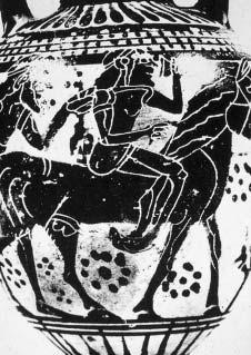
Abstract: Hephaestus, the Olympian god, was the divine smith, famed for inventions, who taught men glorious crafts. The fixed epithet for Hephaestus, used from the eighth century B.C. by Homer and Hesiod, and other ancient authors up to the fifth century A.D., is “·ÌÊÈÁ˘‹ÂȘ”, i.e. with both feet crooked. He is also called “΢ÏÏÔÔ‰›ˆÓ”, i.e. clubfooted. His body posture and his typical gait are characteristically described by Homer and help in making a diagnosis. His anomaly is described as being congenital, and both his sons are reported as having deformed feet. For this disablement, several interpretations and contemporary medical diagnoses have been proposed. In this article a combination of the information derived from ancient Greek texts and illustrative arts is made and it is concluded that Hephaestus' disability was congenital bilateral talipes equinovarus, i.e. clubfoot.
Key words: Ancient history of medicine, Genetics medical history, Hephaestus, Hephaistos, congenital bilateral clubfoot.
Pathology and Oral Surgery Sector, Dental School of University of Athens
Correspondence: ∂fterpi Bazopoulou∫yrkanidou ebazopou@dent.uoa.gr Pathology and Oral Surgery Sector, Dental School of University of Athens
36. Apollonius Rhodius. The Argonautica. I. 202-206, III. 37, IV. Translated by Seaton RC (1912), Loeb Classical Library. Vol. I. London: Heinemann/Cambridge, MA: Harvard University Press; 1954.
37. Lexicon Iconographicum Mythologiae Classicae (LIMC). Artemis Verlag Zurich und Munchen; 1988. IV. 1, p. 627628, 650-659, 681-684; IV. 2, p. 386-405.
38. Flaxman J. The Iliad of Homer. London: Longman, Hurst, Rees and Orme; 1805. Fig. 30.
39. Horton WA, Rimoin DL, Lachman RS, Skovby F, Hollister DW, Spranger J, et al. The phenotypic variability of diastrophic dysplasia. J Pediatr 1978;93:609-613.
40. Little W, Fowler HW, Coulson J. The Oxford University Dictionary Illustrated. Onions CT, editor. Oxford, at the Clarendon Press: The Caxton Publishing Co.; 1967.
41. Homer. The Iliad. New York: Penguin Books; 1991. Translated by Fagles R. Interview in the newspaper “∏
(The Daily) (Athens), November 3rd 1996.
42. Wang JH, Palmer RM, Chung CS. The role of major gene in clubfoot. Am J Hum Genet 1988;42:772-776.
43. Fowler M. The Myth of Erichthonius. Class Phil 1943;38: 28-32.
44. Euripides. Ion. 20-26, 258-274.
45. Antigonus Carystius. Mirabilia. xii.
46. Ovidius Publius Naso. Metamorphoses. ii. 52.
47. Lactantius L. Caelius Firmianus. Divinae Institutiones. i. 17.
48. Augustine, Saint. De Civitate Dei. xvii. 12.
49. Fulgentius. Mythologiae. ii. 14.
50. Probus Marcus Valerius. On Virgil’s Georgics. iii. 113.
51. Hyginus. Fabulae. 166, Astronomica. ii. 13.
52. Servius. On Virgil’s Georgics. iii. 113.
53. Scholia Bernesia ad Vergili Bucolica (Georgico, iii. 113).
54. Eudokia Makrembolitissa. Violarium. p. 7, I.
55. Philargyrius Junius. On Virgil’s Georgics. iii. 113.
56. Powell B. Erichthonius and the Three Daughters of Cecrops. Ithaca, New York: Cornell University/The Macmillan Co.; 1906. p. 1-7, 55-65.
Age-related serial plasma citrulline levels in preterm neonates
H.P. Ioannou1, E. Diamanti2, Z. Tsampoura2, V. Drossou-Agakidou2, P. Augoustides-Savvopoulou1
Abstract
Introduction: Citrulline is a non-essential amino acid that is synthesized almost exclusively in the small intestine. In adults and children with short-bowel syndrome, citrulline has served as a reliable index of the remaining small intestinal length. Citrulline is also a precursor of arginine, the role of which is crucial for neonatal metabolism and growth.
Objective: The aim of this study was to determine serial plasma citrulline levels in stable preterm neonates and to investigate potential changes in relation to time and intestinal maturation.
Methods: Serial plasma citrulline levels were measured in 18 clinically stable preterm neonates with gestational age ≤32 weeks and birth weight ≤1,500 g on days of life 2, 7, 14, 21 and 28. Quantitative analysis of plasma citrulline levels was performed using cation-exchange high performance liquid chromatography with postcolumn derivatization.
Results: In the study population, mean plasma citrulline levels showed a statistically significant progressive increase from 19±4 Ìmol/L on day 2 and 20±4 Ìmol/L on day 7 to 23±4 Ìmol/L on day 14, 29±5 Ìmol/L on day 21, and 31±5 Ìmol/L on day 28 (P<0.01). The route of feeding did not appear to have an affect on plasma levels of citrulline (similar values were obtained from neonates who were fed enterally and parenterally on day 7).
Conclusions: Citrulline levels in normal preterm neonates seem to be age-related and may serve as reference values, facilitating the evaluation of compromised intestinal function in preterm neonates with severe gastrointestinal problems.
Citrulline is a non-essential amino acid which is not incorporated in proteins and was once considered mainly as a metabolic intermediate in the urea cycle. However, recent studies have underlined the importance of this amino acid in both cellular metabolism and in the monitoring of small intestinal function (1). Citrulline is synthesized in the liver and the small intestine but only the latter contributes significantly to circulating citrulline levels (2). Thus citrulline has been proposed as a potential marker of intestinal function. In the last decade, several clinical studies involving patients with short bowel syndrome (3,4) villous atrophy-associated intestinal disease (5) and small intestine transplantation (6,7) have demonstrated that citrulline levels correlate with functional intestinal mass.
Moreover, it has been shown that citrulline is the immediate precursor of arginine, an amino acid with unique properties and with key roles in protein synthesis, ammonia detoxification, creatine and polyamine synthesis and NO produc-
tion (8). In an effort to elucidate the mechanism of arginine deficiency in preterm neonates (9) with the neonatal pig as a model for studying infant nutrition and metabolism, Wu et al. found that both citrulline and arginine production was significantly less in enterocytes of preterm piglets than in term piglets.
Little is known about citrulline concentrations in neonates and published data regarding plasma levels of citrulline in preterms, as far as we are aware, is scarce.
The aim of this study was to determine serial plasma citrulline levels in stable preterm neonates and to investigate potential changes in relation to time and intestinal maturation.
Subjects and methods
All neonates participating in the study were admissions to the 1st Neonatal Intensive Care Unit of Hippokration General Hospital. Eighteen preterm neonates of a gestational age ≤32 weeks and birth weight ≤1,500 g with a stable clinical condition, were eligible for the study and were prospectively followed during the study period. Exclusion criteria included congenital abnormalities, evidence of liver and kidney dysfunction, inborn errors of
Table 1. Patient characteristics
No. of patients
18 male:female11:7
Birth weight (g) (mean ± SD)1288±165
Gestational age (wk)30.7±1.1
SGA
IVH (any grade)
3 (17%)
3 (17%)
indomethacin0 (0%)
sepsis3 (17%)
RDS6 (33%)
SGA=small for gestational age, IVH=intraventricular hemorrhage, RDS=respiratory distress syndrome
metabolism and exchange transfusion during the investigation period.
This study was approved by the ethics committee of the Hospital. Total parenteral nutrition (TPN) was provided according to nursery protocol and all decisions were made independently, by the attending neonatologist. Amino acid supply started on the first postnatal day (Primene 10%). The initial regimen of 1.5 g/kg/d was increased each day by 0.5 g/kg/d until 3 g/kg/d or full enteral feeding with preterm formula (Pre Aptamil - Milupa) was achieved.
EDTA blood samples (up to 500 ÌL) were collected consecutively on days 2, 7, 14, 21 and 28 of life, at the time of sampling for clinically indicated laboratory tests. In order to avoid postprandial amino acid fluctuations in enterally fed neonates, blood samples were drawn immediately before a feeding whereas sampling in the exclusively parenterally fed neonates was random. Blood samples were transported to the laboratory on ice and centrifuged within 30 min at 3000 revs./min for 10 min at 4ÆC. The plasma was deproteinized using sulfosalicylic acid and the deproteinized samples were subsequently
Table 3. Plasma
Essential AA (Ìmol/L)
Table 2. Amino acid (AA) intakes on the day of blood sampling
*DOL 2DOL 7DOL 14DOL 21DOL 28
Total AA intake 1.8±0.53.6±1.63.6±0.33.7±0.23.6±0.1 (g/kg/day)
AA intake via PN 1.6±0.42.1±1.30.6±1.10.3±10 (g/kg/day)
AA intake via EN0.2±21.6±1.33±1.23.1±1.23.6±0.1 (g/kg/day)
DOL=day of life, AA=amino acids, PN=parenteral nutrition, EN=enteral nutrition
stored at -80ÆC until the day of analysis. Quantitative measurements of plasma amino acids were performed by HPLC (High performance Liquid Chromatography) using a cation exchange column with Li buffers and post column derivitization with ortho-phthalaldehyde.
The results were analyzed by one-way ANOVA for repeated measurements. Differences between the means were determined by linear contrasts. Statistical analysis was performed using SPSS (12.0) for Windows. Probability values <0.05 were taken to indicate statistical significance.
Results
Clinical characteristics of the study population are shown in ∆able 1. Amino acid intakes on the days of blood sampling and plasma amino acid levels are presented in Tables 2 and 3, respectively. Daily protein intake of 3 g/kg/day was reached at 4.3±0.7 days of age. Enteral feeds were begun at 2.4±0.8 days and full enteral feeds were established by 11.8±3.9 days. Plasma levels of branched-chain amino acids (valine,
(AA) concentrations of the studied infants (mean ± SD) DOL 2DOL
Histidine73±2277±2081±1977±3180±1634-119
Isoleucine51±1468±1768±1969±3374±2227-90
Leucine88±22111±20121±33118±40133±3753-169
Lysine170±46203±60224±68221±73213±6180-232
Methionine35±1235±742±1142±1642±1522-50
Phenylalanine73±1760±963±1963±2960±1622-72
Taurine107±62125±43105±3397±4173±33
Threonine135±51114±47194±65214±87210±10570-197
Valine145±35185±46168±44152±43150±3888-222
Non-Essential AA
Alanine210±75200±73256±88284±103258±73125-647
Asparagine41±1933±1554±2066±2869±23
Aspartate11±313±710±510±410±416-81
Citrulline19±421±524±629±730±714-32
Glutamine395±131326±74412±83461±146419±96142-851
Glutamate33±1345±1951±1752±2080±6824-243
Glycine238±74227±85235±86222±55174±4977-376
Ornithine85±26106±42118±106112±8077±310-157
Serine124±42131±22152±29144±48123±360-326
Tyrosine98±7252±3195±42105±57107±4338-119
amino acid
Figure 1. Postnatal changes of plasma citrulline levels. Values are presented as means ± SD for 18 neonates.
leucine, isoleucine) increased between days 2 and 7 in parallel with the increase of daily protein intake (1.8±0.5 g/kg/d, 3.7±0.4 g/kg/d, respectively).
Transition to enteral nutrition (day 14) appeared to substantially increase threonine and tyrosine levels. Plasma amino acid profiles on days 21 and 28 were similar to amino acid profiles of breast-fed term neonates (10). As shown in Table 3, significant differences were observed in plasma amino acid profiles of neonates on total parenteral nutrition and neonates on enteral feeding at the same time point (day 7). Among these, valine, isoleucine, ornithine and lysine were significantly higher and tyrosine significantly lower in the parenterally fed group (p<0.05). In parenterally fed neonates tyrosine levels were well below reference values (11).
Citrulline levels presented a steady increase in relation to time during the first month of life. Mean plasma concentrations increased from 19 Ìmol/L on day 2 and 20 Ìmol/L on day 7, to 23 Ìmol/L on day 14, 29 Ìmol/L on day 21, and to 34 Ìmol/L on day 28. Comparison of citrulline levels revealed statistically significant differences between days 7 and 14 (p<0.018)and
Table 4. Comparison of amino acid profiles of parenterally and enterally-fed neonates on day of life 7
Amino acid (Ìmol/L)*PN groupEN groupp
Valine 229±26145±27p<0.01
Histidine 94±1964±18p=0.03
Isoleucine 81±1654±12p<0.01
Ornithine 137±4585±30p=0.06
Lysine 263±51166±31p<0.01
Glycine 307±85176±15p<0.05
Phenylalanine 74±1461±9p=0.13
Citrulline 21±322±4p=0.67
Tyrosine 28±880±34p<0.01
* Values are expressed as means ± standard deviation
between days 14 and 21 (p<0.004). In contrast, differences in citrulline values between days 2 and 7 and between days 21 and 28 were not significant. On day 7, neonates on total parenteral nutrition and neonates on enteral feeding showed similar plasma citrulline concentrations (p=0.6). A graphical presentation of plasma citrulline levels in relation to age is shown in Figure 1.
Citrulline levels correlated significantly with the percentage of amino acids administered enterally (R2=0.76, p<0.05). However, regression analysis with plasma citrulline as the response, and percentage of enterally administered amino acids or age as predictors, showed that the influence of age on citrulline levels was the stronger of the two parameters (R2=0.96, P<0.001, Figures 2 and 3).
Discussion
The results of this study suggest that plasma citrulline levels in normal preterm neonates increase in relation to age. In addition, a significant linear relationship between the amount of amino acids administered enterally and citrulline levels was found. This
y=2,8271x + 15,902
Figure 2. Correlation of mean plasma citrulline (Ìmol/L) with age
Figure 3. Correlation of mean plasma citrulline levels (Ìmol/L) and amount of amino acids (g/kg/day) administered enterally at time of measurement.
relationship indicates that intestinal maturation, expressed as enteral feeding tolerance, correlates with citrulline values. Interestingly, comparison of citrulline levels between parenterally and enterally fed neonates on day 7 of life, revealed similar values in both groups, suggesting that the route of feeding does not have an effect on plasma citrulline levels.
This was in contrast to the higher concentrations of the majority of plasma amino acids measured on day 7 in parenterally fed neonates, which is to be expected as parenteral nutrition bypasses gut digestion and results in aminoacidemia (Table 4). The pattern of plasma amino acids in neonates maintained on parenteral nutrition reflects the amino acid pattern of the infused mixture, whereas plasma amino acids of enterally fed neonates do not merely reflect the pattern of protein intake but also the extent of metabolism in enteric mucosa and liver (12). It is increasingly being recognized that the small intestine plays a pivotal role in modulating the amino acids delivered to the rest of the body and their circulating levels (13-15). In enteric mucosa amino acids are converted to other amino acids and the latter are released into the bloodstream in order to support ongoing protein synthesis. Another important observation was the substantially reduced plasma tyrosine concentration in neonates on total parenteral nutrition which has been reported by several investigators and is attributed to the poor solubility of tyrosine in parenteral amino acid mixtures (11,1617). As shown in Table 4, progression to enteral feeding normalized tyrosine values.
The gastrointestinal tract (GIT) undergoes marked structural and functional maturation in the periods immediately before and after birth (18). However, a reliable marker of intestinal maturation which would serve as a guide in the feeding or treatment of the immature or compromised gut is lacking. Due to its unique metabolism, citrulline has recently emerged as a promising marker of enterocyte function and as a potential therapeutic supplement for oral, enteral, and parenteral nutrition of gut-compromised patients. More evidence is needed to support this concept.
Immature infants may have numerous complications including necrotizing enterocolitis. Low plasma arginine concentrations have been described in neonates with necrotizing enterocolitis (NEC). Zamora et al. (19) reported reduced arginine levels immediately before the onset of NEC, suggesting that the availability of L-arginine may be a factor limiting nitric oxide production, predisposing the immature gut to NEC. Becker et al. (20) observed reduced amino acid levels in premature infants with NEC, especially glutamine and arginine, suggesting that
pathogenesis of NEC may partly be attributed to glutamine and arginine deficiencies.
Recent research has shown that enterocytes (epithelial absorptive cells of the small intestine) are the principal cells responsible for the net synthesis of citrulline and arginine from glutamine and proline in neonates (21,22). This metabolic pathway is crucial for maintaining arginine homeostasis in the neonate. Wu et al. demonstrated that both citrulline production from glutamine and citrulline conversion into arginine are limited in the enterocytes of preterm piglets as a result of immaturity of key enzymes in the biosynthetic pathway. Impaired intestinal synthesis of citrulline most likely accounts for the reduced availability of citrulline in preterm infants.
Drawing meaningful conclusions about amino acid metabolism based on plasma amino acids is often difficult, as the plasma pool of amino acids is minute in comparison to the free intracellular and proteinbound pools (13). As mentioned above, citrulline is a non-protein amino acid synthesized from glutamine and proline only in the intestine. Recent studies have indicated that citrulline seems to be a reliable index of intestinal function in cases of short bowel syndrome (SBS) both in adults and children (3,4). Citrulline has been shown to have a positive correlation with percent enteral calories in infants with SBS and a cut-off value of ≥19 Ìmol/L had a 94% sensitivity and a 67% specificity for termination of total parenteral nutrition (4).
In conclusion, the present study demonstrates that in preterm neonates there appears to be a progressive increase in plasma citrulline levels in relation to time. Establishment of reference values is a prerequisite for the evaluation of compromised intestinal function in preterm neonates. It has been shown that conditions or disorders associated with ATP depletion or lactic acidosis, such as sepsis and mucosal ischemia, reduce enterocyte citrulline synthesis in neonatal pigs (23). Whether plasma citrulline is useful for the evaluation of such conditions in preterm neonates needs further assessment in prospective studies of neonates with normal and compromised intestinal function.
References
1. Curis E, Nicolis I, Moinard C, Osowska S, Zerrouk N, Bénazeth S, et al. Almost all about citrulline in mammals. Amino Acids 2005;29:177-205.
2. Windmueller HG, Spaeth AE. Source and fate of circulating citrulline. Am J Physiol 1981;241:E473-E480.
3. Crenn P, Coudray-Lucas C, Thuillier F, Cynober L, Messing B. Postabsorptive plasma citrulline concentration is a marker of absorptive enterocyte mass and intestinal failure in humans. Gastroenterology 2000;119:1496-1505.
4. Rhoads JM, Plunkett E, Galanko J, Lichtman S, Taylor L, Maynor A, et al. Serum citrulline levels correlate with enteral
tolerance and bowel length in infants with short bowel syndrome. J Pediatr 2005;146:542-547.
5. Crenn P, Vahedi K, Lavergne-Slove A, Cynober L, Matuchansky C, Messing B. Plasma citrulline: A marker of enterocyte mass in villous atrophy-associated small bowel disease. Gastroenterology 2003;124:1210-1219.
6. Gondolesi G, Fishbein T, Chehade M, Tschernia A, Magid M, Kaufman S, et al. Serum citrulline is a potential marker for rejection of intestinal allografts. Transplant Proc 2002;34:918-920.
7. Pappas PA, G Tzakis A, Gaynor JJ, Carreno MR, Ruiz P, Huijing F, et al. An analysis of the association between serum citrulline and acute rejection among 26 recipients of intestinal transplant. Am J Transplant 2004;4:1124-1132.
8. Morris SM Jr. Arginine: beyond protein. Am J Clin Nutr 2006;83:508S-512S.
9. Wu G, Jaeger LA, Bazer FW, Rhoads JM. Arginine deficiency in preterm infants: biochemical mechanisms and nutritional implications. J Nutr Biochem 2004;15:442-451.
10. Wu PY, Edwards N, Storm MC. Plasma amino acid pattern in normal term breast-fed infants. J Pediatr 1986;109: 347-349.
11. Roberts SA, Ball RO, Moore AM, Filler RM, Pencharz PB. The effect of graded intake of glycyl-L-tyrosine on phenylalanine and tyrosine metabolism in parenterally fed neonates with an estimation of tyrosine requirement. Pediatr Res 2001;49:111-119.
12. Rassin DK. Amino acids in human milk, formulas and parenteral solutions. In: Cynober LA, editor. Metabolic and therapeutic aspects of amino acids in clinical nutrition. 2nd ed. Boca Raton, FL: CRC Press; 2004. p. 474-478.
14. Baracos VE. Animal models of amino acid metabolism: a focus on the intestine J Nutr 2004;134:1656S-1659S.
15. Riedijk MA, van Goudoever JB. Splanchnic metabolism of ingested amino acids in neonates. Curr Opin Clin Nutr Metab Care 2007;10:58-62.
16. Heird WC. Amino acids in pediatric and neonatal nutrition. Curr Opin Clin Nutr Metab Care 1998;1:73-78.
17. Brunton JA, Ball RO, Pencharz PB. Current total parenteral nutrition solutions for the neonate are inadequate. Curr Opin Clin Nutr Metab Care 2000;3:299-304.
18. Commare CE, Tappenden KA. Development of the infant intestine: implications for nutrition support. Nutr Clin Pract 2007;22:159-173.
19. Zamora SA, Amin HJ, McMillan DD, Kubes P, Fick GH, Bützner JD, et al. Plasma L-arginine concentrations in premature infants with necrotizing enterocolitis. J Pediatr 1997;131:226-232.
20. Becker RM, Wu G, Galanko JA, Chen W, Maynor AR, Bose CL, et al. Reduced serum amino acid concentrations in infants with necrotizing enterocolitis. J Pediatr 2000;137: 785-793.
21. Wu G, Morris SM Jr. Arginine metabolism: nitric oxide and beyond. Biochem J 1998;336:1-17.
22. Bertolo RF, Brunton JA, Pencharz PB, Ball RO. Arginine, ornithine, and proline interconversion is dependent on small intestinal metabolism in neonatal pigs. Am J Physiol Endocrinol Metab 2003;284:E915-E922.
23. Dillon EL, Knabe DA, Wu G. Lactate inhibits citrulline and arginine synthesis from proline in pig enterocytes. Am J Physiol 1999;276:G1079-G1086.
1 Department of Radiation Oncology
2 Department of Oncology
3 Serology Laboratory
4 Department of Allergy and Immunology, “P. & A. Kyriakou”
Children’s Hospital, Athens, Greece
Correspondence: Helen Vasilatou-Kosmidis helkosm@yahoo.com
Department of Oncology, “P. & A. Kyriakou”
Children’s Hospital
Thivon and Levadias St., 115 27, Athens, Greece
Immune status and immune recovery in children with lymphoma at the end of treatment (chemotherapy and/or radiotherapy) and on follow-up evaluation
S. Kosmidis1, A. Pourtsidis2, D. Bouhoutsou2, M. Baka2, M. Varvoutsi2, D. Doganis2, C. Kallergi3, N. Douladiris4, M. Synodinou1, F. Saxoni-Papageorgiou4, H. Vasilatou-Kosmidis2
Abstract
Background: Evaluation of the immune status of children with lymphoma after completion of chemotherapy and/or radiotherapy.
Methods: Humoral and cellular immunity were prospectively evaluated in 22 children with lymphoma (11 with Hodgkin's disease and 11 with non-Hodgkin's lymphoma), at the completion of therapy and at 6month intervals for 18 months.
Results: Immunoglobulin (Ig) levels were normal before the onset of therapy in all but one child. At the end of therapy Ig levels had decreased in most children: IgM in 18, IgG in 12, and IgA in 7 children, and 17 of the 22 had decreased CD19 levels. In Hodgkin’s disease, IgG and CD19 levels increased significantly after radiotherapy (P=0.013 and 0.004, respectively). IgM levels remained abnormally low in 16 of 22 children for up to 18 months after treatment completion. At the end of treatment, helper T lymphocyte (CD4) levels were low in 20 children and remained low in 18, and suppressor cell (CD8) levels were elevated in 13 children and remained high in 12 for a period of 6 to 18 months after therapy. Some immunized children became non immune: to polio (15), mumps (6), rubella (5), and measles (1).
Conclusions: In children with lymphoma, IgM levels remained low for long periods after completion of treatment. Helper T lymphocyte levels were low and suppressor cell levels were high at the end of therapy. Suppressor cells normalized faster, while helper cell levels remained abnormally low for a long period. Most children became non immune to polio, whereas the majority retained antibodies to ªªR. Despite depressed immunity, serious infections were not documented.
Despite major advances in treating childhood cancer, infections still represent a significant cause of morbidity and mortality, due to immune damage from treatment (1-4). Children with various types of lymphoma may have additional reasons for impaired immunity. Children with Hodgkin’s disease may exhibit abnormalities of the immune system on diagnosis, and in particular enhanced sensitivity of the T suppressor lymphocytes, resulting in impaired cellular immunity (5,6). In addition, polymorphonuclear chemotaxis and metabolic reactivity both decrease during therapy, resulting in enhanced susceptibility to bacterial infection (7). In children with inherited abnormalities of the immune system the incidence of non-Hodgkin’s lymphoma is about 128 times more frequent than in the general population. Lymphomas occur more frequently in patients with WiscottAldrich and ataxia-telengiectasia syndromes, in which both cellular and humoral immunity are
impaired (8,9). After successful treatment for lymphoid malignancies the rate of immune reconstitution varies depending on the type of underlying disease and specific therapy (10,11). In this prospective study, the immune status and the rate of recovery were evaluated in order to determine the role of chemotherapy and/or radiotherapy in children with lymphomas.
Patients and Methods
The protocol of this study, including informed consent, was approved by the Ethical Committee of the “P. & A. Kyriakou” Children’s Hospital of Athens. Twenty two children with lymphomas were enrolled and studied at the end of their outlined therapy scheme and at 6, 12 and 18 months after completion of treatment. In addition, the children with Hodgkin’s disease were evaluated before and after radiotherapy. For the evaluation of humoral immunity, serum levels of IgG, IgA and IgM were studied by nephelometry and expressed in mg/dl, and CD19 levels were studied by flow cytometry. In addition qualitative and quantitative response to previous immunization for polio and MMR was studied: Values of <5 IU/ml for rubella a nd titers of <1/20 for polio were
Site above diaphragm86 below diaphragm33 bones/CNS2
* NS: nodular sclerosis
** MC: mixed cellularity
*** duration of treatment
considered as loss of immunity. Evaluation of cellular immunity included enumeration of the lymphocyte subpopulation (CD3, CD4, CD8, CD16, CD3HLADR, CD45RO and CD45RA) by flow cytometry using a FACScan instrument (Becton-Dickinson, Mountain View, CA). The results were expressed as median values of the percent of total lymphocyte count and were analyzed using the non-parametric Mann-Whitney U test.
Of the 22 children, 11 had Hodgkin’s disease and 11 had non-Hodgkin’s lymphoma. The characteristics of the patients are shown in Table 1. Children with Hodgkin’s disease were treated according to the SFOP HD 90/93 protocol, which included 4-6 courses of chemotherapy the type of which depended on the stage of disease, followed by radiotherapy 20Gy to the primary disease site, plus to the spleen in 8/11 patients in whom B general symptoms were present (12). Children with nonHodgkin’s lymphoma were treated with chemotherapy alone according to the histology and immunophenotyping, and the duration of treatment ranged from 4 to 24 months (13).
Results
Before RT After RT 6 months 12 months 18 months
All children but one had normal immunoglobulin levels prior to therapy. In both groups at the end of chemotherapy the humoral immunity was depressed in most children: IgG was decreased in 13, IgA in 8, IgM in 19 and CD19 in 17 of 22 children. Also, 15 children had completely lost their immunity to polio, 6 to mumps, 5 to rubella and one to measles, and these findings did not change throughout the observation period. Evaluation of cellular immunity showed increased T suppressor cells CD8 in 15, decreased helper cells CD4 in 15 and increased natural killer cells CD16 in 18 of 22 children. In children with Hodgkin’s disease, evaluation before and after radiotherapy showed significant increase of IgG (p=0.013) and of CD19 (p=0.004). Other changes in cellular immunity were also documented (increased suppressors in 6, decreased helpers in 10 and increased natural killers in 7 children) although statistically these were non significant. Statistically significant changes of cellular immunity persisted at 6, 12 and 18 months after completion of treatment, and specifically: a) end of treatment versus 6 months later, decrease in memory cells CD45RO
Figure 2. Median CD4 and CD8 values of patients with Hodgkin’s disease at various time-points. RT: radiotherapy.
Figure 1. Median immunoglobulin levels of patients with Hodgkin's disease at various time-points. RT: radiotherapy.
Table 1. Patient characteristics of children with lymphomas
(p=0.020) and increase in naïve cells CD45RA (p=0.002); b) end of treatment versus 18 months later, increase in activated cells CD3DR (p=0.048), decrease in memory cells CD45RO (p=0.011) and increase in naïve cells CD45RA (p=0.031). In Figures 1 and 2 median immunoglobulin and helper and suppressor cell levels in the study population are shown, compared to normal median values. In children with nonHodgkin’s lymphoma, comparison of values at the end of therapy and 6 months later showed significant decrease in natural killer cells CD16 (p=0.007) and memory cells CD45RO (p=0.003) and significant increase in naïve cells CD45RA (p=0.005). Comparison of values at the end of treatment and 12 months later showed significant increase in IgG (p=0.015) and IgM (p=0.011), although the latter was still very low, and significant decrease in activated CD3DR cells (p=0.02) and memory cells CD45RO (p=0.006). In both groups of patients IgM levels remained low and CD16 levels
kept decreasing, but with a slight increase in Hodgkin’s disease towards 18 months after completion of treatment (Figures 3 and 4). Memory and naïve cells followed the same curve in Hodgkin’s disease and nonHodgkin’s lymphoma, with gradually decreasing CD45RO and gradually increasing CD45RA (Figure 5). Two children with Hodgkin’s disease developed Herpes zoster infection within the radiation field, but serious infections in children with non-Hodgkin’s lymphoma were documented only during neutropenia.
Discussion
Lymphomas in children are currently treated effectively with chemotherapy, and Hodgkin’s disease with a combination of chemotherapy and radiotherapy. It has been shown that after completion of treatment patients continue to be immunosuppressed for variable periods of time (14,15). Most studies refer to children with leukaemia, in whom severe and long
Figure 3. Median IgM values at various time-points. NHL: non-Hodgkin’s lymphoma, HD: Hodgkin’s disease.
Figure 4. Median CD16 values at various time-points. NHL: non-Hodgkin’s lymphoma, HD: Hodgkin’s disease.
lasting damage of the immune system has been shown (10,14,16). In a large study evaluating children with various malignancies it was shown that at the time of diagnosis patients with Hodgkin’s disease had the highest IgG and IgA levels and those with nonHodgkin’s lymphoma had the highest IgM levels. IgG levels declined after chemotherapy in Hodgkin’s disease, and this drop lasted longer than that observed in leukaemia. Also IgM was observed to be profoundly decreased in non-Hodgkin’s lymphoma and to remain low for a long period of time (17). All but one of the patients in the present study had normal immunoglobulin levels at the time of diagnosis, but all 3 subclasses showed a decrease after chemotherapy in the majority of patients. IgM levels remained low for as long as 18 months after treatment completion. In previous studies, at the time of discontinuation of therapy in children with Hodgkin’s disease 38% of patients had low IgG and 89% had low IgM levels which gradually recovered (11). In contrast, in the present study, although similarly low at therapy completion, IgM levels both in Hodgkin’s disease and non-Hodgkin’s lymphoma, remained low. The significant increase of both IgG and CD19 levels after radiotherapy probably reflects humoral recovery after chemotherapy completion rather than a positive effect of radiotherapy, although the number of children studied is small. It has also been reported that levels of natural killer cells (CD16), CD4 and CD8 were abnormal at therapy completion in the majority of Hodgkin’s disease patients and subsequently improved over time (11). This study, as others, shows that suppressor CD8 cells recovered faster than helper CD4 cells, and this may reflect CD8 regeneration via an independent thymic pathway (10,18,19).
CD45RO
CD45RO
CD45RA
CD45RA
Previous studies have shown that radiation in dosages of up to 20Gy increases the activity of natural killer cells and that their activity is normal in patients with lymphoma after therapeutic cervical irradiation, whereas other studies suggest that the increase natural killer activity in irradiated leukaemia patients may be attributed to humoral factors (1921). The present study shows that in both Hodgkin’s disease and non-Hodgkin’s lymphoma natural killer cells start at high levels at the end of therapy and gradually decrease by 6 and 12 months post-treatment, after which they either keep on dropping (nonHodgkin’s lymphoma) or start increasing (Hodgkin’s disease) at 18 months. Literature reports suggest that most children with leukaemia lose memory to prior immunization (22) in contrast to the experience of this group published earlier (10). Children with Hodgkin’s disease may lose their immunity to pertussis and tetanus from prior immunization, and less frequently to polio (11). In this study, 68% of immunized patients had lost their immunity to polio by the end of therapy. The absence of antibodies to polio persisted 18 months after completion of treatment. This group has previously shown that in children with leukaemia CD45RO memory cells continue to decline significantly for up to 18 months post therapy (10). In the present study in both Hodgkin’s disease and non-Hodgkin’s lymphoma, CD45RO memory cells gradually dropped, reaching very low levels 18 months after therapy completion while over the same period the naïve CD45RA cells were gradually increasing. Various factors such as age, sex, duration of therapy could not be evaluated separately due to the small number of our group of patients.
In conclusion, in children with lymphoma, the
Figure 5. Median CD45RO and CD45RA values at various time-points. NHL: non-Hodgkin’s lymphoma, HD: Hodgkin’s disease.
immunoglobulin levels had decreased at the end of therapy and IgM levels remained low for a prolonged period of time. Helper T lymphocyte levels were low and suppressor cell levels were high at the end of therapy. Suppressor cells normalized faster, whereas helper cell levels remained abnormally low for a long period of time. The improvement of humoral immunity following radiotherapy for Hodgkin’s disease could be attributed to recovery from the earlier chemotherapy. Both CD16 natural killers and CD45RO memory cells decreased following completion of treatment. Most of the children became non immune to polio, whereas the majority retained antibodies to ªªR. Despite their depressed immunity, and even though the spleen was irradiated in the majority of children with Hodgkin’s disease, serious infections attributable to impaired immunity were not documented.
References
1. Lehrnbecher T, Foster C, Va ã zquez N, Mackall CL, Chanock SJ. Therapy induced alterations in host defense in children receiving therapy for cancer. J Pediatr Hematol Oncol 1997;19:399-417.
2. Pizzo PA, Rubin M, Freifeld A, Walsh TJ. The child with cancer and infection. II. Nonbacterial infections. J Pediatr 1991;119:845-857.
3. Pizzo PA. Fever in immunocompromised patients. N Engl J Med 1999;341:893-900.
4. Reilly A. Infections in children with cancer-old approaches and new. Eur J Cancer 2003;39:652-653.
5. Tan CT, De Sousa M, Good RA. Distinguishing features of the immunology of Hodgkin’s disease in children. Cancer Treat Rep 1982;66:969-975.
6. Slivnick DJ, Ellis TM, Nawrocki JF, Fisher RI. The impact of Hodgkin’s disease on the immune system. Semin Oncol 1990;17:673-682.
7. Cairo MS, Mallett C, VandeVen C, Kempert P, Bennetts GA, Katz J. Impaired in vitro polymorphonuclear function secondary to the chemotherapeutic effects of vincristine, adriamycin, cyclophosphamide, and actinomycin D. J Clin Oncol 1986;4:798-804.
8. Shad A, Magrath IT. Malignant Non-Hodgkin’s Lymphomas. In: Pizzo PA, Poplack DG, editors. Principles and Practice of Pediatric Oncology. 3rd ed. Philadelphia: Lippincott; 1997. p. 545-589.
9. Seibel N, Cossman J, Magrath IT. Lymphoproliferative Disorders. In: Pizzo PA, Poplack DG, editors. Principles and Practice of Pediatric Oncology. 3rd ed. Philadelphia: Lippincott; 1997. p. 589-615.
10. Kosmidis S, Baka M, Bouhoutsou D, Doganis D, Kallergi C, Douladiris N, et al. Longitudinal assessment of im-
munological status and rate of immune recovery following treatment in children with ALL. Pediatr Blood Cancer 2008;50:528-532.
11. Mustafa MM, Buchanan GR, Winick NJ, McCracken GH, Tkaczewski I, Lipscomb M, et al. Immune recovery in children with malignancy after cessation of chemotherapy. J Pediatr Hematol Oncol 1998;20:451-457.
12. Oberlin O, Leverger G, Pacquement H, Raquin MA, Chompret A, Habrand JL, et al. Low-dose radiation therapy and reduced chemotherapy in childhood Hodgkin’s disease: the experience of the French Society of Pediatric Oncology. J Clin Oncol 1992;10:1602-1608.
13. Patte C, Auperin A, Gerrard M, Michon J, Pinkerton R, Sposto R, et al. Results of the randomized international FAB/LMB96 trial for intermediate risk B-cell nonHodgkin lymphoma in children and adolescents: it is possible to reduce treatment for the early responding patients. Blood 2007;109:2773-2780.
14. Mackall CL, Fleisher TA, Brown MR, Magrath IT, Shad AT, Horowitz ME, et al. Lymphocyte depletion during treatment with intensive chemotherapy for cancer. Blood 1994;84:2221-2228.
15. Clevers HC, Owen MJ. Towards a molecular understanding of T-cell differentiation. Immunol Today 1991;12:8692.
16. Ek T, Mellander L, Andersson B, Abrahamsson J. Immune reconstitution after childhood acute lymphoblastic leukemia is most severely affected in the high risk group. Pediatr Blood Cancer 2005;44:461-468.
17. Abrahamsson J, Marky I, Mellander L. Immunoglobulin levels and lymphocyte response to mitogenic stimulation in children with malignant disease during treatment and follow-up. Acta Paediatr 1995;84:177-182.
18. Mackall CL, Fleisher TA, Brown MR, Andrich MP, Chen CC, Feuerstein IM, et al. Distinctions between CD8+ and CD4+ T-cell regenerative pathways result in prolonged Tcell subset imbalance after intensive chemotherapy. Blood 1997;89:3700-3707.
19. Alanko S, Salmi TT, Pelliniemi TT. Recovery of natural killer cells after chemotherapy for childhood acute lymphoblastic leukemia and solid tumors. Med Pediatr Oncol 1995;24:373-378.
20. Yamada S, Kawai H, Miyagawa Y, Komiyama A. Effect of Radiotherapy on Natural Killer Activity in Childhood Acute Lymphoblastic Leukemia and Lymphoma. Leukemia and Lymphoma1991;4:343-349.
21. Ault KA, Antin JH, Ginsburg D, Orkin SH, Rappeport JM, Keohan ML, et al. Phenotype of recovering lymphoid cell populations after marrow transplantation. J Exp Med 1985;161:1483-1502.
22. Nilsson A, De Milito A, Engström P, Nordin M, Narita M, Grillner L, et al. Current chemotherapy protocols for childhood acute lymphoblastic leukemia induce loss of humoral immunity to viral vaccination antigens. Pediatrics 2002;109:e91.
Clinico-epidemiological data on children hospitalized with acute gastroenteritis due to rotavirus
(1984-2007)
O. Tsiatsiou1, √. Papagiannopoulou1, Aik. Sarantopoulou1, C. Alexandratos1, D. Karabaxoglou2, π. Kavaliotis1
Abstract
Background: Rotavirus is one of the most important causes of acute gastroenteritis in infancy. The purpose of the study was to evaluate the burden of the disease in the Thessaloniki region, based on admission data.
Methods: The charts of hospitalized patients for the period 1984-2007 were identified by diagnosis codes and reviewed retrospectively. During the study period 2,530 children with rotavirus gastroenteritis (positive for stool Rotazyme test) were hospitalized. Information was recorded for several variables: demographic data, clinical features, biochemical findings, complications, etc.
Results: Of the patients in the study group, 1,374 (54%) were male and 58% were aged <24 months. An increased number of cases was observed in the years 1989, 1992, 1998, 2002 and 2007, and in the months from December to May. The clinical picture of the disease was mild. Fever developed in 80% of the patients, with a mean duration of 2.5 days. Blood in the stool was observed in the 8% Ôf the cases, and only 1% had severe dehydration. The mean hospitalization was 3.9 days. Extraintestinal manifestations were observed in 22% of the patients, and specifically upper respiratory system symptoms in 14% and rash in 9%. Complications developed in 111 patients (4.4%): seizures 91, lactose intolerance 51, hyponatraemia 12, milk protein allergy 11, picture of acute abdomen 9, encephalitis 3, aseptic meningitis 2, sepsis 1, haemolytic-uraemic syndrome 1. One patient aged 1.5 years with trisomy 21 and a sepsis-like syndrome died.
Conclusions: Acute gastroenteritis caused by Rotavirus is a seasonal disease with mild, acute symptoms. When admission to hospital is necessary, this is for a short period of time. Complications are not common and usually not serious.
3. Parashar UD, Hummelman EG, Bresee JS, Miller MA, Glass RI. Global illness and deaths caused by rotavirus disease in children. Emerg Infect Dis 2003;9:565-572.
4. The Pediatric ROTavirus European CommitTee (PROTECT). The paediatric burden of rotavirus disease in Europe. Epidemiol Infect 2006;134:908-916.
5. Parashar UD, Alexander JP, Glass RI; Advisory Committee on Immunization Practices (ACIP), Centers for Disease Control and Prevention (CDC). Prevention of rotavirus gastroenteritis among infants and children. Recommendations of the Advisory Committee on Immunization Practice (ACIP). MMWR Recomm Rep 2006; 55:1-13.
6. Haffejee IE. The epidemiology of rotavirus infections: a global perspective. J Pediatr Gastroenterol Nutr 1995;20:275-286.
7. Albano F, Bruzzese E, Bella A, Cascio A, Titone L, Arista S, et al. Rotavirus and not age determines gastroenteritis severity in children: a hospital-based study. Eur J Pediatr. 2007;166: 241-247.
8. Khuri-Bulos N, Al Khatib M. Importance of rotavirus as a cause of gastroenteritis in Jordan: a hospital based study. Scand J Infect Dis 2006;38:639-644.
9. Schultz R. Rotavirus gastroenteritis in the Northern Territory, 1995-2004. Med J Aust 2006;185:354-356.
10. Newall AT, MacIntyre R, Wang H, ∏ull B, Macartney K. Burden of severe rotavirus disease in Australia. J Paediatr Child Health 2006;42:521-527.
Abstract: Sudden infant death syndrome (SIDS) is defined as the sudden unexplained death of an infant at the age of 1 month-1 year. It usually occurs in previously healthy infants, and the cause of death remains unexplained despite a thorough case investigation, including complete autopsy, death scene investigation and review of the clinical history. It is a disturbance in which genetic, environmental, and behavioural/ sociocultural factors all play a role. The American Academy of Pediatrics announced in 2005 its latest recommendations for reducing the incidence of SIDS, which today is estimated at 0.3-0.6/1,000 live births. Individual medical conditions may warrant a physician recommending precautions to parents, after weighing the relative risks and benefits.
2nd Department of Paediatrics, “Venizeleio” General Hospital of Heraklion ∂ÈÛ·ÁˆÁ‹ ∆Ô Û‡Ó‰ÚÔÌÔ ·ÈÊÓ›‰ÈÔ˘ ‚ÚÂÊÈÎÔ‡ ı·Ó¿ÙÔ˘ ÂÍ·ÎÔÏÔ˘ı› Ó· Â›Ó·È Ë ÚÒÙË ·ÈÙ›· ı·Ó¿ÙÔ˘ Û ˘ÁÈ‹ ÙÂÏÂÈfiÌËÓ· ‚Ú¤ÊË, ËÏÈΛ·˜ 1 ÌËÓfi˜ ¤ˆ˜ 1 ¤ÙÔ˘˜ ÛÙȘ ·ÓÂÙ˘Á̤Ó˜ ¯ÒÚ˜ (1,2).
Key words: Sudden infant death syndrome, recommendations of American Academy of Pediatrics.
2. Lahr MB, Rosenberg KD, Lapidus JA. Bedsharing and maternal smoking in a population-based survey of new mothers. Pediatrics 2005;116:e530-e542.
3. Changing concepts of sudden infant death syndrome: implications for infant sleeping environment and sleep position. American Academy of Pediatrics. Task Force on Infant Sleep Position and Sudden Infant Death Syndrome. Pediatrics 2000;105:650-656.
4. Moon RY, Fu LY. Sudden infant death syndrome. Pediatr Rev 2007;28:209-214.
5. Carpenter RG, Irgens LM, Blair PS, England PD, Fleming P, Huber J, et all. Sudden unexplained infant death in 20 regions in Europe: case control study. Lancet 2004;363: 185-191.
6. Platt MW, Blair PS, Fleming PJ, Smith IJ, Cole TJ, Leach CE, et al. A clinical comparison of SIDS and explained sudden infant deaths: how healthy and how normal?
CESDI SUDI Research Group. Confidential Inquiry into Stillbirths and Deaths in Infancy study. Arch Dis Child 2000; 82:98-106.
7. Wang DW, Desai RR, Crotti L, Arnestad M, Insolia R, Pedrazzini M, et al. Cardiac sodium channel dysfunction in sudden infant death syndrome. Circulation 2007;115: 368-376.
8. Thach B. Tragic and sudden death. Potential and proven mechanisms causing sudden infant death syndrome. EMBO Rep 2008;9:114-118.
9. Blair PS, Fleming PJ. Recurrence risk of sudden infant death syndrome. Arch Dis Child 2008;93:269-270.
10. Hunt CE. Sudden infant death syndrome and other causes of infant mortality: diagnosis, mechanisms, and risk for recurrence in siblings. Am J Respir Crit Care Med 2001;164:346-357.
11. Sahni R, Schulze KF, Kashyap S, Ohira-Kist K, Fifer WP, Myers MM. Sleeping position and electrocortical activity in low birthweight infants. Arch Dis Child Fetal Neonatal Ed 2005;90:F311-F315.
12. American Academy of Pediatrics Task Force on Sudden Infant Death Syndrome. The changing concept of sudden infant death syndrome: diagnostic coding shifts, controversies regarding the sleeping environment, and new variables to consider in reducing risk. Pediatrics 2005;116:1245-1255.
13. Persing J, James H, Swanson J, Kattwinkel J; American Academy of Pediatrics Committee on Practice and Ambulatory Medicine, Section on Plastic Surgery and Section on Neurological Surgery. Prevention and management of positional skull deformities in infants. American Academy of Pediatrics Committee on Practice and Ambulatory Medicine, Section on Plastic Surgery and Section on Neurological Surgery. Pediatrics 2003;112: 199-202.
14. Blair PS, Ball HL. The prevalence and characteristics associated with parent-infant bed-sharing in England. Arch Dis Child 2004;89:1106-1110.
15. McGarvey C, McDonnell M, Hamilton K, O’Regan M, Matthews T. An 8 year study of risk factors for SIDS: bedsharing versus non-bed-sharing. Arch Dis Child 2006;91: 318-323.
16. Chen A, Rogan WJ. Breastfeeding and the risk of postneonatal death in the United States. Pediatrics 2004;113: e435-e439.
17. K iechl-Kohlendorfer U, Hof D, Peglow UP, TrawegerRavanelli B, Kiechl S. Epidemiology of apparent life threatening events. Arch Dis Child 2005;90:297-300.
18. McGovern MC, Smith MB. Causes of apparent life threatening events in infants: a systematic review. Arch Dis Child 2004;89:1043-1048.
19. Mitchell EA, Thompson JM. Parental reported apnoea, admissions to hospital and sudden infant death syndrome. Acta Paediatr 2001;90:417-422.
20. Fu LY, Moon RY. Apparent life-threatening events (ALTEs) and the role of home monitors. Pediatr Rev 2007;28:203-208.
21. Committee on Fetus and Newborn. American Academy of Pediatrics. Apnea, sudden infant death syndrome, and home monitoring. Pediatrics 2003;111:914-917.
22. McNamara F, Sullivan CE. Obstructive sleep apnea in infants: relation to family history of sudden infant death
23. Kahn A; European Society for the Study and Prevention of Infant Death. Recommended clinical evaluation of infants with an apparent life-threatening event. Consensus document of the European Society for the Study and Prevention of Infant Death, 2003. Eur J Pediatr 2004;163: 108-115.
24. Levene S, Bacon CJ. Sudden unexpected death and covert homicide in infancy. Arch Dis Child 2004;89:443-447.
25. Moon RY, Patel KM, Shaefer SJ. Sudden infant death syndrome in child care settings. Pediatrics 2000;106:295-300.
26. Saririan S, Hauck FR. New recommendations to reduce the risk of SIDS: what should we advise parents? Am Fam Physician 2006;74:1839-1840.
Posterior reversible encephalopathy syndrome: a report of two cases and literature review
V. Askiti1, ∞. Mitsioni1, S. ªastroyianni2, P. Gourtzelidis3, π. Nikas4, C. Stefanidis1
Abstract: Reversible posterior encephalopathy syndrome (PRES) is a recently described form of acute encephalopathy with typical reversible radiological findings of bilateral grey and white matter abnormalities suggestive of oedema in the posterior regions of the cerebral hemispheres. The causes are miscellaneous, the commonest being an abrupt increase in blood pressure and treatment with immunosuppressive drugs. The most common clinical symptoms are seizures, severe headaches and mental and visual disturbances. The combination of the clinical features and neuroimaging findings is diagnostic. In most cases, with early diagnosis and appropriate treatment the leukoencephalopathy is reversible within 1-2 weeks. The majority of patients with PRES are adults and it has rarely been described in children. Two children with end stage renal disease and hypertension-induced PRES are presented. Paediatricians must be aware of this syndrome, as early diagnosis, appropriate therapy and removal of the offending medication may prevent progression to irreversible brain damage.
1. Hinchey J, Chaves C, Appignani B, Breen J, Pao L, Wang A, et al. A reversible posterior leukoencephalopathy syndrome. N Engl J Med 1996;334:494-500.
2. Ozcakar ZB, Ekim M, Fitoz S, Teber S, Hizel S, Acar B, et al. Hypertension induced reversible posterior leukoencephalopathy syndrome: a report of two cases. Eur J Pediatr 2004;163:728-730.
3. ∞ntunes NL, Small TN, George D, Boulad F, Lis E. Posterior leukoencephalopathy syndrome may not be reversible. Pediatr Neurol 1999;20:241-243.
4. Dillon WP, Rowley H. The reversible posterior cerebral edema syndrome. AJNR Am J Neuroradiol 1998;19:591.
5. Prasad N, Gulati S, Gupta RK, Kumar R, Sharma K, Sharma KR. Is reversible posterior leukoencephalopathy with severe hypertension completely reversible in all patients? Pediatr Nephrol 2003;18:1161-1166.
6. Onder AM, Lopez R, Teomete U, Francoeur D, Bhatia R, Knowbi O, et al. Posterior reversible encephalopathy syndrome in the pediatric renal population. Pediatr Nephrol 2007;22:1921-1929.
7. Pavlakis SG, Frank Y, Kalina P, Chandra M, Lu D. Occipital-parietal encephalopathy: a new name for an old syndrome. Pediatr Neurol 1997;16:145-148.
9. Kwon S, Koo J, Lee S. Clinical spectrum of reversible posterior leukoencephalopathy syndrome. Pediatr Neurol 2001; 24:361-364.
10. Henderson JN, Noetzel MJ, McKinstry RC, White DA, Armstrong M, DeBaun MR. Reversible posterior leukoencephalopathy syndrome and silent cerebral infarcts are associated with severe acute chest syndrome in children with sickle cell disease. Blood 2003;101:415-419.
11. Ishikura K, Ikeba M, Hamasaki Y, Hataya H, Shishido S, Asanuma H, et al. Posterior reversible encephalopathy syndrome in children: its high prevalence and more extensive imaging findings. Am J Kidney Dis 2006;48:231-238.
12. Stott VL, Hurrell MA, Anderson TJ. Reversible posterior leukoencephalopathy syndrome: a misnomer reviewed. Intern Med J 2005;35:83-90.
13. Taylor MB, Jackson A, Weller JM. Dynamic susceptibility contrast enhanced MRI in reversible posterior leukoencephalopathy syndrome associated with haemolytic uraemic syndrome. Br J Radiol 2000;73:438-442.
14. Idilman R, De Maria N, Kugelmas M, Colantoni A, Van Thiel DH. Immunosuppressive drug-induced leukoencephalopathy in patients with liver transplant. Eur J Gastroenterol Hepatol 1998;10:433-436.
15. Tweddle DA, Windebank KP, Hewson QC, Yule SM. Cyclosporin neurotoxicity after chemotherapy. BMJ 1999; 318:1113.
V. Aggelakou1, K. Anyfantakis2, K. Stamataki3, H. Kokori1, P. Papachileos4, C. Hadjiathanassiou†
Abstract: The classical triad of the McCune-Albright syndrome (MAS) consists of bone fibrous dysplasia, skin hyperpigmentation (café-au-lait spots) and autonomous endocrine hyperfunction, frequently seen in females as peripheral precocious puberty. The molecular basis of MAS is postzygotic somatic missense mutation in the gene enconding the alpha subunit of the stimulatory Gs protein (Gsa), Ôn the long arm of chromosome 20. The clinical presentation of each individual is dependent of the particular distribution of cells carrying the mutation in the affected tissues, causing a broad spectrum of endocrine and nonendocrine manifestations. The case is reported of MAS in a girl with peripheral precocious puberty presenting at the age of 14 months, who subsequently developed bone fibrous dysplasia. Blood DNA analysis for Gsa mutation was negative. The patient responded well to letrozole treatment.
1 1st Department of Paediatrics, “Venizeleio” General Hospital, Heraklion, Crete 2 Department of Endocrinology, Nikaia Hospital 3 Department of Endocrinology, “Venizeleio” General Hospital, Heraklion, Crete 4 Department of Endocrinology, “P. & A. Kyriakou” Children’s Hospital, Athens, Greece
1. Happle R. The McCune-Albright syndrome: a lethal gene surviving by mosaicism. Clin Genet 1986;29:321-324.
2. Weinstein LS, Shenker A, Gejman PV, Merino MJ, Friedman E, Spiegel AM. Activating mutations of the stimulatory G protein in the McCune-Albright syndrome. N Engl J Med 1991;325:1688-1695.
3. De Sanctis L, Delmastro L, Russo MC, Matarazzo P, Lala R, de Sanctis C. Genetics of McCune-Albright syndrome. J Pediatr Endocrinol Metab 2006;19:577-582.
4. Lumbroso S, Paris F, Sultan C; European Collaborative Study. Activating Gsalpha mutations: analysis of 113 patients with signs of McCune-Albright syndrome--a European Collaborative Study. J Clin Endocrinol Metab 2004; 89:2107-2113.
5. Ringel MD, Schwindinger WF, Levine MA. Clinical implications of genetic defects in G proteins. The molecular basis of McCune-Albright syndrome and Albright hereditary osteodystrophy. Medicine (Baltimore) 1996;75:171-184.
6. Albers N, Jörgens S, Deiss D, Hauffa BP. McCune-Albright
syndrome--the German experience. J Pediatr Endocrinol Metab 2002;15:897-901.
7. Wasniewska M, Matarazzo P, Weber G, Russo G, Zampolli M, Salzano G, et al. Clinical presentation of McCune-Albright syndrome in males. J Pediatr Endocrinol Metab 2006;19:619-622.
8. Diaz A, Danon M, Crawford J. McCune-Albright syndrome and disorders due to activating mutations of GNAS1. J Pediatr Endocrinol Metab 2007;20:853-880.
9. Matarazzo P, Lala R, Andreo M, Einaudi S, Altare F, Viora E, et al. McCune-Albright syndrome: persistence of autonomous ovarian hyperfunction during adolescence and early adult age. J Pediatr Endocrinol Metab 2006;19:607-617.
10. Haddad N, Eugster E. An update on the treatment of precocious puberty in McCune-Albright syndrome and testotoxicosis. J Pediatr Endocrinol Metab 2007;20:653-661.
11. Feuillan P, Calis K, Hill S, Shawker T, Robey PG, Collins MT. Letrozole treatment of precocious puberty in girls with McCune-Albright syndrome: a pilot study. J Clin Endocrinol Metab 2007;92:2100-2106.
12. Völkl TM, Dörr HG. McCune-Albright syndrome: clinical picture and natural history in children and adolescents. J Pediatr Endocrinol Metab 2006;19:551-559.
13. Defilippi C, Chiappetta D, Marzari D, Mussa A, Lala R. Image diagnosis in McCune-Albright syndrome. J Pediatr Endocrinol Metab2006;19:561-570.
14. Ippolito E, Bray EW, Corsi A, De Maio F, Exner UG, Robey PG, et al. Natural history and treatment of fibrous dysplasia of bone: a multicenter clinicopathologic study promoted by the European Pediatric Orthopaedic Society. J Pediatr Orthop B 2003;12:155-177.
15. Leet AI, Chebli C, Kushner H, Chen CC, Kelly MH, Brillante BA, et al. Fracture incidence in polyostotic fibrous dysplasia and the McCune-Albright syndrome. J Bone Miner Res 2004;19:571-577.
16. Lala R, Matarazzo P, Andreo M, Marzari D, Bellone J, Corrias A, et al. Bisphosphonate treatment of bone fibrous dysplasia in McCune-Albright syndrome. J Pediatr Endocrinol Metab 2006;19:583-593.
17. Akintoye SO, Chebli C, Booher S, Feuillan P, Kushner H, Leroith D, et al. Characterization of gsp-mediated growth hormone excess in the context of McCune-Albright syndrome. J Clin Endocrinol Metab 2002;87:5104-5112.
18. De Sanctis L, Romagnolo D, Greggio N, Genitori L, Lala R, de Sanctis C. Searching for Arg201 mutations in the GNAS1 gene in Italian patients with McCune-Albright syndrome. J Pediatr Endocrinol Metab 2002;15:883-889.
1. Salenius P, Vankka E. The development of the tibiofemoral angle in children. J Bone Joint Surg Am 1975;57:259-261.
2. Segev E, Hayek S, Lokiec F, Ezra E, Issakov J, Wientroub S. Primary chronic sclerosing (Garré’s) osteomyelitis in children. J Pediatr Orthop B 2001;10:360-364.
3. Belli E, Matteini C, Andreano T. Sclerosing osteomyelitis of Garré periostitis ossificans. J Craniofac Surg 2002;13:765-768.
4. Pettas N, Apostolopoulos A, Fliger I, Antonis K, Samaras P, Leonidou O. Chronic sclerosing osteomyelitis Garré. Case report presentation and review of the literature. Acta Orthop Trauma Hell 2007;58:220-223.
5. Styczynãski J, Bilinãski P, Lasek W, Gajewska-Guryn A, Wysocki M, Balcar-Boron ã A. Garré type periostitis ossificans: effective antibiotic therapy case report and literature review. Case Rep Clin Pract Rev 2002; 3:46-50.