ª¤ÏË Ù˘ ¢ÈÂıÓÔ‡˜ ™˘ÓÙ·ÎÙÈ΋˜ ∂ÈÙÚÔ‹˜ ñ Members of the International Editorial Board
Alexis Arzimanoglou, Paris, France
Ellis D. Avner, Milwaukee, USA
Swati Bhave, New Delhi, India
Alberto Bissot, Panama, Panama
David Branski, Jerusalem, Israel
Francesco Chiarelli, Chieti, Italy
Chok-Wan Chan, Hong Kong, China
Denis Daneman, Toronto, Canada
Jochen Ehrich, Hannover, Germany
Demetrius Ellis, Pittsburgh, USA
Yoshikatsu Eto, Tokyo, Japan
Richard N. Fine, Stony Brook, USA
Margaret C. Fisher, Philadelphia, USA
Raif Geha, Boston, USA
Adenike Grange, Lagos, Nigeria
Judith G. Hall, Vancouver, Canada
Patricia Hamilton, London, UK
Enver Hasanoglu, Ankara, Turkey
Christer Holmberg, Helsinki, Finland
Lewis B. Holmes, Boston, USA
Peter Hoyer, Essen, Germany
Jan Janda, Prague, Czech Republic
Jan Kimpen, Ultrecht, Netherlands
Craig B. Langman, Chicago, USA
John Manis, Boston, USA
Manuel Moya, Alicante, Spain
Hugh O'Brodovich, Toronto, Canada
Ross Petty, Vancouver, Canada
Willem Proesmans, Leuven, Belgium
Jose Ramet, Antwerp, Belgium
Nikolai Shabalov, St. Petersburg, Russia
Alan Sinaiko, Minneapolis, USA
Nick J. Spencer, Coventry, UK
Alfred Tenore, Udine, Italy
Alkis Togias, Bethesda, USA
Eva Tsalikian, Iowa City, USA
Catherine Weil-Olivier, Paris, France
Max Zach, Graz, Austria
Zheng-Yan Zhao, Hangzhou, China
Johannes Zschocke, Heidelberg, Germany
Gene therapy for the kidney using viral vectors
T. Akbulut1, F. Park1,2
Abstract: Many paediatric diseases have reached a therapeutic plateau using currently available surgical and pharmacological approaches. Gene therapy has emerged as an exciting new technology for manipulating cells in the mammalian system, and in some cases, this method has achieved impressive therapeutic benefits. Compared with other organs, such as the brain, liver and lung, methods for genetically modifying renal cells have received relatively little attention. This review discusses the challenges and important developments regarding gene therapy for the kidney, and relates the recent successes and failures to the future potential of gene therapy as a treatment modality in the context of paediatric renal disease.
The developing kidney is known to be subject to numerous inherited and/or acquired diseases, of which many are developmentally debilitating and in some cases, lethal (1,2). Conventional surgical and pharmacological methods capable of ameliorating or even providing a cure for many of these diseases have yet to be developed, because of a lack of information regarding their aetiology. For this reason, the treatment of various paediatric renal diseases has reached a plateau, and alternate strategies are required, including the development of gene therapy vectors derived from viruses.
Gene therapy vectors based on viruses have been studied over the past 30 years, but at present, their use of viral vectors in modifying renal cells has been limited, especially in the study of paediatric diseases. The biology of developing cells can vary greatly in comparison to that of terminally differentiated, adult cells, and so the requirements in the design and development of viral vectors in the treatment of paediatric diseases present a different challenge. For those genetic and environmental diseases that require long-term gene expression to ameliorate their aberrant phenotypes, viral vectors will need to be capable of maintaining their presence within the cell, since most of the normal and pathological cells in paediatric patients will be in a continual and random state of mitotic flux. Other proliferative diseases, such as renal cell carcinoma or nephroblastoma (Wilms’ tumour), may not require long-term persistence by the vector as long as the duration of the therapeutic effect is sufficient to eliminate the oncogenic cells prior to the loss of the vectors from the targeted cells.
It is important to note that a further compli-
cating issue in developing a treatment regimen for the kidney using viral vectors is the complex anatomical structure of this organ. The kidney is comprised of various types of distinct vascular, interstitial and tubular cells, and the access to some of these cells can be limited due to their location within the kidney and lack of adequate blood flow. The route of delivery into the kidney will prove to be an important factor regardless of the vector being considered for use as a gene therapy vehicle.
In this review, the pros and cons of the well known viral vector systems will be discussed in the context of the kidney, along with other gene therapy applications that may provide therapeutic potential in the future.
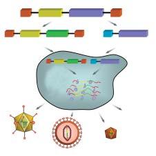
Adenoviral vectors. Adenoviral (Ad) vectors have been one of the most well characterized and extensively studied gene therapy systems over the past 20 years. Characteristically, Ad vectors are predominantly non-integrating episomal, non-enveloped, and double-stranded DNA viruses that have demonstrated broad tropism to numerous cell types in vivo, including the kidney (3,4,6). Early-generation Ad vectors had moderate cloning capacities (~8 kb), but more recently developed helper-dependent (also known as “gutted”) Ad vectors have extremely large cloning capacities (~37 kb) (5). The moderateto-large cloning capacities constitute a clear advantage for these vectors for inserting large promoter and/or transgene fragments for gene expression studies. A generic schematic of viral vector production is shown in Figure 1.
Through Ad vectorology, this virus has been modified to generate both replication-defective and replication-competent systems for treating various types of genetic- and environmentally-
1 Department of Physiology, Medical College of Wisconsin, Milwaukee, WI
2 Department of Medicine, Kidney Disease Center, Medical College of Wisconsin
Correspondence: Frank Park fpark@mcw.edu
Department of Medicine
Kidney Disease Center
Medical College of Wisconsin 8701 Watertown Plank Rd., HRC 4100 Milwaukee, WI 53226
based pathologies in many different organs (4), including the kidney. However, the majority of the studies to date have focused on the replication-defective vectors for gene therapy applications in the kidney (6). In general, most of the pre-clinical experiments using Ad vectors have investigated the transduction efficiency in the kidneys of small and large adult animal models, but their efficiency has been relatively poor. One reason for this is related to the complexity of the renal vascular and tubular architecture, which has been found to make the route of administration into the kidney (as shown in Figure 2) an important factor for transduction of distinct cell types (7-21). Numerous studies over the years have shown a fairly poor transduction into various renal tubular, vascular, glomerular and interstitial cells, regardless of whether the Ad vector was administered anterograde through the renal artery (7,8), retrograde into the ureter (7), or directly into the renal interstitial parenchyma (9). Even prolonged infusion of the Ad vector in vivo and ex vivo into pig (10,11), rat (21) or isolated human kidneys (12) has not markedly increased the transduction efficiency regardless of the species. In general, most of the intravenous and intrarenal infusion studies have shown a propensity for the Ad vector to be more effective in transducing renal epithelial cells than other cell types in the kidney. Another reason for the lack of efficient transduction may be attributed to the low vector titer that reaches the target cells. It has been found that the liver and spleen remove a large percentage of the vector from circulation following a single pass (22,23). By blocking the portal circulation, Ye et al. (24,25) achieved ~85±10% glomerular transduction efficiency by intravenous administration of Ad vector. Prolonged infusion of hightiter Ad vector (>7.5 x 10¹¹) into young Sprague Dawley rats resulted in predominant expression of lacZ transgene in glomeruli (30-70%) (15,25), whereas lower vector titers were unable to transduce the glomerular cells efficiently. Similar high levels of glomerular transduction were also observed in pigs (10,11). There may be differences between the species and cell-type localization of the receptors needed to promote Ad vector internalization resulting in varying degrees of transduction efficiency (26).
In order to optimize transduction of renal cells in vivo, a molecular strategy to re-target the Ad vector is needed. Ad vector binding to target cells and its subsequent internalization require particular receptors and co-receptors (27,28). Until recently, the tropism of the Ad5 vector has been attributed largely to the fibre region on the capsid (29). Manipulation of the fibre region by pseudotyping the Ad5 capsid with ei-
Figure 1. General strategy for viral vector production. Wild type viral genome is composed of mostly non-overlapping cis- and trans-acting elements which could be genetically engineered and spliced into two or more components to create viral vectors. Production of viral vectors generally involves two or more plasmids. The first plasmid, known as the transfer plasmid, contains minimal yet vital parts of the virus genome (cis-acting elements) and the viral genes are replaced with an expression cassette (i.e., promoter and full-length cDNA or short hairpin RNA sequences). Cis-acting elements include non-coding viral DNA required for vector packaging (PD) and end-terminal regions needed for vector stability (ETR). The other plasmid(s) known as helper plasmid(s) contain trans-acting elements (TAE) that encode for essential viral genes required for virus replication and structural integrity. In some cases, TAE can be inserted into the packaging cell lines to simplify vector production. These viral genes can be cloned into a single plasmid or fragmented into multiple plasmids to increase the biosafety of the vector system. Each of the vector plasmids are subsequently transfected into the packaging cell line, which are generally 293-based cells, and after a period of time, the cells and/or supernatant are collected with the functioning vector particles. Abbreviations: ETR: End-terminal regions, PD: Packaging Domain, TAE: trans-acting elements, EC: Expression cassette, P: Promoter, Ad: Adenoviral vector, LENTI: Lentiviral vector, AAV: Adenoassociated viral vector; Other: Other viral vector systems.
ther wild-type Ad-19p fibre (30) or Ad-19p fibre modified to include various kidney specific peptides (31) resulted in a striking lack of hepatic tropism. Instead, successful transduction into renal glomeruli and/or tubular structures was achieved, even with systemic administration and did not require direct renal infusion. Recent studies have demonstrated that coagulation factor X (FX) can be vital to Ad vector targeting to the liver (32,33), and that blockade of the Ad-FX interaction results in the re-targeting of the vector to extra-hepatic organs. Waddington et al. (34) in seminal work demonstrated that the binding
can be transduced within distinct regions of the kidney, and the transduction efficiency appears to be dependent on the route of administration. It may be concluded that the slow perfusion of a high-titer Ad vector at a dose greater than 1 x 10⁹ I.U. via the renal artery or ureter, which allows for the maximization of the contact time between the vector and target cell, provides the best possible method to modify renal cells in vivo
Adeno-associated viral (AAV) vectors. AAV was originally discovered as a “contaminant” of the adenovirus, and has become a popular virus for study in the development of gene therapy application for a number of reasons: Firstly, AAV has been found to be nonpathogenic in humans (48). Secondly, AAV genomes can persist following infection in host cells, either as multimeric concatemers or as an integrant into the host genome. However, the latter event is extremely rare for AAV vectors (49), and the lack of integration minimizes the chance for insertional mutagenesis unlike retroviral vectors (see following section on Integrating viral vectors). It is interesting that wild-type AAV can integrate into a specific region of chromosome 19, but the replication-defective AAV vectors that are used for gene therapy applications have been found to integrate randomly into the genome (49). One of the main limitations to the use of AAV vectors for gene therapy is its relatively small packaging capacity (~4.5 kb), and so extremely large cDNAs cannot be cloned into the vector and packaged efficiently (50). In a similar way to that of Ad vectors, an innate and/or humoral response against the vector has been shown to be developed (48), although alterations in the viral vector could help weaken the anti-vector immune response by removing the antigenic proteins in the AAV capsids. The replacement of the capsid proteins from alternative serotypes, such as AAV7 and AAV8, would minimize the humoral response in most patients (51,52). A generic schematic of viral vector production is shown in Figure 1.
Although many gene therapy applications have been performed over the past decade in different organ systems, the number of studies targeted at the kidney with various serotypes of AAV has been limited. Similar to the findings of experiments performed with adenoviral vectors, the efficiency of transduction was dependent on the route of administration (see Figure 2). Retrograde infusion of AAV serotype 2 (AAV2) by Ito et al. (53) resulted in moderate expression exclusively in the tubular cells in the renal medulla. Renal arterial infusion into rat kidneys showed restricted transduction into the S3 segment of the proximal tubules and intercalated cells of the renal
medulla with no expression detected in the glomeruli (54). To circumvent the complex architecture of the kidney, Lipkowitz et al. (55) attempted to genetically modify renal cells by direct intraparenchymal injection of AAV2 into mouse kidneys, but this resulted in only low expression of tubular structures near the needle track with no expression in the glomeruli or vasculature. This indicated that the AAV vector did not spread efficiently following direct infusion into the kidney. In a comparative study using serotypes of AAV1 to AAV5, it was found that AAV2 was the best transducing system in the kidney among the various serotypes (56).
Recent experiments by numerous researchers have isolated novel serotypes with the potential of enhancing the transduction efficiency into various organs in vivo, including the kidney. Nakai and associates have shown that AAV8 and AAV9 could infect mouse kidneys as determined by the relatively high AAV genome copy numbers using Southern blot analysis (57,58). Transgene expression in the kidney as a result of AAV infection was not determined in these studies. More recently, Bostick et al. (59) demonstrated that AAV9 transduction into the kidney was age-dependent, and that only adult kidneys, but not newborn kidneys were capable of being infected by this particular serotype when administered intravenously. This relates to early studies by Lipshutz et al. (60,61), which demonstrated decreasing expression of transgene during the first month of life, followed by AAV2mediated in utero administration. Even though longterm expression was maintained throughout the life of the animal at detectable levels, the initial loss in transgene expression poses as a potential problem for the use of this vector to treat chronic, debilitating diseases in the developing kidney. Considerable work is needed to elucidate the relative importance of the alternative serotypes of AAV, and their potential role for therapeutic applications for the kidney.
Integrating viral vectors: Retroviral and lentiviral vectors. Biologically, simple and complex retroviruses contain single-stranded RNA genomes that are converted into double stranded DNA through a reverse transcription phase prior to integrating into the host genome. For this reason, these viral vectors have been widely studied as gene therapy tools for a number of different diseases. More detailed vectorology of the retroviral and lentiviral vectors can be read elsewhere (3) and a generic schematic of vector production is shown in Figure 1. Because active proliferation occurs during embryonic and adolescent development, retroviruses would be ideal genetic modifiers for treating paediatric diseases, since copies of
the integrated provirus would be capable of propagation in progeny cells, unlike the non-integrating vector systems.
i. Oncoretroviral vectors. The first clinical gene therapy trial in SCID-ADA children was performed using ex vivo genetically manipulated cells using a simple retrovirus derived from the Moloney leukaemia virus (MLV) (62). Subsequent studies attempted to investigate the efficiency of retroviral vector transduction into the kidney, but the absolute number of genetically modified cells was extremely low. One of the reasons that in vivo gene therapy using retroviruses has been limited is the necessity of nuclear membrane breakdown to render the vector capable of genomic DNA integration (63,64). However, retroviral vector transduction remained extremely low even if it was administered in actively proliferating renal cells in vitro using metanephric tissue (66) or in vivo using chemically injured mouse kidneys (65). In addition to the poor transduction, serious adverse effects in children with X-linked SCID (67-70) following treatment with MLV-transduced haematopoietic stem cells ex vivo has led to renewed efforts to develop alternative complex retroviruses (also known as lentiviruses) isolated from either humans (71) or other species (72-74).
ii. Lentiviral vectors. A few studies have documented the efficiency of lentiviral vectors pseudotyped with vesicular stomatitis virus G protein (VSV-G) into the kidneys in vivo (75,76). Compared to retroviral vectors, lentiviral vectors have been shown to be more efficient in transducing terminally differentiated cells, yet actively cycling cells administered with lentiviral vectors can further increase their efficiency (77,78). Details regarding the vector design have been published elsewhere (3,79). In adult mouse kidneys, Gusella et al. (75,76) found that renal artery perfusion resulted in tubular cell transduction in the outer medullary region of the kidney, whereas retrograde infusion into the ureter led to exclusive collecting duct transduction in the medullary region. The laboratory of the authors has infused higher titers of VSVG pseudotyped lentiviral vectors (>109 T.U./kidney) into rodent renal arteries and found results similar to those of Gusella et al. (75,76) except for random glomeruli expressing the EGFP marker gene (unpublished observations). Due to the relatively moderate transduction in low mitotic renal cells, it is likely that increased efficiency would be obtained if the lentiviral vector were applied in younger, actively proliferating kidneys, as previously found in mouse livers and other organs (9,80), or even in early embryogenesis (73,81).
With increased genetic screening for kidney-based diseases (82), such as autosomal dominant or recessive polycystic kidney disease, there may come a time when genetic modification of early progenitor cells may become a viable method to circumvent the need to abort the foetus, or give birth to a child with a lethal or severely debilitating disorder. Recent studies have manipulated fertilized zygotes using lentiviral vectors to produce transgenic animals with global transgene expression in the kidneys (81). To minimize the offtarget effects of expressing transferred genes, vectors would need to be designed with organ- or even celltype specific promoters to produce exclusive expression in the kidney. Another possibility may be to manipulate renal cells during foetal development as shown by Waddington et al. (73), although in that instance only a few renal cells were found to express the marker gene.
One of the limitations of lentiviral vector transduction into the kidney is probably due to the currently used envelopes for coating the vector system. In general, the VSV-G protein that was isolated from the rhabdovirus has been widely used to study lentiviral vector transduction. However, the native virus is neurotropic and so swapping with renal-tropic envelope proteins (also known as pseudotyping) may provide significant enhancement for renal cell transduction. Another benefit of pseudotyping is that alternative envelopes may circumvent the activation of the immune system whereby the VSV-G envelope has been shown to stimulate humoral responses against the vector (83). Advances in lentiviral vectorology will inevitably demonstrate the viability of this vector system for renal-based diseases.
To date, human clinical trials have been conducted using lentiviral vectors in which haematopoietic stem cells were transduced to conditionally express antisense RNA sequences against HIV genes (84,85). The first human trial using lentiviral vectors documented several biosafety features of this system to help promote its future use for other gene therapy applications (85). Although there are no current trials including children using lentiviral vectors, the findings from the adult trials may provide an important treatment for paediatric AIDS, and this should open the door to many new trials for other lentiviral vector applications to the kidney.
Non-viral vector systems and other potential approaches. Alternative methods consisting of naked DNA transfection with liposomes (86,87) or inactivated Sendai virus (the haemagglutinating virus of Japan, HJV) (88-90) have been examined as potential vehicles for transfection of the kidney in vivo, but in general, the
transgene expression and transfection efficiency remain poor and transient. In many cases, they still lag far behind the currently discussed viral vector. However, the HVJ-liposome vector approach remains one of the few methods to target the glomeruli (88,89), and even with the relatively low transfection efficiencies, it has shown to mediate biological changes. Moreover, retrograde infusion of a modified HJV-liposome complex using an artificial viral envelope (AVE) has also been found to transfect interstitial cells (90), which may be important as a potential tool for studying therapies for interstitial fibrosis.
It is likely that alternative viruses will be studied and developed as gene therapy vectors for the treatment of renal diseases, since a viral vector that can be effectively used for renal gene therapy has yet to be found. Candidate viruses that may become important players could be human foamy virus vectors (91) or other viruses not yet developed from the polyoma family, such as BK (92,93), or the bunyavirus family, such as hantavirus (94). The latter two viruses are both known to have renal cell tropism. It will be interesting to observe what new vector systems will become available for use in renal gene therapy as the field of vectorology continues to evolve. However, in the near future, it may be necessary to use genetically modify extra-renal sites that secrete proteins into the circulation for targeting the kidney to mediate its therapeutic effect, until more efficient vector systems are designed and developed for direct renal gene therapy (74).
Perspective on renal gene therapy.
Although the effectiveness of viral vectors in manipulating renal cells has not been extremely successful, unlike other organ systems, there is a definite need to continue the investigation of vectors for effectively modifying this important organ. There are many diseases that originate in the kidney, particularly in newborns and young infants, for the treatment of which evolution of viral vector technology must culminate in successful vectors. The field of viral vectorology is still in its infancy with respect to the kidney, so there is great optimism for success especially with the expanding information regarding genomics and gene therapy trials. Continued research into viral vectorology will ultimately produce a vector system, possibly derived in some form from those discussed in this review, to treat paediatric renal diseases in the future.
References
1. Feigin RD. Prospects for the future of child health through research. JAMA2005;294:1373-1379.
2. Rimoin DL, Hirschhorn K. A history of medical genetics in pediatrics. Pediatr Res 2004;56:150-159.
3. Park F, Gow KW. Gene therapy: future or flop. Pediatr Clin North Am 2006;53:621-638.
4. Pfeifer A, Verma IM. Gene therapy: promises and problems. Annu Rev Genomics Hum Genet 2001;2:177-211.
5. Palmer DJ, Ng P. Helper-dependent adenoviral vectors for gene therapy. Hum Gene Ther 2005;16:1-16.
6. Isaka Y. Gene therapy targeting kidney diseases: routes and vehicles. Clin Exp Nephrol 2006;10:229-235.
7. Moullier P, Friedlander G, Calise D, Ronco P, Perricaudet M, Ferry N. Adenoviral-mediated gene transfer to renal tubular cells in vivo. Kidney Int 1994;45:1220-1225.
8. Zhu G, Nicolson AG, Cowley BD, Rosen S, Sukhatme VP. In vivo adenovirus-mediated gene transfer into normal and cystic rat kidneys. Gene Ther 1996;3:298-304.
9. Choi YK, Kim YJ, Park HS, Choi K, Paik SG, Lee YI, et al. Suppression of glomerulosclerosis by adenovirus-mediated IL-10 expression in the kidney. Gene Ther 2003;10: 559-568.
10. Heikkila P, Parpala T, Lukkarinen O, Weber M, Tryggvason K. Adenovirus-mediated gene transfer into kidney glomeruli using an ex vivo and in vivo kidney perfusion system - first steps towards gene therapy of Alport syndrome. Gene Ther 1996;3:21-27.
11. Parpala-Sparman T, Lukkarinen O, Heikkila P, Tryggvason K. A novel surgical organ perfusion method for effective ex vivo and in vivo gene transfer into renal glomerular cells. Urol Res 1999;27:97-102.
12. Zeigler ST, Kerby JD, Curiel DT, Diethelm AG, Thompson JA. Molecular conjugate-mediated gene transfer into isolated human kidneys. Transplantation 1996;61: 812-817.
13. Abraham NG, Jiang S, Yang L, Zand BA, LaniadoSchwartzman M, Marji J, et al. Adenoviral vector-mediated transfer of human heme oxygenase in rats decreases renal heme-dependent arachidonic acid epoxygenase activity. J Pharmacol Exp Ther 2000;293:494-500.
14. Blydt-Hansen TD, Katori M, Lassman C, Ke B, Coito AJ, Iyer S, et al. Gene transfer-induced local heme oxygenase-1 overexpression protects rat kidney transplants from ischemia/reperfusion injury. J Am Soc Nephrol 2003;14: 745-754.
15. Ye X, Liu X, Li Z, Ray PE. Efficient gene transfer to rat renal glomeruli with recombinant adenoviral vectors. Hum Gene Ther 2001;12:141-148.
16. Chetboul V, Klonjkowski B, Lefebvre HP, Desvaux D, Laroute V, Rosenberg D, et al. Short-term efficiency and safety of gene delivery into canine kidneys. Nephrol Dial Transplant 2001;16:608-614.
17. Ke B, Coito AJ, Kato H, Zhai Y, Wang T, Sawitzki B, et al. Fas ligand gene transfer prolongs rat renal allograft survival and down-regulates anti-apoptotic Bag-1 in parallel with enhanced Th2-type cytokine expression. Transplantation 2000;69:1690-1694.
18. Swenson KM, Ke B, Wang T, Markowitz JS, Maggard MA, Spear GS, et al. Fas ligand gene transfer to renal allografts in rats: effects on allograft survival. Transplantation 1998;65: 155-160.
19. Chiang-Ting C, Tzu-Ching C, Ching-Yi T, Song-Kuen S, Ming-Kuen L. Adenovirus-mediated bcl-2 gene transfer inhibits renal ischemia/reperfusion induced tubular oxidative stress and apoptosis. Am J Transplant 2005;5: 1194-1203.
20. Tomasoni S, Azzollini N, Casiraghi F, Capogrossi MC, Remuzzi G, Benigni A. CTLA4Ig gene transfer prolongs survival and induces donor-specific tolerance in a rat renal allograft. J Am Soc Nephrol 2000;11:747-752.
21. Benigni A, Tomasoni S, Lutz J, Amuchastegui S, Capogrossi MC, Remuzzi G. Nonviral and viral gene transfer to the kidney in the context of transplantation. Nephron 2000;85:307-316.
22. Fujishiro J, Takeda S, Takeno Y, Takeuchi K, Ogata Y, Takahashi M, et al. Gene transfer to the rat kidney in vivo and ex vivo using an adenovirus vector: factors influencing transgene expression. Nephrol Dial Transplant 2005;20:1385-1391.
23. Huard J, Lochmuller H, Acsadi G, Jani A, Massie B, Karpati G. The route of administration is a major determinant of the transduction efficiency of rat tissues by adenoviral recombinants. Gene Ther 1995;2:107-115.
24. Ye X, Jerebtsova M, Ray PE. Liver bypass significantly increases the transduction efficiency of recombinant adenoviral vectors in the lung, intestine, and kidney. Hum Gene Ther 2000;11:621-627.
25. Ye X, Jerebtsova M, Liu XH, Li Z, Ray PE. Adenovirus-mediated gene transfer to renal glomeruli in rodents. Kidney Int 2002;61:16-23.
26. Fechner H, Haack A, Wang H, Wang X, Eizema K, Pauschinger M, et al. Expression of coxsackie adenovirus receptor and alphav-integrin does not correlate with adenovector targeting in vivo indicating anatomical vector barriers. Gene Ther 1999;6:1520-1535.
27. Bergelson JM, Krithivas A, Celi L, Droguett G, Horwitz MS, Wickham T, et al. The murine CAR homolog is a receptor for coxsackie B viruses and adenoviruses. J Virol 1998;72:415-419.
28. Hong SS, Karayan L, Tournier J, Curiel DT, Boulanger PA. Adenovirus type 5 fiber knob binds to MHC class I alpha2 domain at the surface of human epithelial and B lymphoblastoid cells. EMBO J 1997;16:2294-2306.
29. Nicklin SA, Baker AH. Tropism-modified adenoviral and adeno-associated viral vectors for gene therapy. Curr Gene Ther 2002;2:273-293.
30. Denby L, Work LM, Graham D, Hsu C, von Seggern DJ, Nicklin SA, et al. Adenoviral serotype 5 vectors pseudotyped with fibers from subgroup D show modified tropism in vitro and in vivo. Hum Gene Ther 2004;15:1054-1064.
31. Denby L, Work LM, Seggern DJ, Wu E, McVey JH, Nicklin SA, et al. Development of renal-targeted vectors through combined in vivo phage display and capsid engineering of adenoviral fibers from serotype 19p. Mol Ther 2007;15: 1647-1654.
33. Shayakhmetov DM, Gaggar A, Ni S, Li ZY, Lieber A. Adenovirus binding to blood factors results in liver cell infection and hepatotoxicity. J Virol 2005;79:7478-7491.
35. Yang Y, Nunes FA, Berencsi K, Furth EE, Gonczol E, Wilson JM. Cellular immunity to viral antigens limits E1deleted adenoviruses for gene therapy. Proc Natl Acad Sci U S A 1994;91:4407-4411.
36. Lipshutz GS, Sarkar R, Flebbe-Rehwaldt L, Kazazian H, Gaensler KM. Short-term correction of factor VIII deficiency in a murine model of hemophilia A after delivery of adenovirus murine factor VIII in utero. Proc Natl Acad Sci USA 1999;96:13324-13329.
37. Feng M, Jackson WH Jr, Goldman CK, Rancourt C, Wang M, Dusing SK, et al. Stable in vivo gene transduction via a novel adenoviral/retroviral chimeric vector. Nat Biotechnol 1997;15:866-870.
38. Soifer H, Higo C, Kazazian HH Jr, Moran JV, Mitani K, Kasahara N. Stable integration of transgenes delivered by a retrotransposon-adenovirus hybrid vector. Hum Gene Ther 2001;12:1417-1428.
39. Yant SR, Ehrhardt A, Mikkelsen JG, Meuse L, Pham T, Kay MA. Transposition from a gutless adeno-transposon vector stabilizes transgene expression in vivo. Nat Biotechnol 2002;20:999-1005.
40. Lieber A, Steinwaerder DS, Carlson CA, Kay MA. Integrating adenovirus-adeno-associated virus hybrid vectors devoid of all viral genes. J Virol 1999;73:9314-9324.
41. Marshall E. Gene therapy death prompts review of adenovirus vector. Science 1999;286:2244-2245.
42. Raper SE, Chirmule N, Lee FS, Wivel NA, Bagg A, Gao GP, et al. Fatal systemic inflammatory response syndrome in a ornithine transcarbamylase deficient patient following adenoviral gene transfer. Mol Genet Metab 2003;80:148-158.
43. Lipshutz GS, Flebbe-Rehwaldt L, Gaensler KM. Reexpression following readministration of an adenoviral vector in adult mice after initial in utero adenoviral administration. Mol Ther 2000;2:374-380.
44. Pulkkanen KJ, Parkkinen JJ, Laukkanen JM, Kettunen MI, Tyynela K, Kauppinen RA, et al. HSV-tk gene therapy for human renal cell carcinoma in nude mice. Cancer Gene Ther 2001;8:529-536.
45. Haviv YS, Blackwell JL, Kanerva A, Nagi P, Krasnykh V, Dmitriev I, et al. Adenoviral gene therapy for renal cancer requires retargeting to alternative cellular receptors. Cancer Res 2002;62:4273-4281.
46. Jongmans W, van den Oudenalder K, Tiemessen DM, Molkenboer J, Willemsen R, Mulders PF, et al. Targeting of adenovirus to human renal cell carcinoma cells. Urology 2003;62:559-565.
47. Guse K, Ranki T, Ala-Opas M, Bono P, Sarkioja M, Rajecki M, et al. Treatment of metastatic renal cancer with capsid-modified oncolytic adenoviruses. Mol Cancer Ther 2007;6:2728-2736.
48. Bessis N, GarciaCozar FJ, Boissier MC. Immune responses to gene therapy vectors: influence on vector function and effector mechanisms. Gene Ther 2004;11:S10-S17.
49. Nakai H, Montini E, Fuess S, Storm TA, Grompe M, Kay MA. AAV serotype 2 vectors preferentially integrate into active genes in mice. Nat Genet 2003;34:297-302.
50. Kay MA, Glorioso JC, Naldini L. Viral vectors for gene therapy: the art of turning infectious agents into vehicles of therapeutics. Nat Med 2001;7:33-40.
51. Gao G, Vandenberghe LH, Wilson JM. New recombinant serotypes of AAV vectors. Curr Gene Ther 2005;5:285-297.
52. Gao GP, Alvira MR, Wang L, Calcedo R, Johnston J, Wilson JM. Novel adeno-associated viruses from rhesus monkeys as vectors for human gene therapy. Proc Natl Acad Sci USA 2002;99:11854-11859.
53. Ito K, Chen J, Khodadadian JJ, Vaughan ED Jr, Lipkowitz
M, Poppas DP, et al. Adeno-associated viral vector transduction of green fluorescent protein in kidney: effect of unilateral ureteric obstruction. BJU Int 2008;101:376-381.
54. Chen S, Agarwal A, Glushakova OY, Jorgensen MS, Salgar SK, Poirier A, et al. Gene delivery in renal tubular epithelial cells using recombinant adeno-associated viral vectors. J Am Soc Nephrol 2003;14:947-958.
55. Lipkowitz MS, Hanss B, Tulchin N, Wilson PD, Langer JC, Ross MD, et al. Transduction of renal cells in vitro and in vivo by adeno-associated virus gene therapy vectors. J Am Soc Nephrol 1999;10:1908-1915.
56. Takeda S, Takahashi M, Mizukami H, Kobayashi E, Takeuchi K, Hakamata Y, et al. Successful gene transfer using adeno-associated virus vectors into the kidney: comparison among adeno-associated virus serotype 1-5 vectors in vitro and in vivo. Nephron Exp Nephrol 2004;96:e119-e126.
57. Inagaki K, Fuess S, Storm TA, Gibson GA, Mctiernan CF, Kay MA, et al. Robust systemic transduction with AAV9 vectors in mice: efficient global cardiac gene transfer superior to that of AAV8. Mol Ther 2006;14:45-53.
58. Nakai H, Fuess S, Storm TA, Muramatsu S, Nara Y, Kay MA. Unrestricted hepatocyte transduction with adeno-associated virus serotype 8 vectors in mice. J Virol 2005;79: 214-224.
59. Bostick B, Ghosh A, Yue Y, Long C, Duan D. Systemic AAV-9 transduction in mice is influenced by animal age but not by the route of administration. Gene Ther 2007;14: 1605-1609.
60. Lipshutz GS, Gruber CA, Cao Y, Hardy J, Contag CH, Gaensler KM. In utero delivery of adeno-associated viral vectors: intraperitoneal gene transfer produces long-term expression. Mol Ther 2001;3:284-292.
61. Lipshutz GS, Titre D, Brindle M, Bisconte AR, Contag CH, Gaensler KM. Comparison of gene expression after intraperitoneal delivery of AAV2 or AAV5 in utero. Mol Ther 2003;8:90-98.
62. Blaese RM, Culver KW, Miller AD, Carter CS, Fleisher T, Clerici M, et al. T lymphocyte-directed gene therapy for ADA- SCID: initial trial results after 4 years. Science 1995; 270:475-480.
63. Miller DG, Adam MA, Miller AD. Gene transfer by retrovirus vectors occurs only in cells that are actively replicating at the time of infection. Mol Cell Biol 1990;10:4239-4242.
64. Roe T, Reynolds TC, Yu G, Brown PO. Integration of murine leukemia virus DNA depends on mitosis. EMBO J 1993;12:2099-2108.
65. Bosch RJ, Woolf AS, Fine LG. Gene transfer into the mammalian kidney: direct retrovirus-transduction of regenerating tubular epithelial cells. Exp Nephrol 1993;1:49-54.
66. Woolf AS, Bosch RJ, Fine LG. Gene transfer into the mammalian kidney: microtransplantation of retrovirus-transduced metanephric tissue. Exp Nephrol 1993;1:41-48.
67. Quigley R, Baum M, Reddy KM, Griener JC, Falck JR. Effects of 20-HETE and 19(S)-HETE on rabbit proximal straight tubule volume transport. Am J Physiol Renal Physiol 2000;278:F949-F953.
68. Hacein-Bey-Abina S, von Kalle C, Schmidt M, Le Deist F, Wulffraat N, McIntyre E, et al. A serious adverse event after successful gene therapy for X-linked severe combined immunodeficiency. N Engl J Med 2003;348:255-256.
69. Hacein-Bey-Abina S, Von Kalle C, Schmidt M, McCormack MP, Wulffraat N, Leboulch P, et al. LMO2-associated
clonal T cell proliferation in two patients after gene therapy for SCID-X1. Science 2003;302:415-419.
70. Kohn DB, Sadelain M, Dunbar C, Bodine D, Kiem HP, Candotti F, et al. American Society of Gene Therapy (ASGT) ad hoc subcommittee on retroviral-mediated gene transfer to hematopoietic stem cells. Mol Ther 2003;8:180-187.
71. Naldini L, Blömer U, Gallay P, Ory D, Mulligan R, Gage FH, et al. In vivo gene delivery and stable transduction of nondividing cells by a lentiviral vector. Science 1996;272: 263-267.
72. Curran MA, Kaiser SM, Achacoso PL, Nolan GP. Efficient transduction of nondividing cells by optimized feline immunodeficiency virus vectors. Mol Ther 2000;1:31-38.
73. Waddington SN, Mitrophanous KA, Ellard FM, Buckley SM, Nivsarkar M, Lawrence L, et al. Long-term transgene expression by administration of a lentivirus-based vector to the fetal circulation of immuno-competent mice. Gene Ther 2003;10:1234-1240.
74. Waddington SN, Nivsarkar MS, Mistry AR, Buckley SM, Kemball-Cook G, Mosley KL, et al. Permanent phenotypic correction of hemophilia B in immunocompetent mice by prenatal gene therapy. Blood 2004;104:2714-2721.
76. Gusella GL, Fedorova E, Marras D, Klotman PE, Klotman ME. In vivo gene transfer to kidney by lentiviral vector. Kidney Int 2002;61:S32-S36.
77. Park F, Ohashi K, Kay MA. Therapeutic levels of human factor VIII and IX using HIV-1-based lentiviral vectors in mouse liver. Blood 2000;96:1173-1176.
78. Tsui LV, Kelly M, Zayek N, Rojas V, Ho K, Ge Y, et al. Production of human clotting Factor IX without toxicity in mice after vascular delivery of a lentiviral vector. Nat Biotechnol 2002;20:53-57.
79. Cockrell AS, Kafri T. Gene delivery by lentivirus vectors. Mol Biotechnol 2007;36:184-204.
80. VandenDriessche T, Thorrez L, Naldini L, Follenzi A, Moons L, Berneman Z, et al. Lentiviral vectors containing the human immunodeficiency virus type-1 central polypurine tract can efficiently transduce nondividing hepatocytes and antigen-presenting cells in vivo. Blood 2002;100: 813-822.
81. Michalkiewicz M, Michalkiewicz T, Geurts AM, Roman RJ, Slocum GR, Singer O, et al. Efficient transgenic rat production by a lentiviral vector. Am J Physiol Heart Circ Physiol 2007;293:H881-H894.
82. Molecular genetic testing in pediatric practice: A subject review. Committee on Genetics. Pediatrics 2000;106:14941497.
83. Follenzi A, Battaglia M, Lombardo A, Annoni A, Roncarolo MG, Naldini L. Targeting lentiviral vector expression to hepatocytes limits transgene-specific immune response and establishes long-term expression of human antihemophilic factor IX in mice. Blood 2004;103:3700-3709.
84. Humeau LM, Binder GK, Lu X, Slepushkin V, Merling R, Echeagaray P, et al. Efficient lentiviral vector-mediated control of HIV-1 replication in CD4 lymphocytes from diverse HIV+ infected patients grouped according to CD4 count and viral load. Mol Ther 2004;9:902-913.
85. Manilla P, Rebello T, Afable C, Lu X, Slepushkin V, Humeau LM, et al. Regulatory considerations for novel
gene therapy products: a review of the process leading to the first clinical lentiviral vector. Hum Gene Ther 2005;16: 17-25.
86. Boletta A, Benigni A, Lutz J, Remuzzi G, Soria MR, Monaco L. Nonviral gene delivery to the rat kidney with polyethylenimine. Hum Gene Ther 1997;8:1243-1251.
87. Lien YH, Lai LW. Liposome-mediated gene transfer into the tubules. Exp Nephrol 1997;5:132-136.
88. Isaka Y, Fujiwara Y, Ueda N, Kaneda Y, Kamada T, Imai E. Glomerulosclerosis induced by in vivo transfection of transforming growth factor-beta or platelet-derived growth factor gene into the rat kidney. J Clin Invest 1993; 92:2597-2601.
89. Tomita N, Higaki J, Morishita R, Kato K, Mikami H, Kaneda Y, et al. Direct in vivo gene introduction into rat kidney. Biochem Biophys Res Commun 1992;186:129-134.
90. Tsujie M, Isaka Y, Ando Y, Akagi Y, Kaneda Y, Ueda N, et al. Gene transfer targeting interstitial fibroblasts by the artificial viral envelope-type hemagglutinating virus of Japan liposome method. Kidney Int 2000;57:1973-1980.
91. Rethwilm A. Foamy virus vectors: an awaited alternative to gammaretro- and lentiviral vectors. Curr Gene Ther 2007;7: 261-271.
92. Dall A, Hariharan S. BK virus nephritis after renal transplantation. Clin J Am Soc Nephrol 2008;3:S68-S75.
93. Hariharan S. BK virus nephritis after renal transplantation. Kidney Int 2006;69:655-662.
94. Peters CJ, Simpson GL, Levy H. Spectrum of hantavirus infection: hemorrhagic fever with renal syndrome and hantavirus pulmonary syndrome. Annu Rev Med 1999;50: 531-545.
1 Paediatric Intensive Care Unit, Hippokration Hospital, Thessaloniki, Greece
2 Hypertension Center, 3rd Department of Medicine, Papageorgiou Hospital, Aristotle University of Thessaloniki, Greece
Pediatric Intensive Care Unit, Hippokration Hospital, Thessaloniki, Greece
Left ventricular mass index in hypertensive children and adolescents
S. Stabouli1, V. Kotsis2
Abstract: The increasing trend of high blood pressure in children and adolescents has raised interest in blood pressure measurement in the paediatric setting. Evidence of target organ damage, predominantly an increased left ventricular mass index (LVMI), even in children and adolescents with mild hypertension, has indicated the risk of adverse cardiovascular outcome in early adulthood. Left ventricular hypertrophy is a readily assessed hypertensive target organ damage because of the wide availability of echocardiography. The necessity to assess subtle alterations in cardiac structure in children has led to the use of percentile distributions for LVMI. The relationship between LVMI and blood pressure values in children and adolescents has been the focus of several studies. Although most studies have demonstrated associations between systolic blood pressure and LVMI across a wide range of blood pressure levels in children and adolescents, data correlating blood pressure values with target organ damage and cardiovascular morbidity and mortality are not yet available. However the presence of increased LVMI is generally accepted as an indication for antihypertensive therapy. Published guidelines and new techniques aim to facilitate the early diagnosis of hypertension in paediatric patients and introduce new elements into treatment strategies. Using blood pressure values obtained both in and outside of the clinic setting along with evidence of cardiac structural alterations could lead to successful reduction of cardiovascular risk.
Key words: Blood pressure, essential hypertension, left ventricular mass index.
Abbreviations
BPBlood pressure
LVLeft ventricular
LVMILeft ventricular mass index
LVHLeft ventricular hypertrophy
ABPAmbulatory blood pressure
Introduction
Hypertension is a well-established risk factor for cardiovascular morbidity and mortality in adults. Sustained elevation of blood pressure (BP) impacts the vascular wall, the heart, the kidney and the central nervous system (CNS), producing hypertension induced target organ damage (1,2). Left ventricular hypertrophy (LVH) is a target organ damage of hypertension, which is independently associated with increased risk for myocardial infraction, congestive heart failure and sudden death (3). De Simone et al., showed that left ventricular mass index (LVMI) greater than 51 g/m2.7 is associated with a fourfold increase in risk of adverse cardiovascular events in adults with hypertension (4). In the Framingham study, LVMI and age were strong and consistent predictors of all cause mortality, cardiac death and coronary heart disease (5).
During the last few years, the recognition of
increasing trends of essential hypertension in children and adolescents has posed concerns for the long-term health risks in this age group (6). Due to the relatively short period of hypertension, the presence of clinical manifestations such as congestive heart failure, renal insufficiency or acute encephalopathy in children and adolescents is rare and consequence of very high BP values (7,8). Epidemiological studies have shown that the levels of elevated BP in children and adolescents are usually mild with a predominance of isolated systolic hypertension (9,10). As morbid cardiovascular events are rare in pediatric patients except in severely hypertensive children and adolescents, investigation has relied on surrogate markers of hypertensive target organ damage such as LVH in order to identify hypertensive children and adolescents at risk for complications later in life (10-13).
Increased LVMI has also been associated with other conditions independent of hypertension such as obesity and chronic kidney disease (14,15). LVH remains the best documented target organ damage of hypertension in young populations. However, the level and duration of BP elevation that results in hypertensive target organ damage in children and adolescents remains
poorly defined. In the Fourth report on the diagnosis, evaluation and treatment of high BP in children and adolescents, it is recommended that pediatric patients with established hypertension should have echocardiographic assessment of left ventricular mass at diagnosis and periodically thereafter. In addition the presence of increased LVMI is an indication to initiate or intensify antihypertensive therapy (10).
Pathogenesis of left ventricular hypertrophy in human hypertension
Abnormal increases in left ventricular (LV) mass occur in response to pathological changes in the haemodynamic load (16). Increased wall stress and strain, invoked by increased BP, result in molecular changes, cellular hyperplasia and increased thickness of LV myocardium (17). These changes aim to protect the myocardium from excessive wall tension by minimizing oxygen consumption and provide sufficient strength to maintain adequate cardiac output. Two factors in the pathogenesis of hypertension itself, obesity and increased dietary sodium intake can result in volume overload and possible activation of renin angiotensin system. However, the pathogenesis of LVH in hypertension seems to be mediated by the contribution of non-haemodymamic factors that also stimulate cardiac muscle growth. Extremely high LVMI inappropriate for body size and hemodynamic load have been associated with an adverse cardiovascular phenotype independently of BP values. The genetic background in terms of ethnicity and genotype has been demonstrated to play a significant role in predicting hypertrophic process (18). Biological mediators such as insulin growth factor 1 and neurohormones (angiotensin II, aldosterone, endothelin) may also involve in the pathogenesis of LVH in human hypertension (17).
Echocardiographic measurements of left ventricular
mass
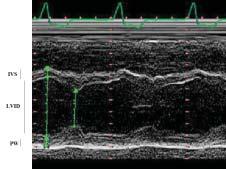
LV mass is determined from echocardiographic measurements of the left ventricle by standard techniques. 2-dimensional guided M-mode echocardiographic measurements of the left ventricle internal dimension, interventricular septal thickness, and posterior wall thickness are made during diastole according to methods established by American Society of Echocardiography (Figure 1) (19). Devereux et al., equation is used to calculate LV mass:
LVIDd is left ventricular internal diameter in di-
astole, IVSTd is intraventricular septal thickness in diastole, and PWTd is posterior wall thickness in diastole (20). Cross-sectional studies have shown that body size and gender are important determinants of left ventricular growth. LVMI was determined as the most appropriate method to remove the effect of normal variation of body size from the clinical evaluation of LV mass. LVMI is calculated by dividing LV mass by height in meters raised to the power of 2.7. LVMI in paediatric patients is then compared with standards and percentiles based on measurements on normal children and adolescents (21,22). LVH is defined as LVMI ≥95th percentile for normal children and adolescents. The cutpoint of 51 g/m2.7 for LVMI which has been determined as indicative of fourfold greater risk for cardiovascular disease in adults is above the 99th percentile for LVMI in normal children and adolescents.
Abnormalities in LV mass can be defined both by a standard measure above which mass is considered excessive for body size and by geometric patterns associated with increased morbidity. Classification of hypertensive paediatric patients by their ventricular geometry pattern (concentric, eccentric hypertrophy, or concentric remodeling) may improve ability to predict cardiovascular risk (23-25).
Blood pressure measurements and definition of hypertension in children and adolescents
Diagnosis of hypertension is critically dependent on accurate BP measurement (26-29). Accurate BP measurement is important in children and adolescents because misdiagnosis and undertreament can result in life-long adverse impact on cardiovascular
IVS
LVID
PW
Figure 1. Measurements of LV mass by M-mode echocardiography. LVID: left ventricle internal dimension, IVS: interventricular septum, PW: posterior wall.
Table 1. Classification of hypertension in children and adolescents
CategorySystolic and/or Diastolic BP percentile for gender, age, and height
Normotension<90th percentile
Prehypertension≥90th and <95th percentile or BP≥120/80 mm Hg
Stage 1 hypertension95th-99th percentile +5 mm Hg
Stage 2 hypertension>99th percentile + 5 mm Hg
health. Diagnosis of hypertension in children and adolescents also requires knowledge of the normal values of BP in this age group (10,11). BP should be preferably measured with a standard mercury sphygmomanometer because BP reference tables are based on auscultatory measurements. However, there is an increase in the use of automated devices for measuring BP in children as these devices are easier to use and are alternative instruments for BP measurement when use of mercury sphygmomanometers is not permitted for ecological reasons (10,13).
Diagnostic criteria for elevated BP in children and adolescents are based on the concept that BP increases with age and body size. According to the Fourth report on the diagnosis, evaluation and treatment of high BP in children and adolescents, hypertension is defined as average systolic BP and/or diastolic BP ≥95th percentile for gender, age, and height on 3 or more occasions (10). Stage 1 hypertension is defined as systolic BP and/or diastolic BP ≥95th to 99th percentile for gender, age, and height + 5 mm Hg and stage 2 hypertension as systolic BP and/or diastolic BP ≥99th percentile for gender, age, and height + 5 mm Hg. Furthermore, children and adolescents with an average systolic and/or diastolic BP ≥90th but <95th percentile for gender, age, and height are characterized as prehypertensive (Table 1).
Accumulating evidence suggests that ambulatory blood pressure (ABP) monitoring is a more accurate method for diagnosis of hypertension than clinic BP measurement, and it is better associated with target organ damage (29-36). ABP monitoring is a noninvasive, fully automated technique in which BP is recorded over an extended period of time, typically 24 hours with minimal intrusion into the daily activities of the patient. Therefore it better represents minute-tominute BP alterations in response to various environmental stimuli throughout the day. The use of ABP in clinical practice has also increased the awareness of BP patterns that are not easily apparent using traditional techniques of BP measurement. ABP monitoring is especially helpful in identifying individuals with nocturnal hypertension, white-coat hypertension (el-
Linear Regression
Male gender
Female gender
24-hours systolic BP (mm Hg)
Figure 2. Linear relationship of LVMI and 24-hours systolic BP in normotensive and hypertensive children and adolescents (Stabouli et al. unpublished data).
evated BP in the clinical setting but normal ABP), and masked hypertension. The latter is a condition that applies to patients whose clinic BP is normal, but ABP is elevated and has been associated with increased LVMI in both adults and children (36). The Fourth report on the diagnosis, evaluation and treatment of high BP in children and adolescents acknowledges the use of ABP monitoring in the diagnosis and management of hypertension in children, but it outlines that it has to be used by experts in its use and interpretation. Reference values for ABP measurement in children and adolescents based on the statistical parameters of BP distribution, indexed by gender and height, establish the 50th, 90th, and 95th percentiles for 24hours and daytime BP, separately (37-40).
Relations between BP and LVMI in children and adolescents
Many studies have investigated the relationship between BP and LV mass in children and adolescents (Table 2). In hypertensive children and adolescents the reported prevalence of LVH ranges from 10% to 46% depending on the method correcting for body size (adjusted for height, body surface area, weight and height raised to various powers) and the characteristics of the study population. In the Bogalusa study, Burke et al., reported in 654 healthy subjects, aged 7-22 years, that after adjustment for body size, LV wall thickness was associated with the level of systolic BP (41). In the Muscantine study, in 904 normotensive subjects, aged 6-17 years, a strong positive association was found between LV mass and both systolic and diastolic BP (42). The same relation between BP and LV mass was demonstrated in hypertensive children and adolescents. Daniels et al., showed that
Table 2. Studies on LVH in children and adolescents with essential hypertension
Author (ref)CountryYearNo of SubjectsPrevalence of LVHSetting
Daniels et al. (43)USA199813046%Hypertension Clinic
Belsha et al. (45)USA19986234%Hypertension Clinic
Sorof et al. (44)USA20023727%Hypertension Clinic
Hanevold et al. (46)USA200412941.1%Hypertension Clinic
Sorof et al. (52)USA20045428%General population
Litwin et al. (47)Poland200517541.6%Hypertension Clinic
McNiece et al. (48)USA200716016.8%Hypertension Clinic
Stabouli et al. (49)Greece20076710%Hypertension Clinic
Seeman et al. (55)Czech Republic20072142%Hypertension Clinic
Asadi (56)USA20026436%Hypertension Clinic
increased LVMI is relatively prevalent in pediatric patients with hypertension (43). They reported in a study of 130 hypertensive children and adolescents that 46% of the patients had LVMI greater than the 95th percentile and 8% had LVMI above the adult cut point of 51 g/m2.7. Among the patients with LVH, 64% had eccentric hypertrophy and 36% had concentric hypertrophy. They also reported that male sex, increased body mass index, and low heart rate during exercise were significant predictors of severe LVH. Sorof et al., found a 27% prevalence of LVMI above the adult cut point of 51 g/m2.7 in 37 untreated severely hypertensive children (44). Belsha et al., performed ABP monitoring and echocardiography in untreated adolescents with mild essential hypertension and reported a 34% prevalence of LVH (45). Clinic systolic and diastolic BP, daytime systolic BP but not diastolic BP were correlated with LVMI. Nighttime systolic BP demonstrated the closest correlation with LVMI. Other investigators underlined the effect of ethnicity in the prevalence of LVH in paediatric hypertension (46). LVH is more prevalent in Hispanic and African American children compared to white children. In a European study Litwin et al., included children with newly diagnosed essential hypertension and reported a 41.6% prevalence of LVH. 13.2% of the children had LVMI greater than the adult cutpoint (47). They also found that LVMI was correlated with 24-hours systolic BP and 24-hours heart rate.
Two studies investigated the relationship between BP and LVMI according to the severity of hypertension. McNiece et al., performed a retrorespective analysis of data from 2 cross-sectional studies using the Fourth report on the diagnosis, evaluation and treatment of high BP in children and adolescents staging criteria for hypertension (48). A total of 163 adolescents were analyzed. LVH prevalence significantly differed between the normotensive, stage 1 and stage 2 hypertension groups with the greater risk for LVH seen among those with stage 2 hypertension. Stabouli
et al., studied 67 children and adolescents, age range 5-20 years, referred for evaluation of essential hypertension (49). They demonstrated that LVMI was increasing from the normotensive to the prehypertensive and the hypertensive group. Statistically significant differences were found between the normotensive and the hypertensive group and between the normotensive and the prehypertensive group. In the same study BP group and male sex were independently associated with LVMI. In the two aforementioned studies LVMI was greater in groups with higher ABP. This finding encompasses the known relationship between LVMI and systolic BP, which becomes more evident when BP is measured using ABP monitoring (Figure 2) (50,51).
In all previous studies in hypertensive children and adolescents, subjects were referral population from hypertension clinics. Therefore, they may overestimate the frequency of LVH in hypertensive children and adolescents as patients referred for evaluation may have more severe hypertension than patients recruited from population based studies. Sorof et al., compared the LVMI between hypertensive children referred for evaluation by primary care providers and hypertensive children identified through communitybased screening and found greater LVMI and higher prevalence of LVH in referral subjects (49% vs. 28%) (52). The overall prevalence of LVH was 37%. After correcting for body mass index z score, which was significantly higher in the referral group, the differences between the 2 groups did not persist.
BP reduction results in LVH regression in many clinical trials in adult populations. Brilla et al., in a study that included myocardial biopsies before and during antihypertensive treatment, found reduction in interstitial fibrosis and myocyte diameter by angiotensin converting enzyme inhibitor and diuretic treatment, respectively (53). In the TOMHS study, lifestyle changes that led to BP control achieved LVMI reduction (54). Recent studies have also provided
ev idence that LVMI decreases in hypertensive pediatric patients with BP lowering therapy. Seeman et al., demonstrated that angiotensin converting enzyme inhibitor monotherapy in paediatric patients reduced not only BP but also LVMI levels (55). In another study, BP lowering by angiotensin converting enzyme inhibitor treatment was associated with a significant decrease in LVMI in hypertensive children and adolescents (56). There was more positive therapeutic response with regard to LVH among the patients with stage 2 compared to those with stage 1 hypertension. In multivariate regression analysis, the percentage changes in microalbuminuria, body mass index, and systolic BP were the only significant predictors of the decrease in LVMI.
Conclusions
Increased LVMI can be seen in children and adolescents with BP elevation and it is an easily assessed target organ damage of hypertension. It appears that male, obese hypertensive children and adolescents are more prone to severe LVH. The recognition of early cardiac structure alterations and initiation of pharmacological treatment could help to reduce hypertension-induced consequences later in life. Moreover, monitoring changes in LVMI during antihypertensive treatment can offer valuable information about the success of the treatment itself. Better BP assessment, using ABP monitoring when indicated, may contribute to a more successful approach to reduce cardiovascular risk.
References
1. MacMahon S, Peto R, Cutler J, Collins R, Sorlie P, Neaton J, et al. Blood pressure, stroke, and coronary heart disease. Part 1, Prolonged differences in blood pressure: prospective observational studies corrected for the regression dilution bias. Lancet 1990;335:765-774.
2. Verdecchia P, Clement D, Fagard R, Palatini P, Parati G. Blood Pressure Monitoring. Task force III: Target-organ damage, morbidity and mortality. Blood Press Monit 1999;4:303-317.
3. Levy D, Garrison RJ, Savage DD, Kannel WB, Castelli WP. Prognostic implications of echocardiographically determined left ventricular mass in the Framingham Heart Study. N Engl J Med 1990;322:1561-1566.
4. De Simone G, Devereux RB, Daniels SR, Koren MJ, Meyer RA, Laragh JH. Effect of growth on variability of left ventricular mass: assessment of allometric signals in adults and children and their capacity to predict cardiovascular risk. J Am Coll Cardiol 1995;25:1056-1062.
5. Kannel WB. Role of blood pressure in cardiovascular disease: the Framingham Study. Angiology 1975;26:1-14.
6. Muntner P, He J, Cutler JA, Wildman RP, Whelton PK. Trends in blood pressure among children and adolescents. JAMA 2004;291:2107-2113.
7. Vogt BA. Hypertension in children and adolescents: defin-
ition, pathophysiology, risk factors and long-term sequelae. Curr Therap Res 2001;62:283-297.
8. Kay JD, Sinaiko AR, Daniels SR. Pediatric hypertension. Am Heart J 2001;142:422-432.
9. Sorof JM. Prevalence and consequence of systolic hypertension in children. Am J Hypertens 2002;15:57S-60S.
10. National High Blood Pressure Education Program Working Group on High Blood Pressure in Children and Adolescents. The fourth report on the diagnosis, evaluation, and treatment of high blood pressure in children and adolescents. Pediatrics 2004;114:555-576.
11. Luma GB, Spiotta RT. Hypertension in children and adolescents. Am Fam Physician 2006;73:1558-1568.
12. Daniels SR. Cardiovascular sequelae of childhood hypertension. Am J Hypertens 2002;15:61S-63S.
13. O'Brien E, Asmar R, Beilin L, Imai Y, Mancia G, Mengden T, et al. Practice guidelines of the European Society of Hypertension for clinic, ambulatory and self blood pressure measurement. J Hypertens 2005;23:697-701.
14. De Simone G, Daniels SR, Devereux RB, Meyer RA, Roman MJ, de Divitiis O, et al. Left ventricular mass and body size in normotensive children and adults: assessment of allometric relations and impact of overweight. J Am Coll Cardiol 1992;20:1251-1260.
15. Mitsnefes MM. Cardiovascular disease in children with chronic kidney disease. Adv Chronic Kidney Dis 2005;12: 397-405.
16. Perlini S. Determinants of left ventricular mass in hypertensive heart disease: a complex interplay between volume, pressure, systolic function and genes. J Hypertens 2007;25: 1341-1342.
17. De Simone G, Pasanisi F, Contaldo F. Link of nonhemodynamic factors to hemodynamic determinants of left ventricular hypertrophy. Hypertension 2001;38:13-18.
18. De Simone G, Tang W, Devereux RB, Hunt SC, Kitzman DW, Rao DC, et al. Assessment of the interaction of heritability of volume load and left ventricular mass: the HyperGEN offspring study. J Hypertens 2007;25:13971402.
19. Sahn DJ, DeMaria A, Kisslo J, Weyman A. Recommendations regarding quantitation in M-mode echocardiography: results of a survey of echocardiographic measurements. Circulation 1978;58:1072-1083.
20. Devereux RB, Alonso DR, Lutas EM, Gottlieb GJ, Campo E, Sachs I, et al. Echocardiographic assessment of left ventricular hypertrophy: comparison to necropsy findings. Am J Cardiol 1986;57:450-458.
21. Daniels SR, Meyer RA, Liang YC, Bove KE. Echocardiographically determined left ventricular mass index in normal children, adolescents and young adults. J Am Coll Cardiol 1988;12:703-708.
22. Daniels SR. Hypertension-induced cardiac damage in children and adolescents. Blood Press Monit 1999;4:165-170.
23. Verdecchia P, Schillaci G, Borgioni C, Ciucci A, Battistelli M, Bartoccini C, et al. Adverse prognostic significance of concentric remodeling of the left ventricle in hypertensive patients with normal left ventricular mass. J Am Coll Cardiol 1995;25:871-878.
24. Krumholz HM, Larson M, Levy D. Prognosis of left ventricular geometric patterns in the Framingham Heart Study. J Am Coll Cardiol 1995;25:879-884.
31. Portman RJ, Yetman RJ, West MS. Efficacy of 24-hour ambulatory blood pressure monitoring in children. J Pediatr 1991;118:842-849.
32. Flynn JT. Impact of ambulatory blood pressure monitoring on the management of hypertension in children. Blood Press Monit 2000;5:211-216.
33. Graves JW, Althaf MM. Utility of ambulatory blood pressure monitoring in children and adolescents. Pediatr Nephrol 2006;21:1640-1652.
34. Stabouli S, Kotsis V, Zakopoulos N. Ambulatory blood pressure monitoring and target organ damage in pediatrics. J Hypertens 2007;25:1979-1986.
35. Stabouli S, Kotsis V, Papamichael C, Constantopoulos A, Zakopoulos N. Adolescent obesity is associated with high ambulatory blood pressure and increased carotid intimalmedial thickness. J Pediatr 2005;147:651-656.
36. Stabouli S, Kotsis V, Toumanidis S, Papamichael C, Constantopoulos A, Zakopoulos N. White-coat and masked hypertension in children: association with target-organ damage. Pediatr Nephrol 2005;20:1151-1155.
37. Lurbe E, Redon J, Liao Y, Tacons J, Cooper RS, Alvarez V. Ambulatory blood pressure monitoring in normotensive children. J Hypertens 1994;12:1417-1423.
38. Harshfield GA, Alpert BS, Pulliam DA, Somes GW, Wilson DK. Ambulatory blood pressure recordings in children and adolescents. Pediatrics 1994;94:180-184.
39. Soergel M, Kirschstein M, Busch C, Danne T, Gellermann J, Holl R, et al. Oscillometric twenty-four-hour ambulatory blood pressure values in healthy children and adolescents: a multicenter trial including 1141 subjects. J Pediatr 1997;130:178-184.
40. Wuhl E, Witte K, Soergel M, Mehls O, Schaefer F; German Working Group on Pediatric Hypertension. Distribution of 24-h ambulatory blood pressure in children: normalized reference values and role of body dimensions. J Hypertens 2002;20:1995-2007.
41. Burke GL, Arcilla RA, Culpepper WS, Webber LS, Chiang YK, Berenson GS. Blood pressure and echocardiographic measures in children: the Bogalusa Heart Study. Circulation 1987;75:106-114.
42. Malcolm DD, Burns TL, Mahoney LT, Lauer RM. Factors affecting left ventricular mass in childhood: the Muscatine Study. Pediatrics 1993;92:703-709.
43. Daniels SR, Loggie JM, Khoury P, Kimball TR. Left ventricular geometry and severe left ventricular hypertrophy in children and adolescents with essential hypertension. Circulation 1998;97:1907-1911.
44. Sorof JM, Cardwell G, Franco K, Portman RJ. Ambulatory blood pressure and left ventricular mass index in hypertensive children. Hypertension 2002;39:903-908.
45. Belsha CW, Wells TG, McNiece KL, Seib PM, Plummer JK, Berry PL. Influence of diurnal blood pressure variations on target organ abnormalities in adolescents with mild essential hypertension. Am J Hypertens 1998;11:410-417.
46. Hanevold C, Waller J, Daniels S, Portman R, Sorof J; International Pediatric Hypertension Association. The effects of obesity, gender, and ethnic group on left ventricular hypertrophy and geometry in hypertensive children: a collaborative study of the International Pediatric Hypertension Association. Pediatrics 2004;113:328-333.
47. Litwin M, Niemirska A, Sladowska J, Antoniewicz J, Daszkowska J, Wierzbicka A, et al. Left ventricular hypertrophy and arterial wall thickening in children with essential hypertension. Pediatr Nephrol 2006;21:811-819.
48. McNiece KL, Gupta-Malhotra M, Samuels J, Bell C, Garcia K, Poffenbarger T, et al; Left ventricular hypertrophy in hypertensive adolescents: analysis of risk by 2004 National High Blood Pressure Education Program Working Group staging criteria. Hypertension. 2007;50:392-395.
49. Stabouli S, Kotsis V, Karagianni C, Toumanidis S, Zakopoulos N, Constantopoulos A. Left ventricular mass index in hypertensive children and adolescents [Abstract]. Pediatrics 2008;121:S96.
50. Kotsis V, Stabouli S, Pitiriga V, Papamichael C, Toumanidis S, Zakopoulos N. Impact of gender on 24-h ambulatory blood pressure and target organ damage. J Hum Hypertens 2006;20:658-665.
51. Kotsis V, Stabouli S, Pitiriga V, Toumanidis S, Papamichael C, Zakopoulos N. Ambulatory blood pressure monitoring and target organ damage: effects of age and sex. Blood Press Monit 2006;11:9-15.
52. Sorof JM, Turner J, Martin DS, Garcia K, Garami Z, Alexandrov AV, et al. Cardiovascular risk factors and sequelae in hypertensive children identified by referral versus school-based screening. Hypertension 2004;43:214-218.
53. Brilla CG. Regression of myocardial fibrosis in hypertensive heart disease: diverse effects of various antihypertensive drugs. Cardiovasc Res 2000;46:324-331.
54. Liebson PR, Grandits GA, Dianzumba S, Prineas RJ, Grimm RH Jr, Neaton JD, et al. Comparison of five antihypertensive monotherapies and placebo for change in left ventricular mass in patients receiving nutritional-hygienic therapy in the Treatment of Mild Hypertension Study (TOMHS). Circulation 1995;91:698-706.
55. Seeman T, Gil›k J, Vondraãk K, Simkova ã E, FlögelovaãH, Hlad›kova ã M, et al. Regression of left-ventricular hypertrophy in children and adolescents with hypertension during ramipril monotherapy. Am J Hypertens 2007;20:990-996.
56. Assadi F. Effect of microalbuminuria lowering on regression of left ventricular hypertrophy in children and adolescents with essential hypertension. Pediatr Cardiol 2007;28:27-33.
1st Department of Paediatrics, VenizeleioPananeio General Hospital of Heraklion
G. Niotakis, G. Vlahaki, H. Kokkori
Abstract: Methaemoglobinaemia refers to the excessive oxidation of ferrous iron to the ferric state within the haemoglobin molecule. This reaction impairs the capacity of haemoglobin for oxygen transportation to the peripheral tissues. Cyanosis is its major clinical sign. Methaemoglobin formation increases most commonly due to the oxidative stress produced by certain toxic drugs, other chemicals or dietary products, or, infrequently due to congenital deficiency of the reduction enzymic system of cytochrome-b5-reductase or hereditary types of reduction resisting haemoglobin (haemoglobin M). High levels of methaemoglobin may be life threatening, and require immediate intervention. The cyanotic presentation and the non-specific diagnostic tests may easily confuse the diagnosis. New diagnostic devices based on the spectrophotometric characteristics of methaemoglobin are available for direct and accurate diagnosis. Molecular genetic analysis provides functional insight into the congenital/hereditary types. Prenatal diagnosis offers important information in families affected with the severe types of the disease. Methylene blue is the antidote of choice, although it is contraindicated in G6PD deficiency. New alternative antidotes are still under investigation.
1. Ash-Bernal R, Wise R, Wright SM. Acquired methemoglobinemia: a retrospective series of 138 cases at 2 teaching hospitals. Medicine (Baltimore) 2004;83:265-273.
2. Rehman HU. Methemoglobinemia. West J Med 2001;175: 193-196.
3. Wright RO, Lewander WJ, Woolf AD. Methemoglobinemia: etiology, pharmacology, and clinical management. Ann Emerg Med 1999;34:646-656.
4. Guyton AC. Transport of oxygen and carbon dioxide in the blood and body fluids. In: Guyton AC. Textbook of Medical Physiology. 8th ed. Philadelphia: WB Saunders Company; 1991. p. 433.
5. Mansouri A, Lurie AA. Concise review: methemoglobinemia. Am J Hematol 1993;42:7-12.
6. Verine M, Kumar M, Sarnaik SA, Windle ML, Bergstrom SK, Gross S, Coppes MJ. Methemoglobinemia. eMedicine [Internet]. 2007 April: Webpage: http://www.emedicine.com/ ped/topic1432.htm
7. Percy MJ, McFerran NV, Lappin TR. Disorders of oxidised haemoglobin. Blood Rev 2005;19:61-68.
8. Schiff DE, Roberts WD, Sue YJ. Methemoglobinemia associated with dapsone therapy in a child with pneumonia
9. Dahshan A, Donovan GK. Severe methemoglobinemia complicating topical benzocaine use during endoscopy in a toddler: a case report and review of the literature. Pediatrics 2006;117:e806-e809.
10. Neuhaeuser C, Weigand N, Schaaf H, Mann V, Christophis P, Howaldt HP, Heckmann M. Postoperative methemoglobinemia following infiltrative lidocaine administration for combined anesthesia in pediatric craniofacial surgery. Paediatr Anaesth 2008;18:125-131.
11. Logan BK, Gordon AM. Death of an infant involving benzocaine. J Forensic Sci 2005;50:1486-1488.
12. Gunter JB. Benefit and risks of local anesthetics in infants and children. Paediatr Drugs 2002;4:649-672.
13. Hwang JB, Lee SH, Kang YN, Kim SP, Suh SI, Kam S. Indexes of suspicion of typical cow’s milk protein-induced enterocolitis. J Korean Med Sci 2007;22:993-997.
14. Sager S, Grayson GH, Feig SA. Methemoglobinemia associated with acidosis of probable renal origin. J Pediatr 1995;126:59-61.
15. Powlson DS, Addiscott TM, Benjamin N, Cassman KG, de Kok TM, van Grinsven H, L’Hirondel JL, Avery AA, van Kessel C. When does nitrate become a risk for humans? J Environ Qual 2008;37:291-295.
16. Sadeq M, Moe CL, Attarassi B, Cherkaoui I, Elaouad R, Idrissi L. Drinking water nitrate and prevalence of methemoglobinemia among infants and children aged 1-7 years in Moroccan areas. Int J Hyg Environ Health 2007 Dec 21. [Epub ahead of print]
17. Maric P, Ali SS, Heron LG, Rosenfeld D, Greenwood M. Methaemoglobinaemia following ingestion of a commonly available food additive. Med J Aust 2008;188:156-158.
18. Geller RJ, Barthold C, Saiers JA, Hall AH. Pediatric cyanide poisoning: causes, manifestations, management, and unmet needs. Pediatrics 2006;118:2146-2158.
19. Fewtrell L. Drinking-water nitrate, methemoglobinemia, and global burden of disease: a discussion. Environ Health Perspect 2004;112:1371-1374.
20. Avery AA. Infantile methemoglobinemia: reexamining the role of drinking water nitrates. Environ Health Perspect 1999;107:583-586.
21. Weinberger B, Laskin DL, Heck DE, Laskin JD. The toxicology of inhaled nitric oxide. Toxicol Sci 2001;59:5-16.
22. Kedar PS, Warang P, Nadkarni AH, Colah RB, Ghosh K. A novel G143D mutation in the NADH-cytochrome b(5) reductase gene in an Indian patient with type I recessive hereditary methemoglobinemia. Blood Cells Mol Dis 2008;40:323-327.
23. Davis CA, Crowley LJ, Barber MJ. Cytochrome b5 reductase: the roles of the recessive congenital methemoglobinemia mutants P144L, L148P, and R159*. Arch Biochem Biophys 2004;431:233-244.
24. Ewenczyk C, Leroux A, Roubergue A, Laugel V, Afenjar A, Saudubray JM, Beauvais P, Billette de Villemeur T, Vidailhet M, Roze E. Recessive hereditary methemoglobinemia, type II: delineation of the clinical spectrum. Brain 2008; 131:760-761.
25. Percy MJ, Crowley LJ, Roper D, Vulliamy TJ, Layton DM, Barber MJ. Identification and characterization of the novel FAD-binding lobe G75S mutation in cytochrome b(5) reductase: an aid to determine recessive congenital
methemoglobinemia status in an infant. Blood Cells Mol Dis 2006;36:81-90.
26. Brennan SO, Matthews JR. Hb Auckland [alpha 87(F8) His‡Asn]: a new mutation of the proximal histidine identified by electrospray mass spectrometry. Hemoglobin 1997;21:393-403.
27. Hojas-Bernal R, McNab-Martin P, Fairbanks VF, Holmes MW, Hoyer JD, McCormick DJ, Kubik KS. Hb Chile [beta28(B10)Leu‡Met]: an unstable hemoglobin associated with chronic methemoglobinemia and sulfonamide or methylene blue-induced hemolytic anemia. Hemoglobin 1999;23:125-134.
28. Bradberry SM. Occupational methaemoglobinaemia. Mechanisms of production, features, diagnosis and management including the use of methylene blue. Toxicol Rev 2003;22:13-27.
29. Haymond S, Cariappa R, Eby CS, Scott MG. Laboratory assessment of oxygenation in methemoglobinemia. Clin Chem 2005;51:434-444.
30. Barker SJ, Curry J, Redford D, Morgan S. Measurement of
Quiz
carboxyhemoglobin and methemoglobin by pulse oximetry. Anesthesiology 2006;105:892-897.
33. Mokhlesi B, Corbridge T. Toxicology in the critically ill patient. Clin Chest Med 2003;24:689-711.
34. Jansen T, Barnung S, Mortensen CR, Jansen EC. Isobutylnitrite-induced methemoglobinemia; treatment with an exchange blood transfusion during hyperbaric oxygenation. Acta Anaesthesiol Scand 2003;47:1300-1301.
35. Lindenmann J, Matzi V, Kaufmann P, Krisper P, Maier A, Porubsky C, Smolle-Juettner FM. Hyperbaric oxygenation in the treatment of life-threatening isobutyl-nitrite-induced methemoglobinemia - a case report. Inhal Toxicol 2006;18:1047-1049.
36. Toelle SP, Boltshauser E, Mössner E, Zurbriggen K, Eber S. Severe neurological impairment in hereditary methaemoglobinaemia type 2. Eur J Pediatr 2004;163:207-209.
Abstract: Non febrile seizures constitute the most common paediatric neurological problem, as 410% of children will have at least one episode before the age of 16 years. At the first episode of non febrile seizures, the clinician needs to treat the child (if the episode is in progress) and to decide whether this is epilepsy or a non-epileptic paroxysmal disorder of childhood. Detailed history in combination with a full physical examination (with specific emphasis on the examination of the nervous and cardiovascular system and the skin) have proved to be very helpful in the differential diagnosis. As a second step, the underlying cause of the seizures needs to be detected for evaluation of the possibility of recurrence and the long term prognosis. Several diagnostic tests can be used. Blood and cerebral spinal fluid (CSF) tests should not be used as a routine, but sleeping and non-sleeping EEG, performed soon after the non febrile seizure episode, is considered to be the appropriate test, as it helps in distinguishing between epileptic and non-epileptic disorders, predicting recurrence and deciding about the type of epilepsy and whether to proceed to additional diagnostic tests or to commence antiepileptic treatment. Brain MRI is the neuroimaging test of choice, as it reveals possible defects of the CNS, important for diagnosis and prognosis. All children, of whatever age, with Todd’s paralysis or prolonged (more than a few hours) recovery time after the convulsions, should have urgent brain MRI, for the early detection and treatment of serious causes (such as CNS haemorrhage or local pressure phenomena). Cerebral CT scan is adequate if MRI is not available.
Key words: A first non febrile seizure, childhood, evaluation.
Department of Paediatrics, Child Health Department, Medical School, University of Ioannnina
Correspondence: Meropi Tzoufi mtzoufi@uoi.gr
Department of Paediatrics, Child Health Department, Medical School, University of Ioannnina 451 10 Ioannina – Greece P.O. Box 1186
P, et al. Practice parameter: evaluating a first nonfebrile seizure in children: report of the quality standards subcommittee of the American Academy of Neurology, The Child Neurology Society, and The American Epilepsy Society. Neurology 2000;55;616-623.
3. √lafsson ∂, Ludvigsson P, Gudmundsson G, Hesdorffer D, Kjartansson O, Hauser WA. Incidence of unprovoked seizures and epilepsy in Iceland and assessment of the epilepsy syndrome classification: a prospective study. Lancet Neurol 2005;4:627-634.
4. Kotsopoulos I, de Crom M, Kessels F, Lodder J, Troost J, Twellaar M, et al. Incidence of epilepsy and predictive factors of epileptic and non-epileptic seizures. Seizure 2005;14:175-182.
5. Hirtz D, Berg A, Bettis D, Camfield C, Camfield P, Crumrine P, et al. Practice parameter: treatment of the child with a first unprovoked seizure: Report of the Quality Standards Subcommittee of the American Academy of Neurology and the Practice Committee of the Child Neurology Society. Neurology 2003;60;166-175.
6. Proposal for revised classification of epilepsies and epileptic syndromes. Commission on Classification and Terminology of the International League Against Epilepsy. Epilepsia 1989;30:389-399.
7. Rauchenzauner M, Haberlandt E, Foerster S, Ulmer H, Laimer M, Ebenbichler CF, et al. Brain-type natriuretic peptide secretion following febrile and afebrile seizures - a new marker in childhood epilepsy? Epilepsia 2007;48:101-106.
8. Caraballo RH, Fejerman N. Aetiologies of epilepsies. In: Panayiotopoulos CP. A practical guide to childhood epilepsies. The educational kit on epilepsies. Volume 1. Medicinae; 2006. p. 27-32.
9. Arzimanoglou A, Guerrini R, Aicardi J, editors. Aicardi’s Epilepsy in Children. 3rd ed. Philadelphia: Lippincott Williams & Wilkins; 2004.
10. Engel J Jr; International League Against Epilepsy (ILAE). A proposed diagnostic scheme for people with epileptic seizures and with epilepsy: report of the ILAE Task Force on Classification and Terminology. Epilepsia 2001;42:796-803.
11. Scarfone RJ, Pond K, Thompson K, Fall I. Utility of laboratory testing for infants with seizures. Pediatr Emerg Care 2000;16:309-312.
12. Farrar HC, Chande VT, Fitzpatrick DF, Shema SJ. Hyponatremia as the cause of seizures in infants: a retrospective analysis of incidence, severity, and clinical predictors. Ann Emerg Med 1995;26:42-48.
13. Bloom E, Klein EJ, Shushan D, Feldman KW. Variable presentations of rickets in children in the emergency department. Pediatr Emerg Care 2004;20:126-130.
14. Keating JP, Schears GJ, Dodge PR. Oral water intoxication in infants. An American epidemic. Am J Dis Child 1991;145:985-990.
15. Saudubray JM, Nassogne MC, de Lonlay P, Touati G. Clinical approach to inherited metabolic disorders in neonates: an overview. Semin Neonatol 2002;7:3-15.
16. Sadleir LG, Connolly MB, Applegarth D, Hendson G, Clarke L, Rakshi C, et al. Spasms in children with definite and probable mitochondrial disease. Eur J Neurol 2004;11: 103-110.
17. Nissenkorn A, Zeharia A, Lev D, Watemberg N, FattalValevski A, Barash V, et al. Neurologic presentations of mitochondrial disorders. J Child Neurol 2000;15:44-48.
18. Prego-Lopez M, Devinsky O. Evaluation of a first seizure. Is it epilepsy? Postgrad Med 2002;111:34-36, 43-48.
20. Stroink H, van Donselaar CA, Geerts AT, Peters AC, Brouwer OF, van Nieuwenhuizen O, et al. Interrater agreement of the diagnosis and classification of a first seizure in childhood. The Dutch Study of Epilepsy in Childhood. J Neurol Neurosurg Psychiatry 2004;75:241-245.
21. Gilbert DL, DeRoos S, Bare MA. Does sleep or sleep deprivation increase epileptiform discharges in pediatric electroencephalograms? Pediatrics 2004;114: 658-662.
22. Pohlmann-Eden B, Beghi E, Camfield C, Camfield P. The first seizure and its management in adults and children. BMJ 2006;332:339-342.
23. Shinnar S, Kang H, Berg AT, Goldensohn ES, Hauser WA, Moshé SL. EEG abnormalities in children with a first unprovoked seizure. Epilepsia 1994;35:471-476.
24. Stroink H, Brouwer OF, Arts WF, Geerts AT, Peters AC, van Donselaar CA. The first unprovoked, untreated seizure in childhood: a hospital based study of the accuracy of the diagnosis, rate of recurrence and long term outcome after recurrence. Dutch study of epilepsy in childhood. J Neurol Neurosurg Psychiatry 1998;64:595-600.
25. Martinovicã Z, Jovicã N. Seizure recurrence after a first generalized tonic-clonic seizure, in children, adolescents and young adults. Seizure 1997;6:461-465.
26. Shinnar S, Berg AT, Moshe SL, O’Dell C, Alemany M, Newstein D, et al. The risk of seizure recurrence after a first unprovoked afebrile seizure in childhood: an extended follow-up. Pediatrics 1996;98:216-225.
27. Recommendations for neuroimaging of patients with epilepsy. Commission on Neuroimaging of the πnternational League Against Epilepsy. Epilepsia 1997;38:1255-1256.
28. Guissard G, Damry N, Dan B, David P, Sékhara T,
Ziereisen F, Christophe C. [Imaging in paediatric epilepsy]. Arch Pediatr 2005;12:337-346.
29. Sharma S, Riviello JJ, Harper MB, Baskin MN. The role of emergent neuroimaging in children with new-onset afebrile seizures. Pediatrics 2003;111:1-5.
30. McAbee GN, Barasch ES, Kurfist LA. Results of computed tomography in “neurologically normal” children after initial onset of seizures. Pediatr Neurol 1989;5:102-106.
31. Warden CR, Brownstein DR, Del Beccaro MA. Predictors of abnormal findings of computed tomography of the head in pediatric patients presenting with seizures. Ann Emerg Med 1997;29:518-523.
32. Beghi E, De Maria G, Gobbi G, Veneselli E. Diagnosis and treatment of the first epileptic seizure: guidelines of the Italian League against Epilepsy. Epilepsia 2006;47:2-8.
33. Gaillard WD, Weinstein S, Conry J, Pearl PL, Fazilat S, Vezina LG, et al. Prognosis of children with partial epilepsy: MRI and serial 18FDG-PET. Neurology 2007;68:655-659.
34. Riviello JJ Jr, Ashwal S, Hirtz D, Glauser T, Ballaban-Gil K, Kelley K, et al. Practice parameter: diagnostic assessment of the child with status epilepticus (an evidence-based review): report of the Quality Standards Subcommittee of the American Academy of Neurology and the Practice Committee of the Child Neurology Society. Neurology 2006;67;1542-1550.
35. Practice parameter: neuroimaging in the emergency patient presenting with seizure (summary statement). American College of Emergency Physicians, American Academy of Neurology, American Association of Neurological Surgeons, American Society of Neuroradiology. Ann Emerg Med 1996;28:114-118.
36. King MA, Newton MR, Jackson GD, Fitt GJ, Mitchell LA, Silvapulle MJ, et al. Epileptology of the first-seizure presentation: a clinical, electroencephalographic, and magnetic resonance imaging study of 300 consecutive patients. Lancet 1998;352:1007-1011.
1 Institute of Child Health, Department of Social & Developmental Paediatrics, “Aghia Sophia” Children’s Hospital, Athens, Greece
2 Department of Neurology, “P. & A. Kyriakou” Children’s Hospital, Athens, Greece
Institute of Child Health, Department of Social & Developmental Paediatrics, “Aghia Sophia” Children’s Hospital, Athens, Greece
Behavioural and emotional problems in children with idiopathic generalized epilepsy and well-controlled seizures
A. Prassouli1, A. Attilakos1, E. Katsarou2, J. Sarafidou1, S. Mastroyianni2, K. Voudris2, A. Skardoutsou2, I. Antoniadou1
Abstract
Background: Children with epilepsy are at increased risk of developing behavioural and emotional problems. The aim of this study was to evaluate behavioural and emotional problems in children with idiopathic generalized epilepsy (IGE) and well-controlled seizures, and to investigate whether their problems are associated with demographic and epilepsy-related factors.
Methods: The study included 37 children (18 boys and 19 girls, mean age 8.29±1.00 years) with IGE treated with sodium valproate monotherapy, 22 of whom had generalized convulsive seizures and 15 absence epilepsy. All the children attended mainstream schools and their seizures were well controlled, with no seizures in the past year. The Child Behaviour Checklist (CBCL) by Achenbach was used to assess parentreported behavioural and emotional problems.
Results: The mean duration of epilepsy and treatment were 3.5±1.9 and 3.0±1.8 years, respectively. Sodium valproate levels were within therapeutic limits at the time of evaluation. Behavioural and emotional problems were found in 45.9% of the children, who had significantly higher scores on almost all syndrome scales (except Aggressive behaviour) and on Internalizing, Externalizing and Total problems scales. Male gender was correlated with high scores on the Anxious/Depressed (p=0.009), Social problems (p=0.033), Thought problems (p=0.002), Attention problems (p=0.004), Delinquent behaviour (p=0.015), Aggressive behaviour (p=0.003), Internalizing problems (0.039), Externalizing problems (p=0.003) and Total problems scales (p=0.012).
Conclusions: The study demonstrated a high prevalence of behavioural and emotional problems in children with IGE and well-controlled seizures. The findings emphasize the necessity of evaluating psychosocial problems in children with idiopathic epilepsy, even when their seizures are well controlled.
Epilepsy is the most common paediatric neurological disorder, with a prevalence of about 0.5% to 1% of all children from birth to 16 years of age (1,2). It is a heterogeneous disorder that consists of clinical syndromes characterized by differences in the types of seizures, underlying aetiology, diagnostic criteria, treatment strategies and longitudinal outcome (3). Idiopathic epilepsy has a genetic origin and occurs in the absence of any macroscopic brain abnormalities, while symptomatic epilepsy results from one or more identifiable structural lesions of the brain (4).
It is now well accepted that childhood epilepsy is associated with high rates of behavioural problems and psychiatric disorders. The prevalence of mental health problems in chil-
dren with epilepsy ranges from 16% to 77% (5), compared with 11% in children with diabetes and 9% in the general paediatric population (6). Numerous investigators have attempted to identify specific factors contributing to and predictive of behavioural impairment in children with epilepsy, but with conflicting findings because investigation of the precise relationship between epilepsy and behavioural dysfunction is confounded by several variables, including the occurrence of seizures of various types, the pathophysiology underlying epilepsy, the underlying brain damage, the effect of antiepileptic drug treatment and family-related parameters (7,8). Neurological deficit, drug-resistant epilepsy and family-related factors have more often been found to be associated with the occurrence of behavioural disturbance in children with seizures (5-9). Idiopathic epilepsy is ideal for investigating the effects of the underlying “seizure condition” on behaviour, since it is not due to any specific brain lesion.
Table 1. Demographic characteristics and medical epilepsyrelated variables of 37 children with IGE treated with sodium valproate monotherapy
Sex (M/F)18/19
Age (mean±SD, years)8.29±1.00
Socio-economic status (low/medium/high)6/15/16
Generalized convulsive seizures
(No of children)22
Absence epilepsy (No of children)15
Age at first seizure (mean±SD, years)4.80±1.87
Duration of epilepsy (mean±SD, years)3.48±1.88
Duration of abnormal EEG (mean±SD, years)0.91±0.91
Abnormal EEG at time of evaluation (No of children)9/37
Duration of therapy (mean±SD, years)2.96±1.80
Sodium valproate levels at time of evaluation (mg/L)68.15±12.90
IGE= idiopathic generalized epilepsy, EEG= electroencephalogram, SD= standard deviation.
The aim of this study was to evaluate behavioural and emotional problems in a group of children with idiopathic generalized epilepsy (IGE) and well-controlled seizures and to investigate whether these problems are associated with demographic and epilepsyrelated factors.
Methods
The study group consisted of 37 children with IGE treated with sodium valproate monotherapy, 22 with generalized convulsive seizures and 15 with absence epilepsy. All the children attended mainstream schools and their seizures were well controlled, with no seizures in the preceding year.
The study design was cross-sectional and it was approved by the Institutional Review Board. When the families arrived at the outpatient neurological office for the children’s regular follow-up appointments, the parents were approached about participating in the study. All the parents agreed to participate and gave written informed consent. The Child Behaviour Checklist (CBCL) by Achenbach (10) was used to assess parent-reported behavioural and emotional problems, using a Greek translation version and standardization provided by Roussos et al. (11). The CBCL is a 113-item parental report used to obtain standardized reports of children’s behaviour as observed by parents. The CBCL allows for the evaluation of a total problem score, eight syndrome scales and two broadband groups of syndromes designated as internalizing (Withdrawn, Somatic Complaints and Anxious/Depressed) and externalizing (Aggressive Behaviour and Delinquent Behaviour Scales). Social problems, Thought problems and Attention
Table 2. Numbers (N) and percentages (%) of 37 children with IGE with CBCL scores in the clinical range. Statistical comparison between the percentage of the children with IGE and the percentage of the healthy Greek children with CBCL scores in the clinical range
CBCLChildren withHealthy scalesIGEchildren N (%)(%)P
problems are additional clinical scales. In addition to the clinical scales and to the internalizing and externalizing problem scales, three social competence scales (School Achievement, Peer Relation and Activities) are derived from the CBCL data. A parent rates the child on each item using an 0, 1, 2 scale (0= not true, 1 = sometimes true and 2 = often true) for behaviours in the preceding 6 months. Scores on eight clinical scales and three total scales were used as a dependent variable in the statistical analysis.
Data were expressed as mean ± SD values and were analyzed using the statistical package for social sciences (SPSS 13.0, Chicago IL). One-way analysis of variance (ANOVA) and t-tests were used to compare means and Spearman’s rank correlation coefficients were used to evaluate the association between CBCL t-scores and demographic and epilepsy-related variables.
Results
The sample consisted of 18 boys and 19 girls, aged 6.5 to 10 years. The mean duration of epilepsy and treatment were 3.5±1.9 and 3.0±1.8 years, respectively. Sodium valproate levels were within therapeutic limits at the time of evaluation (68.1±12.9 mg/L). The demographic characteristics and medical epilepsy-related variables of the study group are shown in Table 1. No sex-based differences in medical epilepsy-related variables were observed.
The numbers and percentages of the children with IGE with CBCL scores in the clinical range, and the statistical difference between these percentages and the percentages of the healthy Greek children with
CBCL scores in the clinical range are shown in Figure 1 and Table 2.
Behavioural and emotional problems were reported in 17 children (45.9%) (CBCL-Total score in the clinical range, p<0.0001), 4 children (10.8%) on the Withdrawn scale (p<0.0001), 11 children (29.7%) on the Somatic Complaints scale (p<0.0001), 4 children (10.8%) on the Anxious/Depressed scale (p<0.0001), 16 children (43.2%) on the Social and Thought problems scales (p<0.0001), 5 children (13.5%) on the Attention problems scale (p<0.0001), 10 children (27%) on the Delinquent behaviour scale (p<0.0001), 13 children (35.1%) on the Internalizing problems scale (p<0.0001) and 11 children (29.7%) on the Externalizing problems scale (p<0.0001) had scores in the clinical range.
Among demographic and epilepsy-related variables (see Table 1), only the male gender was correlated with high scores on the Anxious/Depressed scale (p=0.009), Social problems (p=0.033), Thought problems (p=0.002), Attention problems (p=0.004), Delinquent behaviour (p=0.015), Aggressive behaviour (p=0.003), Internalizing problems (0.039), Externalizing problems (p=0.003) and Total problems scale (p=0.012). Post-hoc subgroup analysis to assess whether there were differences in the CBCL scores between children with generalized tonic-clonic seizures and those with absence epilepsy was negative.
Discussion
In this study behavioural and emotional problems were assessed in a group of Greek children with IGE
and well-controlled seizures, and investigation was made of whether these problems were associated with specific epilepsy-related factors. A high proportion of children (45.9%) were found to have behavioural and emotional problems, using the CBCL instrument by Achenbach (10). These children had significantly elevated scores on almost all CBCL syndrome scales (with the exception of the Aggressive behaviour scale) and on the Internalizing, Externalizing and Total problems scales.
CBCL is the instrument most frequently used for assessing behavioural adjustment in patients with epilepsy. In a meta-analysis by Rondenburg et al. (12), 31 of 46 studies assessed behavioural and emotional problems using the CBCL. Furthermore, recent studies have shown that valid and reliable results can be obtained using the CBCL for children with epilepsy (13,14).
It is now accepted that children with epilepsy are at increased risk of behavioural problems compared with both children from the general population and children with other chronic illnesses not involving the central nervous system (5,8,9,12). The meta-analysis by Rondenburg et al. (12), which included 2,434 children with epilepsy, indicated that attention problems, thought problems and social problems tended to be specific to children with epilepsy, while withdrawal, somatic complaints, anxiety/depression, delinquency and aggression were at a level similar to that found in their healthy siblings or in children with other chronic disorders. The prevalence of mental health problems
Figure 1. Percentage (%) of children with idiopathic generalized epilepsy (N=37) and healthy children with CBCL scores in the clinical range.
in children with epilepsy ranges from 16% to 77%, depending on the study (5). This wide range of psychopathology probably reflects methodological differences between studies. Despite these differences, most authors agree that children with epilepsy have a 3 to 9 times higher risk for psychopathology than healthy children and children with non-neurological chronic illnesses.
The findings of this study are comparable with the results of other studies evaluating behaviour problems in children with epilepsy using the CBCL. The presence of behavioural disturbances has been demonstrated in 21-57% of children with epilepsy using CBCL in other studies (15-18). Although symptomatic epilepsy and drug-resistant epilepsy have more usually been associated with the occurrence of behavioural disturbance in children with seizures (5-9), this study showed that children with IGE and well-controlled seizures are also at higher risk for developing psychopathology compared with healthy children. Several studies have shown that behavioural problems may precede, coincide with or follow a diagnosis of childhood epilepsy (5-9,15,16). A recent study documenting behaviour problems prior to the time of first recognition of seizures (15), suggests that for some children epilepsy is a pervasive condition that includes both seizures and behaviour difficulties. This may explain the high prevalence of behavioural and emotional problems found in this study group which consisted of children with IGE and well-controlled seizures. Regarding antiepileptic drug treatment, studies conducted on children treated with sodium valproate monotherapy have demonstrated minimal drug-related effects on behaviour (5-9).
Less is known about specific epilepsy- and nonepilepsy-related factors associated with mental health problems in childhood epilepsy. The majority of studies have failed to demonstrate that specific epilepsyrelated factors, such as age at seizure onset, seizure frequency, seizure type and lateralization of seizure focus, are predictors of psychopathology in children with epilepsy (5-9,19). Recent studies have shown moderating and mediated effects of family-related risk factors for the development of behaviour disturbance in children with epilepsy (20,21). This study did not show an association between behavioural problems and specific epilepsy-related factors, and only the male gender was found to be associated with the occurrence of mental health problems in these children with IGE and well-controlled seizures. These findings are in agreement with some earlier studies (22,23), although others reported that girls present more behavioural problems than boys (17,24).
There are limitations to this study: the number of patients studied was small and the cross-sectional design did not allow causal conclusions to be derived. In addition, the study design did not include assessment of family-related variables that are likely to contribute to the degree of psychosocial disturbances in children with epilepsy. The strengths of this study are the use of a validated and reliable instrument, namely the CBCL, and inclusion of a relatively homogeneous sample of children with IGE and well-controlled seizures.
In conclusion, the results of the present study demonstrated a high prevalence of behavioural and emotional problems in Greek children with IGE and well-controlled seizures. These results further support the potentially negative effect of the underlying “seizure condition” on behaviour and argue against the benign nature of IGE. Routine monitoring of psychological adjustment should be a standard part of the neurological follow-up of children with idiopathic epilepsy, even when their seizures are well controlled. Early intervention could prevent the additional disability that emotional and behavioural problems can cause and could improve the quality of life of these patients.
References
1. Fejerman N. Epilepsy in children and adolescents. Epilepsia 2002;43:44-46.
2. Berg AT, Testa FM, Levy SR, Shinnar S. The epidemiology of epilepsy. Past, present, and future. Neurol Clin 1996; 14:383-398.
3. Shinnar S, Pellock JM. Update on the epidemiology and prognosis of pediatric epilepsy. J Child Neurol 2002;17: S4-S17.
4. Engel J Jr. ILAE classification of epilepsy syndromes. Epilepsy Res 2006;70:S5-S10.
5. Plioplys S, Dunn DW, Caplan R. 10-year research update review: psychiatric problems in children with epilepsy. J Am Acad Child Adolesc Psychiatry 2007;46:1389-1402.
6. Davies S, Heyman I, Goodman R. A population survey of mental health problems in children with epilepsy. Dev Med Child Neurol 2003;45:292-295.
7. Berg AT, Smith SN, Frobish D, Beckerman B, Levy SR, Testa FM, et al. Longitudinal assessment of adaptive behavior in infants and young children with newly diagnosed epilepsy: influences of etiology, syndrome, and seizure control. Pediatrics 2004;114:645-650.
9. Austin JK, Caplan R. Behavioral and psychiatric comorbidities in pediatric epilepsy: toward an integrative model. Epilepsia 2007;48:1639-1651.
10. Achenbach TM. Manual for the Child Behavior Checklist/4-18 and 1991 Profile. Burlington. VT: University of Vermont, Department of Psychiatry; 1991.
11. Roussos A, Karantanos G, Richardson C, Hartman C, Karajiannis D, Kyprianos S, et al. Achenbach's Child Behavior
Checklist and Teachers' Report Form in a normative sample of Greek children 6-12 years old. Eur Child Adolesc Psychiatry 1999;8:165-172.
12. Rodenburg R, Stams GJ, Meijer AM, Aldenkamp AP, Dekovic ã M. Psychopathology in children with epilepsy: a meta-analysis. J Pediatr Psychol 2005;30:453-468.
13. Crijnen AA, Achenbach TM, Verhulst FC. Problems reported by parents of children in multiple cultures: the Child Behavior Checklist syndrome constructs. Am J Psychiatry 1999;156:569-574.
14. Gleissner U, Fritz NE, Von Lehe M, Sassen R, Elger CE, Helmstaedter C. The validity of the Child Behavior Checklist for children with epilepsy. Epilepsy Behav 2008;12:276-280.
15. Austin JK, Harezlak J, Dunn DW, Huster GA, Rose DF, Ambrosius WT. Behavior problems in children before first recognized seizures. Pediatrics 2001;107:115-122.
16. Dunn DW, Austin JK, Huster GA. Behaviour problems in children with new-onset epilepsy. Seizure 1997;6:283-287.
17. Austin JK, Dunn DW, Huster GA. Childhood epilepsy and asthma: changes in behavior problems related to gender and change in condition severity. Epilepsia 2000;41:615-623.
18. Dunn DW, Austin JK, Huster GA. Symptoms of depression in adolescents with epilepsy. J Am Acad Child Adolesc Psychiatry 1999;38:1132-1138.
19. Caplan R, Siddarth P, Gurbani S, Ott D, Sankar R, Shields WD. Psychopathology and pediatric complex partial seizures: seizure-related, cognitive, and linguistic variables. Epilepsia 2004;45:1273-1281.
20. Austin JK, Dunn DW. Progressive behavioral changes in children with epilepsy. Prog Brain Res 2002;135:419-427.
21. Baum KT, Byars AW, deGrauw TJ, Johnson CS, Perkins SM, Dunn DW, et al. Temperament, family environment, and behavior problems in children with new-onset seizures. Epilepsy Behav 2007;10:319-327.
22. Ho / ie B, Mykletun A, Sommerfelt K, Bjo / rnaes H, Skeidsvoll H, Waaler PE. Seizure-related factors and non-verbal intelligence in children with epilepsy. A population-based study from Western Norway. Seizure 2005;14:223-231.
23. Hermann BP, Whitman S, Hughes JR, Melyn MM, Dell J. Multietiological determinants of psychopathology and social competence in children with epilepsy. Epilepsy Res 1988;2:51-60.
24. Austin JK, Risinger MW, Beckett LA. Correlates of behavior problems in children with epilepsy. Epilepsia 1992;33: 1115-1122.
¶ÚfiÏË„Ë ÙˆÓ
ËÏÈΛ·
ηډȷÁÁÂÈ·ÎÒÓ ÓÔÛËÌ¿ÙˆÓ ·fi ÙËÓ ·È‰È΋
A. ÛÙÔÁÏÔ˘1, K. ª·Î¤‰Ô˘1, M. ∞ÓÙˆÓ›ÙÛË1, A. ∫Ô‡ÚÙ˘1, ™. µ·ÚÏ¿Ì˘2, °. µ·ÚÏ¿Ì˘2
Prevention of cardiovascular disease from childhood
A. Hitoglou1, K. Makedou1, M. Antonitsi1, A. Kourtis1, S. Varlamis2, G. Varlamis2
Abstract
Objectives: The aim of this study was to investigate the relationship between lipid parameters and history (personal-family) of the children in the Outpatient Clinic for Cardiovascular Disease Prevention. Methods: Between 1993 and 2007, 599 children with a positive family history of cardiovascular disease or hyperlipidaemia (Group A) were evaluated for serum levels of total cholesterol (TC), LDL-cholesterol (LDL-C), HDL-cholesterol (HDL-C), triglycerides (TG), apolipoprotein B100 (apoB100), apolipoprotein A1 (apoA1) and lipoprotein (a) [Lp(a)]. Their levels were compared with those of a control group of 96 children of comparable age (Group B).
Results: In Group A, the mean levels ± SD were: TC 213.2±69.5 mg/dl, LDL-C 40.8±67.7 mg/dl and Lp(a) 31.3±27.6 mg/dl. In group B, the corresponding levels were: 190.2±55.1 mg/dl, 117.9±47.4 mg/dl and 20±17.5 mg/dl, respectively, which were all significantly lower than those of Group A (p<0.05). In group A, 24.6% of the children of were at intermediate risk (TC 170-199 mg/dl) and 45.4% at high risk (TC≥200 mg/dl) for cardiovascular disease.
Conclusions: Children with a positive family history for cardiovascular disease or hyperlipidaemia are likely to have high levels of TC, LDL-C and Lp(a). Evaluation of the serum lipid parameters in these children is necessary in order to recognize those at high risk for atherosclerosis.
1 Outpatient Clinic for Cardiovascular Disease Prevention, 2nd Department of Paediatrics of the Aristotle University of Thessaloniki, AHEPA University Hospital 2 4th Department of Paediatrics of the Aristotle University of Thessaloniki, Papageorgiou Hospital
Correspondence: Areti Hitoglou aretimak@med.auth.gr Outpatient Clinic for Cardiovascular Disease Prevention, 2nd Department of Paediatrics of the Aristotle University of Thessaloniki, AHEPA University Hospital, Thessaloniki, Greece
1. McNamara JJ, Molot MA, Stremple JF, Cutting RT. Coronary artery disease in combat casualties in Vietnam. JAMA 1971;216:1185-1187.
2. Strong JP, Malcom GT, McMahan CA, Tracy RE, Newman WP 3rd, Herderick EE, et al. Prevalence and extent of atherosclerosis in adolescents and young adults: implications for prevention from the Pathobiological Determinants of Atherosclerosis in Youth Study. JAMA 1999;281:727-735.
3. Castelli WP, Garrison RJ, Wilson PW, Abbott RD, Kalousdian S, Kannel WB. Incidence of coronary heart disease and lipoprotein cholesterol levels. The Framingham Study. JAMA 1986;256:2835-2838.
4. Pocock SJ, Shaper AG, Phillips AN, Walker M, Whitehead TP. High density lipoprotein cholesterol is not a major risk factor for ischaemic heart disease in British men. Br Med J (Clin Res Ed) 1986;292:515-519.
5. Kannel WB, Neaton JD, Wentworth D, Thomas HE, Stamler J, Hulley SB, et al. Overall and coronary heart disease mortality rates in relation to major risk factors in 325,348 men screened for the MRFIT. Multiple Risk Factor Intervention Trial. Am Heart J 1986;112:825-836.
6. National Cholesterol Education Program: Report of the Expert Panel on Blood Cholesterol Levels in Children and Adolescents. Pediatrics 1992;89:525-584.
7. Makedou A, Kourti M, Makedou K, Lazaridou S, Varlamis G. Lipid profile of children with a family history of coronary heart disease or hyperlipidemia: 9-year experience of an outpatient clinic for the prevention of cardiovascular diseases. Angiology 2005;56:391-395.
8. Bistritzer T, Rosenzweig L, Barr J, Mayer S, Lahat E, Faibel H, et al. Lipid profile with paternal history of coronary heart disease before age 40. Arch Dis Child 1995;73:62-65.
9. Relationship of atherosclerosis in young men to serum lipoprotein cholesterol concentrations and smoking. A preliminary report from the Pathobiological Determinants of Atherosclerosis in Youth (PDAY) Research Group. JAMA 1990;264:3018-3024.
10. ∫lag MJ, Ford DE, Mead LA, He J, Whelton PK, Liang KY,
et al. Serum cholesterol in young men and subsequent cardiovascular disease. N Engl J Med 1993;328:313-318.
11. Freedman DS, Srinivasan SR, Cresanta JL, Webber LS, Berenson GS. Cardiovascular risk factors from birth to 7 years of age: the Bogalusa Heart Study. Serum lipids and lipoproteins. Pediatrics 1987;80:789-796.
12. Maher VM, Brown BG, Marcovina SM, Hillger LA, Zhao XQ, Albers JJ. Effects of lowering elevated LDL cholesterol on the cardiovascular risk of lipoprotein(a). JAMA 1995;274:1771-1774.
14. Sveger T, Flodmark CE, Nordborg K, Nilsson-Ehle P, Borgfors N. Hereditary dyslipidemias and combined risk factors in children with a family history of premature coronary artery disease. Arch Dis Child 2000;82:292-296.
15. Terres W, Tatsis E, Pfalzer B, Beil FU, Beisiegel U, Hamm CW. Rapid angiographic progress of coronary artery disease in patients with elevated lipoprotein(a). Circulation 1995;91:948-950.
16. Maher VM, Brown BG. Lipoprotein (a) and coronary heart disease. Curr Opin Lipidol 1995;6:229-235.
17. ∏ennermann JB, Herwig J, März W, Asskali F, Böhles HJ. Lipid and lipoprotein profiles in children with familial hypercholesterolaemia: effects of therapy. Eur J Pediatr 1998;157:912-918.
18. Hickman TB, Briefel RR, Carroll MD, Rifkind BM, Cleeman JI, Maurer KR, et al. Distributions and trends of serum lipid levels among United States children and adolescents ages 4-19 years: data from the Third National Health and Nutrition Examination Survey. Prev Med 1998;27:879-890.
Schoolchildren’s awareness and teachers’ views about active and passive smoking
V. Aivazis1, G. Zardava1, D. Aivazi2, E. Bourli1, A. Vasiliadis2, T. Thomaidis2, E. Fountzila2, E. Gialama1, P. Argyropoulou-Pataka2
Abstract
Background: The aim of this study was to investigate the opinions about smoking of primary school pupils (6th grade) and the views of their teachers
Methods: Questionnaires were distributed to students and teachers of primary schools in Thessaloniki. Overall, 2,237 students’ questionnaires (97.2%) and 615 teachers’ questionnaires of were completed and sent back.
Results: Fathers of pupils were smokers in 73.5% of the cases and mothers in 50.7%. Male teachers were smokers in 45.2% and female teachers in 40.6% of cases. Of the pupils, 97.2% were aware of the repercussions of active smoking and 95.1% of passive smoking. Male teachers acknowledge the effects of smoking in a percentage 98.5% and female teachers 97.7%. A high proportion (94.6%) of the teachers are aware that they present a negative model for their students. The schoolchildren’s models are first their parents and second their teachers (83.5%). Of the non-smoking teachers (the passive smokers) 26.5% leave their office where the other teachers smoke, while 13.6% argue with their active- smoker colleagues about the issue.
Conclusions: Parents and teachers constitute basic negative models for schoolchildren. The 6th grade pupils (11-13 years) are aware of the harmful effects of smoking and consider doctors as the most appropriate persons to inform them about the dangers.
1 1st Department of Paediatrics, Aristotle University of Thessaloniki, Hippokration General Hospital, Thessaloniki 2 Hellenic Antismoking Society, Thessaloniki Annexe
Correspondence: Victor Th. Aivazis aivazis@med.auth.gr 1st Department of Paediatrics, Aristotle University of Thessaloniki, Hippokration General Hospital, Thessaloniki, Greece
1. WHO. Combating the Tobacco Epidemic. In: The World Health Report 1999. Geneva, World Health Organization, 1999. (Webpage: http://www.who.int/toh).
2. Sichletidis L, Tsiotsios I, Chloros D, Gavriilidis A, Kottakis I. The prevalence of smoking in high-school students in Northern Greece. Pneumon 2005;18:99-104.
3. Aivazi D, Mavroudi A, Varlami V, Karagiannis G, Koskinas K, Aivazis V. Epidemiological data concerning smoking habits of medical students and technical university students of Aristotle University of Thessaloniki. Eur Respir J 2004;284:496s.
6. Hackshaw A. Passive smoking: paper does not diminish conclusion of previous reports. BMJ 2003;327:501-502.
7. BrÔ / nnum-Hansen H, Juel K. Abstention from smoking extends life and compress morbitity: a population based study of health expectancy among smokers and never smokers in Denmark. Tob Control 2001;10:273-278.
8. Stavem K, Aaser E, Sandvik L, BjÔ / rnholt JV, Erikssen G, Thaulow E, et al. Lung function, smoking and mortality in a 26-year follow-up of healthy middle-aged males. Eur Respir J 2005;25:618-625.
9. Pelkonen M, Notkola IL, Tukiainen H, Tervahauta M, Tuomilehto J, Nissinen A. Smoking cessation, decline in pulmonary function and total mortality: a 30 year follow up study among the Finnish cohorts of the Seven Countries Study. Thorax 2001;56:703-707.
10. Hodge S, Hodge G, Holmes M, Reyn olds PN. Increased airway epithelial and T-cell apoptosis in COPD remains despite smoking cessation. Eur Respir J 2005;25:447-454.
11. Environmental tobacco smoke: a hazard to children. American Academy of Pediatrics Committee on Environmental Health. Pediatrics 1997;99:639-642.
12. Adair-Bischoff CE, Sauve RS. Environmental tobacco smoke and middle ear disease in preschool-age children. Arch Pediatr Adolesc Med 1998;152:127-133.
13. ª·˘ÚÔ˘‰‹ ∞, ∞˚‚¿˙˘ µ. ∂ÈÙÒÛÂȘ ÙÔ˘
ÃÚÔÓÈο 2002;2:21-26.
14. Young S, Sherrill DL, Arnott J, Diepeveen D, LeSouëf PN, Landau LI. Parental factors affecting respiratory function during the first year of life. Pedriatr Pulmonol 2000;29: 331-340.
15. Reid DJ, McNeill AD, Glynn TJ. Reducing the prevalence of smoking in youth in western countries: an international review. Tob Control 1995;4:266-277.
17. Poulsen LH, Osler M, Roberts C, Due P, Damsgaard MT, Holstein BE. Exposure to teachers smoking and adolescent smoking behavior: analysis of cross sectional data from Denmark. Tob Control 2002;11:246-251.
18. De Vries H, Engels R, Kremers S, Wetzels J, Mudde A. Parents’ and friends’ smoking status as predictors of smoking onset: findings from six European countries. Health Educ Res 2003;18:627-636.
19. Dalton MA, Sargent JD, Beach ML, Titus-Ernstoff L, Gibson JJ, Ahrens MB, et al. Effect of viewing smoking in movies on adolescent smoking initiation: a cohort study. Lancet 2003;362:281-285.
20. Watson JM, Scarinci IC, Klesges RC, Murray DM, Vander Weg M, DeBon M, et al. Relationships among smoking status, ethnicity, socioeconomic indicators, and lifestyle variables in a biracial sample of women. Prev Med 2003;37: 138-147.
21. Taylor AE, Johnson DC, Kazemi H. Environmental tobac-
co smoke and cardiovascular disease. A position paper from the Council on Cardiopulmonary and Critical Care, American Heart Associaton. Circulation 1992;86:699-702.
22. Danilovic M, Milojkovic L, Percinkovski M, Krstic S, Popevic S. Pulmonary function in children of smoking parents. Eur Respir J 2006;28:471s.
23. Fujino Y, Mizoue T, Tokui N, Kikuchi S, Hoshiyama Y, Toyoshima H, et al. Cigarette smoking and mortality due to stomach cancer: findings from the JACC Study. J Epidemiol 2005;15:S113-S119.
25. Wakefield MA, Chaloupka FJ, Kaufman NJ, Orleans CT, Barker DC, Ruel EE. Effect of restrictions on smoking at home, at school and in public places on teenage smoking: cross sectional study. BMJ 2000;321:333-337.
26. Zaric D, Perin B. Tobacco prevention lessons and smoking adolescents: contradiction or reality? Eur Respir J 2006; 28:472s.
Department of Medical Genetics, “Aghia Sofia” Children’s Hospital, University of Athens, Thivon and Levadias, 115 27, Athens, Greece
S. Psoni, G. Leventopoulos, S. Kitsiou-Tzeli, E. Kanavakis, H. Fryssira
Abstract
Background: Bardet-Biedl syndrome (BBS) is an autosomal recessive disease characterized by 1) abdominal obesity, 2) mental retardation, 3) dysmorphic extremities, 4) retinal dystrophy or pigmentary retinopathy, 5) hypogonadism or hypogenitalism, and 6) renal structural abnormalities or functional impairment. The presence of at least 4 of the above 6 criteria is required for diagnosis. The purpose of this study is to present a series of cases of BBS, which is a rare cause of infantile obesity among children with genetic disorder - and stress the cardinal clinical features.
Methods: Eleven children with definite or possible BBS were reevaluated and diagnosis was established on clinical criteria. Genetic molecular testing was not used because of the genetic heterogeneity of the disorder (12 BBS loci). The median age was 5 years, ranging from two months to 16 years. Nine were male and two female. One died as a neonate of severe congenital cardiopathy and therefore laboratory tests were not completed. Karyotype analysis was normal in all the other cases and Prader-Willi syndrome was excluded by DNA analysis.
Results: At the time of assessment, 9/11 patients had truncal obesity with early onset, 5/11 retinopathy pigmentosa, 8/11 post-axial polydactyly, 1/11 nephropathy, 6/9 hypogenitalism (small penis, cryptorchidism) and 7/11 mental retardation.
Conclusions: The diagnosis of BBS continues to be clinically based due to its genetic heterogenicity. Correct diagnosis is useful for monitoring the patient, e assessment of complications and genetic counselling of the families.
1. Beales PL, Elcioglu N, Woolf AS, Parker D, Flinter FA. New criteria for improved diagnosis of Bardet-Biedl syndrome: results of a population survey. J Med Genet 1999;36:437-446.
2. Blacque OE, Leroux MR. Bardet-Biedl syndrome: an emerging pathomechanism of intracellular transport. Cell Mol Life Sci 2006;63:2145-2161.
3. Beales PL, Warner AM, Hitma GA, Thakker R, Flinter FA. Bardet-Biedl syndrome: a molecular and phenotypic study of 18 families. J Med Genet 1997;34:92-98.
4. Farag TI, Teebi AS. High incidence of Bardet-Biedl syndrome among the Bedouin. Clin Genet 1989;36:463-464.
5. Moore SJ, Green JS, Fan Y, Bhogal AK, Dicks E, Fernandez BA, et al. Clinical and genetic epidemiology of BardetBiedl syndrome in Newfoundland: a 22-year prospective, population-based, cohort study. Am J Med Genet A 2005;132: 352-360.
6. Katsanis N, Beales PL, Woods MO, Lewis RA, Green JS, Parfrey PS, et al. Mutations in MKKS cause obesity, retinal dystrophy and renal malformations associated with Bardet-Biedl syndrome. Nat Genet 2000;26:67-70.
7. Nishimura DY, Searby CC, Carmi R, Elbedour K, Van Maldergem L, Fulton AB, et al. Positional cloning of a novel gene on chromosome 16q causing Bardet-Biedl syndrome (BBS2). Hum Mol Genet 2001;10:865-874.
8. Mykytyn K, Braun T, Carmi R, Haider NB, Searby CC, Shastri M, et al. Identification of the gene that, when mutated, causes the human obesity syndrome BBS4. Nat Genet 2001;28:188-191.
9. Stoetzel C, Laurier V, Davis EE, Muller J, Rix S, Badano J, L et al. BBS10 encodes a vertebrate-specific chaperonin-like protein and is a major BBS locus. Nat Genet 2006;38:521-524.
10. Chiang AP, Beck JS, Yen HJ, Tayeh MK, Scheetz TE, Swiderski RE, et al. Homozygosity mapping with SNP arrays identifies TRIM32, an E3 ubiquitin ligase, as a BardetBiedl syndrome gene (BBS11). Proc Natl Acad Sci USA 2006;103:6287-6292.
11. Beales PL, Badano JL, Ross AJ, Ansley SJ, Hoskins BE, Kirsten B, et al. Genetic interaction of BBS1 mutations with alleles at other BBS loci can result in non-Mendelian BardetBiedl syndrome. Am J Hum Genet 2003;72:1187-1199.
12. Katsanis N, Ansley SJ, Badano JL, Eichers ER, Lewis RA, Hoskins BE, et al. Triallelic inheritance in Bardet-Biedl syndrome, a Mendelian recessive disorder. Science 2001;293: 2256-2259.
13. Ansley SJ, Badano JL, Blacque OE, Hill J, Hoskins BE, Leitch CC, et al. Basal body dysfunction is a likely cause of pleiotropic Bardet-Biedl syndrome. Nature 2003;425:628633.
14. Avidor-Reiss T, Maer AM, Koundakjian E, Polyanovsky A, Keil T, Subramaniam S, et al. Decoding cilia function: defining specialized genes required for compartmentalized cilia biogenesis. Cell 2004;117:527-539.
15. Parisi MA, Bennett CL, Eckert ML, Dobyns WB, Gleeson JG, Shaw DW, et al. The NPHP1 gene deletion associated
with juvenile nephronophthisis is present in a subset of individuals with Joubert syndrome. Am J Hum Genet 2004; 75:82-91.
16. Karmous-Benailly H, Martinovic J, Gubler MC, Sirot Y, Clech L, Ozilou C, et al. Antenatal presentation of BardetBiedl syndrome may mimic Meckel syndrome. Am J Hum Genet 2005;76:493-504.
17. Badano JL, Mitsuma N, Beales PL, Katsanis N. The ciliopathies: an emerging class of human genetic disorders. Annu Rev Genomics Hum Genet 2006;7:125-148.
18. Essner JJ, Amack JD, Nyholm MK, Harris EB, Yost HJ. Kupffer’s vesicle is a ciliated organ of asymmetry in the zebrafish embryo that initiates left-right development of the brain, heart and gut. Development 2005;132:1247-1260.
19. Stoetzel C, Muller J, Laurier V, Davis EE, Zaghloul NA, Vicaire S, et al. Identification of a novel BBS Gene (BBS12) highlights the major role of a vertebrate-specific branch of chaperonin-related proteins in Bardet-Biedl syndrome. Am J Hum Genet 2007;80:1-11.
20. Blacque OE, Reardon MJ, Li C, McCarthy J, Mahjoub MR, Ansley SJ, et al. Loss of C. elegans BBS-7 and BBS-8 protein function results in cilia defects and compromised intraflagellar transport. Genes Dev 2004;18:1630-1642.
21. Ross AJ, May-Simera H, Eichers ER, Kai M, Hill J, Jagger DJ, et al. Disruption of Bardet-Biedl syndrome ciliary proteins perturbs planar cell polarity in vertebrates. Nat Genet 2005;37:1135-1140.
22. Katsanis N. The oligogenic properties of Bardet-Biedl syndrome. Hum Mol Genet 2004;13:R65-R71.
23. Badano JL, Kim JC, Hoskins BE, Lewis RA, Ansley SJ, Cut-
ler DJ, et al. Heterozygous mutations in BBS1, BBS2 and BBS6 have a potential epistatic effect on Bardet-Biedl patients with two mutations at a second BBS locus. Hum Mol Genet 2003;12:1651-1659.
24. Katsanis N, Lupski JR, Beales PL. Exploring the molecular basis of Bardet-Biedl syndrome. Hum Mol Genet 2001;10: 2293-2299.
25. Hidaka K, Ashizawa N, Endoh H, Watanabe M, Fukumoto S. Fine structure of the cilia in the pancreatic duct of WBN/Kob rat. Int J Pancreatol 1995;18:207-213.
26. Vickers SP, Dourish CT. Serotonin receptor ligands and the treatment of obesity. Curr Opin Investig Drugs 2004;5: 377-388.
27. Mak HY, Nelson LS, Basson M, Johnson CD, Ruvkun G. Polygenic control of Caenorhabditis elegans fat storage. Nat Genet 2006;38:363-368.
28. Benzinou M, Walley A, Lobbens S, Charles MA, Jouret B, Fumeron F, et al. Bardet-Biedl syndrome gene variants are associated with both childhood and adult common obesity in French Caucasians. Diabetes 2006;55:2876-2882.
29. Nishimura DY, Fath M, Mullins RF, Searby C, Andrews M, Davis R, et al. Bbs2-null mice have neurosensory deficits, a defect in social dominance, and retinopathy associated with mislocalization of rhodopsin. Proc Natl Acad Sci USA 2004;101:16588-16593.
30. Hampshire DJ, Ayub M, Springell K, Roberts E, Jafri H, Rashid Y, et al. MORM syndrome (mental retardation, truncal obesity, retinal dystrophy and micropenis), a new autosomal recessive disorder, links to 9q34. Eur J Hum Genet 2006;14:543-548.
Abstract: Nebulised adrenaline (epinephrine) is used in paediatric practice in the management of acute laryngotracheobronchitis and acute bronchiolitis. Nebulised adrenaline constitutes the only therapeutic intervention of proven immediate efficacy in the subglottic oedema of acute laryngotracheobronchitis. Although it has no anti-inflammatory properties, its immediate pharmacological action helps to improve respiratory distress in the short term while co-administered corticosteroids begin to exert their antiinflammatory effects. The L-isomer of adrenaline (1:1000 solution) is used in Europe in a 2.5 ml dose in infants and preschool children and a 5 ml dose in older children. The frequency of administration should not exceed 1-2 hourly, and the patient should be monitored carefully after drug administration since rebound oedema may occur when the action of the drug wears off. The use of inhaled bronchodilators in acute bronchiolitis is a highly debated issue. Recent studies and meta-analyses fail to confirm their role in the improvement of clinical scores, haemoglobin oxygen saturation and duration of hospitalization. Due to its combined ·- and ‚-adrenergic actions, nebulised adrenaline has been proposed as the ideal bronchodilatory/angioconstrictive drug for counteracting the pathophysiological effects of the disease. Although some studies show beneficial effects, more recent data and systematic reviews do not support the role of nebulised adrenaline in the management of acute bronchiolitis. However, in moderate and severe cases, a trial of 2.5 ml of nebulised L-epinephrine (solution 1:1000) is recommended, which can be repeated as needed if it is found to be effective after careful clinical assessment.
2. Ledwith CA, Shea LM, Mauro RD. Safety and efficacy of nebulized racemic epinephrine in conjunction with oral dexamethasone and mist in the outpatient treatment of croup. Ann Emerg Med 1995;25:331-337.
3. Gardner HG, Powell KR, Roden VJ, Cherry JD. The evaluation of racemic epinephrine in the treatment of infectious croup. Pediatrics 1973;52:52-55.
4. Kaditis AG, Wald ER. Viral croup: current diagnosis and treatment. Pediatr Infect Dis J 1998;17:827-834.
7. Bourchier D, Dawson KP, Fergusson DM. Humidification in viral croup: a controlled trial. Aust Paediatr J 1984;20: 289-291.
8. Lavine E, Scolnik D. Lack of efficacy of humidification in the treatment of croup: Why do physicians persist in using an unproven modality? CJEM 2001;3:209-212.
9. Neto GM, Kentab O, Klassen TP, Osmond MH. A randomized controlled trial of mist in the acute treatment of moderate croup. Acad Emerg Med 2002;9:873-879.
10. Scolnik D, Coates AL, Stephens D, Da Silva Z, Lavine E, Schuh S. Controlled delivery of high vs low humidity vs mist therapy for croup in emergency departments: a randomized controlled trial. JAMA 2006;295:1274-1280.
11. Rodriguez GE, Branch LB, Cotton EK. The use of humidity in asthmatic children. J Allergy Clin Immunol 1975;56: 133-140.
12. Henry R. Moist air in the treatment of laryngotracheitis. Arch Dis Child 1983;58:577.
13. Moore M, Little P. Humidified air inhalation for treating croup. Cochrane Database Syst Rev 2006;3:CD002870.
14. Waisman Y, Klein BL, Boenning DA, Young GM, Chamberlain JM, O’Donnell R, et al. Prospective randomized double-blind study comparing L-epinephrine and racemic epinephrine aerosols in the treatment of laryngotracheitis (croup). Pediatrics 1992;89:302-306.
15. Sparrow A, Geelhoed G. Prednisolone versus dexamethasone in croup: a randomised equivalence trial. Arch Dis Child 2006;91:580-583.
16. Duman M, Ozdemir D, Atasever S. Nebulised L-epinephrine and steroid combination in the treatment of moderate to severe croup. Clin Drug Investig 2005;25:183-189.
17. Bjornson CL, Johnson DW. Croup-treatment update. Pediatr Emerg Care 2005;21:863-870.
19. Wang EE, Law BJ, Boucher FD, Stephens D, Robinson JL, Dobson S, et al. Pediatric Investigators Collaborative Network on Infections in Canada (PICNIC) study of admission and management variation in patients hospitalized with respiratory syncytial viral lower respiratory tract infection. J Pediatr 1996;129:390-395.
20. Kimpen JL, Schaad UB. Treatment of respiratory syncytial virus bronchiolitis: 1995 poll of members of the European Society for Paediatric Infectious Diseases. Pediatr Infect Dis J 1997;16:479-481.
21. Brand PL, Vaessen-Verberne AA. Differences in management of bronchiolitis between hospitals in The Netherlands. Dutch Paediatric Respiratory Society. Eur J Pediatr 2000;159:343-347.
22. Barben JU, Robertson CF, Robinson PJ. Implementation of evidence-based management of acute bronchiolitis. J Paediatr Child Health 2000;36:491-497.
23. Kellner JD, Ohlsson A, Gadomski AM, Wang EE. Efficacy of bronchodilator therapy in bronchiolitis. A meta-analysis. Arch Pediatr Adolesc Med 1996;150:1166-1172.
24. Everard ML. Acute bronchiolitis and pneumonia in infancy resulting from the respiratory syncytial virus. In: Taussig LM, Landau L, Le Souëf PN, Morgan WJ, Martinez FD, Sly PD, editors. Pediatric Respiratory Medicine. St Louis: Mosby; 1999. p. 580-595.
26. Dobson JV, Stephens-Groff SM, McMahon SR, Stemmler MM, Brallier SL, Bay C. The use of albuterol in hospitalized infants with bronchiolitis. Pediatrics 1998;101:361-368.
27. Kornecki A, Shemie SD. Bronchodilators and RSV-induced respiratory failure: agonizing about beta2 agonists. Pediatr Pulmonol 1998;26:4-5.
28. Dawson K, Kennedy D, Asher I, Cooper D, Cooper P, Francis P, et al. The management of acute bronchiolitis. Thoracic Society of Australia and New Zealand. J Paediatr Child Health 1993;29:335-337.
29. Flores G, Horwitz RI. Efficacy of beta2-agonists in bronchiolitis: a reappraisal and meta-analysis. Pediatrics 1997; 100:233-239.
31. American Academy of Pediatrics Subcommittee on Diagnosis and Management of Bronchiolitis. Diagnosis and management of bronchiolitis. Pediatrics 2006;118:1774-1793.
32. O’Callaghan C, Milner AD, Swarbrick A. Paradoxical deterioration in lung function after nebulised salbutamol in wheezy infants. Lancet 1986;2:1424-1425.
33. Ho L, Collis G, Landau LI, Le Souef PN. Effect of salbutamol on oxygen saturation in bronchiolitis. Arch Dis Child 1991;66:1061-1064.
34. Hughes DM, Lesouëf PN, Landau LI. Effect of salbutamol on respiratory mechanics in bronchiolitis. Pediatr Res 1987;22:83-86.
35. Prendiville A, Green S, Silverman M. Paradoxical response to nebulised salbutamol in wheezy infants, assessed by partial expiratory flow-volume curves. Thorax 1987;42:86-91.
36. Wohl ME, Chernick V. State of the art: bronchiolitis. Am Rev Respir Dis 1978;118:759-781.
37. Meates M. Towards evidence-based medicine for paediatricians. In: Phillips B, ed. Archimedes. Arch Dis Child 2002; 87:548-550.
38. Wainwright C, Altamirano L, Cheney M, Cheney J, Barber S, Price D, et al. A multicenter, randomized, double-blind, controlled trial of nebulized epinephrine in infants with acute bronchiolitis. N Engl J Med 2003;349:27-35.
39. Hartling L, Wiebe N, Russell K, Patel H, Klassen TP. Epinephrine for bronchiolitis. Cochrane Database Syst Rev 2004;(1):CD003123.
40. Mull CC, Scarfone RJ, Ferri LR, Carlin T, Salvaggio C, Bechtel KA, et al. A randomized trial of nebulized epinephrine vs albuterol in the emergency department treatment of bronchiolitis. Arch Pediatr Adolesc Med 2004;158:113-118.
41. Ralston S, Hartenberger C, Anaya T, Qualls C, Kelly HW. Randomized, placebo-controlled trial of albuterol and epinephrine at equipotent beta-2 agonist doses in acute bronchiolitis. Pediatr Pulmonol 2005;40:292-299.
42. Langley JM, Smith MB, LeBlanc JC, Joudrey H, Ojah CR, Pianosi P. Racemic epinephrine compared to salbutamol in hospitalized young children with bronchiolitis; a randomized controlled clinical trial [ISRCTN46561076]. BMC Pediatr 2005;5:7.
43. Mandelberg A, Tal G, Witzling M, Someck E, Houri S, Balin A, et al. Nebulized 3% hypertonic saline solution treatment in hospitalized infants with viral bronchiolitis. Chest 2003;123:481-487.
44. Tal G, Cesar K, Oron A, Houri S, Ballin A, Mandelberg A. Hypertonic saline/epinephrine treatment in hospitalized infants with viral bronchiolitis reduces hospitalization stay: 2 years experience. Isr Med Assoc J 2006;8:169-173.
Abstract: Turner syndrome is characterized by short stature and gonadal dysgenesis. The karyotype confirms the clinical diagnosis. The phenotype of Turner syndrome is the result of complete or partial absence of one of the two X chromosomes in females. Monosomy X is not inherited provided that the parents have normal karyotypes. The recurrence risk for the family is not considered higher than the prevalence in live births (1:2,500). Prenatal diagnosis of Turner syndrome is usually an unexpected finding when amniocentesis or chorionic villus sampling (CVS) is performed because of advanced maternal age or abnormal prenatal biochemical or ultrasound (US) screening tests. In such a case, a detailed US study of the foetus with Turner syndrome is strongly indicated. The parents should receive appropriate genetic counselling and be referred to a paediatric endocrinologist for further information about the problem of their unborn daughter.
ñ www.turner-syndrome-us.org (Turner Syndrome Society of the United States)
ñ www.tss.org.uk (Turner Syndrome Support Society).
µÈ‚ÏÈÔÁÚ·Ê›·
1. Milunsky A. Genetic Disorders and the Fetus: Diagnosis, Prevention, and Treatment. 5th ed. Baltimore: The Johns Hopkins University Press; 2004.
2. Bondy CA, Turner Syndrome Study Group. Care of girls and women with Turner syndrome: A guideline of the Turner Syndrome Study Group. J Clin Endocrinol Metab 2007;92:10-25.
3. Leka SK, Kitsiou-Tzeli S, Kalpini-Mavrou A, Kanavakis E. Short stature and dysmorphology associated with defects in the SHOX gene. Hormones (Athens) 2006;5:107-118.
4. Zinn AR, Ross JL. Critical regions for Turner syndrome phenotypes on the X chromosome. In: Saenger P, Pasquino AM, eds. Optimizing health care for Turner patients in the 21st century. Elsevier; 2000. p. 19-28.
Hypoplastic left heart syndrome in a neonate. Case report and literature review
1 Department of NeonatÔlogy, University of Crete
2 Paediatric Cardiology Unit, Department of Paediatrics, University of Crete
Correspondence: Christina Giannakopoulou aligisak@med.uoc.gr Department of NeonatÔlogy, University of Crete
E. Korakaki1, J. Germanakis2, A. Savvidou1, A. Manoura1, E. Chatzidaki1, E. Saitakis1, K-M. Margari1, C. Giannakopoulou1
Abstract: Hypoplastic left heart syndrome (HLHS) refers to the abnormal development of the left-sided cardiac structures. The syndrome includes underdevelopment of the left ventricle, aorta and aortic arch, as well as mitral atresia or stenosis. HLHS usually presents in the first 24 to 48 hours of life and its clinical manifestations, like those of other congenital heart diseases, are non-specific, and a high index of suspicion is necessary to make an early diagnosis. Prenatal echocardiography can identify the foetus with HLHS between 18 and 22 weeks of gestation, which may have a favourable impact on the early management and surgical outcome. Although advances in ultrasound diagnosis and surgical techniques have resulted in improved outcome in this group of patients, the mortality and morbidity remain high. The case is presented of a 2 day-old girl with HLHS. The clinical presentation, aetiology, diagnostic evaluation, treatment and long-term outcome of HLHS are reviewed.
10. Drinkwater DC Jr, Aharon AS, Quisling SV, Dodd D, Reddy VS, Kavanaugh-McHugh A, et al. Modified Norwood operation for hypoplastic left heart syndrome. Ann Thorac Surg 2001;72:2081-2087.
11. Malec E, Januszewska K, Kolcz J, Mroczek T. Right ventricle-to-pulmonary artery shunt versus modified BlalockTaussig shunt in the Norwood procedure for hypoplastic left heart syndrome - influence on early and late haemodynamic status. Eur J Cardiothorac Surg 2003;23:728-734.
12. Azakie T, Merklinger SL, McCrindle BW, Van Arsdell GS, Lee KJ, Benson LN, et al. Evolving strategies and improving outcomes of the modified Norwood procedure: a 10year single-institution experience. Ann Thorac Surg 2001;72:1349-1353.
13. Pizarro C, Malec E, Maher KO, Januszewska K, Gidding SS, Murdison KA, et al. Right ventricle to pulmonary artery conduit improves outcome after stage I Norwood for hypoplastic left heart syndrome. Circulation 2003;108:II 155-II160.
1. Connor JA, Thiagarajan R. Hypoplastic left heart syndrome. Orphanet J Rare Dis 2007;2:23.
2. Sano S, Ishino K, Kawada M, Arai S, Kasahara S, Asai T, et al. Right ventricle-pulmonary artery shunt in first-stage palliation of hypoplastic left heart syndrome. J Thorac Cardiovasc Surg 2003;126:504-510.
3. Hoffman TM, Wernovsky G, Atz AM, Kulik TJ, Nelson DP, Chang AC, et al. Efficacy and safety of milrinone in preventing low cardiac output syndrome in infants and children after corrective surgery for congenital heart disease. Circulation 2003;107:996-1002.
4. Goldberg CS, Gomez CA. Hypoplastic left heart syndrome: new developments and current controversies. Semin Neonatol 2003;8:461-468.
5. Tabbutt S, Ramamoorthy C, Montenegro LM, Durning SM, Kurth CD, Steven JM, et al. Impact of inspired gas mixtures on preoperative infants with hypoplastic left heart syndrome during controlled ventilation. Circulation 2001;104:I159-I164.
6. Gaynor JW, Mahle WT, Cohen MI, Ittenbach RF, DeCampli WM, Steven JM, et al. Risk factors for mortality after the Norwood procedure. Eur J Cardiothorac Surg 2002;22: 82-99.
7. El-Zein C, Ilbawi MN. Recent advances in neonatal cardiac surgery. World J Surg 2008;32:340-345.
8. Jenkins KJ, Gauvreau K, Newburger JW, Spray TL, Moller JH, Iezzoni LI. Consensus-based method for risk adjustment for surgery for congenital heart disease. J Thorac Cardiovasc Surg 2002;123:110-118.
9. Tibballs J, Kawahira Y, Carter BG, Donath S, Brizard C, Wilkinson J. Outcomes of surgical treatment of infants with hypoplastic left heart syndrome: an institutional ex-
14. Hughes ML, Shekerdemian LS, Brizard CP, Penny DJ. Improved early ventricular performance with a right ventricle to pulmonary artery conduit in stage 1 palliation for hypoplastic left heart syndrome: evidence from strain Doppler echocardiography. Heart 2004;90:191-194.
15. Ashburn DA, McCrindle BW, Tchervenkov CI, Jacobs ML, Lofland GK, Bove EL, et al. Outcomes after the Norwood operation in neonates with critical aortic stenosis or aortic valve atresia. J Thorac Cardiovasc Surg 2003;125:10701082.
16. Ungerleider RM, Shen I, Yeh T, Schultz J, Butler R, Silberbach M, et al. Routine mechanical ventricular assist following the Norwood procedure -- improved neurologic outcome and excellent hospital survival. Ann Thorac Surg 2004; 77:18-22.
17. Gandhi SK, Canter CE, Kulikowska A, Huddleston CB. Infant heart transplantation ten years later -- where are they now? Ann Thorac Surg 2007;83:169-171.
18. Creighton DE, Robertson CM, Sauve RS, Moddemann DM, Alton GY, Nettel-Aguirre A, et al. Neurocognitive, functional, and health outcomes at 5 years of age for children after complex cardiac surgery at 6 weeks of age or younger. Pediatrics 2007;120:e478-e486.
19. Renella P, Chang RK, Ferry DA, Bart RD, Sklansky MS. Hypoplastic left heart syndrome: attitudes among pediatric residents and nurses toward fetal and neonatal management. Prenat Diagn 2007;27:1045-1055.
20. Mahle WT, Clancy RR, McGaurn SP, Goin JE, Clark BJ. Impact of prenatal diagnosis on survival and early neurologic morbidity in neonates with the hypoplastic left heart syndrome. Pediatrics 2001;107:1277-1282.
Kikuchi-Fujimoto disease or histiocytic necrotizing lymphadenitis. Case report and literature review
E. I. Christianakis1, C. Papantzimas2, G. Tapaki2, N. Myriokefalitakis2
Abstract: Kikuchi-Fujimoto disease or histiocytic necrotizing lymphadenitis is a self-limiting disease which affects young people, particularly women. Clinically, it usually manifests as lymphadenopathy, mainly in the cervical area (70-90% of patients), and approximately 50% of patients present with fever. Anorexia, nausea, weight loss, arthralgia, myalgia, skin rashes and night sweats may be observed. Abnormal laboratory findings include leucopenia, thrombocytopenia with lymphocytosis, atypical lymphocytes, neutropenia and elevated ESR. The diagnosis is established by biopsy of affected lymph nodes, and the most common histological findings are large confluent areas with eosinophilic necrosis, extracellular karyorrhectic debris and crescent lymphocytes. A variety of agents, such as viruses and bacteria, have been widely postulated as aetiological factors. The case is reported of a 14 year-old boy with the clinical and laboratory characteristics of Kikuchi-Fujimoto disease, investigated because of persistent fever, lymphadenopathy and skin rash.
1 Children’s Surgical Department, General Children’s Hospital of Penteli, P. Penteli, Athens 2 1st Department of Paediatrics, General Children’s Hospital of Penteli, P. Penteli, Athens
Correspondence: Efstratios π. Christianakis xristianakis@in.gr Children’s Surgical Department, General Children’s Hospital of Penteli, P. Penteli, Athens
5. Lee KY, Yeon YH, Lee BC. Kikuchi-Fujimoto disease with prolonged fever in children. Pediatrics 2004;114:e752-e756. (Webpage: http://pediatrics.aappublications.org/cgi/content/full/114/6/e752).
6. Shusang V, Marelli L, Beynon H, Davies N, Patch D, Dhillon AP, et al. Autoimmune hepatitis associated with Kikuchi-Fujimoto’s disease. Eur J Gastroenterol Hepatol 2008;20:79-82.
7. Lazzareschi I, Barone G, Ruggiero A, Liotti L, Maurizi P, Larocca LM, et al. Paediatric Kikuchi-Fujimoto disease: a benign cause of fever and lymphadenopathy. Pediatr Blood Cancer 2008;50:119-123.
8. Chan JK, Saw D. Histiocytic necrotizing lymphadenitis (Kikuchi’s disease): a clinicopathologic study of 9 cases. Pathology 1986;18:22-28.
9. Imai K, Yokozeki H, Nishioka K. Kikuchi’s disease (histiocytic necrotizing lymphadenitis) with cutaneous involvement. J Dermatol 2002;29:587-592.
10. ∂ÌÔÚÈ¿‰Ô˘ M, ∆Ú·ÁÈ·ÓÓ›‰Ë˜ A, ∆˙Ô˘‚ÂϤ΢ °, ºÈ‰¿ÓË §, ∫ˆÛÙfiÔ˘ÏÔ˜ I, ∞ı·Ó·ÛÈ¿‰Ô˘ º. ¡fiÛÔ˜ Kikuchi-Fujimoto. Clinical Quiz. ¶·È‰È·ÙÚÈ΋ 2007;70:246, 252.
11. Smith KG, Becker GJ, Busmanis I. Recurrent Kikuchi’s disease. Lancet 1992;340:124.
13. OãNeill D, OãGrady J, Variend S. Child fatality associated with pathological features of histiocytic necrotizing lymphadenitis (Kikuchi-Fujimoto disease). Pediatr Pathol Lab Med 1998;18:79-88.
14. Hedia G, Jamel A, Maher A, Hanadi A, Agnes H, Nidhameddine K. Kikuchi-Fujimoto disease associated with systemic lupus erythematosus. J Clin Rheumatol 2005;11: 341-342.
15. Rao GS, Vohra D, Kuruvilla M. Is Kikuchi-Fujimoto disease a manifestation of systemic lupus erythematosus? Int J Dermatol 2006;45:454-456.
Greek paediatric research in international journals, 2007-2008
E. Galanakis
Department of Paediatrics, University of Crete
A small sample of studies from Greece, of general paediatric interest, recently published in international medical journals and covering material from the laboratory to clinical and social fields and from the neonatal age through infancy and childhood to adolescence are presented in brief in this issue. ¶·È‰È·ÙÚÈ΋
classification of systemic lupus erythematosus. Arthritis Rheum 1982;25:1271-1277.
3. Jönsen A, Bengtsson AA, Nived O, Ryberg B, Sturfelt G. Outcome of neuropsychiatric systemic lupus erythematosus within a defined Swedish population: increased morbidity but low mortality. Rheumatology (Oxford) 2002;41:1308-1312.
5. Yu HH, Lee JH, Wang LC, Yang YH, Chiang BL. Neuropsychiatric manifestations in pediatric systemic lupus erythematosus: a 20-year study. Lupus 2006;15:651-657.
6. Nived O, Sturfelt G, Liang MH, De Pablo P. The ACR nomenclature for CNS lupus revisited. Lupus 2003;12:872-876.
7. Sibbitt WL Jr, Brandt JR, Johnson CR, Maldonado ME, Patel SR, Ford CC, Bankhurst AD, Brooks WM. The incidence and prevalence of neuropsychiatric syndromes in pediatric onset systemic lupus erythematosus. J Rheumatol 2002;29:1536-1542.
1. Klein-Gitelman MS, Miller ML. Systemic lupus erythematosus. In: Behrman RE, Kliegman RM, Jenson HB, editors. Nelson textbook of pediatrics. 17th ed. Philadelphia: Saunders; 2002. p. 809-813.
2. Tan EM, Cohen AS, Fries JF, Masi AT, McShane DJ, Rothfield NF, et al. The 1982 revised criteria for the
8. The American College of Rheumatology nomenclature and case definitions for neuropsychiatric lupus syndromes. Arthritis Rheum 1999;42:599-608.
9. Harel L, Sandborg C, Lee T, von Scheven E. Neuropsychiatric manifestations in pediatric systemic lupus erythematosus and association with antiphospholipid antibodies. J Rheumatol 2006;33:1873-1877.
2-4 πÔ˘Ó›Ô˘ 2008
Perinatal Medicine 2008
Harrogate,
Contact: Kate Melton England, Tel.: 02-0-89-798-300 United Kingdom
Fax: 02-0-89-796-700
E-mail: kmelton@hamptonmedical.com
13
πÔ˘Ó›Ô˘ 2008 Séminaire de Dermatologie Pédiatrique Paris, France de l'Hôpital Necker Enfants Malades
Contact: SDPHN 2008
Tel.: 33-0-153-858-262
Fax: 33-0-153-858-283
E-mail: sdphn2008info@mci-group.com
13-15 πÔ˘Ó›Ô˘ 2008 46Ô ¶·ÓÂÏÏ‹ÓÈÔ
COREXPO ¶ÏËÚÔÊÔڛ˜: AC&C International ∂ÎıÂÛÈ·Îfi ∫¤ÓÙÚÔ