ʤ˜ Ù˘ International Committee of Medical Journal
Editors/ Uniform Requirements for Manuscripts Submitted to Biomedical Journals, (http://www.icmje.org Î·È http:// www.icmje.org/icmje.pdf). OÈ Û˘ÓÙÌ‹ÛÂȘ ÙˆÓ Ù›ÙÏˆÓ ÙˆÓ ÂÚÈÔ‰ÈÎÒÓ Á›ÓÔÓÙ·È Ì ‚¿ÛË ÙÔ Cumulated Index Medicus [List of Journals Indexed in Index Medicus (http://www.nlm.nih.gov/bsd/uniform requirements.html)].
Proesmans W. Bartter syndrome and its neonatal variant. Eur J Pediatr 1997;156:669-679.
™˘ÌÏËڈ̷ÙÈÎfi Ù‡¯Ô˜ ÂÚÈÔ‰ÈÎÔ‡:
Flyvbjerg A. Role of growth hormone, insulin-like growth factors (IGFs) and IGF-binding proteins in the renal complications of diabetes. Kidney Int 1997;52 (60 Suppl):S12-S19.
Èڛ˜ Û˘ÁÁڷʤ·:
National Institutes of Health Consensus Development Conference. Neurofibromatosis conference statement. Arch Neurol 1988;45:575-578.
¶ÚÔÛ‰ÈÔÚÈÛÌfi˜ Ù‡Ô˘ ¿ÚıÚÔ˘:
Schreiner GF, Lange L. Ethanol modulation of macrophage influx in glomerulonephritis [Abstract]. J Am Soc Nephrol 1991;2:562.
Should antileukotriene therapies be used instead of inhaled corticosteroids in asthma? [Editorial]. Am J Respir Crit Care Med 1998;158:1697-1701.
Laux-End R, Inaebnit D, Gerber HA, Bianchetti MG. Vasculitis associated with levamisole and circulating autoantibodies [Letter]. Arch Dis Child 1996;75:355-356.
II. µπµ§π∞
∫ÂÊ¿Ï·ÈÔ Û ‚È‚Ï›Ô:
Clark AG, Barratt TM. Steroid-responsive nephrotic syndrome. In: Barratt TM, Arner ED, Harmon WE, editors. Pediatric Nephrology. 4th ed. Baltimore: Lippincott William Wilkins; 1999. p. 742.
™‡ÁÁÚ·ÌÌ· ‹ ÌÔÓÔÁÚ·Ê›·:
Gorlin RJ, Cohen MM, Levin LS. Syndromes of the head and neck. 3rd ed. New York: Oxford University Press; 1990.
¢ËÌÔÛ›Â˘ÛË Û ÙfiÌÔ Ú·ÎÙÈÎÒÓ: Bauer AW. The two definitions of bacterial resistance. In: Smith AJ, Rogers CA, eds. Proceedings of the Third International Congress of Chemotherapy; 1962 May 29-31; New York: International Society of Chemotherapy; 1963. p. 484-500.
Kaplan SJ. Post hospital home health care: the elderly’s access and utilization [dissertation]. St. Louis (Mo): Washington Univ.; 1995.
πππ. CD-ROM
Anderson SC, Poulsen KB. Anderson’s electronic atlas of hematology [CD-ROM]. Philadelphia: Lippincott Williams & Wilkins; 2002.
IV.™∆O ¢π∞¢π∫∆ÀO ÕÚıÚÔ Û ÂÚÈÔ‰ÈÎfi:
Abood S. Quality improvement initiative in nursing homes: the ANA acts in an advisory role. Am J Nurs [Internet]. 2002 Jun: Webpage: http://www.nursingworld.org/AJN/2002/june/Wawatch.htm
ªÔÓÔÁÚ·Ê›·:
Foley KM, Gelband H, editors. Improving palliative care for cancer [Monograph, Internet]. Washington: National Academy Press; 2001. Webpage: http://www.nap.edu/books/0309074029/html
πÛÙÔÛÂÏ›‰Â˜: Cancer-Pain.org [Webpage, Internet]. New York: Association of Cancer Online Resources, Inc.; 2002: http://www.cancer-pain.org/
The Greek Paediatric Society is the owner of “Paediatriki”, its official scientific journal, which is distributed to its members. Its objectives are the publication of paediatric scientific work and the continuing education of paediatricians. For this purpose, it publishes a variety of articles, and in particular:
1.Editorials (upon invitation by the Editorial Board).
2.Review articles.
3.Award-winning articles.
4.Original articles.
5.Clinical Quiz
6.Round tables.
7.Current issues.
8.Issues of healthcare management and education.
9.Case reports.
10.News.
11.Brief reports.
12.Letters to the editor.
13.Abstracts.
14.Future congresses and events.
15.Book reviews.
The Editorial Board reserves the right to publish articles of special scientific interest and articles on current issues without observing submission order. In addition it publishes upon decision original papers presented at the Annual Paediatric Conference, presentations of special interest - in whole or in part, and letters - in whole or in part - referring to scientific articles published in the journal. Regarding papers on current issues, the author’s request for immediate publication should be quoted on the first page. The Editorial Board reserves the right to accept such papers for immediate publication.
All manuscripts should not have been published previously, in whole or in part, and not be under consideration by another publication. Manuscripts should acknowledge any funding, sponsorship or other financial support. All clinical research should have been conducted following informed consent of participants or of their legal representatives according to the Declarations of Helsinki and Tokyo. In addition, the US National Institute of Health guide for the care and use of laboratory animals (DHEW Publication, NIH, 80-23) should have been observed. Clinical trials should have been approved by the Ethics Committee of the Hospital.
Authors’ opinions and conclusions expressed in the published papers do not necessarily reflect those of the journal. The Greek Paediatric Society, the Editorial Board and the Publisher of the journal do not necessarily approve the content of the advertisements appearing in the journal.
The copyright of all published papers is held by “Paediatriki” and their reproduction in whole or in part is authorized only following written consent of the journal.
B. Manuscript Preparation
“Paediatriki” suggests compliance with the “Uniform Requirements for Manuscripts Submitted to Biomedical Journals”, recently modified and published on the websites:
http://www.icmje.org and http://www.icmje.org/icmje.pdf
The entire paper (including figure legends and tables)
should be typed on one side of blank paper format A4 (21x29.7 cm), double line spacing and minimum indent 2.5 cm on both sides.
The paper should have the following structure: title page, short title, abstract in Greek and English, list of abbreviations, text, acknowledgements and quoting of grants, sponsorships or other financial support sources, references, tables, figures, figure legends. Each of these sections should be started on a new page. Pages should be numbered consecutively, beginning with the title page.
Text length shall be:
ñreview articles 2000-3000 words;
ñoriginal articles and case reports 1500-2500 words;
ñbrief reports1000-1500 words;
ñletters 250-500 words.
The title page should include:
ñthe title (<14 words) and the short title (<5 words) of the article. No abbreviations are permitted in the title;
ñthe name and surname of all authors;
ñthe centre (institution, clinic, laboratory) of origin of the paper. If there is no affiliation with a specific centre, the status of the author(s) should be cited (e.g., private paediatrician) and home address;
ñthe complete address, e-mail and telephone number of the author to whom correspondence should be addressed.
Abstracts
The abstract should summarize the objectives, methodology, main results and conclusions of the study.
ñIt should contain at least 200 words, and not exceed 250 words.
ñIt should consist of the following paragraphs: background, methods, results and conclusions.
The English abstract should cite at the beginning the title of the paper and the authors’ names in English. The content of the text should consist of the following paragraphs: background, methods, results and conclusions. The abstract in English should not differ in content from the corresponding Greek abstract.
Beneath the Greek and English abstracts, three to five key words in the respective language should be supplied, to be used in the thematic index.
Text
Original articles include: introduction, methods, results and discussion. The introduction includes the latest research data on the subject and the main references and the objectives of the paper. The description of the methods should be precise and detailed so as to enable reproduction by other researchers. In addition, the statistical methods of analysis and evaluation of the results should be described. Results should be presented clearly, together with the appropriate statistical analysis. Discussion should cover the results ensuing from the research, their significance and possible associations with the observations of other researchers.
Case reports comprise a short introduction, case description and brief discussion, with emphasis on differential diagnosis.
The structure of all other articles is free, according to the judgment of the authors.
Thanks or acknowledgements (reference to grants, sponsorships or other sources of financial support) should be quoted at the end of the text, before references.
Units of measures of laboratory analyses
Laboratory analyses should be expressed in the Système International (SI) units and in the metric (Conventional) system in parentheses. See conversion tables on the websites: http://www.icmje.org and http://www.icmje.org/icmje.pdf
Abbreviations
All issues of the journal contain internationally established abbreviations. Complex or long terms often repeated in the text may be replaced by abbreviations explained by the authors in a list submitted with the paper. Abbreviations are reported in parentheses only in abstracts.
References
The reference section contains all references numbered in the order in which they appear in the text. In the text, references are to be indicated by Arabic numerals in parentheses. References should be no more than:
ñ70 in review articles;
ñ30 in original articles;
ñ12 in current issues and case reports;
ñ5 in brief reports and letters.
In listing references follow the recently modified standards of the InternationalCommitteeofMedicalJournalEditors/Uniform Requirements for Manuscripts Submitted to Biomedical Journals, (http://www.icmje.org and http:// www.icmje.org/icmje.pdf). Abbreviated names of journals should conform to the Cumulated Index Medicus [List of Journals Indexed in Index Medicus (http://www.nlm.nih.gov/bsd/uniform requirements.html)].
Examples of reference style
I. JOURNALS
All authors are cited if they are six or less; if they are 7 or more, the first six are cited, followed by “et al”.
Regular edition:
Proesmans W. Bartter syndrome and its neonatal variant. Eur J Pediatr 1997;156:669-679.
Supplement issue:
Flyvbjerg A. Role of growth hormone, insulin-like growth factors (IGFs) and IGF-binding proteins in the renal complications of diabetes. Kidney Int 1997;52 (60 Suppl):S12-S19.
No author:
National Institutes of Health Consensus Development Conference. Neurofibromatosis conference statement. Arch Neurol 1988;45:575-578.
Article type specification:
Schreiner GF, Lange L. Ethanol modulation of macrophage influx in glomerulonephritis [Abstract]. J Am Soc Nephrol 1991;2:562.
Should antileukotriene therapies be used instead of inhaled corticosteroids in asthma? [Editorial]. Am J Respir Crit Care Med 1998;158:1697-1701.
Laux-End R, Inaebnit D, Gerber HA, Bianchetti MG. Vasculitis associated with levamisole and circulating autoantibodies [Letter]. Arch Dis Child 1996;75:355-356.
II. BOOKS
Chapter in book:
Clark AG, Barratt TM. Steroid-responsive nephrotic syndrome. In: Barratt TM, Arner ED, Harmon WE, editors. Pediatric Nephrology. 4th ed. Baltimore: Lippincott William Wilkins; 1999. p. 742.
Book or monograph:
Gorlin RJ, Cohen MM, Levin LS. Syndromes of the head and neck. 3rd ed. New York: Oxford University Press; 1990.
Publication in a volume of proceedings:
Bauer AW. The two definitions of bacterial resistance. In: Smith AJ, Rogers CA, eds. Proceedings of the Third International Congress of Chemotherapy; 1962 May 29-31; New York: International Society of Chemotherapy; 1963. p. 484-500.
Doctoral dissertation:
Kaplan SJ. Post hospital home health care: the elderly’s access and utilization [dissertation]. St. Louis (Mo): Washington Univ.; 1995.
πππ. CD-ROM
Anderson SC, Poulsen KB. Anderson’s electronic atlas of hematology [CD-ROM]. Philadelphia: Lippincott Williams & Wilkins; 2002.
IV. ON THE INTERNET
Article in journal
Abood S. Quality improvement initiative in nursing homes: the ANA acts in an advisory role. Am J Nurs [Internet]. 2002 Jun: Webpage: http://www.nursingworld.org/ AJN/2002/june/Wawatch.htm
Monograph
Foley KM, Gelband H, editors. Improving palliative care for cancer [Monograph, Internet]. Washington: National Academy Press; 2001. Webpage: http://www.nap.edu/books/0309074029/html
Websites
Cancer-Pain.org [Webpage, Internet]. New York: Association of Cancer Online Resources, Inc.; 2002: http://www.cancer-pain.org/
Tables and Figures
Three copies should be submitted (original plus 2 copies). Their width should either be equal to the width of one column (7.5 cm) or to the width of the page (15.5 cm). Their maximum length, titles included, should not exceed 22 cm.
Tables are numbered with Arabic numerals in the order in which they appear in the text. They should have a short title and abbreviations should be listed at the bottom. Vertical lines in tables should be avoided.
All illustration material is considered as figures (graphs, pictures, etc.). They should be of excellent quality. Also, at the back of every picture, the number of the picture and the name of the first author should be noted in pencil, with an arrow showing the top of the picture. The identity of patients should not be recognizable from their pictures nor should their names be stated.
C. Manuscript Submission and Publication
All manuscripts should be accompanied by a floppy disk or CD, as well as by a letter, signed by all the authors, in which it is stated that the paper has not been published in part or in whole, or is not under consideration by another journal and that the authors accept its publication in “Paediatriki”. Any funding, sponsorship or other financial support should be acknowledged.
Once the manuscript has been accepted, the corrected version, rewritten according to the reviewers’ recommendations should be submitted to the Editorial Board accompanied by a floppy disk or CD, containing the paper in Word format, along with a covering letter specifying in detail the modifications or objections to the reviewers’ suggestions.
Delay in submission of the modified paper exceeding 30 days entails new submission.
Authors will be charged film and reprint expenses, paid upon dispatch of the first proof directly to the printer.
Manuscripts of papers which have not been approved for publication are not returned to the author. The accompanying figures and photographs can be returned upon request within six months.
Manuscripts submitted for review and publication in “Paediatriki” should be sent in three copies to the following address:
Editorial Board
Greek Paediatric Society 92, Michalakopoulou Street 115 28 Athens, Greece
Before submitting your paper, make sure it contains:
1.3 copies of the text of the paper, printed according to instructions.
2.A floppy disk or CD with the entire material of the paper (text, tables, pictures).
3.A covering letter and a statement that the paper has not been previously published.
4.The title page (on a separate page), which includes: a.the title and short title of the paper; b.the name and surname (full name)of the author(s); c.the academic centre(s) of origin; d.the corresponding authors’ name, address and telephone number;
5.English and Greek abstracts, with the following structure: background, methods, results and conclusions (double space, separate page) and keywords.
6.List of abbreviations (double space, separate page).
7.Text (double space, separate page).
8.Acknowledgements and reference to funding, sponsorships or other financial sources.
9.References (double space, separate page).
10.Tables (one per page) in three copies.
11.Figures with an arrow at the back showing the top, numbered, in two copies.
12.Figure titles (double space - on separate pages) in three copies.
1.Luster AD. Chemokines - chemotactic cytokines that mediate inflammation. N Engl J Med 1998;338:436-445.
2.Rollins BJ. Chemokines. Blood 1997;90:909-928.
3.Rossi D, Zlotnik A. The biology of chemokines and their receptors. Ann Rev Immunol 2000;18:217-242.
4.Zlotnik A, Yoshie O. Chemokines: a new classification system and their role in immunity. Immunity 2000;12:121-127.
5.Murphy PM. International Union of Pharmacology. XXX. Update on chemokine receptor nomenclature. Pharmacol Rev 2002;54:227-229.
6.Huber AR, Kunkel SL, Todd RF 3rd, Weiss SJ. Regulation of transendothelial neutrophil migration by endogenous interleukin-8. Science 1991;254:99-102.
7.Jiang Y, Beller DI, Frendl G, Graves DT. Monocyte chemoattractant protein-1 regulates adhesion molecule expression and cytokine production in human monocytes. J Immunol 1992;148:2423-2428.
8.Vaddi K, Newton RC. Regulation of monocyte integrin expression by beta-family chemokines. J Immunol 1994;153:4721-4732.
9.Bischoff SC, Krieger M, Brunner T, Dahinden CA. Monocyte chemotactic protein 1 is a potent activator of human basophils. J Exp Med 1992;175:1271-1275.
10.Clark RA. Activation of the neutrophil respiratory burst oxidase. J Infect Dis 1999;179 (Suppl 2):S309-S317.
11.Weiss SJ. Tissue destruction by neutrophils. N Engl J Med 1989;320:365-376.
12.Strieter RM, Standiford TJ, Huffnagle GB, Colletti LM, Lukacs NW, Kunkel SL. “The good, the bad, and the ugly”. The role of chemokines in models of human disease. J Immunol 1996;156:3583-3586.
13.Greenberger MJ, Strieter RM, Kunkel SL, Danforth JM, Laichalk LL, McGillicuddy DC et al. Neutralization of macrophage inflammatory protein-2 attenuates neutrophil recruitment and bacterial clearance in murine Klebsiella pneumonia. J Infect Dis 1996;173:159-165.
15.Rhoades ER, Cooper AM, Orme IM. Chemokine response in mice infected with Mycobacterium tuberculosis. Infect Immun 1995;63:3871-3877.
16.Cocchi F, DeVico AL, Garzino-Demo A, Arya SK, Gallo RC, Lusso P. Identification of RANTES, MIP-1 alpha, and MIP-1 beta as the major HIV-suppressive factors produced by CD8+ T cells. Science 1995;270:1811-1815.
17.Bleul CC, Farzan M, Choe H, Parolin C, Clark-Lewis I, Sodroski J et al. The lymphocyte chemoattractant SDF-1 is a ligand for LESTR/fusin and blocks HIV-1 entry. Nature 1996;382:829-833.
18.Paxton WA, Martin SR, Tse D, O’Brien TR, Skurnick J, VanDevanter NL et al. Relative resistance to HIV-1 infection of CD4 lymphocytes from persons who remain uninfected despite multiple high-risk sexual exposure. Nat Med 1996;2:412-417.
19.Arvanitakis L, Geras-Raaka E, Varma A, Gershengorn MC, Cesarman E. Human herpesvirus KSHV encodes a constitutively active G-protein-coupled receptor linked to cell proliferation. Nature 1997;385:347-350.
20.Call DR, Nemzek JA, Ebong SJ, Bolgos GL, Newcomb DE, Remick DG. Ratio of local to systemic chemokine concentrations regulates neutrophil recruitment. Am J
Pathol 2001;158:715-721.
21.Repine JE, Beehler CJ. Neutrophils and adult respiratory distress syndrome: two interlocking perspectives in 1991. Am Rev Respir Dis 1991;144:251-252.
22.Steinberg KP, Milberg JA, Martin TR, Maunder RJ, Cockrill BA, Hudson LD. Evolution of bronchoalveolar cell populations in the adult respiratory distress syndrome. Am J Respir Crit Care Med 1994;150:113-122.
23.Calkins CM, Bensard DD, Patrick DA, Karrer FM, McIntyre RC. Altered neutrophil function in the neonate protects against sepsis-induced lung injury. J Pediatr Surg 2002;37:1042-1047.
24.Knapp S, Thalhammer F, Locker GJ, Laczika K, Hollenstein U, Frass M et al. Prognostic value of MIP-1 alpha, TGF-beta 2, sELAM-1, and sVCAM-1 in patients with gram-positive sepsis. Clin Immunol Immunopathol 1998;87:139-144.
25.Hageman JR, Caplan MS. An introduction to the structure and function of inflammatory mediators for clinicians. Clin Perinatol 1995;22:251-261.
26.Meadow W, Rudinsky B. Inflammatory mediators and neonatal sepsis. Rarely has so little been known by so many about so much. Clin Perinatol 1995;22:519-536.
27.Grob PM, David E, Warren TC, DeLeon RP, Farina PR, Homon CA. Characterization of a receptor for human monocyte-derived neutrophil chemotactic factor/interleukin-8. J Biol Chem 1990;265:8311-8316.
28.Holmes WE, Lee J, Kuang WJ, Rice GC, Wood WI. Structure and functional expression of a human interleukin-8 receptor. Science 1991;253:1278-1280.
29.Cummings CJ, Martin TR, Frevert CW, Quan JM, Wong VA, Mongovin SM et al. Expression and function of the chemokine receptors CXCR1 and CXCR2 in sepsis. J Immunol 1999;162:2341-2346.
30.Strieter RM, Polverini PJ, Kunkel SL, Arenberg DA, Burdick MD, Kasper J et al. The functional role of the ELR motif in CXC chemokine-mediated angiogenesis. J Biol Chem 1995;270:27348-27357.
31.Arenberg DA, Polverini PJ, Kunkel SL, Shanafelt A, Hesselgesser J, Horuk R et al. The role of CXC chemokines in the regulation of angiogenesis in non-small cell lung cancer. J Leukoc Biol 1997;62:554-562.
32.Keane MP, Arenberg DA, Lynch JP 3rd, Whyte RI, Iannettoni MD, Burdick MD et al. The CXC chemokines, IL-8 and IP-10, regulate angiogenic activity in idiopathic pulmonary fibrosis. J Immunol 1997;159:1437-1443.
33.Garcia-Zepeda EA, Combadiere C, Rothenberg ME, Sarafi MN, Lavigne F, Hamid O et al. Human monocyte chemoattractant protein (MCP)-4 is a novel CC chemokine with activities on monocytes, eosinophils, and basophils induced in allergic and nonallergic inflammation that signals through the CC chemokine receptors (CCR)-2 and -3. J Immunol 1996;157:5613-5626.
34.Minshall EM, Cameron L, Lavigne F, Leung DY, Hamilos D, Garcia-Zepeda EA et al. Eotaxin mRNA and protein expression in chronic sinusitis and allergen-induced nasal responses in seasonal allergic rhinitis. Am J Respir Cell Mol Biol 1997;17:683-690.
35.Luster AD, Rothenberg ME. Role of the monocyte chemoattractant protein and eotaxin subfamily of chemokines in allergic inflammation. J Leukoc Biol 1997;62:620-633.
36.Gillitzer R, Wolff K, Tong D, Muller C, Yoshimura T, Hartmann AA et al. MCP-1 mRNA expression in basal keratinocytes of psoriatic lesions. J Invest Dermatol 1993;101:127-131.
37.Nelken NA, Coughlin SR, Gordon D, Wilcox JN. Monocyte chemoattractant protein-1 in human atheromatous plaques. J Clin Invest 1991;88:1121-1127.
38.Arya M, Patel HR, Williamson M. Chemokines: key play-
ers in cancer. Curr Med Res Opin 2003;19:557-564.
39.Brew R, Erikson JS, West DC, Kinsella AR, Slavin J, Christmas SE. Interleukin-8 as an autocrine growth factor for human colon carcinoma cells in vitro. Cytokine 2000;12:78-85.
40.Miyamoto M, Shimizu Y, Okada K, Kashii Y, Higuchi K, Watanabe A. Effect of interleukin-8 on production of tumor-associated substances and autocrine growth of human liver and pancreatic cancer cells. Cancer Immunol Immunother 1998;47:47-57.
41.Fujisawa N, Sakao Y, Hayashi S, Hadden WA 3rd, Harmon CL, Miller EJ. alpha-Chemokine growth factors for adenocarcinomas; a synthetic peptide inhibitor for alphachemokines inhibits the growth of adenocarcinoma cell lines. J Cancer Res Clin Oncol 2000;126;19-26.
42.Moore BB, Keane MP, Addison CL, Arenberg DA, Strieter RM. CXC chemokine modulation of angiogenesis: the importance of balance between angiogenic and angiostatic members of the family. J Investig Med 1998;46:113-120.
43.Carr R. Neutrophil production and function in newborn infants. Br J Haematol 2000;110:18-28.
44.Mariscalco MM, Tcharmtchi MH, Smith CW. P-Selectin support of neonatal neutrophil adherence under flow: contribution of L-selectin, LFA-1, and ligand(s) for P-selectin. Blood 1998;91:4776-4785.
45.Koenig JM, Simon J, Anderson DC, Smith E, Smith CW. Diminished soluble and total cellular L-selectin in cord blood is associated with its impaired shedding from activated neutrophils. Pediatr Res 1996;39:616-621.
46.Franz AR, Steinbach G, Kron M, Pohlandt F. Interleukin8: a valuable tool to restrict antibiotic therapy in newborn infants. Acta Paediatr 2001;90:1025-1032.
47.Schultz C, Rott C, Temming P, Schlenke P, Moller JC, Bucsky P. Enhanced interleukin-6 and interleukin-8 synthesis in term and preterm infants. Pediatr Res 2002;51:317-322.
48.Martin H, Olander B, Norman M. Reactive hyperemia and interleukin 6, interleukin 8, and tumor necrosis factor-alpha in the diagnosis of early-onset neonatal sepsis. Pediatrics 2001;108:E61.
49.Mehr SS, Doyle LW, Rice GE, Vervaart P, Henschke P. Interleukin-6 and interleukin-8 in newborn bacterial infection. Am J Perinatol 2001;18:313-324.
51.De Dooy JJ, Mahieu LM, Van Bever HP. The role of inflammation in the development of chronic lung disease in neonates. Eur J Pediatr 2001;160:457-463.
52.Sullivan SE, Staba SL, Gersting JA, Hutson AD, Theriaque D, Christensen RD et al. Circulating concentrations of chemokines in cord blood, neonates, and adults. Pediatr Res 2002;51:653-657.
53.Hariharan D, Ho W, Cutilli J, Campbell DE, Douglas SD. C-C chemokine profile of cord blood mononuclear cells: selective defect in RANTES production. Blood 2000;95:715-718.
54.Dollner H, Vatten L, Linnebo I, Zanussi GF, Laerdal A, Austgulen R. Inflammatory mediators in umbilical plasma from neonates who develop early-onset sepsis. Biol Neonate 2001;80:41-47.
55.Ho WZ, Lioy J, Song L, Cutilli JR, Polin RA, Douglas SD. Infection of cord blood monocyte-derived macrophages with human immunodeficiency virus type 1. J Virol 1992;66:573-579.
56.Sperduto AR, Bryson YJ, Chen IS. Increased susceptibility of neonatal monocyte/macrophages to HIV-1 infection. AIDS Res Hum Retroviruses 1993;9:1277-1285.
57.Wasik TJ, Bratosiewicz J, Wierzbicki A, Whiteman VE, Rutstein RR, Starr SE et al. Protective role of betachemokines associated with HIV-specific Th responses against perinatal HIV transmission. J Immunol 1999;162:4355-4364.
1 Neonatology Clinic, University of Crete, Heraklion
2 Paediatric Clinic, University of Crete, Heraklion
Chemokines, a fascinating family of proteins, appear to be involved in a number of physiological and pathological processes. Their role in the pathophysiology of various diseases is under extensive investigation. They are involved at all stages of tumour development. Chemokines can also modulate angiogenesis, which is associated with several chronic inflammatory diseases. Their role in host response to infection is essential, since they control the attraction of leucocytes to tissues. Chemokines may also be involved in the undesirable sequelae of sepsis, including the multiple organ dysfunction syndrome. There is evidence that neutralization of chemokine activity may be of therapeutic value in the septic process. Especially in the neonatal period, when sepsis-related morbidity and mortality is high, the understanding of the role of chemokines in the pathophysiology of the septic process along with the new therapeutic targets that this understanding may provide, is expected to improve the prognosis of perinatal infections.
3.Panayiotopoulos CP. A clinical guide to epileptic syndromes and their treatment. Oxford: Bladon Medical Publishing; 2002. p. 1-278.
4.Jan MM. Assessment of the utility of paediatric electroencephalography. Seizure 2002;11:99-103.
5.Aydin K, Okuyaz C, Serdaroglu A, Gucuyener K. Utility of electroencephalography in the evaluation of common neurologic conditions in children. J Child Neurol 2003;18:394-396.
6.Eeg-Olofsson O, Petersen I, Sellden U. The development of the electroencephalogram in normal children from the age of 1 through 15 years. Paroxysmal activity. Neuropadiatrie 1971;2:375-404.
7.Deonna T, Ziegler AL, Despland PA, van Melle G. Partial epilepsy in neurologically normal children: clinical syndromes and prognosis. Epilepsia 1986;27:241-247.
8.Donat JF, Wright FS. Episodic symptoms mistaken for seizures in the neurologically impaired child. Neurology 1990;40:156-157.
10.Camfield PR, Camfield CS, Dooley JM, Tibbles JA, Fung T, Garner B. Epilepsy after a first unprovoked seizure in childhood. Neurology 1985;35:1657-1660.
11.Shinnar S, Berg AT, Moshe SL, Petix M, Maytal J, Kang H et al. Risk of seizure recurrence following a first unprovoked seizure in childhood: a prospective study. Pediatrics 1990;85:1076-1085.
12.Miller H, Blume WT. Primary generalized seizure disorder: correlation of epileptiform discharges with seizure frequency. Epilepsia 1993;34:128-132.
13.Braathen G, Melander H. Early discontinuation of treatment in children with uncomplicated epilepsy: a prospective study with a model for prediction of outcome. Epilepsia 1997;38:561-569.
14.Ohtahara S. Clinico-electrical delineation of epileptic encephalopathies in childhood. Asian Med J 1978;21:7-17.
15.Drury I, Beydoun A, Garofalo EA, Henry TR. Asymmetric hypsarrhythmia: clinical electroencephalographic and radiological findings. Epilepsia 1995;36:41-47.
16.Blume WT, David RB, Gomez MR. Generalized sharp and slow wave complexes. Associated clinical features and long-term follow-up. Brain 1973;96:289-306.
17.Doose H, Gerken H, Hien-Volpel KF, Volzke E. Genetics of photosensitive epilepsy. Neuropadiatrie 1969;1:56-73.
18.Blume WT, Young GB, Lemieux JF. EEG morphology of partial epileptic seizures. Electroencephalogr Clin Neurophysiol 1984;57:295-302.
19.Dinner DS, Luders H, Rothner AD, Erenberg G. Complex partial seizures of childhood onset: a clinical and encephalographic study. Cleve Clin Q 1984;51:287-291.
20.de Weerd AW, Arts WF. Significance of centro-temporal spikes on the EEG. Acta Neurol Scand 1993;87:429-433.
21.Blom S, Heijbel J. Benign epilepsy of children with centrotemporal EEG foci. Discharge rate during sleep. Epilepsia 1975;16:133-140.
22.Maher J, Ronen GM, Ogunyemi AO, Goulden KJ. Occipital paroxysmal discharges suppressed by eye opening: variability in clinical and seizure manifestations in childhood. Epilepsia 1995;36:52-57.
23.Blume WT. Clinical and electroencephalographic correlates of the multiple independent spike foci pattern in children. Ann Neurol 1978;4:541-547.
24.Jayakar PB, Seshia SS. Electrical status epilepticus during slow-wave sleep: a review. J Clin Neurophysiol 1991;8:299311.
25.Hirsch E, Marescaux C, Maquet P, Metz-Lutz MN, Kiesmann M, Salmon E et al. Landau-Kleffner syndrome: a clinical and EEG study of five cases. Epilepsia 1990;31:756-767.
26.Garg BP, Patel H, Markand ON. Clinical correlation of periodic lateralized epileptiform discharges in children. Pediatr Neurol 1995;12:225-229.
27.Frantzen E, Lennox-Buchthal M, Nygaard A. Longitudinal EEG and clinical study of children with febrile convulsions. Electroencephalogr Clin Neurophysiol 1968;24:197-212.
28.Aicardi J, Chevrie JJ. The significance of electroencephalographic paroxysms in children less than 3 years of age. Epilepsia 1973;14:47-55.
Department of Neurology, “P. & A. Kyriakou” Children’s Hospital, Athens
Correspondence:
Konstantinos A. Voudris
Department of Neurology, “P. & A. Kyriakou” Children’s Hospital
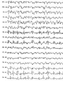
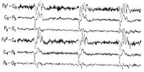
Electroencephalographic investigation of children with epilepsy
K. A. Voudris
Abstract
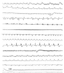
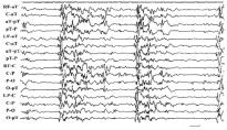
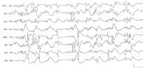
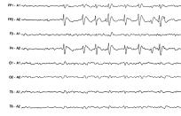
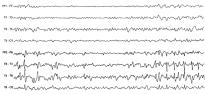
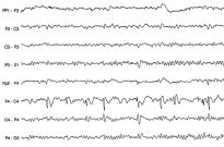
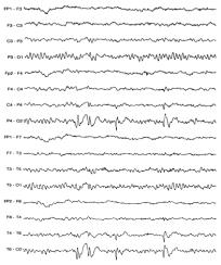
Electroencephalography is the basic technique for the neurophysiological investigation of epilepsy and may help in the diagnosis, study and prognosis of epilepsy. Although epilepsy is a clinical diagnosis, electroencephalographic data may play an important role in support of the clinical impression. Electroencephalography in paediatric epilepsy may be useful for confirmation of the diagnosis, recognition of the types of seizures and underlying epileptic syndromes, and for prediction, both of response to antiepileptic treatment and recurrence of seizures after discontinuation of antiepileptic treatment. Synchronized video-electroencephalographic monitoring offers improvements in the study of epileptic children with persistent epilepsy, and, in particular, may help in the differential diagnosis between epileptic and non-epileptic paroxysmal clinical events. Electroencephalography is a harmless and relatively inexpensive technique, which may be of great benefit for children with epilepsy when it is performed by experienced technicians and interpreted by specialized doctors, and the findings are correlated with clinical and other laboratory data.
stained by four histological methods. Histochem J 1998;30: 539-547.
10.Ornberg RL, Woerner BM, Edwards DA. Analysis of stained objects in histological sections by spectral imaging and differential absorption. J Histochem Cytochem 1999;47:1307-1314.
11.Siboni G, Rothmann C, Ehrenberg B, Malik Z. Spectral imaging of MC540 during murine and human colon carcinoma cell differentiation. J Histochem Cytochem 2001;49:147-153.
12.Rothmann C, Cohen AM, Malik Z. Chromatin condensation in erythropoiesis resolved by multipixel spectral imaging: differentiation versus apoptosis. J Histochem Cytochem 1997;45:1097-1108.
µÈ‚ÏÈÔÁÚ·Ê›·
1.∫ersey JH. Fifty years of studies of the biology and therapy of childhood leukemia. Blood 1997;90:4243-4251.
2.Pui CH. Acute lymphoblastic leukemia. Pediatr Clin North Am 1997;44:831-846.
3.Pui CH. Childhood leukemias. N Engl J Med 1995;332:1618-1630.
4.Skinner JM, Zhao Y, Coventry B, Bradley J. Video image analysis in pathology. Dis Markers 1993;11:53-70.
5.Oberholzer M, Ostreicher M, Christen H, Bruhlmann M. Methods in quantitative image analysis. Histochem Cell Biol 1996;105:333-355.
6.Aziz DC, Barathur RB. Quantitation and morphometric analysis of tumors by image analysis. J Cell Biochem 1994;19 (Suppl):S120-S125.
7.Rothmann C, Malic Z, Bar-Am I, Cabib D. Spectral imaging of fluorescence in cell biology, cancer therapy and cytogenetics. In: Kohen E, editor. Applications of optical engineering to the study of cellular pathology. Research Signpost, India; 1999. p. 135-150.
8.Miller ML, Andringa A, Elliott J, Conwell K 2nd, Dixon K, Carty MP. The morphological and spectral phenotype of apoptosis in HeLa cells varies following exposure to UV-C and the addition of inhibitors of ICE and CPP32. Cell Prolif 1998;31:17-33.
9.Rothmann C, Barshack I, Kopolovic J, Malik Z. Spectrally resolved morphometry of the nucleus in hepatocytes
13.Bain BJ, Lewis SM. Preparation and staining methods for blood and bone marrow films. In: Dacie J, Lewis S, editors. Practical Haematology. 9th ed. Edinburgh, London, Melbourne, New York: Churchill Livingstone; 2001. p. 50.
14.Friedrich K, Seiffert W, Zimmermann HW. Romanowsky dyes and Romanowsky-Giemsa effect. Structural investigations of the purple DNA-AB-EY dye complexes of Romanowsky-Giemsa staining. Histochemistry 1990;93:247256.
15.Galbraith W, Marshall PN, Bacus JW. Microspectrophotometric studies of Romanowsky stained blood cells. I. Subtraction analysis of a standardized procedure. J Microsc 1980;119:313-330.
16.Sorensen FB. Quantitative analysis of nuclear size for objective malignancy grading: a review with emphasis on new, unbiased stereologic methods. Lab Invest 1992;66:4-23.
17.Schultz RA, Nielsen T, Zavaleta JR, Ruch R, Wyatt R, Garner HR. Hyperspectral imaging: a novel approach for microscopic analysis. Cytometry 2001;43:239-247.
18.Boland MV, Murphy RF. Automated analysis of patterns in fluorescence-microscope images. Trends Cell Biol 1999;9:201-202.
19.Anguiano A. Fluorescence in situ hybridization (FISH): overview and medical applications. J Clin Ligand Assay 2000;23:33-42.
20.Malik Z, Rothmann C, Cycowitz T, Cycowitz ZJ, Cohen AM. Spectral morphometric characterization of B-CLL cells versus normal small lymphocytes. J Histochem Cytochem 1998;46:1113-1118.
Spectral imaging study of normal lymphocytes and lymphoblasts of acute lymphoblastic leukaemia in childhood*
N. Katzilakis1, E. Stiakaki1, A. Papadakis2, H. Dimitriou1, K. Balas2, M. Kalmanti1
Abstract
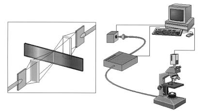
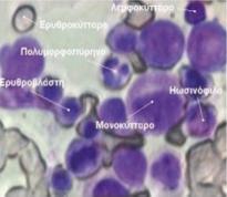
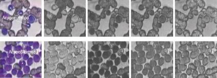
Background: In this study a new method is proposed for distinction between normal lymphocytes and L1 lymphoblasts of acute lymphoblastic leukaemia (ALL), using spectral analysis and imaging. In many cases the morphological distinction of normal lymphocytes from L1 lymphoblasts using light microscopy is difficult. The differentiation and classification is based mainly on cytochemical features and on immunologic, cytogenetic and molecular characteristics.
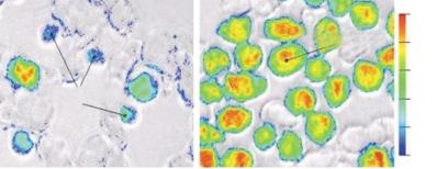
Methods: The method, which is based on the principles of spectral microscope and imaging, assesses the spectral absorbance characteristics of blood cells with variant biochemical composition, which uptake dye in different ways on routine cytological staining. The spectral microscope system is capable of performing imaging of absorbance in a variety of spectral bands, at any spatial point of the image, in a wide spectral range from 400 nm to 1000 nm. Spectral analysis was performed on bone marrow smears of 30 children aged 2-14 years, 12 with ALL and 18 with non malignant disease (prolonged fever, anaemia, thrombopenia or neutropenia) following staining with May-Grunwald-Giemsa stain (MGG).
Results: The analysis showed a statistically significant difference (p<0.00005) between normal lymphocytes and lymphoblasts, based on the detection, identification and mapping of their spectral absorbance. The wavelengths of 535 nm and 545 mn corresponded with the maximum absorbance for lymphocytes and lymphoblasts respectively. The maximum spectral differentiation was observed at the wavelength of 630 nm.
Conclusions: The results of this study support the potential of spectral imaging to provide new quantitative indices correlated with the biochemical, functional and structural status of the cell, and consequently suggest a new method for cell differentiation and classification in acute leukaemia.
Neuropsychological aspects of learning disabilities in epilepsy. Epilepsia 1990;31 (Suppl 4):S9-S20.
7.Sabers A, Moller A, Dam M, Smed A, Arlien-Soborg P, Buchman J et al. Cognitive function and anticonvulsant therapy: effect of monotherapy in epilepsy. Acta Neurol Scand 1995;92:19-27.
8.Piccirilli M, D’Alessandro P, Tiacci C, Ferroni A. Language lateralization in children with benign partial epilepsy. Epilepsia 1988;29:19-25.
9.Weglage J, Demsky A, Pietsch M, Kurlemann G. Neuropsychological, intellectual, and behavioral findings in patients with centrotemporal spikes with and without seizures. Dev Med Child Neurol 1997;39:646-651.
10.Bourgeois BF. Antiepileptic drugs, learning, and behavior in childhood epilepsy. Epilepsia 1998;39:913-921.
11.Meador KJ. Cognitive side effects of medications. Neurol Clin 1998;16:141-155.
12.Glauser TA, Nigro M, Sachdeo R, Pasteris LA, Weinstein S, Abou-Khalil BJ et al. Adjunctive therapy with oxcarbazepine in children with partial seizures. The Oxcarbazepine Pediatric Study Group. Neurology 2000;54:2237-2244.
13.Schmidt D, Elger CE. Oxcarbazepin (Trileptal). An effective and well tolerated new drug as first choice in treatment of focal seizures. Nervenarzt 2000;71:849-855.
14.Commission on Classification and Terminology of the International League Against Epilepsy. Proposal for revised classification of epilepsies and epileptic syndromes. Epilepsia 1989;30:389-399.
15.Wechsler D. Manual for the Wechsler intelligence scale for children. 3rd ed. San Antonio: The Psychological Corporation; 1991.
16.Tymchuk AJ. Comparison of Bender error and time scores for groups of epileptic, retarded, and behavior-problem children. Percept Mot Skills 1974;38:71-74.
18.Diagnostic and statistical manual of mental disordersFourth edition (DSM-IV). Washington D.C.: American Psychiatric Association; 1994.
19.Shalev RS, Gross-Tsur V, Masur D. Cognition, behavior and academic performance in children with epilepsy. In: Shinnar S, Amir N, Branski D, editors. Childhood seizures. Basel, Switzerland: Karger Pub; 1995. p. 170-178.
20.Jambaque I, Dellatolas G, Dulac O, Ponsot G, Signoret JL. Verbal and visual memory impairment in children with epilepsy. Neuropsychologia 1993;31:1321-1337.
21.Stephani U. Typical semiology of benign childhood epilepsy with centrotemporal spikes (BCECTS). Epileptic Disord 2000;2 (Suppl 1):S3-S4.
22.Piccirilli M, D’Alessandro P, Sciarma T, Cantoni C, Dioguardi MS, Giuglietti M et al. Attention problems in epilepsy: possible significance of the epileptogenic focus. Epilepsia 1994;35:1091-1096.
23.Mazurkiewicz-Beldzi‹ska M, Olszewska A. Effects of carbamazepine, phenytoine and oxcarbazepine on cognitive functions in children with epilepsy. Epileptologia 2000;8:1-88.
24.Curran HV, Java R. Memory and psychomotor effects of oxcarbazepine in healthy human volunteers. Eur J Clin Pharmacol 1993;44:529-533.
25.Aikia M, Kalviainen R, Sivenius J, Halonen T, Riekkinen PJ. Congnitive effects of oxcarbazepine and phenytoin monotherapy in newly diagnosed epilepsy: one year follow-up. Epilepsy Res 1992;11:199-203.
26.Carlsson G, Igelbrink-Schulze N, Neubauer BA, Stephani U. Neuropsychological long-term outcome of rolandic EEG traits. Epileptic Disord 2000;2 (Suppl 1):S63-S66.
27.Deonna T. Rolandic epilepsy: neuropsychology of the active epilepsy phase. Epileptic Disord 2000;2 (Suppl 1):S59-S61.
28.Doose H, Neubauer B, Carlsson G. Children with benign focal sharp waves in the EEG: developmental disorders and epilepsy. Neuropediatrics 1996;27:227-241.
29.Schoenfeld J, Seidenberg M, Woodard A, Hecox K, Inglese C, Mack K et al. Neuropsychological and behavioral status of children with complex partial seizures. Dev Med Child Neurol 1999;41:724-731.
30.Staden U, Isaacs E, Boyd SG, Brandl U, Neville BG. Language dysfunction in children with Rolandic epilepsy. Neuropediatrics 1998;29:242-248.
Cognitive functions in children with focal epilepsy and long-term oxcarbazepine therapy
M. Tzitiridou1, T. Panou1, E. Pavlidou1, G. Arsos2, A. Makavos3, E. Michaletou3, C. Panteliadis1
Abstract
Background: Children with epilepsy function quite satisfactorily with appropriate antiepileptic therapy, but they are reported to be at risk of developing cognitive disabilities.
Methods: This study was conducted during the period 1998-2002 in order to evaluate the effect of oxcarbazepine (OXC) on the cognitive function of school-age children with new onset epilepsy. This study included 43 children aged 6-14 years, suffering from either simple idiopathic or focal epilepsy, and 45 matched controls. Cognitive functions were evaluated by two tests: a) WISC-III, and b) Bender-Santucci. The patients and the children in the control group were examined neurologically, both clinically and by CT, MRI imaging, with normal findings. All the tests were performed at the beginning of the study and repeated after 14 months, during which the children with epilepsy were being treated with OXC. No differences were found between the patients and the control group on IQ evaluation pre-treatment.
Results: Six of the 43 patients and three of the 45 children in the control group had learning disabilities. These children had deficits in computational skill and in visual short-term memory and had poor reading skills. Scores for total, verbal and performance IQ of the children with learning disabilities were within the normal range. At re-examination 14 months later both groups showed improvement in logical abstract thinking and in verbal comprehension, which are dependent on cognitive and mental maturation. These findings show that OXC does not effect long-term acquisition and information retrieval. The BenderSantucci test showed increase in the opticokinetic perception, as the children became familiar with the test.
Conclusions: In summary, no effect of OXC on cognitive function was detected. Learning disabilities in a few children were unrelated occurrences.
1. Rosenstein NE, Perkins BA, Stephens DS, Popovic T, Hughes JM. Meningococcal disease. N Engl J Med 2001;344:1378-1388.
2.Jodar L, Feavers IM, Salisbury D, Granoff DM. Development of vaccines against meningococcal disease. Lancet 2002;359:1499-1508.
3. Hubert B, Caugant DA. Recent changes in meningococcal disease in Europe. Euro Surveill 1997;2:69-71.
4.van Deuren M, Brandtzaeg P, van der Meer JW. Update on meningococcal disease with emphasis on pathogenesis and clinical management. Clin Microbiol Rev 2000;13:144-166.
5.Kyaw MH, Clarke SC, Christie P, Jones IG, Campbell H. Invasive meningococcal disease in Scotland, 1994 to 1999, with emphasis on group B meningococcal disease. J Clin Microbiol 2002;40:1834-1837.
6.World Health Organization. Meningococcal disease. WHO Report on global surveillance of epidemic-prone infectious diseases. Chapter 5. 2000.
7.Baines PB, Hart CA. Severe meningococcal disease in childhood. Br J Anaesth 2003;90:72-83.
8.Thorburn K, Baines P, Thomson A, Hart CA. Mortality in severe meningococcal disease. Arch Dis Child 2001;85:382385.
10.American Academy of Pediatrics (Committee on Infectious Diseases). Meningococcal disease prevention and control strategies for practice-based physicians (Addendum: recommendations for college students). Pediatrics 2000;106:1500-1504.
11.Castellanos-Ortega A, Delgado-Rodriguez M. Comparison of the performance of two general and three specific scoring systems for meningococcal septic shock in children. Crit Care Med 2000;28:2967-2973.
12.Rosenstein NE, Fischer M, Tappero JW. Meningococcal vaccines. Infect Dis Clin North Am 2001;15:155-169.
14.Ragunathan L, Ramsay M, Borrow R, Guiver M, Gray S, Kaczmarski EB. Clinical features, laboratory findings and management of meningococcal meningitis in England and Wales: report of a 1997 survey. Meningococcal meningitis: 1997 survey report. J Infect 2000;40:74-79.
15.Tsolia MN, Theodoridou M, Tzanakaki G, Kalabalikis P, Urani E, Mostrou G et al. The evolving epidemiology of invasive meningococcal disease: a two-year prospective, population-based study in children in the area of Athens. FEMS Immunol Med Microbiol 2003;36:87-94.
16.Cartwright K, Noah N, Peltola H. Meningococcal Disease Advisory Board. Meningococcal disease in Europe: epidemiology, mortality, and prevention with conjugate vaccines. Report of a European advisory board meeting Vienna, Austria, 6-8 October 2000. Vaccine 2001;19:43474356.
17.Kremastinou J, Tzanakaki G, Kansouzidou A, Pagalis A, Danielides V, Kouppari G et al. Recent emergence of serogroup C meningococcal disease in Greece. FEMS Immunol Med Microbiol 1999;23:49-55.
21.Antignac A, Ducos-Galand M, Guiyoule A, Pires R, Alonso JM, Taha MK. Neisseria meningitidis strains isolated from invasive infections in France (1999-2002): phenotypes and antibiotic susceptibility patterns. Clin Infect Dis 2003;37:912-920.
23.Surveillance of bacterial meningitis in Europe 1999/2000. Eurosurveill Weekly 2002;6.
24.Newcombe J, Cartwright K, Palmer WH, McFadden J. PCR of peripheral blood for diagnosis of meningococcal disease. J Clin Microbiol 1996;34:1637-1640.
25.Carrol ED, Thomson AP, Riordan FA, Fellick JM, Shears P, Sills JA et al. Increasing microbiological confirmation and changing epidemiology of meningococcal disease on Merseyside, England. Clin Microbiol Infect 2000;6:259-262.
26.Connolly M, Noah N. Is group C meningococcal disease increasing in Europe? A report of surveillance of meningococcal infection in Europe 1993-6. European Meningitis Surveillance Group. Epidemiol Infect 1999;122:41-49.
28.Trotter C, Borrow R, Andrews N, Miller E. Seroprevalence of meningococcal serogroup C bactericidal antibody in England and Wales in the pre-vaccination era. Vaccine 2003;21:1094-1098.
29.Alcala B, Salcedo C, Arreaza L, Berron S, De La Fuente L, Vazquez JA. The epidemic wave of meningococcal disease in Spain in 1996-1997: probably a consequence of strain displacement. J Med Microbiol 2002;51:1102-1106.
30.Faustini A, Fabrizi E, Sangalli M, Bordi E, Cipriani P, Fiscarelli E et al. Role of laboratories in population-based surveillance of invasive diseases in Lazio, Italy, 1998-2000. Eur J Clin Microbiol Infect Dis 2002;21:824-826.
32.Tzanakaki G, Urwin R, Musilek M, Kriz P, Kremastinou J, Pangalis A et al. Phenotypic and genotypic approaches to characterization of isolates of Neisseria meningitidis from patients and their close family contacts. J Clin Microbiol 2001;39:1235-1240.
33.Sbyrakis S, Galanakis E. Meningococcal vaccine and herd immunity. Lancet 1999;354:1733.
34.Fernandez S, Arreaza L, Santiago I, Malvar A, Berron S, Vazquez JA et al. Impact of meningococcal vaccination with combined serogroups A and C polysaccharide vaccine on carriage of Neisseria meningitidis C. J Med Microbiol 2003;52:75-77.
35.Swartley JS, Marfin AA, Edupuganti S, Liu LJ, Cieslak P, Perkins B et al. Capsule switching of Neisseria meningitidis. Proc Natl Acad Sci USA 1997;94:271-276.
36.Jensen ES, Berthelsen L, Lind I, Fussing V, Sorensen HT, Schonheyder HC. Period prevalence and case-fatality rate associated with distinctive clone complexes of Neisseria meningitidis serogroups B and C. Eur J Clin Microbiol Infect Dis 2002;21:506-512.
37.Shlush LI, Behar DM, Zelazny A, Keller N, Lupski JR, Beaudet AL et al. Molecular epidemiological analysis of the changing nature of a meningococcal outbreak following a vaccination campaign. J Clin Microbiol 2002;40:3565-3571.
38.Goldacre MJ, Roberts SE, Yeates D. Case fatality rates for meningococcal disease in an English population, 1963-98: database study. BMJ 2003;327:596-597.
39.Balmer P, Borrow R, Miller E. Impact of meningococcal C conjugate vaccine in the UK. J Med Microbiol 2002;51:717-722.
40.Salleras L, Dominguez A, Cardenosa N. Impact of mass vaccination with polysaccharide conjugate vaccine against serogroup C meningococcal disease in Spain. Vaccine 2003;21:725-728.
42.Salleras L, Dominguez A, Cardenosa N. Dramatic decline of serogroup C meningococcal disease in Catalonia (Spain) after a mass vaccination campaign with meningococcal C conjugated vaccine. Vaccine 2003;21:729-733.
43.Cripps AW, Foxwell R, Kyd J. Challenges for the development of vaccines against Haemophilusinfluenzae and Neisseria meningitidis. Curr Opin Immunol 2002;14:553-557.
E. Apostolou1, E. Galanakis1, G. Vlahaki2, S. Stefanaki3, S. Maraki4, E. Kokori2, A. Tsilimigaki3, †S. Sbyrakis1,5
Abstract
Background: Neisseria meningitidis is a common cause of meningitis and septicaemia worldwide. Despite significant progress in its management, invasive meningococcal disease remains a threat for children of all ages. The aim of this study was to investigate the incidence of meningococcal disease in Crete over a 13-year period.
Methods: The study included all 67 children, residents of the prefectures of Iraklio, Rethimno and Lasithi, who were hospitalized for meningococcal disease in the three local Paediatric Departments and the Paediatric Intensive Care Unit in the period 1990-2002. The children were aged from 1 month to 13 years (mean age 4.66 years).
Results: Morbidity was increased during the winter months. The mean annual incidence was estimated at 5.54/100,000. The local rates for the prefectures of Rethimno, Iraklio and Lasithi were 7.85/100,000, 5.26/100,000 and 4.23/100,000 respectively. The prevalence was higher in infants, where serogroup B was more common. The serogroups was determined in 29 cases, serogroups B and C accounting for 48% and 41.3% respectively. Two periods of increased incidence of meningococcal disease were observed, the first in 1998-1999 with a predominance of serogroup C, and the second in 2002 with a predominance of group B. The fatality rate was estimated at 9%, with no correlation with the serogroup.
Conclusions: Meningococcal disease caused considerable morbidity and mortality in the three prefectures of Crete studied. Monitoring of the epidemiologic trends of this threatening infection is required to ensure rational use of the new vaccines.
2.Hyams JS. The digestive system. In: Behrman R, Kliegman R, Jenson H, editors. Inflammatory bowel disease. Nelson Textbook of Pediatrics. 17th ed. Philadelphia: W.B. Saunders; 2004. p. 1252-1255.
3.Farmer RG, Michener WM. Prognosis of Crohn’s disease with onset in childhood or adolescence. Dig Dis Sci 1979;24:752-757.
4.Harboe ZB, Paerregaard A, Hansen BF, Hesselfeldt P. Crohn disease in early childhood. J Pediatr Gastroenterol Nutr 2001;32:224-225.
5.Hyams JS. Crohn’s disease in children. Pediatr Clin North Am 1996;43:255-277.
6.Shivananda S, Lennard-Jones J, Logan R, Fear N, Price A, Carpenter L et al. Incidence of inflammatory bowel disease across Europe: is there a difference between north and south? Results of the European Collaborative Study on Inflammatory Bowel Disease (EC-IBD). Gut 1996;39:690-697.
7.Sawczenko A, Sandhu BK, Logan RF, Jenkins H, Taylor CJ, Mian S et al. Prospective survey of childhood inflammatory bowel disease in the British Isles. Lancet 2001;357:1093-1094.
8.Marx G, Seidman EG, Martin SR, Deslandres C. Outcome of Crohn’s disease diagnosed before two years of age. J Pediatr 2002;140:470-473.
9.Cohen Z, Weizman Z, Kurtzbart E, Newman N, Kapuller V, Maor E et al. Infantile colonic Crohn’s disease: a report of four cases in one family. J Pediatr Gastroenterol Nutr 2000;30:461-463.
10.Lindberg E, Lindquist B, Holmquist L, Hildebrand H. Inflammatory bowel disease in children and adolescents in Sweden, 1984-1995. J Pediatr Gastroenterol Nutr 2000;30: 259-264.
11.Langholz E, Munkholm P, Krasilnikoff PA, Binder V. Inflammatory bowel diseases with onset in childhood. Clinical features, morbidity, and mortality in a regional cohort. Scand J Gastroenterol 1997;32:139-147.
12.Baldi A, Di Marino MP, Vicidomini G, Baldi F. Crohn’s disease in infancy: a case report. Int Surg 1998;83:154-156.
13.Karlinger K, Gyorke T, Mako E, Mester A, Tarjan Z. The epidemiology and the pathogenesis of inflammatory bowel disease. Eur J Radiol 2000;35:154-167.
14.Moran AP, Prendergast MM. Molecular mimicry in Campylobacter jejuni and Helicobacter pylori lipopolysaccharides: contribution of gastrointestinal infections to autoimmunity. J Autoimmun 2001;16:241-256.
15.Shi HN, Walker A. Bacterial colonization and the development of intestinal defences. Can J Gastroenterol 2004;18: 493-500.
16.Ohkusa T, Nomura T, Sato N. The role of bacterial infection in the pathogenesis of inflammatory bowel disease. Intern Med 2004;43:534-539.
17.Prantera C, Zannoni F, Scribano ML, Berto E, Andreoli A, Kohn A et al. An antibiotic regimen for the treatment of active Crohn’s disease: a randomized, controlled clinical trial of metronidazole plus ciprofloxacin. Am J Gastroenterol 1996;91:328-332.
18.Ishikawa T, Okamura S, Oshimoto H, Kobayashi R, Mori M. Metronidazole plus ciprofloxacin therapy for active Crohn’s disease. Intern Med 2003;42:318-321.
1 Paediatric Clinic of the University of Crete, Heraklion
2 Department of Gastroenterology, 2nd Clinic of Internal Medicine, “Aghia Sophia” Children’s Hospital, Athens
3 Laboratory of Clinical Imaging, University of Crete, Heraklion
E. Papadopoulou1, E. Katsiyiannaki2, E. Mihailidou1, I. Grammatikakis3, E. Mantzouranis1
Abstract
Crohn’s disease is very rare in infancy. The case is presented of an 8 month-old boy who was hospitalized because of fever of undetermined origin and weight loss over the preceding month. A colonoscopy was carried out on the 35th day of hospitalization after episodes of bloody diarrhoea. The biopsy specimens were indicative of inflammatory bowel disease. Campylobacter jejuni was isolated from the stool culture on the 45th day of the hospitalization. Amoxycillin/clavulanic acid was administered for four days, based on the antibiotic sensitivity test, with no clinical response. On the 50th day of hospitalization ciproxin plus metronidazole treatment was started, and the infant became afebrile after ten days. Sulphasalazine was added 12 days later. The ciproxin plus metronidazole was continued for three months and sulphasalazine for one year. The infant remained without fever for eight months, with normal stools and normal weight gain. After a relapse he was rehospitalized at which time colonoscopy and biopsy confirmed the diagnosis of Crohn’s disease. Since then the patient has been under treatment with mesalazine, and has remained symptom-free.
Key words
Crohn’s disease, infancy, fever of unknown origin, Campylobacter jejuni
∆Ú¿¯ËÏÔ˜: Û¿ÓÈ· ÂÓÙfiÈÛË ÙÂÚ·ÙÒÌ·ÙÔ˜
ÛÙ· ·È‰È¿
ª. ÷ÛÈÒÙÔ˘1, ∂. ™·ÌÌÔ‡ÙË2, °. ¶ÈÙÛÔ˘Ï¿Î˘1, ª. µ·Î¿ÎË2, C. van Viet - ∫ˆÓÛÙ·ÓÙÈÓ›‰Ô˘3, ∂. ∫Ô˘‰Ô˘ÌÓ¿Î˘4
Literature review and proposed classification. J Pediatr Surg 1988;23:583-591.
3.Elmasalme F, Giacomantonio M, Clarke KD, Othman E, Matbouli S. Congenital cervical teratoma in neonates. Case report and review. Eur J Pediatr Surg 2000;10:252-257.
4.Hassab MH, Shadi SM, Al-Olayet YF, Al-Bassam AR. Ten years’ experience in the management of cervical teratomas. Pediatr Surg Int 1996;11:536-538.
5.Hajdu SI, Faruque AA, Hajdu EO, Morgan WS. Teratoma of the neck in infants. Am J Dis Child 1966;111:412-416.
6.Rothschild MA, Catalano P, Urken M, Brandwein M, Som P, Norton K et al. Evaluation and management of congenital cervical teratoma. Case report and review. Arch Otolaryngol Head Neck Surg 1994;120:444-448.
7.Wiatrak BJ, Myer CM 3rd, Bratcher GO. Report of a nasopharyngeal teratoma evaluated with magnetic resonance imaging. Otolaryngol Head Neck Surg 1990;102:186-190.
8.Welch KJ. The oropharynx and jaws. In: Welch KJ, Randolph JG, Ravitch MM et al, editors. Pediatric surgery. Vol 1. 4th ed. Chicago: Year Book; 1986. p. 513-515.
9.Abelev GI. Alpha-fetoprotein in ontogenesis and its association with malignant tumors. Adv Cancer Res 1971;14: 295-358.
10.Gundry SR, Wesley JR, Klein MD, Barr M, Coran AG. Cervical teratomas in the newborn. J Pediatr Surg 1983;18:382-386.
11.Dudgeon DL, Isaacs H Jr, Hays DM. Multiple teratomas of the head and neck [letter]. J Pediatr 1974;85:139-140.
1.Green JS, Dickinson FL, Rickett A, Moir A. MRI in the assessment of a newborn with cervical teratoma. Pediatr Radiol 1998;28:709-710.
2.Jordan RB, Gauderer MW. Cervical teratomas: an analysis.
12.Filston HC. Hemangiomas, cystic hygromas, and teratomas of the head and neck. Semin Pediatr Surg 1994;3:147-159.
M. Hasiotou1, ∂. Sammouti2, G. Pitsoulakis1, M. Vakaki2, C. van Viet - Konstadinidou3, E. Koudoumnakis4
Abstract
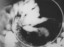
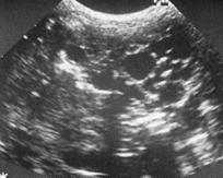
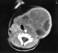
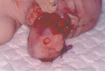
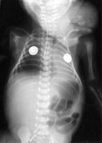
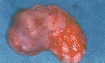
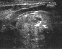
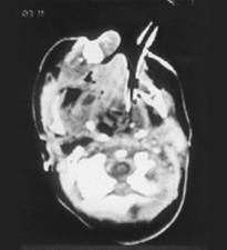
Congenital cervical teratomas are rare, representing 3% of teratomas in childhood. Although mostly benign, because of their location they are associated with a high mortality rate due to respiratory distress, and require immediate surgical excision. Imaging investigation is essential for the exact diagnosis and to assist in preoperative planning. The imaging characteristics of 5 cases (4 boys and 1 girl) of cervical teratoma are presented. All children were operated on and histopathology confirmed the diagnosis of teratoma. Two of the children died soon after surgery.
Key words
Cervical teratomas, children, imaging.
1 CT and MRI Department, “Aghia Sophia” Children’s Hospital, Athens
2.Stringer MD, Dhawan A, Davenport M, Mieli-Vergani G, Mowat AP, Howard ER. Choledochal cysts: lessons from a 20 year experience. Arch Dis Child 1977;73:528-531.
3.Gillet M, Favre S, Fontolliet C, Halkic N, Mantion G, Heyd B. Monolobar Caroli's disease. Apropos of 12 cases. Chirurgie 1999;124:13-19.
4.Caroli J, Couinaud C, Soupault R, Porcher P, Eteve J. A new disease, undoubtedly congenital, of the bile ducts: unilobar cystic dilation of the hepatic ducts. Sem Hop Paris 1958;34:496-502.
5.Barros JL, Polo JR, Sanabia J, Garcia-Sabrido JL, GomezLorenzo FJ. Congenital cystic dilatation of the intrahepatic bile ducts (Caroli’s disease): report of a case and review of the literature. Surgery 1995;85:589-592.
6.Todani T, Watanabe Y, Narusue M, Tabuchi K, Okajima K. Congenital bile duct cysts: classification, operative procedures, and review of thirty-seven cases including cancer arising from choledochal cyst. Am J Surg 1977;134:263-269.
9.Desmet VJ. Pathogenesis of ductal plate abnormalities. Mayo Clin Proc 1998;73:80-89.
10.Tsuchida Y, Sato T, Sanjo K, Etoh T, Hata K, Terawaki K et al. Evaluation of long-term results of Caroli’s disease: 21 years’ observation of a family with autosomal “dominant” inheritance, and review of the literature. Hepatogastroenterology 1995;42:175-181.
11.Pinto RB, Lima JP, da Silveira TR, Scholl JG, de Mello ED, Silva G. Caroli’s disease: report of 10 cases in children and adolescents in southern Brazil. J Pediatr Surg 1998;33: 1531-1535.
12.Sans M, Rimola A, Navasa M, Grande L, Garcia-Valdecasas JC, Andreu H et al. Liver transplantation in patients with Caroli’s disease and recurrent cholangitis. Transpl Int 1997;10:241-244.
13.Guibaud L, Lachaud A, Touraine R, Guibal AL, Pelizzari M, Basset T et al. MR cholangiography in neonates and infants: feasibility and preliminary applications. AJR Am J Roentgenol 1998;170:27-31.
Paediatric Clinic of the University of Thessaly
Correspondence: Eleni Papadimitriou 14 Georgiou Stavrou str. 412 21, Larissa
Date of submission: 01-03-2004
Date of approval: 03-01-2005
Caroli’s disease with congenital hepatic fibrosis. A case report
E. Papadimitriou, G. Matafia, V. Hatzopoulou, V. Mikraki, N. Skenteris
Abstract
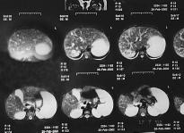
Caroli’s disease is a rare congenital disorder consisting of non-communicating cystic dilatation of the intrahepatic biliary ducts, very often associated with congenital hepatic fibrosis and polycystic kidneys. It is rarely diagnosed during childhood, usually presenting with symptoms of liver or kidney dysfunction. The case is presented of a girl of Ukrainian origin with a history of recurrent urinary tract infections during infancy. Imaging studies had at that time revealed large dysplastic kidneys. At the age of 2.5 years she presented with hepatomegaly of undetermined origin. She was referred for the first time to our clinic at the age of 11 years, for evaluation of prolonged fever. She was well developed, and her physical examination was unremarkable except for hepatosplenomegaly. The liver and kidney blood biochemistry findings were within the normal range, but the CT and MRI findings were consistent with Caroli’s disease. The patient has follow-up visits every six months. She has developed portal hypertension with first degree oesophageal varices and mild cytopenias due to hypersplenism. She is scheduled to undergo liver transplantation since Caroli’s disease carries an increased risk of development of bile duct carcinoma.
1.Frieden IJ, Reese V, Cohen D. PHACE syndrome. The association of posterior fossa brain malformations, hemangiomas, arterial anomalies, coarctation of the aorta and cardiac defects, and eye abnormalities. Arch Dermatol 1996;132:307-311.
2.Burns AJ, Kaplan LC, Mulliken JB. Is there an association between hemangioma and syndromes with dysmorphic features? Pediatrics 1991;88:1257-1267.
3.Pascual-Castroviejo I. Vascular and nonvascular intracranial malformation associated with external capillary hemangiomas. Neuroradiology 1978;16:82-84.
4.Reese V, Frieden IJ, Paller AS, Esterly NB, Ferriero D, Levy ML et al. Association of facial hemangiomas with DandyWalker and other posterior fossa malformations. J Pediatr 1993;122:379-384.
5.Fernandez GR, Munoz FJ, Padron C, Munoz M. Microphthalmos, facial capillary hemangioma and Dandy-Walker malformation. Acta Ophthalmol Scand 1995;73:173-175.
6.Goh WH, Lo R. A new 3C syndrome: cerebellar hypoplasia, cavernous haemangioma and coarctation of the aorta. Dev Med Child Neurol 1993;35:637-641.
7.Quecedo E, Gil-Mateo MP, Pont V, Febrer MI, Aliaga A. Giant hemifacial angioma and PHACE syndrome. Br J Dermatol 1997;136:649-650.
8.Hirsch JF, Pierre-Kahn A, Renier D, Sainte-Rose C, Hoppe-Hirsch E. The Dandy-Walker malformation. A review of 40 cases. J Neurosurg 1984;61:515-522.
9.Coats DK, Paysse EA, Levy ML. PHACE: a neurocutaneous syndrome with important ophthalmologic implications: case report and literature review. Ophthalmology 1999;106:1739-1741.
11.Rizzo R, Micali G, Incorpora G, Parano E, Pavone L. A very aggressive form of facial hemangioma. Pediatr Dermatol 1988;5:263-265.
12.Slavotinek AM, Dubovsky E, Dietz HC, Lacbawan F. Report of a child with aortic aneurysm, orofacial clefting, hemangioma, upper sternal defect, and marfanoid features: possible PHACE syndrome. Am J Med Genet 2002;110: 283-288.
13.White CW, Wolf SJ, Korones DN, Sondheimer HM, Tosi MF, Yu A. Treatment of childhood angiomatous diseases with recombinant interferon alfa-2a. J Pediatr 1991;118: 59-66.
14.Brouty-Boye D, Zetter BR. Inhibition of cell motility by interferon. Science 1980;208:516-518.
An infant with PHACE syndrome
A. Tsitsika, H. Fryssira, A. Lourida, N. Manolaki
Abstract
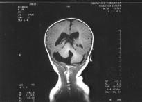
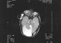
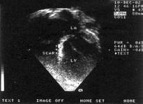
Large facial haemangiomas may be found in combination with dysplasias of the central nervous system (primarily, the Dandy-Walker malformation), the cardiovascular system and the eyes. The name PHACE (an acronym) was proposed for the syndrome of patients whose deformities meet the above criteria. The case reported is of an 8 month-old boy who had a giant, unilateral, plaque-like facial haemangioma, with a coexisting Dandy-Walker variant malformation, pulmonary hypertension, eyelid ptosis and strabismus of the eye on the affected side. The child had received oral corticosteroids as treatment for the rapidly growing facial lesion and at first a slight improvement was observed. After discontinuation of treatment there was a relapse and the baby was referred for further evaluation and treatment. Interferon-alfa was administered at daily dosage of 3x10⁶ U/M2 subcutaneously for 12 months with satisfying results. The coexisting anomalies were also treated. This patient is described as a possible PHACE case, and the pathogenesis, the clinical features, the diagnosis and the treatment options of the syndrome are discussed. It is emphasized that large, unilateral facial haemangiomas may be associated with the anomalies described, and in cases of abnormal findings or rapidly progressing haemangiomatosis further brain, cardiac and ocular investigation is indicated, as the diagnosis of PHACE syndrome must be considered.
1.Cole DE, Clarke LA, Riddell DC, Samson KA, Seltzer WK, Salisbury S. Congenital adrenal hypoplasia, Duchenne muscular dystrophy, and glycerol kinase deficiency: importance of laboratory investigations in delineating a contiguous gene deletion syndrome. Clin Chem 1994;40: 2099-2103.
2.McCabe ER, Towbin J, Chamberlain J, Baumbach L, Witkowski J, van Ommen GL et al. Complementary DNA probes for the Duchenne muscular dystrophy locus demonstrate a previously undetectable deletion in a patient with dystrophic myopathy, glycerol kinase deficiency, and congenital adrenal hypoplasia. J Clin Invest 1989;83:95-99.
3.Ferey S, Merzoug V, Linglart A, Chaussain JL, Kalifa G. A peculiar form of neonatal adrenal insufficiency: the IMAGe association. Two new cases. J Radiol 2003;84:323-325.
4.Lalli E, Bardoni B, Zazopoulos E, Wurtz JM, Strom TM, Moras D et al. A transcriptional silencing domain in DAX1 whose mutation causes adrenal hypoplasia congenita. Mol Endocrinol 1997;11:1950-1960.
5.Tabarin A. Congenital adrenal hypoplasia and DAX-1 gene mutations. Ann Endocrinol 2001;62:202-206.
6.Chrousos GP. The hypothalamic-pituitary-adrenal axis and immune-mediated inflammation. N Engl J Med 1995; 332:1351-1362.
7.Schwarz K, Thwaites R, Minford A, Day C, Butler G. Congenital adrenal hypoplasia presenting as a chronic respiratory condition. Arch Dis Child 2003;88:261-262.
8.Watterberg KL, Scott SM, Backstrom C, Gifford KL, Cook KL. Links between early adrenal function and respiratory outcome in preterm infants: airway inflammation and patent ductus arteriosus. Pediatrics 2000;105:320-324.
9.Watterberg KL, Scott SM. Evidence of early adrenal insufficiency in babies who develop bronchopulmonary dysplasia. Pediatrics 1995;95:120-125.
10.Tabarin A, Achermann JC, Recan D, Bex V, Bertagna X, Christin-Maitre S et al. A novel mutation in DAX1 causes delayed-onset adrenal insufficiency and incomplete hypogonadotropic hypogonadism. J Clin Invest 2000;105: 321-328.
1 Department of Neonatology, "Alexandra" University Hospital, Athens
2 Department of Enzymology and Cellular Function, Institute of Child Health, "Aghia Sophia" Children’s Hospital, Athens
Congenital adrenal hypoplasia presenting as severe respiratory failure. Case report
M. Dasopoulou1, M. Apostolou1, G. Baroutis1, H. Michelakakis2, E. Loukatou1, C. Costalos1
Abstract
Congenital adrenal hypoplasia is a rare condition, which occurs with an incidence of approximately 1 in 12,500 live births, usually presenting with hypoglycaemic convulsions, vomiting, failure to thrive or even collapse in early life. Links between early adrenal insufficiency and adverse respiratory outcome or even bronchopulmonary dysplasia in very low birth weight neonates have also been described. Respiratory problems in the early postnatal period are a rare manifestation of congenital adrenal hypoplasia and could be fatal unless early diagnosis is established, followed by prompt treatment. Aggressive treatment with corticosteroids should start immediately because of the unpredictable outcome of such a serious condition.
1.Keeling JW, Knowles SA. Sudden death in childhood and adolescence. J Pathol 1989;159:221-224.
2.Molander N. Sudden natural death in later childhood and adolescence. Arch Dis Child 1982;57:572-576.
3.Driscoll DJ, Edwards WD. Sudden unexpected death in children and adolescents. J Am Coll Cardiol 1985;5 (6 Suppl):S118B-S121B.
4.Neuspiel DR, Kuller LH. Sudden and unexpected natural death in childhood and adolescence. JAMA 1985;254: 1321-1325.
5.Wren C, O’Sullivan JJ, Wright C. Sudden death in children and adolescents. Heart 2000;83:410-413.
6.Wren C. Sudden death in children and adolescents. Heart 2002;88:426-431.
7.Morentin B, Aguilera B, Garamendi PM, Suarez-Mier MP. Sudden unexpected non-violent death between 1 and 19 years in north Spain. Arch Dis Child 2000;82:456-461.
9.Southall DP, Stebbens V, Shinebourne EA. Sudden and unexpected death between 1 and 5 years. Arch Dis Child 1987;62:700-705.
10.Maron BJ, Shirani J, Poliac LC, Mathenge R, Roberts WC, Mueller FO. Sudden death in young competitive athletes. Clinical, demographic, and pathological profiles. JAMA 1996;276:199-204.
11.Burke AP, Farb A, Virmani R, Goodin J, Smialek JE. Sports-related and non-sports-related sudden cardiac death in young adults. Am Heart J 1991;121:568-575.
12.Berger S, Dhala A, Friedberg DZ. Sudden cardiac death in infants, children, and adolescents. Pediatr Clin North Am 1999;46:221-234.
13.Valsangiacomo E, Schmid ER, Schupbach RW, Schmidlin D, Molinari L, Waldvogel K et al. Early postoperative arrhythmias after cardiac operation in children. Ann Thorac Surg 2002;74:792-796.
14.Sullivan ID, Presbitero P, Gooch VM, Aruta E, Deanfield JE. Is ventricular arrhythmia in repaired tetralogy of Fallot an effect of operation or a consequence of the course of the disease? A prospective study. Br Heart J 1987;58:40-44.
15.Gatzoulis MA, Walters J, McLaughlin PR, Merchant N, Webb GD, Liu P. Late arrhythmia in adults with the Mustard procedure for transposition of great arteries: a surrogate marker for right ventricular dysfunction? Heart 2000;84:409-415.
16.Peters NS, Somerville J. Arrhythmias after the Fontan procedure. Br Heart J 1992;68:199-204.
17.Maron BJ, Epstein SE, Roberts WC. Hypertrophic cardiomyopathy: a common cause of sudden death in the young competitive athlete. Eur Heart J 1983;4 Suppl F:S135-S144.
18.Corrado D, Basso C, Schiavon M, Thiene G. Screening for hypertrophic cardiomyopathy in young athletes. N Engl J Med 1998;339:364-369.
19.McKenna WJ, Deanfield JE. Hypertrophic cardiomyopathy: an important cause of sudden death. Arch Dis Child 1984;59:971-975.
20.Elliott P. Cardiomyopathy. Diagnosis and management of dilated cardiomyopathy. Heart 2000;84:106-112.
21.Maron BJ, Tajik AJ, Ruttenberg HD, Graham TP, Atwood GF, Victorica BE et al. Hypertrophic cardiomyopathy in infants: clinical features and natural history. Circulation 1982;65:7-17.
22.Maron BJ, Olivotto I, Spirito P, Casey SA, Bellone P, Gohman TE et al. Epidemiology of hypertrophic cardiomyopathy-related death: revisited in a large non-referral-based patient population. Circulation 2000;102:858-864.
23.Robbins RC, Stinson EB. Long-term results of left ventricular myotomy and myectomy for obstructive hypertrophic cardiomyopathy. J Thorac Cardiovasc Surg 1996;111:586-594.
24.McKenna WJ, Behr ER. Hypertrophic cardiomyopathy: management, risk stratification, and prevention of sudden death. Heart 2002;87:169-176.
25.Kitaoka H, Takata J, Yabe T, Hitomi N, Furuno T, Doi YL. Low dose dobutamine stress echocardiography predicts the improvement of left ventricular systolic function in dilated cardiomyopathy. Heart 1999;81:523-527.
26.Walker JD, Crawford FA, Kato S, Spinale FG. The novel effects of 3,5,3’-triiodo-L-thyronine on myocyte contractile function and ‚-adrenergic responsiveness in dilated cardiomyopathy. J Thorac Cardiovasc Surg 1994;108:672-679.
27.Tibayan FA, Lai DT, Timek TA, Dagum P, Liang D, Daughters GT et al. Alterations in left ventricular torsion in tachycardia-induced dilated cardiomyopathy. J Thorac Cardiovasc Surg 2002;124:43-49.
28.Sidwell RU, Yates R, Atherton D. Dilated cardiomyopathy in dystrophic epidermolysis bullosa. Arch Dis Child 2000;83:59-63.
29.Lee YH, Lee HD, Lee YA, Lee YS, Jung JA, Hwang GG et al. Ganglioneuroblastoma presenting as dilated cardiomyopathy. Arch Dis Child 2003;88:162-164.
30.Burch M, Siddiqi SA, Celermajer DS, Scott C, Bull C, Deanfield JE. Dilated cardiomyopathy in children: determinants of outcome. Br Heart J 1994;72:246-250.
31.Rivenes SM, Kearney DL, Smith EO, Towbin JA, Denfield SW. Sudden death and cardiovascular collapse in children with restrictive cardiomyopathy. Circulation 2000;102: 876-882.
32.Beekman RH, Rocchini AP, Crowley DC, Schork MA, Rosenthal A. Acute hemodynamic effects of captopril in children with a congestive or restrictive cardiomyopathy. Circulation 1991;83:523-527.
33.Colin AA, Jaffe M, Shapira Y, Ne’eman Z, Gutman A, Korman S. Muscle carnitine deficiency presenting as familial fatal cardiomyopathy. Arch Dis Child 1987;62:1170-1172.
35.Lipshultz SE, Rifai N, Sallan SE, Lipsitz SR, Dalton V, Sacks DB et al. Predictive value of cardiac troponin T in pediatric patients at risk for myocardial injury. Circulation 1997;96:2641-2648.
36.Corrado D, Basso C, Thiene G. Arrhythmogenic right ventricular cardiomyopathy: diagnosis, prognosis, and treatment. Heart 2000;83:588-595.
37.Lee KJ, Mc Crindle BW, Bohn DJ, Wilson GJ, Taylor GP, Freedom RM et al. Clinical outcomes of acute myocarditis in childhood. Heart 1999;82:226-233.
38.Kawai C. From myocarditis to cardiomyopathy: mechanisms of inflammation and cell death: learning from the past for the future. Circulation 1999;99:1091-1100.
39.Oakley CM. Myocarditis, pericarditis and other pericardial diseases. Heart 2000;84:449-454.
40.Case CL. Diagnosis and treatment of pediatric arrhythmias. Pediatr Clin North Am 1999;46:347-354.
41.Antoniadis S, Defterevos E, Papageorgiou G, Stratakis N, Korantzis A. Fetal arrhythmias: diagnosis and treatment. 3rd Balkan Meeting on Pediatric Cardiology and Cardiac Surgery. Athens; 1995.
43.Ward C. Severe arrhythmias in Coxsackie virus B3 myopericarditis. Arch Dis Child 1978;53:174-176.
44.Southall DP, Richards J, Hardwick RA, Shinebourne EA, Gibbens GL, Thelwall-Jones H et al. Prospective study of
fetal heart rate and rhythm patterns. Arch Dis Child 1980;55:506-511.
45.O’Callaghan FJ, Clarke AC, Joffe H, Keeton B, Martin R, Salmon A et al. Tuberous sclerosis complex and Wolff-Parkinson-White syndrome. Arch Dis Child 1998;78:159-162.
46.Campbell RM, Strieper MJ, Frias PA, Collins KK, Van Hare GF, Dubin AM. Survey of current practice of pediatric electrophysiologists for asymptomatic Wolff-Parkinson-White syndrome. Pediatrics 2003;111:245-247.
47.Vignati G, Balla E, Mauri L, Lunati M, Figini A. Clinical and electrophysiologic evolution of the Wolff-ParkinsonWhite syndrome in children: impact on approaches to management. Cardiol Young 2000;10:367-375.
48.Prystowsky EN, Fananapazir L, Packer DL, Thompson K A, German LD. Wolff-Parkinson-White syndrome and sudden cardiac death. Cardiology 1987;74 (2 Suppl):S67S71.
49.De Giovanni JV, Dindar A, Griffith MJ, Edgar RA, Silove ED, Stumper O et al. Recovery pattern of left ventricular dysfunction following radiofrequency ablation of incessant supraventricular tachycardia in infants and children. Heart 1998;79:588-592.
50.Steinbeck G. Should radiofrequency current ablation be performed in asymptomatic patients with the WolffParkinson-White syndrome? Pacing Clin Electrophysiol 1993;16:649-652.
51.Till JA, Rowland E, Shinebourne EA, Ward DE. Treatment of refractory supraventricular arrhythmias with flecainide acetate. Arch Dis Child 1987;62:247-252.
52.Shimizu W, Satomi K, Kamakura S. Images in cardiology: visualization of activation and repolarization in congenital long QT syndrome. Heart 2002;88:190.
53.Zupancic JA, Triedman JK, Alexander M, Walsh EP, Richardson DK, Berul CI. Cost-effectiveness and implications of newborn screening for prolongation of QT interval for the prevention of sudden infant death syndrome. J Pediatr 2000;136:481-489.
54.Huang TC, Cecchin FC, Mahoney P, Portman MA. Corrected QT interval (QTc) prolongation and syncope associated with pseudohypoparathyroidism and hypocalcemia. J Pediatr 2000;136:404-407.
55.Benatar A, Decraene T. Comparison of formulae for heart rate correction of QT interval in exercise ECGs from healthy children. Heart 2001;86:199-202.
56.Sarubbi B, Musto B, Ducceschi V, D’Onofrio A, Cavallaro C, Vecchione F et al. Congenital junctional ectopic tachycardia in children and adolescents: a 20 year experience based study. Heart 2002;88:188-190.
57.Santinga JT, Kirsh MM, Brady TJ, Thrall J, Pitt B. Left ventricular function in patients with ventricular arrhythmias and aortic valve disease. Ann Thorac Surg 1983;35:152-155.
58.Ficker DM, So EL, Shen WK, Annegers JF, O’Brien PC, Cascino GD et al. Population-based study of the incidence of sudden unexplained death in epilepsy. Neurology 1998;51:1270-1274.
59.Langan Y, Nashef L, Sander JW. Certification of deaths attributable to epilepsy. J Neurol Neurosurg Psychiatry 2002;73:751-752.
60.Camfield CS, Camfield PR, Veugelers PJ. Death in children with epilepsy: a population-based study. Lancet 2002;359:1891-1895.
61.Champ CS, Byard RW. Sudden death in asthma in childhood. Forensic Sci Int 1994;66:117-127.
62.Donner EJ, Smith CR, Snead OC 3rd. Sudden unexplained death in children with epilepsy. Neurology 2001;57:430-434.
63.Watanabe N, Akasaka T, Yamaura Y, Akiyama M, Kaji S, Saito Y et al. Intramyocardial coronary flow characteristics in patients with hypertrophic cardiomyopathy: non-invasive assessment by transthoracic Doppler echocardiography. Heart 2003;89:657-658.
64.Burch M, Mann JM, Sharland M, Shinebourne EA, Patton MA, McKenna WJ. Myocardial disarray in Noonan syndrome. Br Heart J 1992;68:586-588.
65.Angelini A, Crosato M, Boffa GM, Calabrese F, Calzolari V, Chioin R et al. Active versus borderline myocarditis: clinicopathological correlates and prognostic implications. Heart 2002;87:210-215.
66.Sato Y, Taniguchi R, Nagai K, Makiyama T, Okada H, Yamada T et al. Measurements of cardiac troponin T in patients with hypertrophic cardiomyopathy. Heart 2003;89: 659-660.
67.Chakrabarti S, Thomas E, Wright JG, Vettukattil JJ. Congenital coronary artery dilatation. Heart 2003;89:595-596.
In sudden death in childhood the cause, in the vast majority of cases, is of cardiac origin. In many cases the underlying cause is congenital heart disease, which may or may not have been diagnosed, and may or may not have been operated on. The death rate is particularly high in post-operative patients where the management is difficult. Sudden death usually occurs during sports or in conditions of extreme fatigue. The cardiomyopathies and especially the hypertrophic form, constitute another high risk category. Acquired heart diseases also play a major part. Apart from these diagnosed causes, it is well known that arrhythmias carry the highest risk in incidents of sudden death when the aetiology remains unknown even after necropsy. For management of the problem, the follow up and treatment of patients known to have heart disease is of paramount importance, along with general preventive measures. A cardiovascular check up is absolutely necessary in every child who is going to be involved in competitive sports and championships, and in any child when the paediatricians have identified suspicious findings including heart murmurs, rhythm disturbances, symptoms or events such as chest pain, syncope, etc. Also a family history of heart disease or sudden death should be taken into account. All the evaluations should be performed and the relevant certificates be issued by a specialist paediatric cardiologist, after clinical examination and tests such as electrocardiogram, colour Doppler and, whenever necessary, 24-hour Holter monitoring or other tests. The issue of certificates given by doctors unrelated to paediatric cardiology could be dangerous. For the time being, screening tests for all children are not considered necessary, but this will be reconsidered following the findings of various large-scale projects currently in progress on the subject.
1.Inwald D, Franklin O, Cubitt D, Peters M, Goldman A, Burch M. Enterovirus myocarditis as a cause of neonatal collapse. Arch Dis Child Fetal Neonatal Ed 2004;89:F461F462.
28 ™ÂÙÂÌ‚Ú›Ô˘- Pre-event to 1st European Conference on ∂Ú¤ÙÚÈ· 1 √ÎÙˆ‚Ú›Ô˘ 2005 Injury Prevention and Safety Promotion “Increasing efficiency in injury prevention”