Melander E, Ekdahl K, Jonsson G, Molstad S ∞fi‰ÔÛË ÛÙ· ∂ÏÏËÓÈο: ∫. ∆ÛÔ˘Ì¿Î·˜
¶ÚÔÛ¯‹ ™˘Ó¤‰ÚÈ·
Left atrial myxoma in a child presenting as an acute cerebral episode
A.N. Kourtesis, C.A. Paphitis, ∂. Latoufi, E. Dragogia, G. Briasoulis, V. Thanopoulos, M. Azariades
∫Ôhler’s disease. A rare condition in children
C. Tsoumakas, A. Papadopoulou, P. Miha-Georgiopoulou
LITERATURE ABSTRACTS
Adolescent suicide attempts. Risks and protectors
Borowsky I, Ireland M, Resuick M Greek translation: C. Tsoumakas
A trial of recombinant human granulocyte colony stimulating factor for the treatment of very low birthweight infants with presumed sepsis and neutropenia
Russel ARB, Emmerson AJB, Wilkinson N, Chant T, Sweet DG, Halliday HL et al Greek translation: S. Fotopoulos
Frequency of penicillin-resistant pneumococci in children is correlated to community utilization of antibiotics
Melander E, Ekdahl K, Jonsson G, Molstad S Greek translation: C. Tsoumakas
H Û‡ÓÙ·ÍË ÙˆÓ ‚È‚ÏÈÔÁÚ·ÊÈÎÒÓ ·Ú·ÔÌÒÓ Á›ÓÂÙ·È Û‡Ìʈӷ Ì ÙȘ ÚԉȷÁڷʤ˜ Ù˘ International Committee of Medical Journal Editors / Uniform Requirements for Manuscripts Submitted to Biomedical Journals (JAMA 1997;277:927-934), http://jama.amaassn.org/info/auinst_req.html. OÈ Û˘ÓÙÌ‹ÛÂȘ ÙˆÓ Ù›ÙÏˆÓ ÙˆÓ ÂÚÈÔ‰ÈÎÒÓ Á›ÓÔÓÙ·È Ì ‚¿ÛË ÙÔ Cumulated Index Medicus (List of Journals Indexed in Index Medicus, http://www.nlm.nih.gov).
Proesmans W. Bartter syndrome and its neonatal variant. Eur J Pediatr 1997;156:669-679.
™˘ÌÏËڈ̷ÙÈÎfi Ù‡¯Ô˜ ÂÚÈÔ‰ÈÎÔ‡:
Flyvbjerg A. Role of growth hormone, insulin-like growth factors (IGFs) and IGF-binding proteins in the renal complications of diabetes. Kidney Int 1997;52 (60 Suppl):S12-S19.
Èڛ˜ Û˘ÁÁڷʤ·:
National Institutes of Health Consensus Development Conference. Neurofibromatosis conference statement. Arch Neurol 1988;45:575-578.
Schreiner GF, Lange L. Ethanol modulation of macrophage influx in glomerulonephritis [abstract].
J Am Soc Nephrol 1991;2:562.
Should antileukotriene therapies be used instead of inhaled corticosteroids in asthma? [Editorial]. Am J Respir Crit Care Med 1998;158:1697-1701.
Laux-End R, Inaebnit D, Gerber HA, Bianchetti MG. Vasculitis associated with levamisole and circulating autoantibodies [letter]. Arch Dis Child 1996;75:355-356.
II. µÈ‚Ï›·
∫ÂÊ¿Ï·ÈÔ Û ‚È‚Ï›Ô:
Clark AG, Barratt TM. Steroid-responsive nephrotic syndrome. In: Barratt TM, Arner ED, Harmon WE, editors. Pediatric Nephrology. 4th ed. Baltimore: Lippincott William Wilkins; 1999. p. 742.
™‡ÁÁÚ·ÌÌ· ‹ ÌÔÓÔÁÚ·Ê›·:
Gorlin RJ, Cohen MM, Levin LS. Syndromes of the head and neck. 3rd ed. New York: Oxford University Press; 1990.
¢ËÌÔÛ›Â˘ÛË Û ÙfiÌÔ Ú·ÎÙÈÎÒÓ:
Bauer AW. The two definitions of bacterial resistance. In: Smith AJ, Rogers CA, eds. Proceedings of the Third International Congress of Chemotherapy; 1962 May 29-31; New York: International Society of Chemotherapy; 1963. p. 484-500.
Abstract: A large number of heterogeneous disorders may lead to thromboembolism of the cerebral vessels. In view of the high incidence of death rate or permanent neurological sequelae, early diagnosis and extensive laboratory investigations are essential. The identification of the causative mechanism may help in the therapeutic management during the acute phase, as well as in the prevention of further episodes.
1. Edwards M, Hoffman H. Cerebral vascular disease in children and adolescents. Baltimore: Williams and Wilkins; 1989.
2. Brett EM. Vascular disorders of the nervous system. In: Brett EM, ed. Paediatric Neurology. 3rd ed. New York: Churchill Livingstone; 1997. p. 571-588.
3. Roach ES. Etiology of stroke in children. Sem Pediatr Neurol 2000;7:244-265.
4. Dusser A, Goutieres F, Aicardi J. Ischaemic strokes in children. J Child Neurol 1986;1:131-136.
5. Gastaut H, Poirier F, Payan H, Saloman G, Toga M, Vigouroux M. HHH syndrome: Hemiconvulsions, hemiplegia, epilepsy. Epilepsia 1960;1:418-447.
6. Youroukos S, Psychou F, Fryssiras S, Paikos P, Nicolaidou P. Idiopathic intracranial hypertension in children. J Child Neurol 2000;15:453-457.
7. Benamer H, Bone I. Cerebral venous thrombosis: anticoagulant or thrombolyic therapy? J Neurol Neurosurg Psychiat 2000;69:427-430.
HÌÂÚÔÌËÓ›· ˘Ô‚ÔÏ‹˜: 20-12-2001
HÌÂÚÔÌËÓ›· ¤ÁÎÚÈÛ˘: 21-12-2001
¢È‡ı˘ÓÛË ·ÏÏËÏÔÁÚ·Ê›·˜: ™ˆÙ‹Ú˘ °ÈÔ˘ÚÔ‡ÎÔ˜
∞’ ¶·È‰È·ÙÚÈ΋ ∫ÏÈÓÈ΋ ¶·ÓÂÈÛÙËÌ›Ô˘ ∞ıËÓÒÓ
¡ÔÛÔÎÔÌÂ›Ô ¶·›‰ˆÓ “∏ ∞Á›· ™ÔÊ›·”
T.K. 115 27, ∞ı‹Ó· E-mail: sotel@hol.gr
£ÚÔÌ‚ÔÂÌ‚ÔÏÈο ÂÂÈÛfi‰È· ÙÔ˘ ÎÂÓÙÚÈÎÔ‡
·ÈÙÈÔÏÔÁ›·˜ ÛÙ· ·È‰È¿. ª›· ÚfiÎÏËÛË
™. ∞ÚÒÓË - µÔ˘ÚÓ¿
Thromboembolic episodes of the central nervous system (CNS) of diverse etiology in children. A challenge for the physician
S. Aronis - Vournas
Abstract: Thromboembolic episodes (TE) of the central nervous system is rare in childhood which, however, has been increasingly recognized during the last few years. They occur with a frequency of 1-3.3/100.000/year in the pediatric population, the incidence being higher in the neonatal period e.g. 1 case per 1000 births of full-term infants. The pathogenesis of TE in children and adolescents has been unclear due to lack of prospective studies. Nevertheless, extensive studies during the past 5 years tend to identify it as being a multifactorial disease. The most common underlying diseases predisposing to CNS thrombosis include congenital heart disease, perinatal asphyxia, malignancies, infections, sickle cell anaemia, vasculitis, nephrotic syndrome, homocysteinaemia, and vascular dysplasia. Probable triggering events are: trauma, major surgery, obesity, dehydration, medication such as corticosteroids, L-asparaginase, central catheters and others. The literature regarding the role of congenital (FV Leiden, FII 20210A, MTHFR, AT, PC, PS) or acquired prethrombotic disorders (Lpa, Lupus anticoagulant, ACA, anti-b2GP1) in the pathogenesis of CNS cerebral infarction is controversial. However recent studies indicate that homozygocity for MTHFR and FV Leiden is closely associated with CNS infarcts and may be used as
ıÚfiÌ‚ˆÛË ÛÙËÓ ·È‰È΋ ËÏÈΛ· Â›Ó·È Û¿ÓÈÔ,
predictors for recurrence of CNS thrombosis. The antiphospholipid syndrome (APS) is strongly associated with arterial or venous thrombosis in various organs, including CNS. Myxoma of the right atrium, a rare condition, usually presents as an acute cerebral syndrome, due to cerebral embolism from thrombus developing around the tumor or from detached particles derived from the tumor. In contrast with the APS, the presence of Lupus anticoagulant (LA) during childhood is not usually associated with thrombosis. The LA is transient and is detected following a viral infection or administration of drugs. In summary, CNS thromboembolic episodes are a rare event during childhood and constitute a real challenge for the physician, due to their diverse aetiology. Besides a good clinical examination, sensitive imaging techniques must be used in order to visualize the location and extent of the thrombus. The necessity and type of therapeutic intervention depends on the underlying disease. Moreover, the presence of one or more congenital or acquired prethrombotic factors will define the duration of anticoagulant therapy.
1. Rosendaal F. Thrombosis in the young: epidemiology and risk factors. A focus on venous thrombosis. Thromb Haemost 1977;78:1-6.
2. de Veber G, Roach ES, Riela AR, Wiznitzer M. Stroke in children: recognition, treatment and future directions. Semin Pediatr Neurol 2000;7:309-317.
3. Estan J, Hope P. Unilateral cerebral infarction in full term infants. Arch Dis Child 1997;76: F88-F93.
4. Kirkham FJ, Prengler M, Hewes DKM, Ganesan V. Risk factors for arterial ischaemic stroke in children. Child Neurol 2000;15:299-307.
5. Strater R, Nowak-Gottl U. Thrombophilia as risk factor in paediatric ischaemic stroke. Abstracts, 47th SSC meeting on Perinatal / Pediatric Hemosatsis, Paris; July 2001.
6. Kirkham FJ, Prengler M, Ganesan V. The incidence of recurrence after a first ischaemic stroke in childhood. Abstracts 47th SSC meeting on Perinatal / Pediatric Hemostasis, Paris; July 2001.
7. Mercuri E, Cowan F, Gupte G, Manning R, Laffan M, Rutherford M et al. Prothrombotic disorders and abnormal neurodevelopmental outcome in infants with neonatal cerebral infarction. Pediatrics 2001;107:1400-1404.
8. Petaja J, Hiltunen L, Fellman V. Increased risk of intraventricular hemorrhage in preterm infants with thrombophilia. Pediatr Res 2001;49:643-646.
13. Wilson W, Gharavi A, Koike T. International consensus statement on preliminary classification criteria for defining antiphosphospholipid syndrome. Arthritis Rheum 1999;42:1309-1311.
14. Brey RL. Differential diagnosis of central venous system manifestions of the APS. J Autoimmun 2000;15:133-138.
¶·È‰È·ÙÚÈ΋ 2002;65:4-8
15. Antiphospholipid antibodies. Abstracts. 9th International Symposium on Antiphospholipid Antibodies. France 2000;15:81-271.
16. Male C, Lechner K, Gichinger S, Kapiotis S, Wank H. Clinical significance of lupus anticoagulants in children. J Pediatr 1999;134:199-205.
17. Becton DL, Stine KC. Transient lupus anticoagulant associated with haemorrhage rather than thrombosis: the haemorrhagic lupus anticoagulant syndrome. J Pediatr 1997;130:998-1000.
18. Amiral J, Aronis S, Adamtziki E, Garoufi A, Karpathios T. Association of lupus anticoagulant with transient antibodies to prothrombin in a patient with hypoprothrombinemia. Thromb Res 1997;86:73-78.
19. Bayir H, Morelli PJ, Smith TH, Bianceniel TA. A left atrial
myxoma presenting as a cerebrovascular accident. Pediatr Neurol 1999;21:569-572.
20. Hishitani T, Ogawa K, Hoshino K, Yoshitake M, Koyanagi K, Namakura Y. Myxoma causing sudden neurologic symptoms including ataxia and facial nerve palsy. J Pediatr 1999;135:652.
Abstract: More than 100 years have passed since Little first suggested that cerebral palsy (CP) was associated with perinatal events; however, its etiology is still poorly understood. Recent research findings on the etiology of CP show that both antenatal and neonatal risk factors contribute to the development of severe neurologic impairment, including CP among very low birth weight (VLBW) infants. This article summarizes the recent research data concerning the etiology of CP in VLBW infants. Antenatal factors associated with increased risk for CP are genetic factors, short interpregnancy interval, intrauterine infections, placental abruption, vaginal bleeding, death of a co-twin in multiple pregnancy. In contrast, antenatal factors associated with decreased risk for CP are delivery without labor (elective cesarean section), delivery at specialized perinatal services, antenatal corticosteroids and preeclampsia. Neonatal factors associated with increased risk for CR are neonatal acidosis, sepsis, hypotension, respiratory distress syndrome, hypocapnia, chronic lung disease, pneumothorax and hypothyroxinemia. In conclusion, neurologic impairments among VLBW infants, including CP, are probably not caused by isolated risk factors, but rather by a complex interaction of many factors which act upon at different stages of development.
Division of Neonatology
General District Hospital “Alexandra”, Athens
§¤ÍÂȘ ÎÏÂȉȿ: ÂÁÎÂÊ·ÏÈ΋ ·Ú¿Ï˘ÛË, ÂÍ·ÈÚÂÙÈο
¯·ÌËÏÔ‡ ‚¿ÚÔ˘˜ Á¤ÓÓËÛ˘ ÓÂÔÁÓ¿, ·Ú¿ÁÔÓÙ˜
ÎÈÓ‰‡ÓÔ˘, ÚÔÁÂÓÓËÙÈÎÔ›, ÓÂÔÁÓÈÎÔ›.
™˘ÓÙÔÌÔÁڷʛ˜
∂õ°¡∂Í·ÈÚÂÙÈο ¯·ÌËÏÔ‡ ‚¿ÚÔ˘˜ Á¤ÓÓËÛ˘ ÓÂÔÁÓ¿
∏∫∏ÏÈΛ· ·ËÛ˘
™∫™¯ÂÙÈÎfi˜ ΛӉ˘ÓÔ˜
™∞¢™‡Ó‰ÚÔÌÔ ·Ó·Ó¢ÛÙÈ΋˜ ‰˘Û¯¤ÚÂÈ·˜
ö¡ÃÚfiÓÈ· Ó¢ÌÔÓÈ΋ ÓfiÛÔ˜
∂ÈÛ·ÁˆÁ‹
Œ¯Ô˘Ó ÂÚ¿ÛÂÈ ÂÚÈÛÛfiÙÂÚ· ·fi 100 ¯ÚfiÓÈ·
·ÊfiÙÔ˘ Ô Little (1) ÚÒÙÔ˜ ¤‰ÂÈÍ fiÙÈ Ë ÂÁÎÂÊ·ÏÈ΋
1. Little WJ. On the influence of abnormal parturition, difficult labor, premature birth and asphyxia neonatorum on the mental and physical condition of the child especially in relation to deformities. Trans Obstet Soc Lond 1862;3:293344.
2. Hogbert B, Flegberg G, Olow I, Wendt LV. The changing panorama of cerebral palsy in Sweden VII prevalence and origin in the birth year period 1987-1990. Acta Paediatr 1996;85:954-960.
3. Deidre J, Sellers S, MacKenzie JZ, Yadkin PL, Johnson AM. Case-control study of antenatal and intrapartum risk factors for cerebral palsy in very preterm singleton babies. Lancet 1995;346:1449-1454.
4. Cummins SK, Nelson KB, Gether JK. Cerebral palsy in four northern California counties, births 1983 through 1985. J Pediatr 1993;123:230-237.
5. O’ Stee TM, Dammann O. Antecedents of cerebral palsy in VLBW infants. Clin Perinatol 2000;27:285-300.
9. McHale DP, Mitchell S, Bundey S, Mognihan L, Campell DA, Woods CG et al. A gene for autosomal recessive symmetrical spastic cerebral palsy maps to chromosome 2q24-25. Am J Human Genet 1999;64:526-532.
10. Mchale DP, Jackson AP, Campell DA, Levene MI, Corry P, Woods GG et al. A gene for ataxic cerebral palsy maps to chromosome 9p12-q12. Eur J Hum Genet 2000;8:267-272.
11. Harum KH, Hoon AH, Casella JF. Factor-V Leiden: a risk factor for cerebral palsy. Dev Med Child Neurol 1999;41:781-785.
12. Harum KH, Hoon AH, Kato GJ, Casella JF, Breiter SN, Johnston MV. Homozygous factor-V mutation as a genetic cause of perinatal thrombosis and cerebral palsy. Dev Med Child Neurol 1999;41:777-780.
13. Palmer L, Petterson B, Blair E, Burton D. Family patterns of gestational age at delivery and growth in utero in moderate and severe cerebral palsy. Dev Med Child Neurol 1994;36:1108-1119.
14. Pinto-Martin J, Cnaan A, Zhao H. Short interpregnancy interval and the risk of disabling cerebral palsy in low birth weight population. J Pediatr 1998;132:818-821.
15. Cooke RW. Cerebral palsy in very low birthweight infants. Arch Dis Child 1990;65:201-206.
16. O’ Shea TM, Klinepeter KL, Dillard RG. Prenatal events and the risk of cerebral palsy in very low birthweight infants. Am J Epidemiol 1998;147:362-369.
18. Grether JK, Nelson KB, Cummins SK. Twinning and cerebral palsy: experience in four Northern California counties, births 1983 through 1985. Pediatrics 1993;92:854-858.
19. Petterson B, Nelson KB, Watson L, Stanley F. Twins, triplets and cerebral palsy in births in Western Australia in the 1980s. BMJ 1993;307:1239-1243.
21. Dammann O, Allred EN, Veelken N. Increase risk in spastic diplegia among very low birth weight children after preterm labor or prelabor rupture of membranes. J Pediatr 1998;132:531-535.
22. Pinto-Martin JA, Riolo S, Cnaan A, Holzman C, Susser WM, Paneth N. Cranial ultrasound prediction of disabling and nondisabling cerebral palsy at age two in a low birth weight population. Pediatrics 1995;95:249-254.
23. Scheller JM, Nelson KB. Twinning and neurologic morbidity. Am J Dis Child 1992;146:1110-1113.
24. Monset Couchard M, de Bethmann O, Relier JP. In vitro fertilization neonates transferred to neonatal medicine. Mid- and long-term outcome in 99 families. J Gynecol Obstet Biol Reprod 1995;24:85-92.
25. Souza SW, Rivlin E, Cadman J, Richards B, Buck P, Lieberman BA. Children conceived by in vitro fertilization after fresh embryo tranfer. Arch Dis Child Fetal Neonatal Ed 1997;76:F70-74.
26. Nelson KB, Grether JK. Can magnesium sulfate reduce the risk of cerebral palsy in low birth weight infants? Pediatrics 1995;95:263-269.
27. Wilson-Costello D, Borawski E, Friedman H, Redline R, Fanaroff A, Hack M. Perinatal correlates of cerebral palsy and other neurologic impairment among very low birth weight children. Pediatrics 1998;102:315-322.
28. Collins M, Paneth N. Preeclampsia and cerebral palsy: Are they are related? Dev Med Child Neurol 1998;40:207-211.
29. Arnold CC, Kramer MS, Hobbs CA, McLean FH, Usher RH. Very low birth weight: A problematic cohort for epidemiologic studies of very small or immature neonates. Am J Epidemiol 1991;134:604-613.
30. Spinillo A, Capuzo E, Cavanelli A, Stornati M, De-Santolo A, Fazzi E. Preeclampsia, preterm delivery and infant cerebral palsy. Eur J Obstet Gynecol Reprod Biol 1998;77:151-155.
31. Schendel DE, Berg CJ, Yeargin-Allsopp M, Boyle CA, Decoufle P. Prenatal magnesium sulfate exposure and the risk for cerebral palsy or mental retardation among very low birth weight children aged 3 to 5 years. JAMA 1996;276:1805-1810.
32. Thordestein M, Bagenholm R, Thiringer K, Kjellmer T. Scavengers of free oxygen radicals in combination with magnesium ameliorate perinatal hypoxic-ischemic brain damage in the rat. Pediatr Res 1993;34:23-26.
33. Goldstein RF, Rouse DJ. Preterm birth, cerebral palsy and magnesium. Nat Med 1997;3:146-147.
34. Grether JK, Nelson KB, Emery ES, Cummins SK. Prenatal and perinatal factors and cerebral palsy in very low birth weight infants. J Pediatr 1996;128:407-414.
35. Dammann O, Leviton A. Maternal intauterine infection, cytokines and brain damage in the preterm newborn. Pediatr Res 1997;42:1-8.
36. Leviton A, Paneth N, Reuss ML. Maternal infection, fetal inflammatory response, and brain damage in very low birth weight infants. Pediatr Res 1999;46:566-575.
37. Baud O, Ville Y, Zupan V, Boithias C, Lacaze-Masmonteil T, Cabilan JC et al. Are neonatal brain lesions due to intrauterine infection related to mode of delivery? Br J Obstet Gynecol 1998;105:121-124.
38. Gilles FH, Leviton A, Kerr CS. Endotoxin leukoencephalopathy in the telecephalon of the newborn kitten. J Neurol Sci 1976;27:183-191.
39. Young RSK, Yagel SK, Towfihti J. Systematic and neuropathologic effects of E. coli endotoxin in newborn dogs. Pediatr Res 1983;17:349-353.
40. Yoon BH, Kim CJ, Romero R. Experimentally induced intrauterine infection causes fetal brain white matter lesions in rabbits. Am J Obstet Gynecol 1997;177:797-802.
41. Yoon BH, Jun JK, Romero R, Park KH, Gomez R, Choi JH. Amniotic fluid inflammatory cytokines (interleukin-6, interleukin-1beta and tumor necrosis factor-alfa), neonatal brain white matter lesions and cerebral palsy. Am J Obstet Gynecol 1997;177:19-26.
42. Yoon BH, Romero R, Yang SH, Jun JK, Kim IO, Choi JH. Interleukin-6 concentrations in umbilical cord plasma are elevated in neonates with white matter lesions associated with periventricular leukomalecia. Am J Obstet Gynecol 1996;174:1433-1440.
43. Gomez R, Romero R, Ghezz F, Yoon BH, Mazor M, Edwin SS et al. The fetal inflammatory response syndrome. Am J Obstet Gynecol 1998;179:194-202.
44. Baud O, Emilie D, Pelletier E. Amniotic fluid concentrations of interleukin-1 beta, interleukin-6 and TNF-alpha in chorioamnionitis before 32 weeks of gestation: histologic associations and neonatal outcome. Br J Obstet Gynecol 1999;106:72-77.
45. Deguchi K, Oguchi K, Akashima S. Characteristic neuropathology of leukomalecia in extremely low birth weight infants. Pediatr Neurol 1997;16:296-300.
46. Yoon BH, Romero R, Kim CJ, Koon JN, Choe G, Syn HC et al. High expression tumor necrosis factor-alfa and interleukin in periventricular leukomalecia. Am J Obstet Gynecol 1997;177:406-411.
47. Crowley PA. Antenatal corticosteroid therapy: a meta analysis of the randomized trials, 1972 to 1994. Am J Obstet Gynecol 1995;173:322-335.
48. Elimian A, Verma U, Canterino J, Shan J, Visintainer P, Tejani N. Effectiveness of antenatal steroids in obstetric subgroups. Obstet Gynecol 1999;93:174-179.
49. Leviton A, Dammann O, Allred EN, Kuban K, Pagano M, VanMarter L. Antenatal corticosteroids and cranial ultrasound abnormalities. Am J Obstet Gynecol 1999;181:1007-1017.
50. Barks JD, Post M, Tuor UI. Dexamethasone prevents hypoxic-ischemic brain damage in the neonatal rat. Pediatr Res 1991;29:558-563.
51. Tuor UI, Simone CS, Barks JD, Post M. Dexamethasone prevents cerebral infraction without affecting cerebral blood flow in neonatal rats. Stroke 1993;24:452-457.
52. Barnes PJ, Karin M. Nuclear factor-kB: A pivotal transcription factor in chronic inflammatory diseases. N Engl J Med 1997;336:1066-1071.
53. Bessler H, Mendel C, Straussberg R, Gurary N, Aloni D, Sirota L. Effects of dexamethasone on IL-1beta, IL-6 and TNF-alfa production by mononuclear cells of newborns and adults. Biol Neonate 1999;75:225-233.
54. Vermillion ST, Soper DE, Chasedunn-Roark J. Neonatal sepsis after betamethasone administration to patients with preterm rupture membranes. Am J Obstet Gynecol 1999;181:320-327.
55. Huang WL, Beazley LD, Quinlivan JA, Evans SF, Newnham JP, Dunlop SA. Effect of cortisteroid on brain growth in fetal sheep. Obstet Gynecol 1999;94:213-218.
56. American College of Obstetricians and Gynecologists. Fetal distress and birth asphyxia. ACOG Committee Opinion 137. Washington DC: ACOG, 1994.
57. Dickinson JE, Eriksen NL, Meyer BA, Parisi VM. The effect of preterm birth on umbilical cord blood gas. Obstet Gynecol 1992;79:575-578.
58. O’ Shee TM, Preisser JS, Klinepeter KL, Dillard RG. Trends in mortality and cerebral palsy in a geographically based cohort of very low birth weight neonates born between 1982 to 1994. Pediatrics 1998;101:642-647.
59. Allan WC, Vohr B, Makuch RW, Katz KH, Ment LR. Antecedents of cerebral palsy in a multicentrial of indomethacin for intraventicular hemorrhage. Arch Pediatr Adolesc Med 1997;151:580-585.
60. O’ Shee TM, Klinepeter KL, Meis PS, Dillard RG. Intrauterine infection and the risk of cerebral palsy in very low birth weight infants. Paediatr Perinatal Epidemiol 1998;12:72-83.
61. O’ Shee TM, Kothadie JM, Klinepeter KL, Goldstein DJ, Jackson BG, Weaver RG et al. Randomized placebocontrolled trial of a 42-day tapering course of dexamethasone to reduce the duration of ventilator dependency in very low birth weight infants: Outcome of study. Pediatrics 1999;104:15-21.
62. Topp M, Landhoff-Roos J, Uldall P, Kristensen J. Intrauterine growth and gestational age in preterm infants with cerebral palsy. Early Hum Dev 1996;44:27-36.
63. Gray RH, O’ Callaghan MJ, Mohay HA, Burns YR, King JF. Maternal hypertension and neurodevelopmental outcome in very preterm infants. Arch Dis Child Fetal Neonatal Ed 1998;79:F88-F93.
64. Klein RZ, Carlton EL, Faix JD, Frank JE, Hermos RJ, Mullaney D et al. Thyroid function in very low birth weight infants. Clin Endocrinol 1997;47:411-417.
65. Reuss ML, Paneth N, Pinto-Martin J, Lorenz JM, Susser M. The relation of transient hypotheroxinemia in preterm infants to neurologic development at two years of age. N Engl J Med 1996;334:821-827.
66. Cashore WJ. The neurotoxicity of bilirubin. Clin Perinatol 1990;19:437-447.
67. O’ Shee TM, Dillard RG, Klinepeter KL, Goldestein DJ. Serum bilirubin levels, intracranial hemorrhage and the risk of developmental problem in very low birth weight neonates. Pediatrics 1992;90:888-892.
68. Wilson-Costello D, Borawski E, Friedman H, Pedline R, Fanaroff AA, Hack M. Perinatal correlates of cerebral palsy and other neurologic impairement among VLBW children. Pediatrics 1998;102:315-322.
ηډȷÎfi˜ ı¿Ó·ÙÔ˜, Û‡Ó‰ÚÔÌÔ ·ÔÎÏÂÈÛÌÔ‡ ‰ÂÍÈÔ‡ ÛΤÏÔ˘˜ Î·È ·Ó¿Û·Û˘ ST ‰È·ÛÙ‹Ì·ÙÔ˜.
Abstract: The Brugada syndrome is characterized by a right bundle branch block, ST segment elevation in the precordial leads V1 to V3, and episodes of syncope or sudden death caused by rapid polymorphic ventricular arrhythmias /ventricular fibrillation in patients with a structurally normal heart. It was first reported in 1992 by Brugada and Brugada. Since then, the number of cases indentified worldwide have increased, with more than 200 cases of Brugada syndrome reported up to date. The patients are mostly male and demonstrate their first arrhythmic event around the fourth decade. Nevertheless, the syndrome should also be considered as a cause of unexplained sudden cardiac death in children and even in infants. Systematic family studies have demonstrated an autosomal dominant inheritance pattern in many cases. Mutations in SCN5A, a gene encoding the cardiac sodium channel, have been detected in several patients. Pharmacological treatment with beta-adrenergic agents and anti-arrhythmic drugs does not protect effectively against recurrent arrhythmias. The implantation of a cardioverter-defibrillator constitutes the only effective therapy for the prevention of sudden death.
Key words: Brugada syndrome, sudden cardiac death, right bundle branch block - ST elevation syndrome.
2. Brugada R, Brugada J, Antzelevitch C, Kirsch GE, Potenza D, Towbin JA et al. Sodium channel blockers identify risk for sudden death in patients with ST-segment elevation and right bundle branch block but structurally normal hearts. Circulation 2000;101:510-515.
3. Alings M, Wilde A. “Brugada” syndrome. Clinical data and suggested pathophysiological mechanism. Circulation 1999;99:666-673.
4. Brugada P, Brugada J. Right bundle branch block, persistent ST segment elevation and sudden cardiac death: a distinct clinical and electrocardiographic syndrome. A multicenter report. J Am Coll Cardiol 1992;20:1391-1396.
5. Brugada P, Brugada R, Brugada J. Sudden death in patients and relatives with the syndrome of right bundle branch block, ST segment elevation in the precordial leads V1 to V3 and sudden death. Eur Heart J 2000;21:321-326.
6. Viskin S, Fish R, Eldar M, Zeltser D, Lesh MD, Glick A et al. Prevalence of the Brugada sign in idiopathic ventricular fibrillation and healthy controls. Heart 2000;84:31-36.
7. Brugada P. Brugada syndrome: an electrocardiographic diagnosis not to be missed. Heart 2000;84:1-2.
8. Wilde AAM, Priori SG. Brugada syndrome and sudden death [letter]. Eur Heart J 2000;21:1483-1484.
9. Priori SG, Napolitano C, Gasparini M, Pappone C, Bella PD, Brignole M et al. Clinical and genetic heterogeneity of right bundle branch block and ST-segment elevation syndrome. A prospective evaluation of 52 families. Circulation 2000;102:2509-2515.
10. Priori S, Napolitano C, Giordano U, Collisani G, Memmi M. Brugada syndrome and sudden cardiac death in children. Lancet 2000;355:808-809.
11. Erickson CC, Jones CS. Pediatric sudden cardiac death: What the pediatrician needs to know. Pediatr Ann 2000;29:509-518.
12. Suzuki H, Torigoe K, Numata O, Yazaki S. Infant case with a malignant form of Brugada syndrome. J Cardiovasc Electrophysiol 2000;11:1277-1280.
13. Hermida JS, Lemoine JL, Aoun FB, Jarry G, Rey JL, Quiret JC. Prevalence of the Brugada syndrome in an apparently healthy population. Am J Cardiol 2000;86:91-94.
14. Matsuo K, Kurita T, Inagaki M, Kakishita M, Aihara N, Shimizu W et al. The circadian pattern of the development of ventricular fibrillation in patients with Brugada syndrome. Eur Heart J 1999;20:465-470.
15. Priori SG, Barhanin J, Hauer RNW, Haverkamp W, Jongsma HJ, Kleber AG et al. Genetic and molecular basis of cardiac arrhythmias: Impact on clinical management. Parts I and II. Circulation 1999;99:518-528.
16. Priori SG, Barhanin J, Hauer RNW, Haverkamp W, Jongsma HJ, Kleber AG et al. Genetic and molecular basis of cardiac arrhythmias: Impact on clinical management. Eur Heart J 1999;20:174-195.
17. Alshinawi C, Mannens M, Wilde A. Mutations in the human cardiac sodium channel gene (SCN5A) in patients with Brugada’s syndrome [abstract]. Eur Heart J 1998;19(Suppl):78.
18. Brugada J, Brugada R, Brugada P. Right bundle-branch block and ST-segment elevation in leads V1 through V3. A marker for sudden death in patients without demonstrable structural heart disease. Circulation 1998;97:457-460.
19. Belhassen B, Viskin S, Fish R, Glick A, Setbon I, Eldar M. Effects of electrophysiologic-guided therapy with Class IA antiarrhythmic drugs on the long-term outcome of patients with idiopathic ventricular fibrillation with or without the Brugada syndrome. J Cardiovasc Electrophysiol 1999;10:1301-1312.
Abstract: The thromboembolic episodes of the central nervous system (CNS) are quite rare in childhood and their pathophysiology still remains unknown mainly because of the restricted amount of available data. In our study, 48 children were included (28 boys, 20 girls), who presented with a CNS thromboembolic episode during the years 1990-1998. Their mean age was 6.3 years. The diagnosis was established using radiologic tests (CT scan, MRI scan, MR angiography etc). The episodes were classified as: ischemic infarcts (n=19), arterial thromboses (n=18) and cerebral venous sinus thromboses (n=11). An underlying disease was detected in 38.65% and a triggering event in 14.6% of the children. Twenty three out of 48 children (47.9%) had one or more thrombophilic disorders (inherited disorder: 8 children, acquired disorder: 18 children). The acquired disorders were, as a rule, temporary and related to the underlying disease. In general, one or more risk factors for thrombosis were detected in 68.75% of the children. Treatment was mainly conservative, except for a few selected cases. Thromboembolic episodes in children appear to be multifactorial. Our experience from the use of anticoagulant and/or thrombolytic therapy in our patient population - mainly in cases with cerebral venous sinus thrombosis - is generally favourable. Nevertheless, comparative studies are necessary
1. Rosendaal F. Thrombosis in the young: epidemiology and risk factors. A focus on venous thrombosis. Thromb Haemost 1997;78:1-6.
2. de Veber G, Roach ES, Riela AR, Wiznitzer M. Stroke in children: recognition, treatment and future directions. Semin Pediatr Neurol 2000;7:309-317.
3. Smith CH, Baumann R. Clinical features and magnetic resonance imaging in congenital and childhood stroke. J Child Neurol 1991;6:263-272.
4. Brower M, Rollins N, Roach ES. Basal ganglia and thalamic infarction in children. Cause and clinical features. Arch Neurol 1996;53:1252-1256.
5. Tosetto A, Ruggeri M, Gastaman G, Rodeghiero F. Inherited abnormalities of blood coagulation in juvenile stroke. A case control study. Blood Coagul Fibrinolysis 1997;8:397-402.
6. Holt Larsen B, Soelberg Sorenses P, Marquardsen J. Transient ischemic attacks in young patients: a thromboembolic or migrainous manifestation? A 10 year follow up study of 46 patients. J Neurol Neurosurg Psychiatry 1990;53:1029-1033.
7. Norman D, Baker L. Cerebrovascular disease. Curr Opin Neurol Neurosurg 1991;4:837-842.
9. µ·rron T, Gusnard D, Zimmerman R, Clancy R. Cerebral venous thrombosis in neonates and children. Pediatr Neurol 1992;8:112-116.
10. Rivkin M, Anderson M, Kaye E. Neonatal idiopathic cerebral venous thrombosis: an unrecognized cause of transient seizures or lethargy. Ann Neurol 1992;32:51-56.
11. Adams R. Sickle cell disease and stroke. J Child Neurol 1995;10:15-76. ¶·È‰È·ÙÚÈ΋ 2002;65:23-31 Paediatriki 2002;65:23-31
12. Perkin GD. Cerebral venous thrombosis: developments in imaging and treatment. J Neurol Neurosurg Psychiatry 1995;59:1-3.
13. Burns A, Wilson E , Harber M, Brunton C, Sweny P. Cerebral venous sinus thrombosis in minimal change nephrotic syndrome. Nephrol Dial Transplant 1995;10:30-34.
14. Divekar A, Ali U, Ronghe M, Singh A, Dalvi R. Superior sagittal sinus thrombosis in a child with nephrotic syndrome. Pediatr Nephrol 1996;10:206-207.
15. Garcia RD, Baker A, Cunningham M, Weber A. Lateral sinus thrombosis associated with otitis media and mastoiditis in children. J Pediatr Infect Dis 1995;14:617-623.
16. Radhakrishnan K, Thacker AK, Maloo JC, el Mangoush MA. Sickle cell trait and stroke in the young adult. Postgrad Med J 1990;66:1078-1080.
17. Feldenzer J, Bueche M, Venes J, Gebarski S. Superior sagittal sinus thrombosis with infarction in sickle cell trait. Stroke 1987;18:656-660.
18. Partington MD, Aronyk KE, Byrd SE. Sickle cell trait and stroke in children. Pediatr Neurosurg 1994;20:148-151.
19. Vielhaber H, Ehrenforth S, Koch H, Scharrer I, van der Werf N, Nowak-Gottl U. Cerebral venous sinus thrombosis in infancy and childhood: role of genetic and acquired risk factors of thrombophilia. Eur J Pediatr 1998;157:555-560.
20. Chan AK, de Veber G. Prothrombotic disorders and ischemic stroke in children. Semin Pediatr Neurol 2000;7:301-308.
21. µÔnduel M, Sciuccati G, Hepner M, Torres AF, Pieroni G, Frontroth Jr. Arch Neurol 1999;56:967-971.
22. Dusser A, Boyer-Neumann C, Wolf M. Temporary protein C deficiency associated with cerebral arterial thrombosis in childhood. J Pediatr 1988;113:849-851.
24. Gonault-Heilmann M, Quetin P, Dreyfus M, Gandrille S, Emmerich J, Leroy-Matheron C, Guesnu M. Massive thrombosis of venous cerebral sinuses in a 2-year old boy with a combined inherited deficiency of Antithrombin III and Protein C. Thromb Haemost 1994;72:782-786.
25. Uttenreuther-Fischer M, Vetter B, Hellmann C, Otting U, Ziemer S, Hausdorf G, Gaedicke G, Kulozik A. Paediatric thromboembolism: the influence of non-genetic factors and the role of activated protein C resistance and protein C deficiency. Eur J Pediatr 1997;156:277-281.
26. ∫irkham FJ, Prengler M, Hewes DK, Ganesan V. Risk factors for arterial ischemic stroke in children. J Child Neurol 2000;15:299-307.
Abstract: The aim of the study was to investigate the clinical significance of coagulasenegative staphylococcus (CONS) isolated from blood cultures (BC) of neonates receiving intensive care, and to evaluate the significance of the clinical and laboratory findings in distinguishing between sepsis and contamination. We reviewed the records of neonates hospitalized at the neonatal intensive care unit (NICU) from 1994 to 1999 who grew CONS from at least one BC. The clinical data and the results of all positive CONS BCs and the antibiotic sensitivity were recorded. An episode of isolation was considered to represent sepsis when the same CONS strain was isolated from two or more BCs. During the study period CONS was isolated from 269 BCs obtained from 184 neonates that corresponded to 200 episodes. Only 135/200 (68%) episodes could be classified either as sepsis (n=41) or contamination (n=94). The incidence of factors predisposing to sepsis and the numbers of neonates with increased CRP were significantly higher in the septic episodes. Clinical and/or laboratory evidence of sepsis were present in 90% of the septic episodes and in 51% of contamination ones. The sensitivity of the clinical and laboratory evidence of sepsis in discriminating between sepsis and contamination was 90%, the specificity 49%, the positive predictive value 43% and the negative predictive value 92%. In conclusion: a) only in 2/3 of the episodes in which CONS was isolated, the BCs could distinguish between sepsis and contamination b) of the episodes that could be
Department of Neonatology Aristotelion University of Thessaloniki Department of Microbiology of Ippokration General Hospital, Thessaloniki
1. Hervas JA, Alomar A, Salva F, Reina J, Benedi VJ. Neonatal sepsis and meningitis in Mallorca, Spain, 1977-1991. Clin Infect Dis 1993;16:719-724.
2. Rodriguez Cervilla J, Fraga JM, Garcia Riestra C, Fernandez Lorenzo JR, Martinez Soto I. Neonatal sepsis: epidemiologic indicators and relation to birth weight and length of hospitalization time. An Esp Pediatr 1998;48:401-408.
3. Kirchhoff LV, Sheagren JN. Epidemiology and clinical significance of blood cultures positive for coagulasenegative staphylococcus. Infect Control 1985:6:479-486.
4. Kumar SP, Delivoria-Papadopoulos M. Infections in newborn infants in a special care unit. A changing pattern of infection. Ann Clin Lab Sci 1985;15:351-356.
5. Neumeister B, Kastner S, Conrad S, Klotz G, Bartmann P. Characterization of coagulase-negative staphylococci causing nosocomial infections in preterm infants. Eur J Clin Microbiol Infect Dis 1995;4:856-863.
6. Patrick CC, Kaplan SL, Baker CJ, Parisi JT, Mason EO Jr. Persistent bacteremia due to coagulase-negative staphylococci in low birth weight neonates. Pediatrics 1989;84:977-985.
7. Villari P, Sarnataro C, Iacuzio L. Molecular epidemiology of staphylococcus epidermidis in a neonatal intensive care unit over a three-year period. J Clin Microbiol 2000;38:1740-1746.
10. Freeman J, Epstein MF, Smith NE, Platt R, Sidebottom DG, Goldmann DA. Extra hospital stay and antibiotic usage with nosocomial coagulase-negative staphylococcal bacteremia in two neonatal intensive care unit populations. Am J Dis Child 1990;144:324-329.
11. Gray JE, Richardson DK, McCormick MC, Goldmann DA. Coagulase-negative staphylococcal bacteremia among very low birth weight infants: relation to admission illness severity, resource use, and outcome. Pediatrics 1995;95:225-230.
12. Savey A, Fleurette J, Salle BL. An analysis of the microbial flora of premature neonates. J Hosp Infect 1992;21:275-289.
13. Szewczyk EM, Piotrowski A, Rozalska M. Predominant staphylococci in the intensive care unit of a paediatric hospital. J Hosp Infect 2000;45:145-154.
14. Archer GL. Coagulase-negative staphylococci in blood cultures: the clinician’s dilemma. Infect Control 1985;6:12-13.
15. Baltimore RS. Is it real or is it a contaminant? A guide to the interpretation of blood culture results. Am J Dis Child 1987;141:241-242.
16. Seo SK, Venkataraman L, De Girolami PC, Samore MH. Molecular typing of coagulase-negative staphylococci from blood cultures does not correlate with clinical criteria for true bacteremia. Am J Med 2000;109:697-704.
17. Fidalgo S, Vazquez F, Mendoza MC, Perez F, Mendez FJ. Bacteremia due to staphylococcus epidermidis: microbiologic, epidemiologic, clinical, and prognostic features. Rev Infect 1990;12:520-528.
18. MacGregor RR, Beaty HN. Evaluation of positive blood cultures: guidelines for early differentiation of contaminated from valid positive cultures. Arch Intern Med 1972;130:84-87.
19. Dominguez-de Villota E, Algora-Weber A, Millan I, Rubio JJ, Galdos P, Mosquera JM. Early evaluation of coagulase negative staphylococcus in blood samples of intensive care unit patients. A clinically uncertain judgement. Intensive Care Med 1987;13:390-394.
20. St. Geme JW 3d, Bell LM, Baumgart S, D’ Angio CT, Harris MC. Distinguishing sepsis from blood culture contamination in young infants with blood cultures growing coagulase-negative staphylococci. Pediatrics 1990;86:157-162.
21. Nataro JP, Corcoran L, Zirin S, Swink S, Taichman N, Goin J et al. Prospective analysis of coagulase-negative staphylococcal infection in hospitalized infants. J Pediatr 1994;125:798-804.
22. Khatib R, Riederer KM, Clark JA, Khatib S, Briski LE, Wilson FM. Coagulase-negative staphylococci in multiple blood cultures: strain relatedness and determinants of same-strain bacteremia. J Clin Microbiol 1995;33:816-820.
23. Ferreiros CM, Souto MJ, Criado MT. Plasmid-pattern analysis in the differentiation of staphylococcus
24. Izard NC, Hachler H, Grehn M, Kayser FH. Ribotyping of coagulase-negative staphylococci with special emphasis on intraspecific typing of staphylococcus epidermidis. J Clin Microbiol 1992;30:817-823.
25. Edwards MS. Coagulase-negative staphylococcal bacteremia in neonates: confusion continued (editorial). Pediatrics 1990;86:320-322.
26. Maayan-Metzger A, Linder N, Marom D, Vishne T, Ashkenazi S, Sirota L. Clinical and laboratory impact of coagulase-negative staphylococci bacteremia in preterm infants. Acta Paediatr 2000;89:690-693.
27. Schmidt BK, Kirpalani HM, Corey M, Low DE, Philip AG, Ford-Jones EL. Coagulase-negative staphylococci as true pathogens in newborn infants: a cohort study. Pediatr Infect Dis J 1987;6:1026-1031.
28. Sandoe JA, Kerr KG, Reynolds GW, Jain S. Staphylo coccus capitis endocarditis: two cases and review of the literature. Heart 1999;82:e1.
29. Zaidi AK, Harrell LJ, Rost JR, Reller LB. Assessment of similarity among coagulase-negative staphylococci from sequential blood cultures of neonates and children by pulsed-field gel electrophoresis. J Infect Dis 1996;174:1010-1014.
30. Noel GJ, Edelson PJ. Staphylococcus epidermidis bacteremia in neonates: further observations and the occurrence of focal infection. Pediatrics 1984;74:832-837.
31. Battisti O, Mitchison R, Davies PA. Changing blood culture isolates in a referral neonatal intensive care unit. Arch Dis Child 1981;56:775-778.
32. Baumgart S, Hall SE, Campos JM, Polin RA. Sepsis with coagulase-negative staphylococci in critically ill newborns. Am J Dis Child 1983;137:461-463.
33. Anday EK, Talbot GH. Coagulase-negative staphylococcus bacteremia - a rising threat in the newborn infant. Ann Clin Lab Sci 1985;15:246-251.
34. Akiyama H, Yamasaki O, Tada J, Arata J. The production of superantigenic exotoxins by coagulase-negative staphylococci isolated from human skin lesions. J Dermatol Sci 2000;24:142-145.
35. Karlowicz MG, Buescher ES, Surka AE. Fulminant lateonset sepsis in a neonatal intensive care unit, 1988-1997, and the impact of avoiding empiric vancomycin therapy. Pediatrics 2000;106:1387-1390.
36. Drossou V, Kanakoudi F, Tzimouli V, Sarafidis K, Taparkou A, Bougiouklis D et al. Impact of prematurity stress and sepsis on the neutrophil respiratory burst activity of neonates. Biol Neonate 1997;72:201-209.
38. Spafford PS, Sinkin RA, Cox C, Reubens L, Powell KR. Prevention of central venous catheter-related coagulasenegative staphylococcal sepsis in neonates. J Pediatr 1994;125:259-263.
39. Raad I, Davis S, Khan A, Tarrand J, Elting L, Bodey GP. Impact of central venous catheter removal on the recurrence of catheter-related coagulase-negative staphylococcal bacteremia. Infect Control Hosp Epidemiol 1992;13:215-221.
40. Worthington T, Lambert PA, Elliott TS. Is hospital-acquired intravascular catheter-related sepsis associated with outbreak strains of coagulase-negative staphylococci? J Hosp Infect 2000;46:130-134.
41. Okada Y, Klein NJ, van Saene HK, Webb G, Holzel H, Pierro A. Bactericidal activity against coagulase-negative staphylococci is impaired in infants receiving long-term parenteral nutrition. Ann Surg 2000;231:276-281.
42. Jensen AG, Wachmann CH, Poulsen KB, Espersen F, Scheibel J, Skinhoj P et al. Risk factors for hospitalacquired staphylococcus aureus bacteremia. Arch Intern Med 1999;159:1437-1444.
43. Herwaldt LA, Geiss M, Kao C, Pfaller MA. The positive predictive value of isolating coagulase-negative staphylococci from blood cultures. Clin Infect Dis 1996;22:14-20.
1Russel ARB, Emmerson AJB, Wilkinson N, Chant T, Sweet DG, Halliday HL et al
A trial of recombinant human granulocyte colony stimulating factor for the treatment of very low birthweight infants with presumed sepsis and neutropenia
Abstract: Eosinophilia is common in the neonatal period. However, its causes, mechanism and clinical significance are still unknown. Previous studies have reported that eosinophilia may be associated with numerous conditions (establishment of an anabolic state, drug reactions, response to foreign antigens, chronic lung disease, erythropoietin treatment and infections). The aim of this study was to evaluate the possible association of various conditions, especially infection, with eosinophilia and to clarify whether recognition of an increased eosinophil count is of any clinical significance in the management of hospitalized neonates. Fifty-six neonates with eosinophilia (AEC >700/mm3) and 55 control neonates matched for gestational age, birth weight and hospitalization days were included in the study. A significant difference between the two groups was found only in blood transfusions, Á-globulin treatment, specific antibiotic treatment and infectious disease. However, neonates who develop septic episodes are treated with specific antibiotics and Á-globulin and are more often transfused. Thus it can be speculated that the main relationship observed is that between eosinophilia and infection whereas the other associations may be secondary. The relative risk factor for infection when the AEC is >700/mm 3 is 1.58, with confidence interval 1.30;1.91. Eosinophilia seems to be a reliable indicator of sepsis while normal AEC does not exclude infection. The exact cause of eosinophilia in neonates remains unclear. However, sepsis seems to be associated with eosinophilia. Infection should be strongly considered in the evaluation of a neonate with eosinophilia.
1. Sullivan S, Calhoun DA. Eosinophilia in the neonatal intensive care unit. Clin Perinatol 2000;27:603-622.
2. Gibson EL, Vaucher Y, Corrigan JJ. Eosinophilia in premature infants: relationship to weight gain. J Pediatr 1979;95:99-101.
3. Hatch DL, Garona JE, Goldman LR, Walker KO. Persistent eosinophilia in an infant with probable intrauterine exposure to L-tryptophan-containing supplements. Pediatrics 1991;88:810-813.
4. Van-Reempts PJ, Van-Overmeire B, Mahieu LM, Vanacker KJ. Clinical experience with ceftriaxone treatment in the neonate. Chemotherapy 1995;41:316-322.
5. Aranda JV, Portuguez-Malavasi A, Collinge JM, Germanson T, Outerbridge EW. Epidemiology of adverse drug reactions in the newborn. Dev Pharmacol Ther
1982;5:173-184.
6. Yamamoto C, Kojima T, Hattori K, Nogi S, Imamura H, Tsubura A et al. Eosinophilia in premature infants: correlation with chronic lung disease. Acta Paediatr 1996;85:1232-1235.
7. Ehara A, Takeda Y, Kida T, Mizukami S, Hagisawa M, Yamada Y. Time-course changes of eosinophil counts in premature infants: no effects of medical manipulation, except erythropoietin treatment, on eosinophilia. Pediatr Int 2000;42:58-60.
8. Bhat AM, Scanlon JW. The pattern of eosinophilia in premature infants. J Pediatr 1981;98:612-616.
9. Patel L, Garvey B, Arnon S, Roberts IA. Eosinophilia in newborn infants. Acta Paediatr 1994;83:797-801.
10. Wolach B, Bogger-Goren S, Whyte R. Perinatal hematological profile of newborn infants with candida antenatal infections. Biol Neonate 1991;59:5-12.
11. Weinberg AG, Rosenfeld CR, Manroe BL, Browne R. Neonatal blood cell count in health and disease. II. Values for lymphocytes, monocytes and eosinophils. J Pediatr 1985;106:462-466.
12. Calhoun DA, Sullivan SE, Lunoe M, Du Y, Christensen RD. Granulocyte-macrophage colony-stimulating factor and interleukin-5 concentrations in premature neonates with eosinophilia. J Perinatol 2000;20:166-171.
13. Fayon M, Babin JP. Hypereosinophilia in premature newborn infants. Pediatrics 1988;43:667-672.
14. Lawrence R Jr, Church JA, Richards W, Lipsey AI. Eosinophilia in the hospitalized neonate. Ann Allergy 1980;44:349-352.
15. Rothenberg ME. Eosinophilia. N Engl J Med 1998;338:1592-1600.
1Melander E, Ekdahl K, Jonsson G, Molstad S Frequency of penicillin-resistant pneumococci in children is correlated to community utilization of antibiotics Pediatr Infect Dis J 2000;19:1172-1177
Metastases in the central nervous system in children with neuroblastoma M. Baka, A. Pourtsidis, A. Stasinopoulou, M. Varvoutsi, D. Bouhoutsou, K. Paidousi, E. Kosmidi
Abstract: Neuroblastoma is the second in frequency malignant solid tumor among children. The majority of children affected by neuroblastoma (approximately 60%), present with distant metastases at the time of diagnosis. Common metastatic sites of neuroblastoma are the bones, bone marrow, liver and in infants the skin. With the use of aggressive chemotherapy, with or without autologous rescue, other metastatic sites have been identified during the recent years namely the CNS, the lungs and the ovaries. In order to study the occurrence of CNS metastases in children with neuroblastoma, we retrospectively reviewed the records of all patients diagnosed with neuroblastoma from 1/1980 to 12/2000 (a total of 102 children), and we describe 3 children with CNS metastases that were detected at the time of relapse. An attempt is also made to outline the possible pathophysiologic mechanisms of this metastatic pattern by reviewing the data reported in the literature.
1. Miller RW, Young JL Jr, Novakovic B. Childhood cancer. Cancer 1995;75:395-405.
2. Brodeur GM, Castleberry RP. Neuroblastoma. In: Pizzo PA, Poplack DG, eds. Principles and practice of pediatric oncology. Philadelphia: JB Lippincott; 1997. p. 739.
3. Cowie F, Corbett R, Pinkerton CR. Lung involvement in neuroblastoma: incidence and characteristics. Med Pediatr Oncol 1997;28:429-432.
4. Stephenson SR, Cook BA, Mease AD, Ruymann FB. The prognostic significance of age and pattern of metastases in stage IV-S neuroblastoma. Cancer 1986;58:372-375.
5. Ladenstein R, Philip T, Lasset C, Hartmann O, Garaventa A, Pinkerton R et al. Multivariate analysis of risk factors in stage 4 neuroblastoma patients over the age of one year treated with megatherapy and stem-cell transplantation: a report from the European Bone Marrow Transplantation Solid Tumor Registry. J Clin Oncol 1998;16:953-965.
6. Dini S, Lamino E, Garavento A, Rogers D, Dallorso S, Viscoli C et al. Multivariate therapy and unpurged autologous bone marrow transplantation for poor prognosis neuroblastoma. J Clin Oncol 1991;9:962-969.
7. Kellie SJ, Hayes FA, Bowman L, Konvar EH, Langstan J, Jenkins JJ et al. Primary extracranial neuroblastoma with central nervous system metastases: characterization by clinicopathologic findings and neuroimaging. Cancer 1991;68:1999-2006.
8. Blatt J, Fitz C, Mirro J. Recognition of central nervous system metastases in children with metastatic primary extracranial neuroblastoma. Pediatr Hematol Oncol 1997;14:233-241.
9. Palasis S, Morris J, Egelhoff J. Central nervous system relapse following bone marrow transplantation in stage IV neuroblastoma. Pediatr Hematol Oncol 1999;16:443-452.
10. Banerjee S, Marwaha RK, Bajwa RPS. Primary pelvic neuroblastoma with central nervous system metastases. Pediatr Hematol Oncol 1995;12:309-312.
11. Astigarraga I, Lajarreta R, Navajas A, Fernanotez-Teijeiro A, Imaz I, Bezanila JL. Secondary central nervous system metastases in children with neuroblastoma. Med Pediatr Oncol 1996;27:529-533.
12. Kramer K, Kushner B, Heller G, Chenug Nai-Kong V. Neuroblastoma metastatic to the central nervous system. The Memorial Sloan-Kettering cancer experience and a literature review. Cancer 2001;91:1510-1519.
13. Gallet BL, Egeihoff JC. Unusual CNS and Orbital metastases of neuroblastoma. Pediatr Radiol 1989;19:287-289.
15. Strauss I, Driscoll SG. Congenital neuroblastoma involving the placenta. Report of two cases. Pediatrics 1964;34:23-31.
16. Shaw PJ, Eden T. Neuroblastoma with intracranial involvement: an ENSG study. Med Pediatr Oncol 1992;20:149-155.
17. Xaidara A, Andrioti M, Mikraki V, Prodromou N, Grafakos S, Arvanitis D et al. Neuroblastoma metastatic to brain. Med Pediatr Oncol 1996;26:208-214.
The effect of a nucleotide supplemented formula on the peptic system of preterm infants V. Skouteri1, C. Costalos1, S. Sevastiadou1, S. Gavrili1, K. Danelatou1, A. Voyiatzi2, C. Economidou2, V. Petrochilou2
Abstract: Studies in animals have shown that nucleotides (NT) have significant effects upon gastrointestinal growth and differentiation. πn the present study we investigated the hypothesis that an NT enriched formula might improve gastrointestinal absorptive function and enteral flora in preterm infants. A double blind randomized controlled trial was designed. Forty two premature infants with a mean gestational age of 32.6 weeks, were randomly assigned into two groups consisting of 21 infants each. The NT group received a standard term formula supplemented with NT, while the WNT group received a special preterm formula unsupplemented with NT. Ten days following the establishment of full enteral feeding, an oral DXylose test was performed. Stools were also collected for faecal fat concentration and culture. With regards to the absorptive capacity of the intestine, there was no significant difference in DXylose blood levels between the 2 groups. Faecal fat was significantly higher in the NT group. With regards to the faecal flora, the addition of NT to the formula was associated with: a) Increased colonization with bacteroides, bacillus bifidus and clostridium b) Decreased colonization with gram(-) bacteria, and staphylococcus sp. c) A significantly larger colony number of gram(-) bacteria, bacteroides and lactobacillus colonies were cultured d) A smaller number of E. coli , staphylococcus sp, bacillus bifidus and clostridium colonies were cultured. In conclusion, the use of an NT enriched standard term formula
1 ¡ÂÔÁÓÔÏÔÁÈÎfi ∆Ì‹Ì·
¶. °ÂÓÈÎfi ¡ÔÛÔÎÔÌÂ›Ô “∞ÏÂÍ¿Ó‰Ú·”, ∞ı‹Ó·
2 ªÈÎÚÔ‚ÈÔÏÔÁÈÎfi ∆Ì‹Ì·
¶. °ÂÓÈÎfi ¡ÔÛÔÎÔÌÂ›Ô “∞ÏÂÍ¿Ó‰Ú·”, ∞ı‹Ó·
1 Department of Neonatal Medicine, “Alexandra” General Hospital, Athens
2 Department of Microbiology, “Alexandra” General Hospital, Athens
in preterm infants, as compared to the use of an usupplemented preterm formula, does not improve gastrointestinal absorptive capacity and does not lead to a stool flora that closely resembles that of breast fed infants. As the composition of the two formulae differed in several nutrients, it is difficult to draw definitive conclusion on the usefulness of NT in preterm nutrition. For this reason more studies are required.
1. Carver JD. Dietary nucleotides: effects on the immune and gastrointestinal systems. Acta Paediatr Suppl 1999;430:83-88.
2. Carver JD, Walker WA. The role of nucleotides in human nutrition. Nutritional Biochemistry 1995;6:58-72.
3. Uauy R. Dietary nucleotides and requirements in early life. In: Lebenthal E, editor. Textbook of Gastroenterology and Nutrition in Infancy. 2nd ed. New
York: Raven Press; 1989. p. 265-280.
4. Gil A, Pita ML, Martinez A, Molina JA, Sanchez-Medina F. Effect of dietary nucleotides on the plasma fatty acids in at-term neonates. Hum Nutr Clin Nutr 1986;40:185-195.
5. Roe JH, Rice EW. A photometric method for the determination of free pentoses in animal tissues. J Biol Chem 1947;173:507-512.
6. Ducker DA, Hughes CA, Warren I, Mc Neish AS. Neonatal gut function, measured by the one hour blood D[+] Xylose test: influence of gestational age and size. Gut 1980;21:133-136.
7. Phuapradit P, Narang A, Mendonca P, Harris DA, Baum JD. The steatocrit: a simple method for estimating stool fat content in newborn infants. Arch Dis Child 1981;56:725-727.
8. Cosgrove M. Nucleotides. Nutrition 1998;14:748-751.
9. Carver JD. Dietary nucleotides: Cellular immune, intestinal and hepatic system effects. J Nutr 1994;124:1445-1485.
10. Uauy R, Stringel G, Thomas R, Quan R. Effect of dietary nucleosides on growth and maturation of the developing gut in the rat. J Pediatr Gastroenterol Nutr 1990;10:497-503.
11. Tsujunaka T, Iijima S, Kido Y, Homma T, Ebisui C, Kan K et al. Role of nucleosides and nucleotide mixture in intestinal mucosal growth under total parenteral nutrition. Nutrition 1993;9:532-535.
12. Yu VYH. The role of dietary nucleotides in neonatal and infant nutrition. Singapore Med J 1998;39:145-150.
13. Nunez MC, Ayudarte MV, Morales D, Suarez MD, Gil A. Effect of dietary nucleotides on intestinal repair in rats with experimental chronic diarrhea. J Parenter Enteral Nutr 1990;14:598-604.
14. Brunsen O, Espinoza J, Araya M, Cruchet S, Gil A. Effect of dietary nucleotide supplementation on diarrhoeal
disease in infants. Acta Paediatr 1994;83:188-191.
15. Cosgrove M, Davies DP, Jenkins HR. Nucleotide supplementation and the growth of term small for gestational age infants. Arch Dis Child 1996;74:F122-F125.
16. Gil A, Pita ML, Martinez A, Molina JA, Sanchez-Medina F. Effects of dietary nucleotides in the plasma fatty acids in at term neonates. Hum Nutr Clin Nutr 1986;40:185-195.
17. Balmer SE, Hanvey LS, Wharton BA. Diet and faecal flora in the newborn: nucleotides. Arch Dis Child 1994;70:F137-F140.
18. Roy CC, Ste-Marie M, Chartrant RT, Weber A, Bard H, Doray B. Correction of the malabsorption of the preterm infant with a medium-chain triglyceride formula. J Pediatr 1975;86:446-450.
19. Auricchio S, Rubino A, Murset G. Intestinal glycosidase activities in the human embryo, fetus and newborn. Pediatrics 1965;35:944-954.
20. Orrhage K, Nord CE. Factors controlling the bacterial colonization of the intestine in breastfed infants. Acta Paediatr Suppl 1999;430:47-57.
21. Rubaltelli F, Biadaioli R, Pecile P, Nicoletti P. Intestinal flora in breast and bottle-fed infants. J Perinat Med 1998;186-191.
22. Leach JL, Baxter JH, Molitor BE, Ramstack MB, Masor ML. Total potentially available nucleosides of human milk by stage of lactation. Am J Clin Nutr 1995;61:1224-1230.
Abstract: The purpose of our study was the clinical and laboratory investigation of all children with acute bronchiolitis (AB) for possible associated cardiac involvement, such as myocarditis and pericarditis. One hundred and thirty three infants and small children aged 2-24 months, hospitalized for AB in the 4th Pediatric Department of Aristotelion University of Thessaloniki, AHEPA Hospital, during the period 1995-1998, were studied. The following investigations were performed on admission to the hospital: total clinical dyspnea score (0-12), oxygen haemoglobin saturation (SaO 2), full haematologic and biochemical examination and serology (1st sample) for respiratory viruses, mycoplasma, chlamydia and rickettsia (the second serology sample after 15-20 days), and total immunoglobulin E. The presence of cardiac disease was confirmed by chest radiography, electrocardiography and echocardiography. Based on clinical and laboratory findings, the patients were divided into two groups. Group A consisted of 120 children with the diagnosis of AB, and Group B of 13 children with the diagnosis of AB with cardiac involvement. The data of the two groups were compared statistically. Cardiac disease was found in 13 children (9.8%): myocarditis in 6 (4.5%), pericarditis in 5 (3.8%) and myopericarditis in 2 (1.5%). During the clinical evaluation the Group B patients had: a) higher whole clinical dyspnea score (p<0.001), b) lower SaO2, c) higher fever, lasting for more days, and d)
1 4th Pediatric Clinic of Aristotelion University of Thessaloniki
2 1st Pediatric Clinic of Aristotelion University of Thessaloniki
3 Microbiology Laboratory, Medical School of Aristotelion University of Thessaloniki (A.U.Th.)
4 Division of Forest and Natural Environment A.U.Th.
1. Erard LM. Acute bronchiolitis and pneumonia in infancy resulting from the RSV. In: Taussing LM, Laudan LI, eds. Pediatric Respiratory Medicine. St Louis Mosby Co; 1999. p. 580-595.
2. Baker KA, Ryan ME. RSV infection in infants and young children. What’s new in diagnosis, treatment, and prevention. Postgrad Med 1999;106:97-99,103-104,107108 passim.
4. Ruuskanen O, Ogra PL. Respiratory syncytial virus. Curr Probl Pediatr 1993;23:50-79.
5. µentur L, Canny G, Shields MD, Kerem E, Schuh S, Reisman JJ et al. Controlled trial of nebulized albuterol in children younger than 2 years of age with acute asthma. Pediatrics 1992;89:133-137.
7. Lipinski JK, Goodman A. Pneumothorax complicating bronchiolitis in an infant. Pediatr Radiol 1980;9:244-246.
8. Thomas JA, Raroque S, Scott WA, Toro Fiqueroa LO, Levin LD. Successful treatment of severe dysrhythmias in infants with respiratory syncytial virus infections: two cases and a literature review. Crit Care Med 1997;25:880-886.
9. Korppi M, Reijonen T, Poysa L, Juntunen Backman K. A 2 to 3 year outcome after bronchiolitis. Am J Dis Child 1993;147:628-631.
10. Hilleman MR. Respiratory syncytial virus. Am Rev Respir Dis 1963;88:181-189.
11. Henderson AT, Rosezweig S. Bronchiolitis in infancy: Clinical study with special emphasis on the cardiac complications. U.S. Armed Forces Med J 1951;2:943-952.
12. Scott LP III, Gutelius MF, Parrott RH. Children with acute respiratory tract infections: An electrocardiographic survey. Am J Dis Child 1970;119:111-113.
15. Rakshi K, Couriel JM. Management of acute bronchiolitis. Arch Dis Child 1994;71:463-469.
16. Pacheco SE, Sheaver WT. Laboratory aspects of immunology. Pediatr Clin N Am 1994;41:623-624.
17. Grist NR, Ross CA, Bell ES, Statt GS. Diagnostic methods
in clinical virology. Oxford: Blackwell; 1979.
18. Sahn GD, DeMaria A, Kisslo G, Weeran Man A. Recommendations regarding questionaire in m-mode echocardiography: Results of a survey of echocardiographic measurements. Circulation 1978;58:1072-1076.
19. Hill AB, Hill ID. In: Brandford Hill’s Principles of Medical Statistics. 12th edition. London: Edward Arnold; 1991.
21. Gowrishankar K, Rajajee S. Varied manifestations of viral myocarditis. Indian J Pediatr 1994;61:75-80.
22. Gouton M, Di Filippo S, Sassolas F, Stamm D, Bozio A, Floret D. Acute infectious myocarditis in children. A propos of 2 series from Lyon. Arch Mal Coeur 1995;88:753-759.
23. Silber EN. Respiratory viruses and heart disease. Ann Intern Med 1958;48:228-241.
24. Adams CW. Post viral myopericarditis associated with the influenza virus. Am J Cardiol 1959;4:56-67.
25. Puchkov GF, MinKovich BM. A case of respiratory syncytial infection in a child complicated by interstitial myocarditis with lethal outcome. Arch Pathol 1972;34:70-73.
26. Gilesman TD, Gohd RS. Respiratory syncytial virus and heart disease. A report of two cases. JAMA 1976;236:1128-1130.
27. Meudiem S. Respiratory syncytial virus and heart block. Cause and effect. Austr NZ J Med 1991;15:55-57.
28. Menahem S. Respiratory syncytial virus and supraventricular tachycardia in an infant. Int J Cardiol 1991;32:249-251.
29. Agarwala BN, Ruschhaupt DE. Complete heart black from mycoplasma pneumoniae infection. Pediatr Cardiol 1991;12:233-236.
30. Grumbach IM, Heim A, Pring-Akerblom P, Vanhof S, Hein WJ, Muller G et al. Adenoviruses and enteroviruses as pathogens in myocarditis and dilated cardiomyopathy. Acta Cardiol 1999;54:83-88.
31. Conway HJ. Myocarditis with influenza B infection. J Pediatr Infect Dis 1997;16:629-630.
32. Donnerstein RL, Berg RA, Shehab Z, Ovadia M. Complex atrial tachycardias and respiratory syncytial virus infections in infants. J Pediatr 1994;125:23-28.
33. Torres F, Anquita M, Tejero I, Gimenez D, Franco M, Zayas R et al. Acute myocarditis with severe cardiac dysfunction in the pediatric population. The evolution and differential characteristics with respect to adult myocarditis. Rev Esp Cardiol 1995;48:660-665.
Chronic disease risk factors in primary school children of Crete following a six year health and nutrition education intervention program* Y. Manios, I. Moschandrea, C. Hatzis, A. Kafatos
Abstract: There is abundant evidence that the roots of chronic disease lie in childhood, during which time certain health related behaviors are established. The objective of the current study was to design, implement and assess the effectiveness of a health and nutrition education program undertaken in primary schools in Crete. The school-based intervention program was applied to all children registered in first grade in 1992 in two counties of Crete and had a total duration of six years. Children registered in a third county served as a control group. In order to assess the effectiveness of the intervention, a variety of biological and behavioral parameters were measured prior to and following completion of the intervention in a randomly selected school-based sample of 602 intervention group and 444 control group pupils. Our results showed that the biochemical indices generally improved significantly more in the intervention group, as compared to the controls, over the six-year period. Similarly, the changes observed in the anthropometric variables in the two groups were in favor of the intervention group. Total energy intake and consumption of total fat and saturated fat increased significantly less in the intervention group, as compared to the control, while time devoted to leisure time physical activity and cardiovascular run test performance increased significantly more in the intervention group. In conclusion, the positive changes in several of the chronic disease risk factors observed in the present
Department of Social Medicine
Preventive Medicine and Nutrition Clinic
University of Crete Medical School *1st Prize of Social Pediatrics 39th Panhellenic Pediatric Meeting,
study, underline the significant role of health education and nutrition programs in health promotion and prevention of chronic diseases, and encourage the implementation of such programs in all the schools of the country.
1. Kafatos A, Mamalakis G. Policies and programs in nutrition and physical fitness in Greece. Simopoulos AP, editor. Nutrition and Fitness in Health and Disease. World Rev Nutr Diet. Basel: Karger; 1993. p. 206-217.
2. Lionis C, Kafatos A, Vlachonikolis J, Vakaki M, Tzortzi M, Petraki A. The effects of a health education intervention
program among Cretan adolescents. Prev Med 1991;20:685-699.
3. Manios Y, Kafatos A, Mamalakis G. The effects of a health education intervention initiated at first grade over a 3 year period: physical activity and fitness indices. Health Educ Res 1998;13:593-606.
4. Manios Y, Moschandreas J, Hatzis C, Kafatos A. Evaluation of a health and nutrition intervention in primary school children of Crete over a three-year period. Prev Med 1999;2:149-159.
5. Williams C, Arnold C, Wynder E. Primary prevention of chronic disease beginning in childhood. The “Know Your Body” programme: Design of the study. Prev Med 1977;6:344-357.
6. Walter HJ, Wynder EL. The development, implementation, evaluation and future directions of a chronic disease prevention program for children: The “Know Your Body” study. Prev Med 1989;18:59-71.
7. Manios Y, Kafatos A, Markakis G. Physical activity of 6 year old children: Validation of two proxy reports. Pediatr Exerc Sci 1998;10:176-188.
8. Committee of Experts on Sports Research. EUROFIT. Rome: Edigraf Editoriale Grafica; 1988.
9. Walter HJ, Hofman A, Connelly PA, Barrett LT, Kost KL. Primary prevention of chronic disease in childhood: Changes in risk factors after one year of intervention. Am J Epidemiol 1985;122:772-781.
10. Walter HJ, Hofman A, Vaughan RD, Wynder EL. Modification of risk factors for coronary heart disease: fiveyear results of the school-based intervention trial. N Engl J Med 1988;318:1093-1100.
11. Arbeit ML, Johnson CC, Mott SD, Harsha DW, Nicklas TA, Webber LS et al. The Heart smart cardiovascular school health promotion: Behavior correlates of risk factor change. Prev Med 1992;21:18-32.
12. Tell G, Vellar O. Noncommunical disease risk factor intervention in Norwegian adolescents: The Oslo youth study. In: Hetzel B, Berenson G, editors. Cardiovascular risk factors in childhood: Epidemiology and Prevention. Amsterdam: Elsevier; 1987. p. 203-217.
13. The Writing Group for the DISC Collaborative Research Group. Efficacy and Safety of Lowering dietary intake of fat and cholesterol in children with elevated low-density lipoprotein cholesterol. JAMA 1995;273:1429-1435.
14. Lytle LA, Stone EJ, Nichaman MZ, Perry CL, Montgomery DH, Nicklas TA et al. Changes in nutrient intakes of elementary school children following a school-based intervention: Results from the CATCH study. Prev Med 1996;25:465-477.
15. McKenzie TL, Nader PR, Stikmiller PK, Yang M, Stone EJ, Perry CL et al. School physical education: Effect of a child and adolescent trial for cardiovascular health. Prev Med 1996;25:423-431.
16. Mamalakis G, Kafatos A. Prevalence of obesity in Greece. Int J Obesity 1996;20:488-492.
17. Resnicow K, Orlandi MA, Vaccaro D, Wynder E. Implementation of a pilot school-site cholesterol reduction intervention. J School Health 1989;59:74-78.
Senn K et al. Family-based cardiovascular risk reduction education among Mexican- and Anglo- Americans. Fam Community Health 1992;15:57-74.
19. Luepker RV, Perry CL, McKinlay SM, Nader PR, Parcel GS, Stone EJ et al. Outcomes of a field trial to improve children’s dietary patterns and physical activity. A child and adolescent trial for cardiovascular health (CATCH). JAMA 1996;275:768-776.
20. Webber LS, Osganian SK, Feldman HA, Wu M, McKenzie TL, Nichaman M et al. Cardiovascular risk factors among children after a 2ó - year intervention - The CATCH study. Prev Med 1996;25:432-441.
21. Lauer RM, Lee J, Clarke WR. Factors affecting the relationship between childhood and adult cholesterol levels: The Muscatine study. Pediatrics 1988;82:309-318.
22. Berenson GS, Epstein FH. Conference on blood lipids in children: Optimal levels for early prevention of coronary artery disease. Prev Med 1983;12:741-797.
23. Bush PJ, Zuckerman AE, Taggart VS, Theiss PK, Peleg EO, Smith SA. Cardiovascular risk factor prevention in black school children: the ”Know Your Body” evaluation project. Prev Med 1989;16:215-227.
24. Resnicow K, Cohn L, Reinhardt J, Cross D, Futterman R, Kirschner E et al. A three-year evaluation of the ”Know Your Body” program in inner-city schoolchildren. Health Educ Q 1992;19:463-480.
25. Tamir D, Feurstein A, Brunner S, Halfon S, Reshef A, Palti H. Primary prevention of cardiovascular disease in childhood: Changes in serum total cholesterol, high density lipoprotein and body mass index after 2 years of intervention in Jerusalem schoolchildren age 7-9 years. Prev Med 1990;19:22-30.
26. Perry CL, Bishop DB, Taylor G, Murray DM, Mays RW,
Dudovitz BS et al. Changing fruit and vegetable consumption among children: The 5-a-day power plus program in St. Paul, Minnesota. Am J Public Health 1998;88:603-609.
27. Nader PR, Sellers DE, Johnson CC, Perry CL, Stone EJ, Cook KC et al. The effect of an adult participation in a school-based family intervention to improve children’s diet and physical activity: The child and adolescent trial for cardiovascular health. Prev Med 1996;25:455-464.
28. Donnelly JE, Jacobsen DJ, Whatley JE, Hill JO, Swift LL, Cherrington A et al. Nutrition and physical activity program to attenuate obesity and promote physical and metabolic fitness in elementary school children. Obesity Research 1996;4:229-243.
29. Harrell JS, McMurray RG, Bangdiwala SI, Frauman AC, Gansky SA, Bradley CB. Effects of school-based intervention to reduce cardiovascular disease risk factors in elementary-school children: The Cardiovascular Health in Children (CHIC) study. J Pediatr 1996;128:797-805.
30. Smolak L, Levine MP, Schermer F. A controlled evaluation of an elementary school primary prevention program for eating problems. J Psychosom Res 1998;44:339-353.
31. Contento IR, Manning AD, Shannon B. Research perspective on school-based nutrition education. J Nutr Educ 1992;24:247-260. ¶·È‰È·ÙÚÈ΋ 2002;65:66-76
Abstract: The lupus anticoagulant (LA) is an acquired inhibitor of blood coagulation. The presence of LA in children is usually transitory and benign, commonly induced by infection or drugs. We report 4 cases of asymptomatic APTT prolongation associated with the development of LA. In 3 cases LA was linked to repeated respiratory infections treated with antibiotics and was identified at screening before adenotonsillectomy. In the 4th case the presence of LA was detected during laboratory investigation due to a family history of hemophilia. In all cases laboratory findings were normalized within a period of a few months.
4. Roubey RA. Autoantibodies to phospholipid-binding plasma proteins: a new view of lupus anticoagulants and other antiphospholipid autoantibodies. Blood 1994;84:2854-2867.
5. Love PE, Santoro SA. Antiphospholipid antibodies: anticardiolipin and the lupus anticoagulant in systemic lupus erythematosus and in non-SLE disorders. Prevalence and clinical significance. Ann Intern Med 1990;112:682-698.
6. Molta C, Meyer O, Dosquet C, Montes de Oca M, Babron MC, Danon F et al. Childhood onset SLE: antiphosphlipid autoantibodies in 37 patients and their first degree relatives. Pediatrics 1993;92:849-853.
7. Seaman DE, Londino AV, Kwoh CK, Medsger TA, Manzi S. Antiphospholipid autoantibodies in pediatric SLE. Pediatrics 1995;96:1040-1045.
2002;65:77-80
8. Montes de Oca MA, Babron MC, Bletry O, Broyer M, Courtecuisse V, Fontaine JL et al. Thrombosis in systemic lupus erythematosus: a French collaboration study. Arch Dis Child 1991;66:713-717.
9. Gattorno M, Buocompagni A, Molinari AC, Barbano GC, Morreale G, Stalla F et al. Antiphospholipid autoantibodies in paediatric SLE, juvenile chronic arthritis and overlap syndromes: SLE patients with both lupus anticoagulants and high-titre anticardiolipin antibodies are at risk for clinical manifestations related to the antiphospholipid syndrome. Br J Rheumatol 1995;34:873-881.
10. Manco J, Nuss R. Lupus anticoagulant in children with thrombosis. Am J Hematol 1995;48:240.
11. Von Scheven E, Athreya BH, Rose CD, Goldsmith DP, Morton L. Clinical characteristics of antiphospholipid antibody syndrome in children. J Pediatr 1996; 129:339-345.
12. Manco-Johnson MJ. Antiphospholipid antibodies in children. Semin Thromb Hemost 1998;24:591-598.
13. Manco J, Nuss R, Key N, Moertel C, Jacobson L, Meech S et al. Lupus anticoagulant and protein S deficiency in children with postvaricella purpura fulminans or thrombosis. J Pediatr 1996;128:319-323.
14. Lee MT, Nardi MA, Hadzi N, Kerpatkin M. Transient hemorrhagic diathesis associated with an inhibitor of prothrombin with lupus anticoagulant in a 1ó year old girl: report of a case and review of literature. Am J Hematol 1996;51:307-314.
associated with hemorrhage rather than thrombosis: the hemorrhagic lupus anticoagulant syndrome. J Pediatr 1997;130:998-1000.
16. Amiral J, Aronis S, Adamtziki E, Garoufi A, Karpathios T. Association of lupus anticoagulant with transient antibodies to prothrombin in a patient with hypoprothrombinemia. Thromb Research 1997;86:73-78.
17. Singh AK, Rao KP, Kizer J, Lazarchick J. Lupus anticoagulants in children. Ann Clin Lab Sci 1988; 18:384-387.
19. Currimboy Z. Transitory anticoagulants in healthy children. Am J Pediatr Hematol Oncol 1984;6:210-212.
20. Echaniz-Laguna A, Thiriaux A, Ruolt-Olivesi I, Marescaux C, Hirsch E. Lupus anticoagulant induced by the combination of valproate and lamotrigine. Epilepsia 1999;40:1661-1663.
21. Muntean W, Peted W. Lupus anticoagulant after measles. Eur J Pediatr 1980;134:135-138.
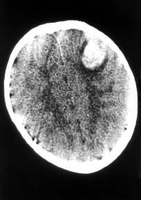
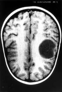
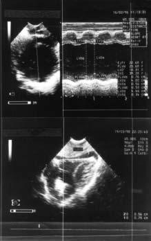
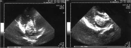
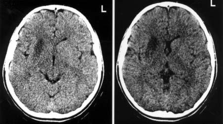
Abstract: The case of an 11 year old girl is presented, who suffered an acute cerebral ischaemic episode due to embolism from an atrial myxoma. The patient, who suffered the episode while dancing, was admitted to the hospital with signs of left hemiplegia, facial palsy and impairment of vision of the right eye. The initial evaluation on admission, which included computerized tomography (CT) of the brain and echocardiography, revealed a small localized area of reduced density at the region of the left internal capsule of the brain, a picture consistent with an old ischaemic lesion, as well as the presence of a left atrial tumor which protruded through the mitral valve into the left ventricle during diastole. A repeat CT scan, 12 hours later, showed a new ischaemic area at the region of the right internal capsule, a picture consistent with the clinical findings of capsular hemiplegia. Nine days after admission, there was significant improvement of the neurological condition of the patient, and allowed the safe surgical removal of the atrial tumor using cardiopulmonary bypass. The tumor consisted of a friable and villous myxoma 4x4 cm in size, originating from the posterior surface of the left atrium above the mitral valve. The patient made an uneventful recovery and was discharged 12 days later. The presence of atrial myxoma is rare during childhood and often has a stormy initial presentation.
Peripheral General Children’s Hospital “Agia Sophia”, Athens
1. Nadas AS, Ellison RC. Cardiac tumors in infancy. Am J Cardiol 1968;21:363-367.
2. Mc Allister HA Jr, Fenoglio JJ Jr. Tumors of the cardiovascular system. In: Hartman WH, Cowan WR, editors. Atlas of tumor pathology. Washington, DC: Armed Forces Institute of Pathology; 1978. p. 432-441.
3. Ha JW, Rim SJ, Chang BC, Chung N, Cho SY. A mitral valve myxoma prolapsing into the left ventricular outflow tract. Clin Cardiol 2001;24:570-573.
4. Pfammatter JP, Iff T, Schupbach P. Imitation of chronic recurrent inflammatory illness by cardiac myxoma. Eur J Pediatr 1996;155:637-639.
5. Salehian O, Demers C, Patel A. Atrial myxoma presenting as isolated unilateral blindness: a case report and review of the literature. Can J Cardiol 2001;17:898-900.
6. Bickford BJ, Egan M, Bryce AG. Surgical treatment of tumors of the heart: a report of two cases. Br J Surg 1956;43:514-517.
7. Dennis MA, Appareti K, Manco-Johnson ML, Clewell W, Wiggins J. The echocardiographic diagnosis of multiple fetal cardiac tumors. J Ultrasound Med 1985;4:327-331.
9. Ota T, Okumura S, Matsuba K, Yamasita M, Ushimaru K. A case of surgically treated left atrial myxoma early after cerebral embolism. Kyobu Geka 1996;49:1094-1096.
10. Waller DA, Ettles DF, Saunders NR, Williams G. Recurrent cardiac myxoma: The surgical implications of two distinct groups of patients. Thorac Cardiovasc Surg 1989;37:226-230.
11. Kigawa I, Fukuda S, Marat D, Tanaka J, Ikeda S, Okiyama M et al. A case of familial cardiac myxoma. Kyobu Geka 1996;49:323-326.
12. Pinede L, Duhaut P, Loire R. Clinical presentation of left atrial cardiac myxoma. A series of 112 consecutive cases. Medicine (Baltimore) 2001;80:159-172.
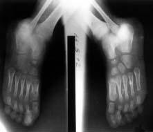
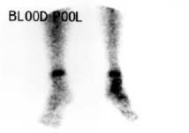
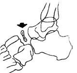
Abstract: Kohler’s disease is a rare disease of the tarsal navicular bone that is characterized by pain and swelling of the affected area, and Ôn radiographic examination, by flattening and sclerosis of the affected bone. It occurs more frequently in boys and there is evidence that it is due to mechanical factors due to compression of the bone before its ossification. Frequently it is associated with abnormal ossification. We describe the case of a 6 year old boy with Kohler’s disease.
3. Karp M. Kohler’s disease of the tarsal scaphoid. J Bone Joint Surg 1937;19:84.
4. Waugh W. The ossification and vascularization of the tarsal navicular and their relation to Kohler’s disease. J Bone Joint Surg 1958;40-B:765.
5. Behrman RE, Kliegman RM, Arvin AM. The foot and toes: osteochondroses. In: Nelson Textbook of Pediatrics. 15th Ed. Philadelphia: WB Saunders Co; 1996. p. 1922.
6. Morrissy RT, Weinstein SL. Osteochondroses. In: Lovell and Winter’s Pediatric Orthopaedics. 4th Ed. LippincottRaven Publishers; 1996. p. 1101.
8. Speed K. Kohler’s disease of the tarsal scaphoid bone. Trans Amer Surg Ass 1927;45:179.
9. Williams GA, Cowell HR. Kohler’s disease of the tarsal navicular. Clin Orthop 1981;158:53.
10. Scaglietti O, Stringa G, Mizzau M. Plus-variant of the astragalus and subnormal scaphoid space, two important findings in ∫ohler’s scaphoid necrosis. Acta Orthop Scand 1962;32:500.
11. England SP. Evaluation and management of pediatric foot deformities. In: The Pediatric Clinics of North America. Common Orthopedic Problems II. 1996;43:1104.
HÌÂÚÔÌËÓ›· ˘Ô‚ÔÏ‹˜: 28-05-2001
HÌÂÚÔÌËÓ›· ¤ÁÎÚÈÛ˘: 06-12-2001
¢È‡ı˘ÓÛË ·ÏÏËÏÔÁÚ·Ê›·˜: ∫ˆÓÛÙ·ÓÙ›ÓÔ˜ ∆ÛÔ˘Ì¿Î·˜
¢È¢ı˘ÓÙ‹˜ ¶·È‰È·ÙÚÈÎÔ‡ ∆Ì‹Ì·ÙÔ˜
¡ÔÛÔÎÔÌÂ›Ô ∫∞∆, ∞ÙÙÈ΋ ¡›Î˘ 2, 145 61, ∫ËÊÈÛÈ¿
17-19 π·ÓÔ˘·Ú›Ô˘ 200220th Annual Sleep DisordersRancho Mirage in Infancy and ChildhoodCalifornia, U.S.A
¶ÏËÚÔÊÔڛ˜: Alice Clark
∆ËÏ.: 760-773-4514
E-mail: registration@annenberg.net
21-23 π·ÓÔ˘·Ú›Ô˘ 2002 31st International SymposiumObergurgl, Austria of Pediatric Surgery
¶ÏËÚÔÊÔڛ˜: Prof. Dr. Ernst Horcher
∆ËÏ.: 431-404-006-837
E-mail: ernst.horcher@akh.wien.ac.at
9-10 ºÂ‚ÚÔ˘·Ú›Ô˘ 2002 3Ë ¶·È‰ÔÓ¢ÌÔÓÔÏÔÁÈ΋
£¤Ì·: “§ÔÈÌÒÍÂȘ Î·È ·ÏÏÂÚÁÈΤ˜
ÂΉËÏÒÛÂȘ ÙÔ˘ ·Ó·Ó¢ÛÙÈÎÔ‡
¶ÏËÚÔÊÔڛ˜: ª. ∞ÓıÚ·ÎfiÔ˘ÏÔ˜-TYPORAMA
∆ËÏ.: (0610) 999 544, 999 612
Fax: (0610) 994 533
18-21 ºÂ‚ÚÔ˘·Ú›Ô˘ 2002 CIPP International CongressNice, France on Pediatric Pulmonology
¶ÏËÚÔÊÔڛ˜: Annie Bidart
∆ËÏ.: 33-0-497-038-597
Fax: 33-0-497-038-598
E-mail: cipp@cipp-meeting.com
8-10 ª·ÚÙ›Ô˘ 2002 7Ô ™˘Ó¤‰ÚÈÔ ÁÈ· ÙÔ∂Ú¤ÙÚÈ·
∂ÏÈÎÔ‚·ÎÙËÚ›‰ÈÔ ÙÔ˘ ¶˘ÏˆÚÔ‡
•ÂÓÔ‰Ô¯Â›Ô HOLIDAYS
¶ÏËÚÔÊÔڛ˜: C&C International SA
∆ËÏ.: 010-68.89.100
21-24 ª·ÚÙ›Ô˘ 2002
29Ô ¶·ÓÂÏÏ‹ÓÈÔ ™˘Ó¤‰ÚÈÔ πˆ¿ÓÓÈÓ·
∂Ó‰ÔÎÚÈÓÔÏÔÁ›·˜ Î·È ªÂÙ·‚ÔÏÈÛÌÔ‡
•ÂÓÔ‰Ô¯Â›Ô Du Lac
∆ËÏ.: 010-77.74.370
23-24 ª·ÚÙ›Ô˘ 2002
£Ú¿Î˘ ¶·È‰È·ÙÚÈ΋
¶ÏËÚÔÊÔڛ˜: Minerva Fairs and Congresses
∆ËÏ.: 010-32.17.456, 32.17.474
Fax: 010-32.20.595
E-mail: minerva@medianet.gr
19-21 ∞ÚÈÏ›Ô˘ 2002
¶·ÓÂÈÛÙ‹ÌÈÔ πˆ·ÓÓ›ÓˆÓ
∆ËÏ.: 06510-97.719
21-24 ∞ÚÈÏ›Ô˘ 2002
VIII International Congress Cannes, France on Immunorehabilitation
“Allergy, Immunology and Global Network”
Congress Secretariat
Tel.: (++7-095) 336-5000
Fax: (++7-095) 336-5000
E-mail: acicis@ibch.ru
Web site: http://www.isir.ru
26-28 ∞ÚÈÏ›Ô˘ 2002
1st Mediterranean CongressLimassol, Cyprus of Neurology