2 minute read
Good to know

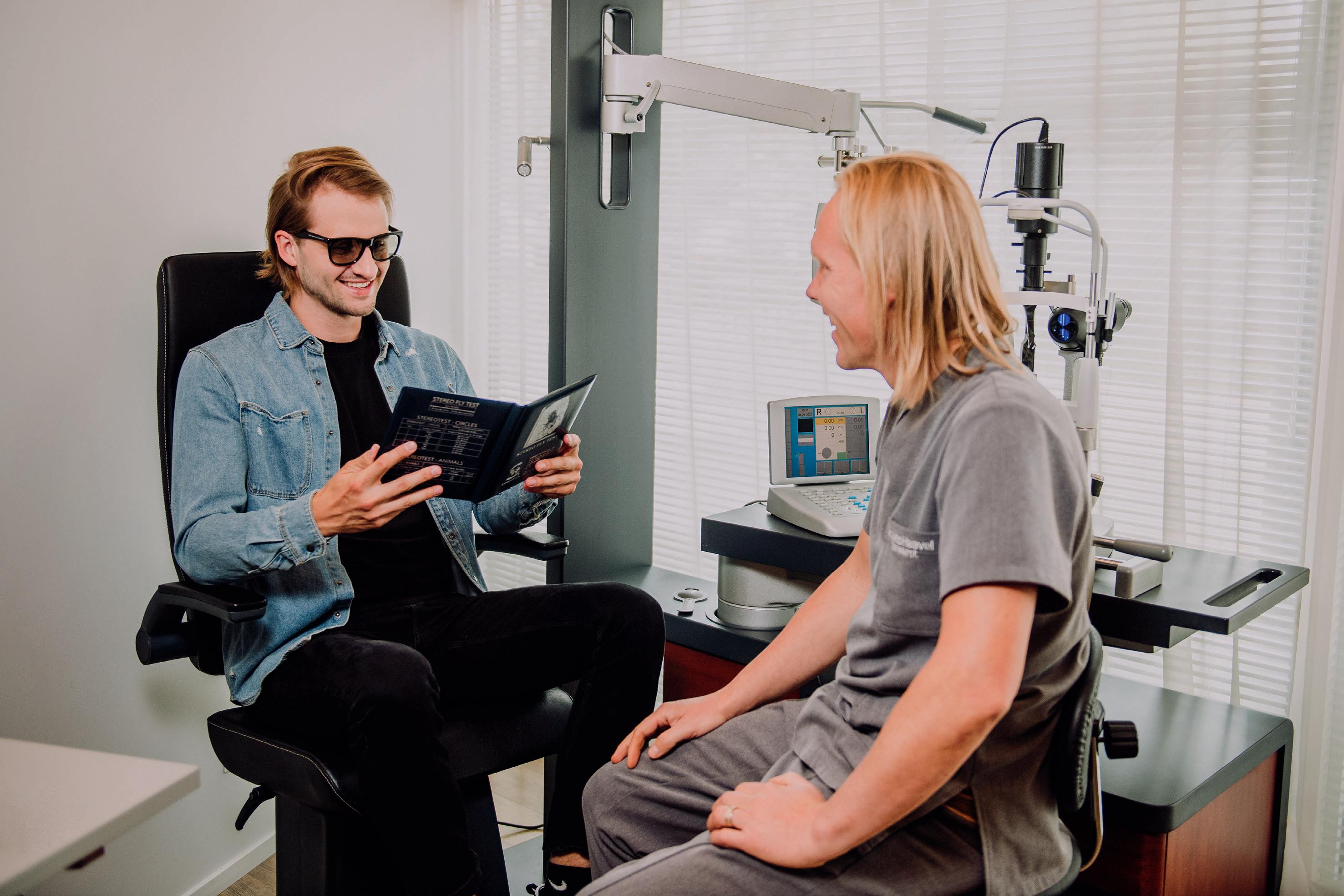
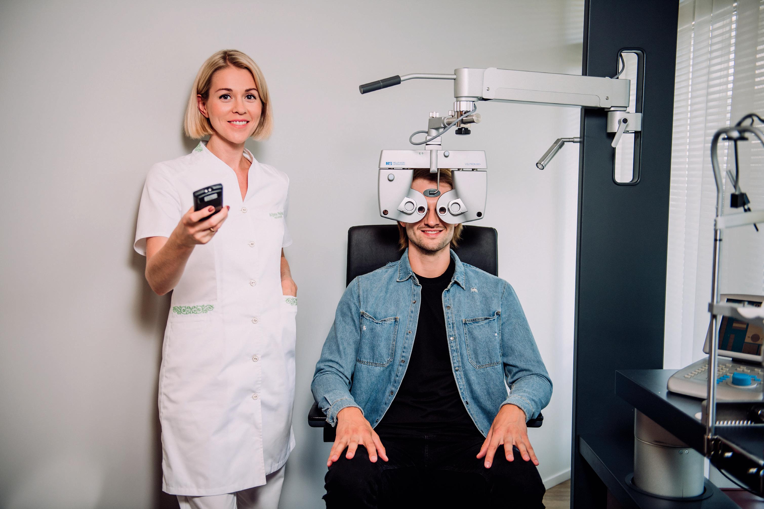
Functioning of the eye and refractive errors: Before undergoing a laser eye procedure it is recommended to know how the eye works. Below is a brief explanation.
An eye is like a camera: the cornea is a clear, curved window that forms the front wall of the eye while the retina is light-sensitive tissue at the back of the eye, which is connected via a network of nerve fibres to the visual centres of the brain. The retina functions like a film in a camera while the cornea acts as a lens that concentrates light on the retina. Upon refraction of light in the eye’s refractive structures, an image appears on the retina, which is forwarded to the brain and later interpreted as visual perception.
Advertisement
The curvatures of the cornea and the intraocular lens determine whether the rays of light that enter the eye from distant objects are concentrated in the macula area of the retina. A refractive error occurs if the eye is not able to focus light exactly on the retina.
Refractive errors are corrected by using a suitable additional lens (e.g. glasses or contact lenses) that converges the light entering the eye exactly on the retina, which results in clear vision.
Refractive surgical procedures with an excimer laser are conducted on the cornea. The cornea is the main transparent refractive structure of the eye. 90 % of the cornea is made up of a tissue that is called the stroma. This is covered by an external layer called epithelium. The shape of the cornea can be changed permanently by polishing the stroma of the cornea with a laser. This improves the cornea’s ability to focus rays of light and results in good and natural visual acuity.
The eye also includes a lens, which also refracts light. The functioning of the lens is similar to that of a camera lens: it is constantly changing the focus depending on whether we are looking at close or distant things. The lens must be elastic in order to function immaculately. The lens loses its elasticity with age and therefore people in their 40s or 50s normally find themselves in a situation where they have difficulty looking at close objects due to the rigidity of the lens. This process is called presbyopia, i.e. age-related far-sightedness. Presbyopic people generally use plus lenses for looking at close objects and reading.
MYOPIA (SHORT-SIGHTEDNESS)
The cornea of a healthy and functioning eye focuses light precisely on the retina, enabling a person to see clearly without glasses or contact lenses. In the case of myopia, the diameter of the eye is a few millimetres longer or the cornea more curved than it should be.
The light concentrates before the retina and is out of focus once it reaches it, forming a blurry image on the fundus. Distant objects seem blurry while close objects may appear clear.
HYPEROPIA (FAR-SIGHTEDNESS)
In the case of hyperopia the eye is shorter or the cornea flatter than it should be. Rays of light converge on a spot behind the retina and are therefore out of focus once they reach the retina. Close objects may seem blurry while distant objects appear more clear. Distant objects may also appear blurry to people with strong hyperopia (more than +2D) and those older than 40 years of age.
Astigmatism
The cornea is normally curved both horizontally and vertically like a football. When rays of light pass through the cornea, they converge in one spot. In the case of astigmatism, the curvature of the cornea is different in either the horizontal or vertical direction, so that it looks like a rugby ball – round on one side and flat on the other. Consequently, the rays of light passing the cornea do not converge in one spot but several. As a result, the vision is distorted and the image on the fundus unclear. Most people with myopia or hyperopia also experience a certain degree of astigmatism.
In all of those cases, the person needs some type of a corrective lens, e.g. glasses or contact lenses, in order to ensure that light is focused correctly. These refractive errors are corrected by using various laser procedure methods.