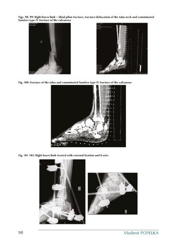
Figs. 98, 99: Right lower limb – tibial pilon fracture, fracture-dislocation of the talar neck and comminuted Sanders type IV fracture of the calcaneus
Fig. 100: Fracture of the talus and comminuted Sanders type IV fracture of the calcaneus
Fig. 101 102: Right lower limb treated with external fixation and K-wire
98
Vladimír POPELKA
Ukázka elektronické knihy, UID: KOS250178
There occurred no wound complications in the patient. K-wires were removed 2 months and external fixator from the right lower leg 4 months postoperatively when radiography showed signs of healing of the fracture of the distal lower leg and the patient was mobilized. The patient was followed up in the outpatient clinic for 2 years. After this period bone union of the right lower leg was confirmed, but non-union of the right talus was noted. As the patient had no problems with weight bearing, the non-union was not treated surgically. The calcaneal fracture healed with a deformity. The left foot showed a healed fracture of the talus but the calcaneal fracture healed with deformity. The position in the talonavicular joint was correct on both sides. Two years after the operation, the patient used close fitting shoes and walked without crutches, with full weight bearing of both lower limbs. Figs. 103 – 105: Outcome of the treatment
15.2 Calcaneal fracture-dislocations It is an unusual injury to the calcaneus, resulting from landing on the foot which is in extreme supination or pronation. Depending on the position of the foot, the posterior articular facet dislocates either laterally or, rarely, also medially. 15.2.1 Lateral dislocation Lateral fracture-dislocations are caused by axial impact in combination with extreme supination of the foot, when the space between the talus and the body of the calcaneus opens and there occurs an extra-articular fracture with the primary fracture line passing between the posterior articular facet and the medial facet, or a fracture involving the medial rim of the posterior articular facet (Sanders type II C fractures). According to the Essex-Lopresti classification, these are tongue type fractures when the entire posterior fragment together with the posterior facet is rotated laterally in the horizontal plane. The other fracture pattern is type III BC, with subluxation of the posterior articular facet, when only the lateral fragment is dislocated and the middle one impacted. The only structure which resists dislocation is the lateral malleolus. With sufficient supination, the fragment misses it. Less opening of the lateral foot, however, results in collision with the distal fibula and fracture of the lateral malleolus, as shown by Figs. 106 and 107. Imaging techniques If only a lateral radiograph is taken in a Sanders type II C fracture-dislocation or extraarticular dislocation, the relative “correct position” may be misinterpreted by an insufficiently experienced surgeon who can see a correct height and length of the calcaneus. The diagnosis can be confirmed only by the axial view and in detail by CT scan.
Treatment of calcaneal fractures
99
Ukázka elektronické knihy, UID: KOS250178
Healing of the fracture in malalignment requires corrective osteotomy. In type IIIB, C, a fracture may be diagnosed also on the lateral radiograph, although an inexperienced surgeon may concentrate only on the fracture of the lateral malleolus and the fracture is confirmed as late as during operation. Treatment In fracture-dislocations, the dislocated fragment exerts pressure on skin and in case of delayed operation there is a risk of development of skin defect. Therefore, these injuries are indicated for emergency surgery. At our institution we use the “Seattle” extended lateral approach to reduce the fracture and its stabilization with plate fixation (Figs. 108 – 109). Figs. 106-109: Lateral subluxation of the posterior articular facet of Sanders type III fracture with fracture of the lateral malleolus after plate fixation of the calcaneus
15.2.2 Medial dislocation Medial fracture-dislocations are rare and result from axial force in combination with extreme pronation of the foot. The medial part of the posterior articular surface is rotated and dislocates medially through the medially opened subtalar joint. There are no reports of this injury in the literature. At our institution we recorded this fracture in Sanders type IIIA, B. Imaging techniques Similarly as in other intra-articular fractures, conclusive evidence is provided by CT examination. Treatment At our institution we treated this injury from the “Seattle” extended lateral approach in combination with the medial McReynolds approach. Reduction was performed using a tibiocalcaneal distractor placed on the medial side. After distraction the medial side of the subtalar joint opened, allowing reduction of the medial fragment and, consequently, of the entire medial wall of the calcaneus. The middle impacted fragment was reduced in the standard way from the lateral approach, namely by retraction of the lateral fragment and its elevation. After radiographic control, internal fixation was performed from the lateral side with a Synthes locking plate (Figs. 110 – 113). At our institution, we treated during the 8-year period a total of 5 fracture-dislocations of the calcaneus, including 4 lateral and one medial dislocation. Lateral fracture-dislocations included two cases of Sanders type IIC and two cases of Sanders type IIIB, C. The medial fracture-dislocation was type IIIA, C. Emergency surgery was performed in all these fractures. Lateral dislocations were treated from the “Seattle” extended lateral approach and the medial dislocation from the extended lateral approach in combination with the McReynolds approach. Fractures were evaluated within the whole group of patients.
100
Vladimír POPELKA
Ukázka elektronické knihy, UID: KOS250178