Preface
ItisourdistinctpleasuretopresentthesecondeditionofMiMB MitochondrialMedicine to theeverincreasingnumberofscientistsandphysicianswhoareasfascinatedbythistiny organelleasweare.WestartedworkingonthefirsteditioninSeptember2014andwereable tobringaboutoneyearlatertwovolumeswithatotalof70chapterstothemarket.Asof today(July2020),195KdownloadshavebeenrecordedforVolumeI1 and90Kdownloads forvolumeII2.InlightoftherapidlygrowingandexpandingfieldofMitochondrial Medicine,wereadilyacceptedtheinvitationtocompileasecondedition,whichwestarted toworkoninMarch2019.Thissecondeditionasofferedhereinvolvesatotalof88chapters with45ofthemwrittenbynewcontributorswhowerenotpartofourfirstedition.Thefirst andsecondeditionscombinedsubsequentlypresentworkfrom115mitochondriallaboratoriesfromaroundtheglobe.Wethereforebelievethesefivevolumescombinedtobethe mostcomprehensivesourceofknow-howinthewide-rangingfieldofMitochondrial Medicine.
Dividing87chaptersequallyoverthreevolumesprovedtobeabitchallenging.We chosethesubtitle TargetingMitochondria forvolumeI, AssessingMitochondria forvolume II,and ManipulatingMitochondriaandDiseaseSpecificApproaches forvolumeIIIwhileof coursebeingwellawareofsignificantoverlapsbetweenthesethreeareasofresearch.For example,itisquiteobviousthatmitochondriaarebeingtargetedforthepurposeofeither assessingthemortomanipulatethem.Wethereforeaskallauthorsnottobetoocritical regardingtheplacementoftheirparticularchapter.Thereaderwebelievewillanyway choosetodownloadachapterofhis/herinterestquiteindependentlyofitsplacementin oneofthethreevolumes.
Allchaptersinthesethreevolumeswerewrittenforgraduatestudents,postdoctoral associates,independentinvestigatorsinacademiaandindustryaswellasphysiciansby leadingexpertsintheirparticularfield.Weareextremelygratefultothemforhaving foundthetimetoeitherupdatetheirchapterfromthefirsteditionortowriteanewchapter. WewillnotforgetthatformanyifnotallofourcontributorstheworldwideCOVID-19 pandemicposedadditionalandunexpectedhurdlestowardsfinishingtheirmanuscriptin duetime.Thankyoutoall!
TheideaforouroriginalbookproposalleadingtothefirsteditionofMiMB MitochondrialMedicine originatedinoureffortstoorganizeaseriesofannualconferenceson TargetingMitochondria(www.targeting-mitochondria.com),thetenthoneofwhichmeanwhilehastakenplaceinNovember2019inBerlin,Germany.Duetotheongoingpandemic, our11thconference(October29–30,2020)willbeavirtualonebutwearesureitwillnot belessexcitingthanallthepreviouseditions.
LastbutnotleastwewouldliketosincerelythankJohnWalker,theserieseditorof MethodsinMolecularBiology,forhavinginvitedustocompilethissecondeditionandforhis
1 https://link.springer.com/book/10.1007/978-1-4939-2257-4.
2 https://link.springer.com/book/10.1007/978-1-4939-2288-8.
unlimitedguidanceandhelpthroughouttheentireprocess.Wealsoowesincerethanksto PatrickMarton,theExecutiveEditoroftheSpringerProtocolSeries,foralwayshavingbeen availableinassistingusthroughouttheentireproject.
Glendale,AZ,USAVolkmarWeissig Paris,FranceMarvinEdeas
Preface.. ...................................................................v
Contributors
1MitochondrialDysfunctioninMitochondrialMedicine:Current Limitations,Pitfalls,andTomorrow.. .....................................1 NaigGueguen,GuyLenaers,PascalReynier, VolkmarWeissig,andMarvinEdeas
2Preparationof“Functional”Mitochondria:AChallengingBusiness...
StefanLehr,SonjaHartwig,andJorgKotzka
3IsolationandQualityControlofFunctionalMitochondria ...................41 SonjaHartwig,JorgKotzka,andStefanLehr
4PurificationofFunctionalPlateletMitochondria UsingaDiscontinuousPercollGradient ...................................57 JacobL.Le´ger,NicolasPichaud,andLucH.Boudreau
5MechanicalPermeabilizationasaNewMethodforAssessment ofMitochondrialFunctioninInsectTissues.. ..............................67 AlessandroGaviraghi,YanAveiro,StephanieS.Carvalho, RodiesleyS.Rosa,MatheusP.Oliveira,andMarcusF.Oliveira
6AnalysisofMitochondrialRetrogradeSignalinginYeastModelSystems .......87 NicolettaGuaragnella,MasˇaZ ˇ dralevic ´ ,ZdenaPalkova ´ , andSergioGiannattasio
7NativeGelElectrophoresisandImmunoblottingtoAnalyze ElectronTransportChainComplexes .....................................103 GiselaBeutnerandGeorgeA.PorterJr.
8MeasuringMitochondrialHydrogenPeroxideLevelsand GlutathioneRedoxEquilibriumin Drosophila NeuronSubtypes UsingRedox-SensitiveFluorophoresand3DImaging .......................113 LoriM.Buhlman,PetrosP.Keoseyan,KathrynHoulihan, andAmberN.Juba
9AssessmentofMitochondrialCellMetabolismbyRespiratory ChainElectronFlowAssays. .............................................129 FlaviaRadogna,De´borahGe´rard,MarioDicato,andMarcDiederich 10Whole-CellandMitochondrialdNTPPoolQuantification fromCellsandTissues ...................................................143 JuanC.Landoni,LiyaWang,andAnuSuomalainen
11Single-ParticleTrackingMethodinFluorescenceMicroscopy toMonitorBioenergeticResponsesofIndividualMitochondria ..............153 CamilleColin,EmmanuelSuraniti,EmmaAbell,AudreySe´mont, NesoSojic,PhilippeDiolez,andSte´phaneArbault
12InvestigationofMitochondrialADP-Ribosylation ViaImmunofluorescence... .............................................165
Ann-KatrinHoppandMichaelO.Hottiger
13AssessmentofMitochondrialCa2+ Uptake... ..............................173
Andra ´ sT.Deak,ClaireJean-Quartier,AlexanderI.Bondarenko, LukasN.Groschner,RolandMalli,WolfgangF.Graier, andMarkusWaldeck-Weiermair
14AssessmentofMitochondrialMembranePotentialandNADH RedoxStateinAcuteBrainSlices .........................................193 AndreyY.Vinokurov,ViktorV.Dremin,GennadiiA.Piavchenko, OlgaA.Stelmashchuk,PlamenaR.Angelova,andAndreyY.Abramov
15EvaluationofMitochondriaContentandFunctioninLive CellsbyMulticolorFlowCytometricAnalysis ..............................203
Hsiu-HanFan,Tsung-LinTsai,IvanL.Dzhagalov, andChia-LinHsu
16AnalysisofMitochondrialDysfunctionDuringCellDeath ...................215 VladimirGogvadzeandBorisZhivotovsky
17ModifiedBlueNativeGelApproachforAnalysisofRespiratory Supercomplexes ........................................................227 SergiyM.Nadtochiy,MeganNgai,andPaulS.Brookes
18Patch-ClampRecordingoftheActivityofIonChannels intheInnerMitochondrialMembrane ....................................235 PiotrBednarczyk,RafałP.Kampa,ShurGałecka, AleksandraSe˛k,AgnieszkaWalewska,andPiotrKoprowski
19AssessmentofMitochondrialProteinGlutathionylation asSignalingforCOPathway .............................................249 AnaS.Almeida,Cla ´ udiaFigueiredo-Pereira, andHelenaL.A.Vieira
203DOpticalCryo-ImagingMethod:ANovelApproach toQuantifyRenalMitochondrialBioenergeticsDysfunction.. ...............259 ShimaMehrvar,AmadouK.S.Camara,andMahsaRanji
21SimultaneousQuantificationofMitochondrialATPandROS ProductionUsingATPEnergyClampMethodology. .......................271 LipingYu,BrianD.Fink,andWilliamI.Sivitz
22High-ThroughputImageAnalysisofLipid-Droplet-Bound Mitochondria ..........................................................285 NathanaelMiller,DaneWolf,NourAlsabeeh,KianaMahdaviani, MayukoSegawa,MarcLiesa,andOrianS.Shirihai
23CellEnergyBudgetPlatformforMultiparametric AssessmentofCellandTissueMetabolism... ..............................305 DmitriB.PapkovskyandAlexanderV.Zhdanov
24Fluorescence-BasedAssayForMeasuringOMA1Activity ....................325 JuliaTobacykandLeeAnnMacMillan-Crow
25StudyingMitochondrialNetworkFormationbyInVivo andInVitroReconstitutionAssay... .....................................333 WanqingDu,XiangjunDi,andQianPeterSu
26ExtractionofFunctionalMitochondriaBasedonMembrane Stiffness... ............................................................343 MdHabiburRahman,QinruXiao,ShiruiZhao, An-ChiWei,andYi-PingHo
27AProtocolforUntargetedMetabolomicAnalysis:From SamplePreparationtoDataProcessing ....................................357 AmandaL.SouzaandGaryJ.Patti
28AMethodforAnalysisofNitrotyrosine-ContainingProteins byImmunoblottingCoupledwithMassSpectrometry ......................383 MatejKohutiarandAdamEckhardt
29InVivoVisualizationandQuantificationofMitochondrial Morphologyin C.elegans ................................................397 R.deBoer,R.L.Smith,W.H.DeVos,E.M.M.Manders, andH.vanderSpek
30AssessingImpactofPlatinumComplexesonMitochondrial Functions.. ............................................................409 SuxingJinandXiaoyongWang
31InSilicoModelingoftheMitochondrialPumpingComplexes withMarkovStateModels.. .............................................425 RogerSpringett
32MonitoringtheMitochondrialPresequenceImportPathway InLivingMammalianCellswithaNewMolecularBiosensor.. ...............441 MaximeJacoupy,EmelineHamon-Keromen,andOlgaCorti Index... ...................................................................453
Contributors
EMMA ABELL • Univ.Bordeaux,INSERM,CentrederechercheCardio-Thoraciquede Bordeaux,U1045&IHULiryc,ElectrophysiologyandHeartModelingInstitute, FondationBordeauxUniversite´,Bordeaux,France;CHUdeBordeaux,Bordeaux,France
ANDREY Y.ABRAMOV • OrelStateUniversity,Orel,Russia;UCLQueenSquareInstituteof Neurology,London,UK
ANA S.ALMEIDA • CEDOC,FaculdadedeCieˆnciaMe´dicas/NOVAMedicalSchool, UniversidadeNovadeLisboa,Lisboa,Portugal
NOUR ALSABEEH • DepartmentofPhysiology,FacultyofMedicine,KuwaitUniversity, KuwaitCity,Kuwait
PLAMENA R.ANGELOVA • UCLQueenSquareInstituteofNeurology,London,UK
STE ´ PHANE ARBAULT • NSysAgroup,Univ.Bordeaux,CNRS,INPBordeaux,ISM,UMR 5255,Talence,France
YAN AVEIRO • FederalUniversityofRiodeJaneiro,InstituteofMedicalBiochemistry LeopoldodeMeis,RioDeJaneiro,RJ,Brazil
PIOTR BEDNARCZYK • DepartmentofPhysicsandBiophysics,InstituteofBiology,Warsaw UniversityofLifeSciences—SGGW,Warsaw,Poland
GISELA BEUTNER • DepartmentofPediatrics-DivisionCardiology,UniversityofRochester, Rochester,NY,USA
ALEXANDER I.BONDARENKO • GottfriedSchatzResearchCenterforCellSignaling, MetabolismandAging,MolecularBiologyandBiochemistry,MedicalUniversityofGraz, Graz,Austria
LUC H.BOUDREAU • DepartmentofChemistryandBiochemistry,UniversitedeMoncton, Moncton,NB,Canada
PAUL S.BROOKES • DepartmentofAnesthesiology,UniversityofRochesterMedicalCenter, Rochester,NY,USA
LORI M.BUHLMAN • ArizonaCollegeofGraduateStudies,MidwesternUniversity,Glendale, AZ,USA
AMADOU K.S.CAMARA • DepartmentofAnesthesiologyandAnesthesiaResearch,Medical CollegeofWisconsin,Wauwatosa,WI,USA
STEPHANIE S.CARVALHO • FederalUniversityofRiodeJaneiro,InstituteofMedical BiochemistryLeopoldodeMeis,RioDeJaneiro,RJ,Brazil
CAMILLE COLIN • NSysAgroup,Univ.Bordeaux,CNRS,INPBordeaux,ISM,UMR5255, Talence,France;Univ.Bordeaux,INSERM,CentrederechercheCardio-Thoraciquede Bordeaux,U1045&IHULiryc,ElectrophysiologyandHeartModelingInstitute, FondationBordeauxUniversite´,Bordeaux,France;CHUdeBordeaux,Bordeaux,France
OLGA CORTI • SorbonneUniversite´,InstitutduCerveau(ICM),InsermU1127,CNRS UMR7225,Paris,France
R. DE BOER • MolecularBiology&MicrobialFoodSafety,SwammerdamInstituteforLife Sciences(SILS),FacultyofScience(FNWI),UniversityofAmsterdam,Amsterdam,The Netherlands
W.H.DE VOS • LaboratoryofCellBiologyandHistology,DepartmentofVeterinarySciences, AntwerpUniversity,Antwerp,Belgium;CellSystemsandImagingResearchGroup, DepartmentofMolecularBiotechnology,GhentUniversity,Ghent,Belgium
ANDRA ´ S T.DEAK • GottfriedSchatzResearchCenterforCellSignaling,Metabolismand Aging,MolecularBiologyandBiochemistry,MedicalUniversityofGraz,Graz,Austria
XIANGJUN DI • InstituteforBiomedicalMaterials&Devices(IBMD),FacultyofScience, UniversityofTechnologySydney,Sydney,NSW,Australia
MARIO DICATO • LaboratoiredeBiologieMole´culaireetCellulaireduCancer,Ho ˆ pital Kirchberg,Luxembourg,Luxembourg
MARC DIEDERICH • CollegeofPharmacy,SeoulNationalUniversity,Seoul,SouthKorea
PHILIPPE DIOLEZ • Univ.Bordeaux,INSERM,CentrederechercheCardio-Thoraciquede Bordeaux,U1045&IHULiryc,ElectrophysiologyandHeartModelingInstitute, FondationBordeauxUniversite´,Bordeaux,France;CHUdeBordeaux,Bordeaux,France
VIKTOR V.DREMIN • OrelStateUniversity,Orel,Russia;AstonUniversity,Birmingham, UK
WANQING DU • StateKeyLaboratoryofMembraneBiology,TsinghuaUniversity-Peking UniversityJointCenterforLifeSciences,SchoolofLifeSciences,TsinghuaUniversity, Beijing,China
IVAN L.DZHAGALOV • InstituteofMicrobiologyandImmunology,NationalYang-Ming University,Taipei,Taiwan
ADAM ECKHARDT • DepartmentofTranslationalMetabolism,InstituteofPhysiology, AcademyofSciencesoftheCzechRepublic,Prague,CzechRepublic
MARVIN EDEAS • Universite´deParis,INSERMU1016,InstitutCochin,CNRSUMR8104, Paris,France;LaboratoryofExcellenceGR-Ex,Paris,France
HSIU-HAN FAN • InstituteofMicrobiologyandImmunology,NationalYang-Ming University,Taipei,Taiwan
CLA ´ UDIA FIGUEIREDO-PEREIRA • CEDOC,FaculdadedeCieˆnciaMe´dicas/NOVAMedical School,UniversidadeNovadeLisboa,Lisboa,Portugal
BRIAN D.FINK • DivisionofEndocrinologyandMetabolism,DepartmentofInternal Medicine,TheUniversityofIowa,IowaCity,IA,USA
SHUR GAŁECKA • LaboratoryofIntracellularIonChannels,NenckiInstituteofExperimental Biology,Warsaw,Poland
ALESSANDRO GAVIRAGHI • FederalUniversityofRiodeJaneiro,InstituteofMedical BiochemistryLeopoldodeMeis,RioDeJaneiro,RJ,Brazil
DE ´ BORAH GE ´ RARD • LaboratoiredeBiologieMole´culaireetCellulaireduCancer,Ho ˆ pital Kirchberg,Luxembourg,Luxembourg
SERGIO GIANNATTASIO • InstituteofBiomembranes,BioenergeticsandMolecular Biotechnologies,CNR,Bari,Italy
VLADIMIR GOGVADZE • DivisionofToxicology,InstituteofEnvironmentalMedicine, KarolinskaInstitutet,Stockholm,Sweden;FacultyofMedicine,MVLomonosovMoscow StateUniversity,Moscow,Russia
WOLFGANG F.GRAIER • GottfriedSchatzResearchCenterforCellSignaling,Metabolismand Aging,MolecularBiologyandBiochemistry,MedicalUniversityofGraz,Graz,Austria
LUKAS N.GROSCHNER • GottfriedSchatzResearchCenterforCellSignaling,Metabolism andAging,MolecularBiologyandBiochemistry,MedicalUniversityofGraz,Graz, Austria
NICOLETTA GUARAGNELLA • InstituteofBiomembranes,BioenergeticsandMolecular Biotechnologies,CNR,Bari,Italy;DepartmentofBiosciences,Biotechnologyand Biopharmaceutics,UniversityofBari“A.Moro”,Bari,Italy
NAIG GUEGUEN • UMRCNRS6015-INSERMU1083,MitoVascInstitute,Universityof Angers,Angers,France;DepartmentofBiochemistryandGenetics,UniversityHospitalof Angers,Angers,France
EMELINE HAMON-KEROMEN • SorbonneUniversite´,InstitutduCerveau(ICM),Inserm U1127,CNRSUMR7225,Paris,France
SONJA HARTWIG • InstituteforClinicalBiochemistryandPathobiochemistry,German DiabetesCenterattheHeinrich-Heine-UniversityDuesseldorf,LeibnizCenterforDiabetes Research,Duesseldorf,Germany;GermanCenterforDiabetesResearch(DZDe.V.), Neuherberg,Germany
YI-PING HO • DepartmentofBiomedicalEngineering,TheChineseUniversityofHong Kong,HongKongSAR,China;CentreforNovelBiomaterials,TheChineseUniversityof HongKong,HongKongSAR,China;ShunHingInstituteofAdvancedEngineering,The ChineseUniversityofHongKong,HongKongSAR,China;TheMinistryofEducationKey LaboratoryofRegenerationMedicine,TheChineseUniversityofHongKong,HongKong SAR,China
ANN-KATRIN HOPP • DepartmentofMolecularMechanismsofDisease(DMMD),University ofZurich,Zurich,Switzerland
MICHAEL O.HOTTIGER • DepartmentofMolecularMechanismsofDisease(DMMD), UniversityofZurich,Zurich,Switzerland
KATHRYN HOULIHAN • ArizonaCollegeofGraduateStudies,MidwesternUniversity, Glendale,AZ,USA
CHIA-LIN HSU • InstituteofMicrobiologyandImmunology,NationalYang-Ming University,Taipei,Taiwan
MAXIME JACOUPY • SorbonneUniversite´,InstitutduCerveau(ICM),InsermU1127,CNRS UMR7225,Paris,France
CLAIRE JEAN-QUARTIER • GottfriedSchatzResearchCenterforCellSignaling,Metabolism andAging,MolecularBiologyandBiochemistry,MedicalUniversityofGraz,Graz, Austria
SUXING JIN • StateKeyLaboratoryofCoordinationChemistry,SchoolofChemistryand ChemicalEngineering,NanjingUniversity,Nanjing,P.R.China
AMBER N.JUBA • ArizonaCollegeofGraduateStudies,MidwesternUniversity,Glendale, AZ,USA
RAFAŁ P.KAMPA • DepartmentofPhysicsandBiophysics,InstituteofBiology,Warsaw UniversityofLifeSciences—SGGW,Warsaw,Poland;LaboratoryofIntracellularIon Channels,NenckiInstituteofExperimentalBiology,Warsaw,Poland
PETROS P.KEOSEYAN • ArizonaCollegeofOsteopathicMedicine,MidwesternUniversity, Glendale,AZ,USA
MATEJ KOHUTIAR • DepartmentofMedicalChemistryandClinicalBiochemistry,2nd FacultyofMedicine,CharlesUniversityinPragueandMotolUniversityHospital,Prague, CzechRepublic
PIOTR KOPROWSKI • LaboratoryofIntracellularIonChannels,NenckiInstituteof ExperimentalBiology,Warsaw,Poland
JORG KOTZKA • InstituteforClinicalBiochemistryandPathobiochemistry,GermanDiabetes CenterattheHeinrich-Heine-UniversityDuesseldorf,LeibnizCenterforDiabetes Research,Duesseldorf,Germany;GermanCenterforDiabetesResearch(DZDe.V.), Neuherberg,Germany
JUAN C.LANDONI • ResearchProgramsUnit,StemCellsandMetabolism,Universityof Helsinki,Helsinki,Finland
JACOB L.LE ´ GER • DepartmentofChemistryandBiochemistry,UniversitedeMoncton, Moncton,NB,Canada
STEFAN LEHR • InstituteforClinicalBiochemistryandPathobiochemistry,GermanDiabetes CenterattheHeinrich-Heine-UniversityDuesseldorf,LeibnizCenterforDiabetes Research,Duesseldorf,Germany;GermanCenterforDiabetesResearch(DZDe.V.), Neuherberg,Germany
GUY LENAERS • UMRCNRS6015-INSERMU1083,MitoVascInstitute,Universityof Angers,Angers,France
MARC LIESA • DivisionofEndocrinology,DepartmentofMedicine,DavidGeffenSchoolof MedicineatUCLA,LosAngeles,CA,USA
LEE ANN MACMILLAN-CROW • DepartmentofPharmacologyandToxicology,Universityof ArkansasforMedicalSciences,LittleRock,AR,USA
KIANA MAHDAVIANI • DepartmentofMedicine,ObesityResearchCenter,BostonUniversity SchoolofMedicine,Boston,MA,USA
ROLAND MALLI • GottfriedSchatzResearchCenterforCellSignaling,Metabolismand Aging,MolecularBiologyandBiochemistry,MedicalUniversityofGraz,Graz,Austria
E.M.M.MANDERS • vanLeeuwenhoekCenterforAdvancedMicroscopy,Universityof Amsterdam,Amsterdam,TheNetherlands
SHIMA MEHRVAR • UniversityofWisconsin-Milwaukee,Milwaukee,WI,USA
NATHANAEL MILLER • DivisionofEndocrinology,DepartmentofMedicine,DavidGeffen SchoolofMedicineatUCLA,LosAngeles,CA,USA;DepartmentofMedicine,Obesity ResearchCenter,BostonUniversitySchoolofMedicine,Boston,MA,USA
SERGIY M.NADTOCHIY • DepartmentofNeuroscience,UniversityofRochesterMedical Center,Rochester,NY,USA;DepartmentofAnesthesiology,UniversityofRochester MedicalCenter,Rochester,NY,USA
MEGAN NGAI • DepartmentofAnesthesiology,UniversityofRochesterMedicalCenter, Rochester,NY,USA
MARCUS F.OLIVEIRA • FederalUniversityofRiodeJaneiro,InstituteofMedical BiochemistryLeopoldodeMeis,RioDeJaneiro,RJ,Brazil
MATHEUS P.OLIVEIRA • FederalUniversityofRiodeJaneiro,InstituteofMedical BiochemistryLeopoldodeMeis,RioDeJaneiro,RJ,Brazil
ZDENA PALKOVA ´ • FacultyofScience,CharlesUniversity,BIOCEV,Prague,CzechRepublic
DMITRI B.PAPKOVSKY • SchoolofBiochemistryandCellBiology,UniversityCollegeCork, Cork,Ireland
GARY J.PATTI • DepartmentofChemistry,WashingtonUniversityinSt.Louis,SaintLouis, MO,USA;DepartmentofMedicine,WashingtonUniversityinSt.Louis,SaintLouis,MO, USA
GENNADII A.PIAVCHENKO • OrelStateUniversity,Orel,Russia;I.M.SechenovFirstMoscow StateMedicalUniversity(SechenovUniversity),Moscow,Russia
NICOLAS PICHAUD • DepartmentofChemistryandBiochemistry,UniversitedeMoncton, Moncton,NB,Canada
GEORGE A.PORTER JR • DepartmentsofPediatrics-DivisionCardiology,Pharmacology andPhysiology,andMedicine(AabCardiovascularResearchInstitute),Universityof Rochester,Rochester,NY,USA
FLAVIA RADOGNA • LaboratoiredeBiologieMole´culaireetCellulaireduCancer,Ho ˆ pital Kirchberg,Luxembourg,Luxembourg
MD HABIBUR RAHMAN • DepartmentofBiomedicalEngineering,TheChineseUniversityof HongKong,HongKongSAR,China;CentreforNovelBiomaterials,TheChinese UniversityofHongKong,HongKongSAR,China
MAHSA RANJI • BiophotonicsLab,FloridaAtlanticUniversity,BocaRaton,FL,USA
PASCAL REYNIER • UMRCNRS6015-INSERMU1083,MitoVascInstitute,Universityof Angers,Angers,France;DepartmentofBiochemistryandGenetics,UniversityHospitalof Angers,Angers,France
RODIESLEY S.ROSA • FederalUniversityofRiodeJaneiro,InstituteofMedicalBiochemistry LeopoldodeMeis,RioDeJaneiro,RJ,Brazil
MAYUKO SEGAWA • DivisionofEndocrinology,DepartmentofMedicine,DavidGeffenSchool ofMedicineatUCLA,LosAngeles,CA,USA
ALEKSANDRA SE˛K • LaboratoryofIntracellularIonChannels,NenckiInstituteof ExperimentalBiology,Warsaw,Poland;FacultyofChemistry,UniversityofWarsaw, Warsaw,Poland
AUDREY SE ´ MONT • Univ.Bordeaux,INSERM,CentrederechercheCardio-Thoraciquede Bordeaux,U1045&IHULiryc,ElectrophysiologyandHeartModelingInstitute, FondationBordeauxUniversite´,Bordeaux,France;CHUdeBordeaux,Bordeaux,France
ORIAN S.SHIRIHAI • DivisionofEndocrinology,DepartmentofMedicine,DavidGeffen SchoolofMedicineatUCLA,LosAngeles,CA,USA
WILLIAM I.SIVITZ • DivisionofEndocrinologyandMetabolism,DepartmentofInternal Medicine,TheUniversityofIowa,IowaCity,IA,USA
R.L.SMITH • AcademischMedischCentrumUniversiteitvan,Amsterdam,TheNetherlands NESO SOJIC • NSysAgroup,Univ.Bordeaux,CNRS,INPBordeaux,ISM,UMR5255, Talence,France
AMANDA L.SOUZA • LifeScienceMassSpectrometryDivision,ThermoFisherScientific,San Jose,CA,USA
ROGER SPRINGETT • CellSpex,Kent,UK
OLGA A.STELMASHCHUK • OrelStateUniversity,Orel,Russia
QIAN PETER SU • InstituteforBiomedicalMaterials&Devices(IBMD),FacultyofScience, UniversityofTechnologySydney,Sydney,NSW,Australia;SchoolofBiomedical Engineering,FacultyofEngineeringandIT,UniversityofTechnologySydney,Sydney, NSW,Australia
ANU SUOMALAINEN • DepartmentofNeurology,UniversityHospital,Helsinki,Finland; NeuroscienceCenter,UniversityofHelsinki,Helsinki,Finland
EMMANUEL SURANITI • NSysAgroup,Univ.Bordeaux,CNRS,INPBordeaux,ISM,UMR 5255,Talence,France
JULIA TOBACYK • DepartmentofPharmacologyandToxicology,UniversityofArkansasfor MedicalSciences,LittleRock,AR,USA
TSUNG-LIN TSAI • InstituteofMicrobiologyandImmunology,NationalYang-Ming University,Taipei,Taiwan
H. VANDER SPEK • MolecularBiology&MicrobialFoodSafety,SwammerdamInstitutefor LifeSciences(SILS),FacultyofScience(FNWI),UniversityofAmsterdam,Amsterdam, TheNetherlands
HELENA L.A.VIEIRA • CEDOC,FaculdadedeCie ˆ nciaMe´dicas/NOVAMedicalSchool, UniversidadeNovadeLisboa,Lisboa,Portugal;UCIBIO,FaculdadedeCieˆncias eTecnologia,UniversidadeNovadeLisboa,Lisboa,Portugal;InstitutodeBiologia ExperimentaleTecnologica(iBET),Oeiras,Portugal
ANDREY Y.VINOKUROV • OrelStateUniversity,Orel,Russia
MARKUS WALDECK -WEIERMAIR • GottfriedSchatzResearchCenterforCellSignaling, MetabolismandAging,MolecularBiologyandBiochemistry,MedicalUniversityofGraz, Graz,Austria
AGNIESZKA WALEWSKA • LaboratoryofIntracellularIonChannels,NenckiInstituteof ExperimentalBiology,Warsaw,Poland
LIYA WANG • DepartmentofAnatomy,PhysiologyandBiochemistry,SwedishUniversityof AgriculturalSciences,Uppsala,Sweden
XIAOYONG WANG • StateKeyLaboratoryofPharmaceuticalBiotechnology,SchoolofLife Sciences,NanjingUniversity,Nanjing,P.R.China
AN-CHI WEI • GraduateInstituteofBiomedicalElectronicsandBioinformatics,National TaiwanUniversity,Taipei,Taiwan
VOLKMAR WEISSIG • DepartmentofPharmaceuticalSciencesandNanocenterofExcellence, MidwesternUniversityCollegeofPharmacyatGlendale,Glendale,AZ,USA
DANE WOLF • DivisionofEndocrinology,DepartmentofMedicine,DavidGeffenSchoolof MedicineatUCLA,LosAngeles,CA,USA;DepartmentofMedicine,ObesityResearch Center,BostonUniversitySchoolofMedicine,Boston,MA,USA
QINRU XIAO • DepartmentofBiomedicalEngineering,TheChineseUniversityofHong Kong,HongKongSAR,China
LIPING YU • DivisionofEndocrinologyandMetabolism,DepartmentofInternalMedicine, TheUniversityofIowa,IowaCity,IA,USA
MAS ˇ A Z ˇ DRALEVIC ´ • FacultyofMedicine,UniversityofMontenegro,Podgorica,Montenegro
SHIRUI ZHAO • DepartmentofBiomedicalEngineering,TheChineseUniversityofHong Kong,HongKongSAR,China;ShunHingInstituteofAdvancedEngineering,The ChineseUniversityofHongKong,HongKongSAR,China
ALEXANDER V.ZHDANOV • SchoolofBiochemistryandCellBiology,UniversityCollegeCork, Cork,Ireland
BORIS ZHIVOTOVSKY • DivisionofToxicology,InstituteofEnvironmentalMedicine, KarolinskaInstitutet,Stockholm,Sweden;FacultyofMedicine,MVLomonosovMoscow StateUniversity,Moscow,Russia
MitochondrialDysfunctioninMitochondrialMedicine: CurrentLimitations,Pitfalls,andTomorrow
NaigGueguen,GuyLenaers,PascalReynier,VolkmarWeissig, andMarvinEdeas
Abstract
Untilrecentlyrestrictedtohereditarymitochondrialdiseases,mitochondrialdysfunctionisnowrecognized asakeyplayerandstrategicfactorinthepathophysiologicalofmanyhumandiseases,rangingfromthe metabolism,vascular,cardiac,andneurodegenerativediseasestocancer.Becauseoftheirparticipationina myriadofcellularfunctionsandsignalingpathways,preciselyidentifyingthecauseofmitochondrial “dysfunctions”canbechallengingandrequiresrobustandcontrolledtechniques.Initiallylimitedtothe analysisoftherespiratorychainfunctioning,theseanalyticaltechniquesnowenlargetotheanalysesof mitochondrialandcellularmetabolism,basedonmetabolomicapproaches.
Here,weaddressthemethodsusedtoassaymitochondrialdysfunction,withahighlightonthe techniquesusedindiagnosisontissuesandcellsderivedfrompatients,theinformationtheyprovide,and theirstrengthandweakness.
Targetingmitochondrialdysfunctionbyvariousstrategiesisahugechallenge,requiresrobustmethodsof evaluation,andshouldbeabletotakeintoconsiderationthemitochondriadynamicsandlocalization.The futureofmitochondrialmedicineisstronglyrelatedtoaperfectcomprehensionofitsdysfunction.
Keywords Mitochondrialdysfunctions,Mitochondriaevaluation,Bioenergetics,Devices, Metabolomics
1Introduction
Thecontributionofmitochondrialdysfunctionsintheonsetand developmentofmanydiseaseshasbeenwidelystudied.These dysfunctionscouldinterferewithmanycellular,metabolic,and homoeostaticfunctions[1].Itisthereforenotsurprisingthat, beyondprimarymitochondrialdisorders,mitochondrialdysfunctionsnowimpactmostareasofmedical.Mitochondrialdysfunction hasbeenfoundinnumerouscommonhumandiseases,including metabolicdiseases[2]suchasobesityanddiabetes,heartdiseases [3],neurodegenerativediseases,suchasParkinson’s,Alzheimer’s
VolkmarWeissigandMarvinEdeas(eds.), MitochondrialMedicine:Volume2:AssessingMitochondria, MethodsinMolecularBiology,vol.2276, https://doi.org/10.1007/978-1-0716-1266-8_1, © SpringerScience+BusinessMedia,LLC,partofSpringerNature2021
[4],orHuntington’s[5],infectiousdiseases[6],inflammation[7], andcancer[8].
Multiplereportshaveshowntheimpactofdysfunctionalmitochondriaontheimmuneresponse.Oneoftheexampleisthe COVID-19pandemiccausedbythecoronavirus(SARS-CoV-2). Manyrecentstudiesrevealedthathumanalveolarepithelialcells withdysfunctionalmitochondriadisplayedincreasedproductionof pro-inflammatorycytokineswhichwerefoundtobeincreasedin COVID-19[9, 10].Recently,weproposedthatnotonlythe intracellularmitochondriadysfunctionisaconsequenceof COVID-19infectionformation[6, 11]butalsothelessexplored extracellularmitochondria(specificallyplateletsmitochondria)may affectbloodcoagulation,clot,andthrombosisformation[10–15].Theseextracellularmitochondriamayrepresentacrucialintercellularmediatorsandmayserveasstrategictherapeutictargetsin COVID-19pathogenesis[11].
Historically,mitochondrialdiseaseshavebeenlargelyaddressed fromthepointofviewofthedefectoftherespiratorychainand thereforeofadefectinenergetichomeostasis.However,mitochondriaarenowrecognizedtoperformmultipleadditionalcellular functionsbeyondenergyproduction.Forexample,electrontransferchain(ETC)activityregulatestheNADH/NAD+redoxstate throughNADHoxidationbycomplexI,[16]whichimpactsto hundredsofcellularreactions[17, 18].Mitochondriaarethemajor sourceofreactiveoxygenspecies(ROS)[19],whichnotonly contributetonormalcellfunctionbutalsoarelinkedtoincreased intracellularoxidativestress.ROSproductionregulatesdifferent signalingpathways,frommetabolicrewiringtoinflammatory responseorcellsurvival[20, 21].Mitochondriaalsointegrate maincellularcatabolicandanabolicpathways.Theyhostnotonly wholeorpartialcomponentsofseveralconvergingcataboliccellular processes,i.e.,glucosemetabolismandthetricarboxylicacid(TCA) cycle,andthe β-oxidationbutalsoseveralbiosyntheticpathways, suchasfolateandsulfurmetabolismorhemebiosynthesis.In additiontogeneratingreducingequivalentsthatfeedthe OXPHOSsystem,theTCAcyclehasnumerousanabolicroles, providingprecursorsforthelipids,proteins,carbohydrates,and nucleotidesbiosynthesis[22].TCAcyclemetabolitesalsoactas signalingmolecules,affectingcriticalcellularprocessesthatcontributetooncogenictransformation,suchasnutrientsignaling, HIF1α-dependentmetabolicreprogramming,orhistoneacetylationanddemethylationbyacetyl-CoAand α-ketoglutarate,respectively[23].Mitochondriaarealsoinvolvedincalciumhomoeostasis [24],stressresponseandqualitycontrol[25],andinitiationof caspase-dependentapoptosis[26].
Developingprecisetechnologiestostudymitochondrialphysiologyisgettingmuchmoreimportantastheprevalenceofcommonandinheritedmitochondrialdiseaseincreases.Targeting
mitochondrialdysfunctionbyvariousstrategiesisahugechallenge andneedarobustmethodsofevaluation[27].
Here,weaddressthetechniquescommonlyusedinclinical diagnosis,theinformationthatcanbeinferredfromthem,their limitations,pits,andpitfalls.
2MethodstoAssessMitochondrialDysfunctions
2.1Basis inBioenergetic
Principlesofmitochondrialbioenergeticshavebeenlargely reviewedoveryears[28];therefore,onlyarapidoverviewwillbe givenhere.Mitochondriaaresurroundedbytheouter(OMM)and inner(IMM)membrane.Theinnermembraneislargelyimpermeantandconstitutesthemajorbarrierbetweenthecytosoland themitochondrialmatrix.IMMformsmultiplecristae,whichhost theETCcomplexesandtheenzymesinvolvedinATPsynthesis,the F0F1-ATPsynthase,theadeninenucleotidetranslocator(ANT), andtheinorganicphosphate(Pi)transporter(PiC).
TheETCsystemiscomposedofmultisubunitsenzymesfunctionallyandphysicallylinkedtogether,thecomplexI(CI,NADH ubiquinonereductase),complexII(CII,succinateubiquinone reductase),complexIII(CIII,ubiquinolcytochromecreductase), andcomplexIV(CIV,cytochromecoxidase),whichtransfersthe electronenergeticpotentialfromNADH/NAD+(CI)and FADH2/FAD+(CII)totheelectrochemicalprotongradient knownastheprotonmotiveforce(Δp).Thisprocessinvolveda seriesofoxidoreductasereactionsinwhichelectronsflowsequentially“downhill”alongtheETCfromareducedtoanoxidized state,endingtomolecularoxygenreductiontoawatermolecule,or “respiration.”Thereleaseoffreeenergyduringelectrontransfer drivestheprotonpumpingacrosstheIMMatcomplexesCI,CIII, andCIV,inturnproducingthe Δp.Otheroxidoreductasesofthe ETCareunableofprotonpumping,particularlythecomplexII, theglycerolphosphatedehydrogenase,andtheelectrontransferringflavoproteinquinoneoxidoreductase(ETF)linkedto fattyacid β-oxidation.Thus,tenprotonsareextrudedforeach electronpairtransferredfromNADHtooxygen,orsixprotons foreachelectronpairtransferredfromFADH2toO2,meaningthat theH+/O2andATP/O2 stoichiometriesdifferaccordingtothe initialsubstrates.
Thefuelingofreducingmolecules,namelyNADHand FADH2,totheETCismainlyensuredbytheTCAcycleandthe β-oxidationpathways.Accordingtotheanapleroticrouteswhich feedTCAortherelativecontributionofTCAor β-oxidation,the resultingNADH/FADH2ratiodiffers,ultimatelymodifyingthe efficiencyofATPsynthesis.Thesubstrateoxidationmoduleconsistsofallthesereactionsinvolvedinsubstratemetabolismsand electrontransport,finallygenerating Δp.
2.2Analyses
ofMaximalETC Activities
The Δpiscomposedofthecharge(Δψm)andchemical(ΔpH) components.Theenergyavailablein Δpdrivesthesynthesisand transportofATP,astheprotonsreturntothematrixthroughthe ATPsynthase[29].TheATPsynthesismoduleincludes,inaddition totheATPsynthetase,theANTthatexchangetheADP/ATPand thePiC.The Δpiscentraltothephysiologicalfunctionsofmitochondria,connectingthesubstrateoxidationmoduletotheATP synthesis.Undermostcircumstances,protonfluxistightlycoupled toOXPHOS.However,the Δpcanalsobedissipatedbyproton leak[30],inwhichprotonsreturnintothematrixindependentlyof ATPsynthesis,orbeusedfortransportprocessesacrosstheIMMof metabolite,ions,orcalcium[31].
Thetotalprotonentryandextrusionareexactlybalanced understeady-stateconditions.ForwardfluxthroughETCcomplexesrequiresathermodynamicdisequilibrium,i.e.,thefree energyavailablefromelectrontransfermustbegreaterthanthat requiredtopumpprotonsagainstthe Δp.The Δpvariationsthus “dictate”theETCactivity:anydecreaseinthe Δpisfollowedbyan increaseinelectrontransferandprotonpumping.Inotherwords, thenetfluxoftheprotoncurrentisreflectedbytherateofmitochondrialoxygenconsumption.Decreaseinthe Δpresultsfrom eitherprotonleakorATPsynthesis,whichisregulatedbythe cellularATPdemand.
Anyalterationinonetheabovedescribedprocessesleadstoa mitochondrialdysfunction.Subsequently,weshalldescribethe conventionalmethodsfordissectingtheseprocesses,andidentifyingtheprimarysiteofimpairment,startingwiththemethodsused inthelaboratoryforthebiochemicaldiagnosisofmitochondrial diseases.Acompletesetoftheseanalyseswilldrawapictureofthe mitochondrialenergy-generatingsystem.
Evaluationofindividualrespiratorychaincomplexactivitiesisa routinebiochemicalapproachforthediagnosisofmitochondrial disorders.AssaystoquantifyCI,CII,CIII,CIV,andATPsynthase enzymaticactivitiesareperformedeitherontissue,mostlymuscle orliverbiopsies,oroncellsasprimaryfibroblastsorlymphocytes. Theyrequirethepreparationoftissuehomogenates,afterthe eliminationofcelldebrisandnuclei,orcelllysates,fromeither freshorfrozensample.However,frozensamplesaremorecommonlyusedbecausefreshsamplesmustbeprocessedimmediately aftersampling.
Isolatedactivityofeachcomplexcanbeanalyzedbyfollowing theoxidation/reductionofspecificsubstratesorsubstrateanalogs byspectrophotometry.Thespectrophotometricenzymeassaysare: l NADH:ubiquinoneoxidoreductase(NUR,EC7.1.1.2,NADH oxidationfollowedat340nm)forCI.
l Succinate:ubiquinoneoxidoreductase(coupledto 2,6-Dichloroindophenolreductionfollowedat600nm)for CII(SUR,EC1.3.5.1).
l Succinate:cytochromecoxidoreductase(CII+CIII,cytochromecreductionat550nm).
l UbiquinolcytochromecoxidoreductaseforCIII(UCCR,EC 1.10.2.2,cytochromecreductionat550nm).
l Cytochrome c oxidaseforCV(COX,EC1.9.3.1,cytochromec oxidationat550nm).
ThemeasurementofcomplexV(oligomycin-sensitiveATPase) ismorechallengingandrequiresmitochondrial-enrichedfractions andisoftenmeasuredinculturedskinfibroblasts.Citratesynthase activityisoftenusedfornormalizationofrespiratorychaincomplex activitiestomitochondrialmass.TheactivitiesnormalizedtoCS and/ortheratiosbetweenmitochondrialenzymesgiveamuch narrowerrangeofnormalvaluescomparedtoactivitiesexpressed withrespecttosampleproteincontent,sincethemitochondrial massarehighlyvariableamongindividualsandsubjectedtomitochondrialbiogenesisregulation.
Whiletheprinciplesofthedifferentprotocolsaresimilar,nowadays,thereisnouniversalassayforspectrophotometricquantificationofETCenzymeactivities.Accordingtolaboratories,assays differwithrespecttobufferconditions,reactiontemperatures, substrateconcentrations,supplementationinCa2+ chelator (EDTA),oradditionofbovineserumalbumin.However,inan attempttofacilitateinterlaboratorycomparisonoftheresults,some referencecentersforthediagnosisofmitochondrialdiseaseshave reevaluatedandstandardizedtheirprotocols[32].
Accurateenzymatictestingoftissueandcellsamplescanbe hamperedbydifferentpitfalls.Afrequentcauseforproblemsis samplecollectionandhandling.Foroptimalpreservationofmitochondrialenzymes,samplesmustbeimmediatelysnap-frozenafter collectionandstoredat 80 C,withoutanythawinguntilanalysis. Otherartifactscomefromlocalanestheticcontaminationofthe sample,particularlylidocaine[32, 33],or,forstudiesonanimals, fromeuthanasiaproceduresusingCO2.Furthermore,oneofthe maindifficultiesinachievingreliableandreproducibledosagesis thequalityofthereagents,withstrongvariabilitybetweenreactive referencesorevenbatchesofreactive.Thus,thesedosagesrequire theinclusionofqualitycontrolsineachseries,inordertovalidate thereactionmix.Thisqualityisusuallyobtainedfromculturecells oranimaltissue[34, 35],sincethehumantissuesarelimited.
Inaddition,assaysthatdeterminetheamountsofOXPHOS complexes,suchasbluenative(BN)gelelectrophoresisfollowedby Westernblotanalysis,shouldbeperformedtodecipheranETC defectrelatedtodecreasedcatalyticactivityorinvolvingalower enzymequantity.
2.3Structural
Analyses:BN-PAGE
ETCenzymesareallmultimericandencodedbyboththemtDNA andnDNA,exceptforcomplexIIthatisfullyencodedbythe nucleargenome.Theirassemblyintoafunctionalcomplexrequires anintricateprocessassistedbychaperones[36].Nowadays,the mechanismsofassemblyforeachcomplexarealmostcompletely resolved[36, 37].Theseassemblyprocessesrequirethestriking coordinationofintra-andextramitochondrialtranscription,translation,proteinimportintomitochondria,andproteinfoldingand incorporationintoassemblyintermediates,uptothefinalcomplex assembly.Anydisturbanceinoneoftheseprocesseswouldalter complexesassemblyandcompromisedETCfunctionandenergy production.Mutationsthatimpairthisassemblyprocessarea frequentcauseofmitochondrialinheriteddiseases[38, 39].Complexmisassemblyisnotonlyassociatedwithanincreasingnumber ofmutationsinETCsubunitgenesorchaperones[40]butCI misassemblyhasalsobeenrecentlyobservedinneurodegenerative diseasessuchasAlzheimer’sandParkinson’sdiseases[41],thus broadeningthespectrumofassemblydefect-associateddiseases, andcouldbeimpliedintheageingprocess[42].
Finally,assemblyofCI,III,andIVtogetherleadstorespiratory supercomplexes,addingalayerofcomplexityinthestructural organizationoftherespiratorychain.Thecrystalstructureofthe supercomplexhasrecentlybeenelucidated[43].Althoughthe pathwaysthatleadtotheirformationandtheirfunctionarestill notcompletelyclear[37, 44],theyarenowrecognizedasthefinal functionalETCunit,or“respirasome.”
“BlueNative”polyacrylamidegelelectrophoresis(BN-PAGE) [45]isarelativelyeasyapproachforanalyzingtheassemblyand abundanceofOXPHOScomplexesforthediagnosisofmitochondrialdiseases[46, 47].Theassemblyprofileprovidesinformation foridentifyingdisease-causingmutationsandfacilitatesmolecular investigationbyhighlightingpotentiallyinvolvedsubunits [39, 48].
BN-PAGEisperformedonisolatedmitochondriaorenrichedmitochondrialfractionsfromeithertissuesorcells.Theprotocols aredescribedintheliterature[46, 47, 49].However,itshouldbe stressedthatresultsdependonthechoiceandconcentrationofthe detergentandonthechoiceoftheantibodies.Aftermitochondrial isolation,themitochondrialmembranesaresolubilizedusingnonionicdetergent.Becausemitochondrialmembraneshavelowcholesterolbuthighcardiolipincontents,[50],digitoninis preferentiallyusedforsupercomplexesanalysiswhile β-dodecylmaltosideshouldbeusedforisolatedcomplexvisualization.However,boththeconcentrationandthedetergenttoproteinratiomustbecarefullyoptimizedtoensurethatdetergent concentrationisabovecriticalmicelleconcentrationbutbelow thedetergent:proteinratiothatwouldsolubilizethecomplexboundcardiolipin,whicharecriticalfortheirstabilityandactivity.
2.4Functional Analyses: RespirationRates
Inaddition,aftertransferoftheelectrophoreticgelsforWestern blotting,thechoiceoftheantibodieswilldeterminewhetherthe quantificationoftheabundanceofthefullyassembledcomplexis privilegedorthedetectionofassemblyintermediates.Inprinciple, anysubunitcanbetargetedandwouldallowthedetectionofthe assembledcomplex,providedthattheepitopeisaccessibleforthe antibody.However,accordingtothelocationofthesubunitwithin thecomplexandtheincorporationstepduringtheassemblyprocess,theassemblyintermediateswhichcouldbedetectedandtheir numberwillvary(Fig. 1).Moreover,duetotherelativelylow dynamicrangeofchemiluminescencedetection,thehighintensity ofthesignalproducedbytheholoenzymecouldbluntotherbands oflowerabundance,astheassemblyintermediates.Toaddressthis issue,thedetectionofassemblyfactorsthatspecificallybindintermediates,butdetachoncetheholoenzymesarefullyassembled shouldbepreferred.Inthiscase,onlytheassemblyintermediates willbevisualized.AnexampleofCIassemblyanalysisusingantibodiestargetingassemblyfactorsisillustratedinFig. 1.
Itisthereforehighlyrecommendedtomastertheassembly pathwayofthecomplexofinterestandthelocationofthedifferent subunitsandassemblyfactorswithinthecomplexstructure/intermediatesbeforeperformingaBN-PAGEanalysis.
However,anormalrespiratorychainenzymeactivity/assembly doesnotexcludeafunctionalimpairmentoftherespiratorychain functioning,whichmaynotbedetectablebyenzymaticmeasurementsorassemblyanalyses.
RespirationstudiesareanefficientwaytoanalyzeETCandmetabolicactivitiesofcellsortissues.Thelast,irreversible,stepof electrontransferalongtheETCisthetransfercatalyzedbythe CIVoffourelectronstoamoleculeofoxygentogeneratetwo moleculesofwater.Thecouplingbetweenelectrontransport(oxidation)andprotonpumpingwithinETCandthetightcoupling betweenoxidationandphosphorylationthroughthe Δp(51,52) meanthatthemitochondrialrespirationrateisanaccuratemeasure ofthetotalETCactivityandmitochondrialATPsynthesisrate. Accordingtotheexperimentaldesign,informationcanbegained onmultipleprocessesrequiredforrespiration,includingsubstrate transportintothemitochondria,reducingequivalentproduction byTCAcycleorbeta-oxidationandelectrondeliverytotherespiratorychain,activitiesofthedifferentcomplexes,ATPsynthesis, protonleak,andmitochondrialmetabolism.Beyondmetabolic analysis,O2 consumptionisnowalsousedtoanalyzecytotoxicity.
2.4.1Devices
TherateofmitochondrialO2 consumptioncouldbedeterminedby anumberofmethods,thetwomainapproachesareamperometric O2 sensors[51]andO2-dependentquenchingofporphyrin-based phosphors[52].Theamperometricapproach,notablydeveloped
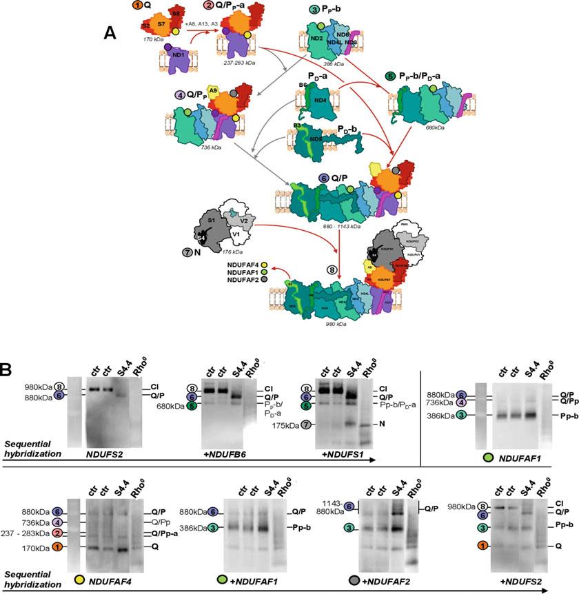
Fig.1 DetectionofCIassemblyintermediatesbytargetingtheCIassemblyfactors.(a)SchematicrepresentationofthemodularassemblyofCITheCIholoenzymeresultsfromthesequentialassemblyoften intermediates(A.I.),assistedbyatleast13CIspecificassemblyfactors.TheQearliestintermediateis composedoftheNDUFS2,NDUFS3,NDUFS7,NDUFS8,andNDUFA5,stabilizedbytwospecificassembly factors,namelyNDUFAF3and NDUFAF4 ina170kDaintermediate(1).ThebindingofQmoduletotheND1 subunitandaccessorysubunitsformsthe237to283kDa(2)intermediatesnamedQ/PP-a, whicharestacked totheinnermembranebyNDUFAF5andNDUFAF6.ThePmoduleisassembledinfourdistinctintermediates, twodistal,namedPD-a andPD-b thatcontainND4andND5subunits,respectively,andtwoproximal,thePP-a andPP-b.ThecentralPP-b intermediate(3)istheentrypointforfourofthesevenMT-DNA-encodedsubunit, i.e.,ND2,ND3,ND6,andND4L,andisboundbysixassemblyfactors,amongwhich, NDUFAF1.Then,the differentmodulesprogressivelycombine(4,5),formingaQ/Pintermediatestackedwiththeassemblyfactor NDUFAF2(6).Inthelaststepoftheprocess,the160kDaNmodule(7),constitutedbythecatalyticNDUFV1, NDUFV2,NDUFS1,andtheaccessoryNDUFA2andNDUFS4subunits,isadded.Finally,oncetheCIassembly iscompleted,alltheassemblyfactorsarereleased,leavingafunctionalholoenzymeof980kDa(8).Nuclear-
TheO2K® Oxygraph
byOroborosInstrument,hashistoricallybeenthemostcommon forinvitroandinvivoinvestigationsofmitochondrialrespiration. Nevertheless,phosphorescentprobesaregaininginterestssincethe introductionoftheXFExtracellularFluxAnalyzerbySeahorse Bioscience[53].
TheO2K® oxygraphdevelopedbyOroborosmeasuresoxygen consumptionbypolarographywithaClark’selectrode.Briefly, oxygendiffusesthroughaTeflonmembrane,whichispermeable tounchargedgases,butnottowater.Aplatinum/silver/KClcoupledelectrodereducesoxygenandoxidizessilver,givingrise toacurrent,whichisproportionaltooxygenconcentrationwithin thephysiologicalrangeofmeasurements.Itoperatesinaclosed chamberof2ml,withastirringmagnettohomogenizeoxygenand biologicalmaterial.Thevolumecanbeadjustedaccordingtothe cells,tissues,ormitochondriaconcentrationstoanalyze. Highresolution designsofOroborosInstrumentsareoptimizedfor highsensitivity,precision,andminimalmeasurementinterferences (typicallythedetectionlimitis~1pmol/sec/mlandthequantificationlimitis~2.5pmol/s/ml,onsiteevaluations).
Fig.1 (continued)encodedsubunitnameswereshortenedbyomittingtheleading“NDUF.”Onlythemain subunitsareindicated.Thenumbersrefertothecorrespondingintermediatesthatcanbedetectedby BN-PAGEinpanel b.(b) StudyofCIassemblybyBN-PAGE intwocontrolfibroblasts(Ctr),onepatientcell linewithaknownassemblydefect[48]andcellslackingMT-DNA(143BRho0).(b1)First,CIassemblywas analyzedusing“classical”antibodiestargetingCIstructuralsubunitslocatedindifferentintermediates,i.e., NDUFS2(Q,leftpanel),NDUFB6(PD-a,middlepanel),andNDUFS1(N,rightpanel).InCtrcells,anti-NDUFS2 antibodyallowedthedetectionofthefullyassembledholoenzyme(8),whileQ/PA.I.(6)accumulatedinthe NDUFS4mutatedcells.NoCIholoenzymeorA.I.couldbedetectedinRho0 FurtherhybridizationwithantiNDUFB6andNDUFS1antibodieshighlightedthepresenceofthePp-b/PD-aat~700kDa(5)andtheNmodule (7),respectively.(b2)A.I.werethenanalyzedbytargetingtheassemblyfactorNDUFAF4(Q),NDUFAF1(Pp-b), andNDUFAF2(Q/P).ThedetectionofNDUFAF1revealedamainbandat~400kDawhichmatchedwiththe describedsizeforthePp-bintermediateandtwohigherfaintbandsjustbelowandabove800kDathat matchedtheQ/PpandQ/Pintermediates,respectively.Asexpected,theseintermediatescontainingthe MT-DNAencodedsubunitswerenotobservedinRho0 cells.NDUFAF4antibodydetectedtheexpectedbands forQ-containingintermediates,i.e.,amainoneforQat170kDa(1)andfainteronesforQ/Pp-a(at~240kDa and~280kDa,(2)),Q/Pp(~750kDa,(4),andQ/P(~880kDa,(6)).Asexpected,onlythesmallestQ intermediates,constitutedonlyofnDNA-encodedsubunits,weredetectedinRho0 cells.ThePp-bintermediatewasdetectedonthesamemembranebyhybridizinganti-NDUFAF1antibody.Finally,additionalhybridizationwithNDUFAF2antibodyclearlyhighlightedtheaccumulationoftheQ/P(6)thatoccurredinNDUFS4cells andhighlighteddifferenthighermolecularweightsupto~1100kDaforthisintermediate.Thus,sincetheydo notbindtheCIholoenzyme,whoseintensesignalcanbluntthepresenceofA.I.,thetargetingoftheassembly factorsallowsamoresensitivedetectionoftheseintermediates.Thetwocombinedapproachesallowthe identificationofsevenoftheassemblyintermediatesinadditiontotheholoenzyme
Thisisallowedby(1)closed,air-tightreactionchambers; (2)highsensitivityofthesensorswithminimalnoise/signalratio; and(3)lowO2-permeablematerials,whichminimizesO2 backdiffusionandoverestimationsofrespiration.Inthesesystems, instrumentalbackgroundO2 consumption(i.e.,nonbiologicalO2 flux,whichconstitutespotentialsourcesofsystematicerror)canbe correctedforaccuratedeterminationsofO2 consumption.The devicecanbefullycalibrated,bothfor0and100%oxygensignals andoxygenconcentrationwithintherespiratorymediumaccordingtotheactual PO2 andO2 solubility.Therefore,real-timeabsolutequantitativemeasurementsofoxygenconcentrationandfluxes canbemeasured,androbustcomparisonofinter-assaysachieved. Normalizationoffluxestoproteincontentorcellconcentration canbeeasilyachievedbyrecoveringthesampleswithinthechamber attheendoftheexperiment.
O2K® oxygraphsareafullyopensystem,allowingasmany injectionsofsubstratesorinhibitorsasneeded,andreoxygenation duringtheexperimenttime-courseifneeded,thusenabling extendedsubstrate–uncoupler–inhibitor–titrationprotocolstobe applied.EachcomplexoftheETCcanbestudiedindependently, onthesamesample,usingdifferentsubstratecombinations.
ThecapabilityoftheO2k® systemhasrecentlyexpanded beyondrespirationtopermitsimultaneousfluorescence-based measurements,potentiometricmeasurementsofH+concentrations(i.e.,pH),of ΔΨmusingtriphenylphosphoniumorfluorescentmembranepotentialprobes,andCa2+,aswellasamperometric measurementofnitricoxide.
Therearehowevermainlimitationswhenconsideringtheuse ofOroborostechnology:First,theO2k® isnotahigh-throughput technology,asonlytwosamplescanbeperformedatonetime,and itisnotautomated.Becauseanalysisusingsubstrate–uncoupler–inhibitor–titrationprotocolstakeapproximatelyonehourtocomplete,large-scaleanalysesaretime-consuming.Second,analysis requirescelldissociationandsuspension.Asmostcellculture–basedstudiesareconductedonadherentcells,thissystemshows limitswhentheexperimentalobjectiveistoassesscellularbioenergeticsinintactculturedcells.Accordingtocelltypes,celldetachmentfromtheextracellularmatrixcouldaltermetabolism[54],in anyway,analysesmustbeperformedimmediatelyandachieved rapidlyaftercelldetachment.Third,despitethehighsensitivity, therelativehighvolumeofthechambers(500 μlto2ml)imposesa nonnegligiblesamplequantity.Typically,10–40 μgofisolated mitochondria,1–2mgpermeabilizedtissueor2–6·106 cells, accordingtocelltypes,areneededforoneexperiment.
TheSeahorse® XFExtracellularFluxAnalyzertechnologyisbased onsolidstatesensorprobesfordetectionofoxygenandH+concentrations,residing200 μmabovethecell/samplemonolayerona multi-wellmicroplateformat.Real-timemeasurementsofoxygen concentrationsaremadebyisolatingasmallvolumeofabout2 μlof mediumaboveamonolayerofsamplesina“transientmicrochamber”withinthemicroplate.Thetechnologybasedonfluorescence quenchingisquitesensitiveandhasbeenoptimizedtoavoidsignal drifting.
ThemainadvantageofSeahorse® technologyreliesonits multi-wellsplateformatandautomatedsystem,allowing highthroughputanalyses.Moreover,thesmallchambervolumeisa greatbenefitwhenlimitedmaterialisavailable.Finally,thissystem representedatechnicalbreakthroughforthefieldofbioenergetics byprovidingthefirstinstrumentwiththecapacitytomeasure invivoO2 consumptiononintactadherentcellsinculture.The XFAnalyzerminimizesthepotentiallimitationofoxygendiffusion foradherentcellsbytransientlylimitingthevolumeofsolutionin whichO2 ismeasuredtojustabovethemonolayerofculturedcells. Moreover,thecapabilityofthesystemexpandsbeyondrespiration andallowssimultaneousmeasurementsofH+extrusion(i.e.,pH), asareflectofglycolyticrates.
However,therearemainlimitationstoconsiderwhenanalyzingmitochondrialrespirationusingtheSeahorse® Analyzer.First, asformanyminiaturizedsetups,high-throughputmethods,the devicelosesinaccuracywhatitgainsinrapidity.Thebiosensorsare calibratedfor100%oxygen,usingthesuppliersolution,notinthe respiratorymediumused.Thecalibrationproceduredoesnotconsidertherealoxygenconcentrationwithinthemedium(oxygen solubilitywithinthemedium,realbarometricpressureduringthe experimentalcourse).However,thetemporarymicroenvironment createdoverthecelllayerisnotcompletelysealedfromatmospheric O2;thus,asO2 concentrationdeclineswithtime,O2 gradient favorsdiffusionfromthemediatothemicro-chambers.O2 gradientcouldalsofavorback-diffusionfromtheplasticofthewells.As thedeviceback-diffusionofoxygenisnotquantifiedandbackgroundcorrectionisperformedrelativetothe“backgroundplate noise”determinedondifferent“control”wells(devoidofsample), thiscouldleadtoanunderestimationofcellularrespirationrate. Thus,thissystemallowsquitequantitativedeterminationofO2 variation,butitdidnotallow absolute quantitativemeasurementsof O2 concentrationandfluxes.Second,inherenttothetechnologies basedonfluorescencequenchingisalsothequitehighvariability betweenthedifferentacquisitions(intra-assayvariability(intraplate,inter-sensors)andmorestrikingly,inter-platesvariability) [55].Thislimitsthecomparisonbetweendifferentexperiments. Third,theSeahorse® setupisnotanopensystem,andonlyfour portssurroundingthesensorareavailablethatmustbeloaded
TheSeahorse® XF
2.4.2Permeabilized TissuesandCells
beforestartingtheanalysis.ThisstronglylimitstheETCsubstrates/inhibitorstitrationprotocols.Itisessentialtoperform robustoptimizationstepspriortoanyrespiratoryassay:theoptimal concentrationofeachinjectablereagentmustbecheckedforeach experimentalcondition.Caremustbealsotakennottooverload thewells,whichcancauseO2 toquicklybecomedepletedunder phosphorylatingconditions,leadingtohypoxia.Finally,thisdevice requiresthatsamplesadheretothebottomofthewell,which correspondstoanadvantagewhenworkingonadherentcells,but couldrequirecoatingofthesamplesinotherconditions.
Toresume,whenaccuratemeasurementofO2 consumption andhighversatilityisaimed,asfordiagnosispurpose,highresolutionO2K® respirometrysystemshouldbeused.However, ifhigh-throughputdosagesareneededasforthescreeningof drugs,orwhenthesamplequantitiesarelimited,theSeahorse® offersamuchhigherthroughputplatformthanthetraditional Clarkelectrode-basedsystems.
Withintissuesorcells,mitochondriaarenotaccessibleformany substratesandinhibitors,andthelargecatabolicprocessesthat mustbeconsideredwhendissectingETCactivityandmitochondrialmetabolismgreatlycomplicatedtheunderstandingofthe results.
Therefore,Isolationofmitochondriathroughtissue/cellculturehomogenizationanddifferentialcentrifugationsisroutinely usedforassessmentofmitochondrialrespiration[56, 57].Isolated mitochondriaremainoneofbestapproachesforstudyingmitochondrialbioenergeticsfreefromtheinfluenceofcellularfactors likethecytoskeleton,endoplasmicreticulum,cellularATPases, togetherwithastrictcontrolonsubstratesupplies.Italsoallows thestudiesofdistinctsubcellularmitochondria,suchassubsarcolemmalandintermyofibrillarmitochondriaofskeletalmuscle, whichdisplaydifferentfunctionalproperties[58].However,the disadvantagesofisolatedmitochondriainclude:
1.Thedisruptionsofmitochondrialstructure[59],ofmitochondrialnetwork,mitochondria–endoplasmicreticulumandmitochondria–cytoskeletoninteractions,whichmayimpact function[60–62].
2.Thepurificationprocessalsobiasthemitochondrialcomposition,byselectinghigh-densitymitochondria,whilediscarding lessdenseonesduringdifferentialcentrifugationsteps[63].
3.Thepurificationprocessrequiresrelativelylargesamplesizes (roughly20 106 cells,or100–150mgwetweightoftissue) toobtainrelevantyields.
4.Thelossofmicro-compartmentalizationandmetabolicchanneling[64, 65].
Analternativeapproachwhenthecontrolofsubstratesupplyis neededistheuseofpermeabilizedtissues,mostlypermeabilized musclefibers,orcellsusinglowconcentrationofplasma-selective detergents[66].Saponinanddigitonin,asaponinderivate,are mostlyselectiveforcholesterollipids,enrichedintheplasmamembrane.Therefore,theuseofsaponin(50–100 μg/ml)fortissue permeabilizationordigitonin(10–30 μg/millioncells)forcell permeabilizationleavesintactallintracellularstructures,including mitochondria.
Permeabilizedcellandskinnedfibertechniqueshaveseveral advantagesforstudiesofmitochondrialfunction:
1.verysmalltissuesamplesarerequired;
2.allmitochondriacanbeinvestigated;
3.moreimportantly,mitochondriaarestudiedintheirnatural surroundings,asthemitochondrialnetworkismaintainedas wellasthepotentialinteractionsbetweenmitochondriaand othersubcellularstructuresandorganelles.
However,themitochondrialintegrityaftersamplepreparations mustbecarefullycontrolledbycheckingtheabsenceofanystimulatoryeffectofcytochromec,witnessingthemaintenanceofthe mitochondrialmembranes’integrity.
TheclassicalrespirationrateexperimentstodeterminemitochondrialbioenergeticfunctionweredefinedbyChanceandWilliams[67].Substrateisaddedintherespiratorymedium(definedas “state2,”noADP),followedbyadditionofADP,allowingthe ATPsynthasetofunction.Thisinducesadropin Δpandthus acceleratestheelectrontransportandprotonpumping(“state 3”).Onpermeabilizedfibersorcells,thepresenceofcellular ATPaseactivitymaintainsahighADP/ATPratiothroughconstant ATPhydrolysisandsubsequentATPrecyclingbymitochondrial synthesis.State4oisachievedbyaddingoligomycin,theATP synthaseinhibitor,toinhibitATPsynthesis,increasingbackthe Δpresultinginslowingdowntherespirationrate.Althoughstate 2andstate4respirationareexperimentallyquiteequivalent,state 4ismoreconvenientlyusedwhenreferringtorespirationunder basal,non-phosphorylatingconditions.Then,additionofaprotonophore,suchasFCCP(carbonylcyanide-p-trifluoromethoxyphenylhydrazone),givestheuncoupledrespiration(“state3u”).
State3uiscontrolledexclusivelybysubstrateoxidationmodule (includingsubstrateuptake,activityoftheNADH/FADH2producingpathway,e.g.,TCAcycle,activityoftheETCcomplexes, poolsizesofubiquinone,andcytochromec).Inhibitionofanyof theseprocesseswilldecreasethestate3urate,butduetothe presenceofcontrolled,unlimitedsubstrates,state3umainlyreflect themaximalETCcapacityforagivensubstrate,eveniftheTCA cycleactivitycouldbesometimesrate-limiting.
State3(ADP)iscontrolled,dependingonthetissueandconditions,byboththeATPsynthesismodule(ANT,PiC,andATP synthase)andsubstrateoxidation.Indeed,insomemitochondria, suchasthosefrombrownfatorliver,theATPsynthaseactivityis limitedanddisplaysastrikingcontrolovertheETCactivity,meaningthatthemaximalETCactivitycanonlybereachedinuncouplingconditions[68].Comparingstate3uandstate3,whenADP isaddedinsaturatingconcentration,willthusprovideinsightinto thecontrolexertedbytheATPsynthesismoduleonthesubstrate oxidationone.
State4iscontrolledpredominantlybytheprotonleakandtoa smallextentbytheactivityofsubstratetransport.Inoptimalcondition,mitochondriamaintainahigh Δpwhichrestrictsproton pumpingandthuselectrontransportleadingtoslowdownofthe respirationrate.Anincreasedstate4orateforagivensubstrate wouldindicatealteredprotonleak.
Importantly,thespecificsubsetofcomplexesanddehydrogenasesengagedbytheseassaysdependsonthesubstrates provided.Theserespiratorystatescanbesequentiallymeasuredin thepresenceofdifferentcombinationsofmitochondrialsubstrates, allowingmultiplemetabolicpathwaysorrespiratorycomplexesto beprobed.
Atypicaltitrationprotocolusedindiagnosispipelines,using substratesofCI,CI+CII,CII,andIV,isasfollowed(Fig. 2):
First,state2(non-phosphorylating)respirationisinitiatedafter addingpyruvateandmalate.
Second,thestate3respirationsaredetailedforthedifferent complexes:theCI-linkedmaximalphosphorylatingrespirationis stimulatedbysaturatingADPconcentration(1.5mM).Succinate (10mM)isaddedtomeasurethecombinedCIandCII-linked respirationwithconvergentCI+IIelectronflowintothe Q-junctioncorrespondingtothemaximalstimulatedphosphorylatingrespiration(OXPHOScapacity).Rotenoneisthenusedto inhibitCIactivityandthustoobtainthemaximalCII-linked phosphorylatingrespiration.
Third,oligomycinisusedtoinhibitF0F1-ATPsynthaseand state4omeasurement.FCCPissequentiallyaddedtouncouple mitochondriaandmeasuretheCII-linkedrespirationinstate3u. AntimycinA(2 μg/ml)isusedtoinhibitcomplexIIIandcheckfor thenon-mitochondrialoxidation.Finally,ascorbate+TMPD,the artificialcomplexIVsubstrates,allowthemeasurementofmaximal COX-drivenrespirationrate,afterinhibitionofcomplexIVby potassiumcyanideandsubtractionofthisnonspecificoxidation.
Therespiratorycontrolratio(RCR),definedasthequotientof maximalstate3tostate4respirations,isoftenusedasanindexof mitochondrialcouplingofoxidationtophosphorylationforagiven substratecombination.TheRCRindeedintegratestheincreasein
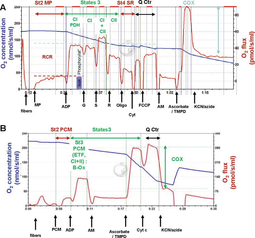
Fig.2 TypicalanalyzetracesofO2consumptiononpermeabilizedfibers.(a)SegmentalanalysisofETC functionusingsequentialsubstratesinjections.Mitochondrialoxygenconsumptionmeasurementswere performedat37 Candatmosphericpressureusingahigh-resolutionoxygraph(O2K,OroborosInstrument, Innsbruck,Austria).Respirationratesonpermeabilizedfibersusing50 μg/mlsaponin(30minutes,4 C)were measuredinrespiratorybuffer(10mMKH2PO4,300mMmannitol,10mMKCl,5mMMgCl2,0.5mMEGTA, and1mg/mlserumalbuminbovine,pH7.2)usingsubstratesofCI,CI+CII,andCIIasfollowed:first,state 2(non-phosphorylating)respirationwasmeasuredafteradding2.5mMpyruvateand5mMmalate.Then,the CI-linkedmaximalphosphorylatingrespirationwasstimulatedbysaturatingADPconcentration(1.5mM)and 3mMNAD+,addedtoavoidTCAlimitationbyNAD+availability.CI-linkedmaximalphosphorylating respirationwasfurtherstimulatedwith5mMglutamate,tocheckforanylimitationofsubstrateavailability byPDHactivity.Succinate(10mM)wasthenaddedtomeasurethecombinedCIandCII-linkedrespiration withconvergentCI+IIelectronflowintotheQ-junctioncorrespondingtothemaximalstimulatedphosphorylatingrespiration(OXPHOScapacity).Rotenone(5 μM)wasusedtoinhibitCIactivityandthustoobtainthe maximalCII-linkedrespiration.Thirdly,oligomycin(F0F1-ATPsynthaseinhibitor,4 μg/ml)andFCCP(carbonyl cyanide-p-trifluoromethoxyphenylhydrazone,amitochondrialuncoupler,1 μM)weresequentiallyaddedto ensurethatthecellswerefullypermeabilized.AntimycinAaddition(2 μg/ml)wasusedtocheckforthe non-mitochondrialoxidation.CIVmaximalrespirationwasinducedwith4mMascorbateand0.3mMTMPD,
2.4.3IntactCells
respirationrateinresponseto ΔpuseforATPsynthesisandthelow respirationratelinkedtoprotonleakinnon-phosphorylatingconditions.RCRvaluesdependonalmosteveryOXPHOSfunctional aspectandcouldthereforebeausefulindicatorofmitochondrial dysfunction.However,RCRdeterminationsaresensitivetoexperimentalinaccuracies;dependingonthequalityofsamplepreparationsandaccuratedeterminationofbackgroundrate,state4rates canbesignificantlyover/underestimated.Moreover,thereareno absoluteRCRvalues,astheseonesaresubstrate-andtissuedependent.Forexample,substratessuchassuccinateorglycerol phosphatetranslocatefewerprotonsperelectronpairthanNADHlinkedsubstrates,sothemaintenanceofthesame Δprequiresa higherrespirationrate.Furthermore,underidenticalsubstrate conditions,differentvaluesmaybeobservedfordifferenttissues, reflectingdifferentsubstrateoxidationkinetics,endogenousprotonleak,orphosphorylatingcapacities[68].Thus,carefulcautions accordingtotheexperimentalconditionsshouldberecommended wheninterpretingRCRvalues.
Permeabilizedcellsorfibersarealsoausefulmodeltoassessthe maximalactivityofthemainsubstrate-providingpathway,i.e.,TCA cycleorbeta-oxidationactivities.Thus,differentTCAcycleintermediatesormitochondrialshuttlessubstrates(malate/aspartate, glycerolphosphateshuttles)canbeusedtodissectspecificregulationswithinthesepathways.Similarly,thecomparisonofmaximal respirationratessustainedinthepresenceofTCAcyclesubstrates (malate,pyruvate,glutamate,andsuccinateforafullyoperating cycle)orinthepresenceofbeta-oxidationsubstrates(short-chain orlong-chainfattyacidscomplexedwithcoenzymeAor L-carnitine)allowsdeterminingthepreferredmetabolicpathways accordingtopathophysiologicalconditions,treatments,ortissues.
Measurementsofmaximalrespiratorychaincomplexactivities, assembly,andlinkedrespirationsarethecornerstoneonwhichthe biochemicaldiagnosisofmitochondrialdisordersisbased.However,thesecanusefullybeimplementedbymoreintegrativemethods,approachingmitochondrialmetabolism.
Theuseofisolatedmitochondriaorpermeabilizedcellshasbeen favoredforyears.Becausetheexperimenterhascontroloverconditions,i.e.,substratesavailabilityandrespiratorystates,this remainsthemethodofchoicetogainmechanisticinsightinto respirationchainfunctionanddysfunctions.However,inthelast
Fig.2 (continued)followedbyCIVinhibitionusing1mMKCNandazide.(b)AnalysisofO2consumptionlinked tothestimulationof β-oxidation. β-oxidationwasstimulatedusingpalmitoyl-L-carnitine,supplementedwith 2.5mMmalate(PCM).AntimycinAaddition(2 μg/ml)wasusedtocheckforthenon-mitochondrialoxidation. CIVmaximalrespirationwasinducedwith4mMascorbateand0.3mMTMPD,followedbyCIVinhibitionusing 1mMKCNandazide
decades,respirationanalysesonintactcellsbecameprominentin thefield.Withwholecellmodels,intrinsicactivityofrespiratory chaincomplexesisdifficulttotestandrespirationratessomehow difficulttointerpret,sincetheiractivitiesareintegratedwitha myriadofparameters.However,overridingthesecomplexitiesis theobvioushigherphysiologicalrelevance.Usingintactcells,mitochondriaarepresentinaphysiologicalenvironmentwithpreserved interactionswiththerestofthecell.Therefore,thismodelisideal toassessmetabolicpathwaysconnecteddirectlyorindirectlyto mitochondrialactivities.
Atypicalexperimentusingintactcellsstartsbythemeasurementofrespiratoryrateintherespiratorymedium,e.g.,cellculture medium,withoutanyadditionsofinhibitorsoruncoupler(Fig. 3). Thisstateisdefinedasthe basalorroutinerespiration.Routine respirationistheminimalrateofoxidativemetabolismrequiredto
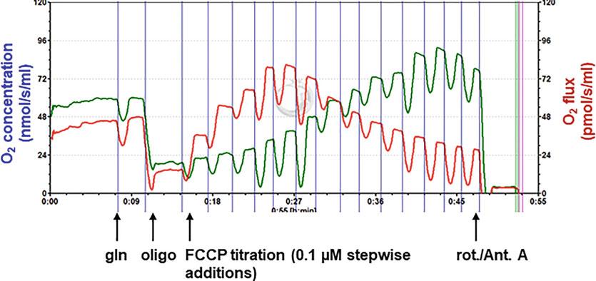
Fig.3 TypicalanalysistracesofO2 consumptiononintactcells.Mitochondrialoxygenconsumptionmeasurementswereperformedat37 Candatmosphericpressureusingahigh-resolutionoxygraph(O2K,Oroboros Instrument,Innsbruck,Austria).RespirationratesonprimaryfibroblastcellsweremeasuredineitherDMEMF12medium(3g/lglucose,0.3mMpyruvate)(redtraces)orinlow-glucosemedium(0.5g/lglucose)(green Traces).4 106 cellswereaddedintheoxygraphicchamberandtheanalysisstartedwithroutinerespiration (R)measurement,whichisdefinedasrespirationinmediumwithoutadditionalsubstratesoreffectors(cell endogenousrespiration,correspondingtothecellularoxidativemetabolism).Then,1mMglutaminewas added,totestforcellmetabolicdependencetoglutaminolysis.F0F1-ATPsynthasewasinhibitedwith oligomycin(4 μg/ml),allowingthemeasurementofnon-phosphorylatingrespiration(leakrespiration).This non-phosphorylatingrespiration(O)wassubtractedfromroutine(R)onetocalculatethecellularphosphorylatingrespiration(R-O).Thiswasfollowedbyuncouplingofoxidativephosphorylationbystepwisetitrationof FCCP(carbonylcyanide-p-trifluoromethoxyphenylhydrazone)uptooptimumconcentrationsallowingthe measurementofthemaximalendogenousrespiration(cellularoxidativecapacity).Thepartofthemaximal capacityuseforoxidativemetabolismwascalculatedasR/F,andthepartofthemaximalcapacityusefor oxidativeATPsynthesiswascalculatedas(R-O)/F.Finally,respirationwasinhibitedbyrotenoneandantimycin A(2.5 μMand2 μg/ml,respectively)tocheckfornon-mitochondrialoxidation
fulfillenergyneedsandsupportcellfunctions.Theroutinerespirationofmostmammaliancellscorrespondsneithertothestate 3(unlimitedavailabilityofsubstrateandADP)nortothestate4o (unlimitedavailabilityofsubstrate,butnoATPsynthesis),butis generallyanintermediatestatebetweentheseones,accordingto theATPdemand.Routinerespirationisusuallystronglycontrolled byATPturnover,butinmanyculturedmammaliancells,aerobic glycolysisalsocontributestothetotalATPturnover[69],meaning thattheroutinerespirationisnotequivalenttocellmetabolicrate. Routinerespirationisfurtherpartlydependentonsubstrateoxidation(includingsubstrateuptake,TCAcycle,activityoftheETC complexes )andprotonleak[70].Therefore,routinerespiration coulddifferaccordingtothesubstrateavailabilityintheincubation medium,totheoxidativeATPsynthesisneedsorinthepresenceof anyagentsdecreasingthe Δp(uncouplers).Forexample,routine respirationcanbeincreasedbydecreasingglucoseconcentration (Fig. 3).Amodificationinroutinerateintegratesanyofthese changesandisthereforequitedifficulttointerpret,butthefollowingstepscanhelptoelucidatethepathwayinvolved.
Routinerespirationmeasurementisfollowedbymitochondrial ATPsynthesisinhibition,througholigomycinaddition.Theresultingrespirationisdefinedas leakrespiration.Indeed,asforpermeabilizedcells,thisrespirationrateinthepresenceofoligomycinis controlledpredominantlynotonlybytheprotonleakbutalso,toa smallerextend,bytheactivityofsubstrateorions(e.g.,Ca2+) transportsandsubstratebeingoxidized.Inintactcells,thesubstratesoxidizedbytheETCarenotunderthecontrolofthe experimenter.Becausetheprotonpumpingisnotequivalent accordingtothesubstrateused,therespirationraterequiredto maintainthe Δpisalsodifferent.Thus,amodestchangeinleak respirationratemayindicateeitherachangeinprotonleakora changein Δpcausedbyalteredsubstrateoxidationortransport. However,alargeincreaseintherespirationratestronglysuggests uncoupledmitochondria.
Thisleakrespirationrateissubtractedfromthepreceding routinestatetoestimatethein-situ phosphorylatingrespiration, i.e.,therespirationratelinkedtoATPsynthesis.However,the protonleakisvoltage-dependent,allthemoreimportantasthe ΔΨ ishigh.SinceATPsynthaseinhibitionresultsinanincreasein ΔΨ,subtractingtheoligomycin-insensitiverespirationfromthe routineoneslightlyovervaluesthepartofprotonleakinvolvedin routinerespirationandunderestimatesATPsynthesis.Thisremains neverthelessoneofthebestapproachestoestimateinsituphosphorylatingrespirationwhenabsolutequantitativevaluesarenot needed[71].Also,calculatingthepartofthephosphorylating versusleakrespirationswithintheroutineonefurtherhelpsto decipherwhichprocessexplainsachangeintheroutinerespiration
rate.Whileanincreaseinphosphorylatingrespirationmostlikely reflectstheresponseofoxidativemetabolismtoincreasedenergy demand,adecreaseinthephosphorylatingrespirationcouldillustrateadecreaseinenergyneeds,ametabolicrewiringfromoxidativemetabolismto“anaerobic”glycolyticone(theso-called Warburgeffect),orablockageintheATPsynthesismodule.
Thenextstepinvolvesuncouplertitration,e.g.,FCCPtitration,toobtainthemaximal uncoupledrespiration rate.Asfor uncoupledrespirationmeasuredonpermeabilizedcells,thisoneis controlbythesubstrateoxidationpathways.However,thereisa markeddifferencehere:whileonpermeabilizedcell,theuncoupled respirationismeasuredinthepresenceofunlimitedavailabilityof definedsubstrate(s),meaningthatuncoupledrespirationismainly areflectofmaximalETCcapacity;onintactcells,thecatabolic pathwayssupplyingETCwithNADHorFADH2 isundercomplex metabolicregulationandcanberate-limiting.Therefore,the uncoupledrespirationrateonintactcellsreflectsthemaximaloverallsubstrateoxidationcapacity,fromsubstrateuptakeandcatabolismtoETCactivity,achievablebycellsundertheassaycondition. Adecreaseinthismaximalrespiratorycapacityisareliableindicator ofmitochondrialdysfunctionbut,becauseofthesecomplexities, cautionshouldbetakenwheninterpretingwhichpathwayis responsibleforthisdysfunction.Comparingthemaximal uncoupledrespirationobtainedonpermeabilizedcellsversusintact cellsprovidesfurthercluestodeterminewhetherareducedETC capacityisinvolvedornot.
CCCPandFCCPareprotonshuttlingcompoundsselectively increasingthepermeabilityoflipidmembranestoprotons.However,CCCPandFCCPalsohavehighreactivitywiththiolgroups. Thiol-combiningagentsuncoupleOXPHOSatlowconcentrations butinhibitrespirationathighconcentrationsbythechemicalmodificationofasmallbutsignificantnumberofmitochondrialthiol groups[72].Moreover,sustainedperturbationof ΔΨm,whichis normallytightlycontrolledtoensurecellproliferationandsurvival, triggerscellularstressresponses,eventuallyleadingtocellapoptosis [72].TheoptimalFCCP/CCCPconcentrationallowingthemaximalrespirationraterecordingdependsoncelltypes,mediumcomposition(e.g.,lowglucoseversushighglucose,fattyacids,or not ),andmitochondriaphysiologicalstate(e.g.,anydrug whichmodifiesthe ΔΨmcouldchangetheoptimalconcentration). Therefore,carefulFCCP/CCCPtitrationmustbeperformedfor eachexperimentalconditiontoensurethefullachievementof uncouplingyetlimitingthedropin ΔΨm.WhenusingaSeahorse Analyzer,onlytwoFCCPinjectionsareallowed,thereforepreliminaryexperimentsmustbeperformedbeforestartinganycomplete experiment.Unappropriateduncouplerdosesareacommonbias observedwhenworkingonintactcells.
Finally,theexperimentendsbytheadditionofETCinhibitors, rotenone,andantimycinAtodetermine residualoxygenconsumption.Residualoxidation(Rox)istherespirationduetooxidative sidereactionsremainingafterfullinhibitionoftheelectrontransfer pathway.MitochondrialrespirationisfrequentlycorrectedforRox asthenon-mitochondrialrespiration.However,Roxmaybepartly relatedtoROSproductionwhichconsumesO2 andisincreased uponETCinhibitoradditions[73].
Fromthesedifferentrespiratoryparameterscanbeinferred importantratios,particularlythesparerespiratorycapacity.The sparerespiratorycapacityistheratiooftheroutinetothe uncoupledrespirations.Withthecautionsaboutthedetermination ofmaximumratesdescribedabove,sparerespiratorycapacityindicatesthepartoftheETCcapacitythatisusedtosustaincell function,i.e.,howclosetoitsbioenergeticlimitcellsareoperating [74].Whilethisratiomaybeparticularlyinformative,again,given thecomplexityofthemechanismsinvolved,cautionsmustbetaken wheninterpretingit.Forexample,adecreaseinsparecapacitymay reflectanincreaseincellularATPrequirementsandasystem increasingitsproduction,oradecreaseinthesubstrateoxidation capacitythatlimitsuncoupledrespiration,dependingonwhether routinerespirationincreasesormaximalrespirationdecreases.Similarly,anincreaseinsparecapacitymaysuggesteitheradecreasein energydemandorametabolicswitchtowardglycolysis,whichthen mostlyensurestheATPproduction,althoughthecapacityofthe ETCremainsunchanged.
Whenworkingwithintactcells,cellmetabolism“determines” whichsubstratesmitochondriacanused.However,theexperimentersetstheextracellularconditions.Thechoiceoftheexact respiratorymediumcomposition,i.e.,thesubstratescomposition andconcentrations,thepresenceofhormones,growthfactors, cytokines,maydeterminetheoutcomeoftheanalysis.Thiscould beapitfall,butthisalsoofferstheopportunitytoperformcell metabolicphenotyping.Highconcentrationofglucoseisoften usedincellculture,butsomecellsprefertooxidizeothercomponentsofthemedium,suchasfattyacidsoraminoacids,particularly glutamine.Forexample,cancercellsundergoprofoundmetabolic rewiringforrapidgrowth.Theynotonlycouldderivemostoftheir energyfromglycolysisratherthanOXPHOSbutalsocoulddepend onoxidativeglutaminolysisormodifiedTCAcycleactivitytosustaintheirbiosynthesis[75].Bymodifyingthemediumcomposition,suchastheglucoseconcentration(Fig. 3),orthepresenceof glutamine,theexperimentercaneasilytestwhichspecificpathwayis preferentiallyusedbycellstosustaintheiroxidativemetabolism. 2.5Mitochondrial
The ΔΨmiscentraltothebioenergeticprocesses.Itnotonly providesthedrivingforceforATPsynthesisanddictatestheETC activityasdetailedabovebutalsoregulatesmetabolitestransport