Conventional Oxygen Therapy
Historical Overview of Oxygen Discovery and Utilization
Dioxygen (O2) was discovered at the end of the eighteenth century by Carl Schiele, Joseph Priestley, and Antoine Lavoisier [27] (Fig. 1.1). More recently, the importance of the work of Michael Sendivogius (1566–1636) was underlined in the path of the discovery of oxygen [28]. More than 150 years before the well-known trio reputed for oxygen discovery, this Polish alchemist could already produce “the food of life” later called oxygen by heating saltpetre (potassium nitrate) [28]. This gas has long been considered as a magical and almost holy gas, initially called the “breath of life” in the Bible, “elixir of life,” “food of life,” “vital air,” and “dephlogisticated air” before Lavoisier named it oxy-gen in 1777, meaning acid generator [29, 30]. Both Priestley and Lavoisier described the major role of oxygen to sustain life and they also described the potential lung toxicity, only few years after initial discoveries [29, 31]. First clinical utilization of oxygen in 1783 is attributed to Caillens, a French physician [32]. After Beddoes and Watt’s work [33], oxygen and other medical gases were studied and administered to patients at a larger scale at the Pneumatic Institution (1798–1802), Bristol, England. Initial results were heterogeneous due to very wide initial indications for oxygen administration (consumptionpulmonary tuberculosis, king’s evil-cervical tuberculosis, asthma, dropsy, palsy …) [34, 35], and possibly due to impurities in the initial oxygen production [36]. Another major obstacle to a large utilization was the diffculties related to oxygen
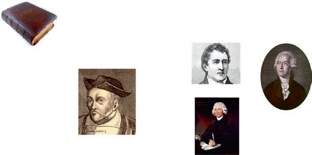
Fig. 1.1 The discovery of oxygen, from the “breath of life” in the Bible to the Oxy/Gen. During centuries, before it was named “oxygen” by Lavoisier, this gas was considered like a magical gas, and it was soon understood that it was vital for all living beings. However, immediately after its discovery, the potential toxicity was suspected by Priestley and Lavoisier 1 Conventional Oxygen
production, transportation, and administration. Sometimes used pure or in combination with nitrous oxide, the frst cylinders for storing oxygen were developed in the second half of the century, in 1868 [34]. Physiologic role of oxygen and modes of administration were extensively studied in the twentieth century and several researchers such as John Scott Haldane (1860–1936) (who described the effects of hypoxia during the Pikes Peak expedition [37, 38] and the “Haldane effect” [39]), his son John Burdon Sanderson Haldane (1892–1964) (who described the neurological effects of hyperoxia in hyperbaric conditions [40, 41]), and Alvan Leroy Barach (1895–1977) [42] provided a scientifc rationale to the medical use of oxygen and improvement in the technical aspect for oxygen administration.
The ways to administer oxygen have changed a lot since its discovery, which may explain the slowness to demonstrate the clinical benefcial effects. Oxygen was initially delivered intermittently due to the fear of lung toxicity. The recommended doses were small, typical therapy being “four gallons [approximately 15 liters] morning and evening, usually delivered in few minutes from a bucket towards the face of the patient” [35]. The intermittent use as well as the device to deliver oxygen may explain in part the initial disappointing results. The frst description of continuous use of oxygen was made by Blodgett in 1890 [43]. At the beginning of the twentieth century, Haldane also recommended the continuous delivery of oxygen to treat injuries caused by chlorine gas during the First World War [44, 45]. Haldane said, “intermittent oxygen therapy is like bringing a drowning man to the surface of the water—occasionally.” In spite of the benefts of continuous administration of oxygen, there was still reluctance on the part of the medical profession to administer oxygen continuously due to the fear of oxygen toxicity. Oxygen was still mainly provided intermittently, until Campbell recommendations in 1960 [46]. Finally, in
1608
Michael Sendivogius (1566-1636)
Carl Scheele (1742-1786)
Joseph Priesltley (1732-1804)
Antoine Lavoisier (1743-1794)
F. Lellouche and E. L’Her
1962 Massaro et al. demonstrated that intermittent administration of oxygen … had only intermittent effects on oxygenation and that oxygen could lead to increased CO2 retention in some patients [47]. In 1973, 34% of the clinicians still provided intermittent oxygen to patients, either often (11%) or rarely (23%) [48]. Currently, almost all physicians use oxygen continuously but this has not always been the standard, and the pendulum is still swinging: after a phase of liberal use, new studies and recommendations are moving towards a more restrictive use and a strict adjustment [6, 13, 14]. The recent recommendations are a turning point in the story of oxygen. The new statement is that healthcare practitioners should be worried about hyperoxemia such as they are with hypoxemia: a turning point … or a return to the initial mitigated feelings of Lavoisier and Priestley for oxygen, a vital and toxic gas!
1.2.2 Updated Recommendations for Oxygen Utilization
The frst goal of oxygen therapy is still to treat hypoxemia in acutely ill patients (mainly during respiratory or cardiac failure) [13, 14]. The other objective which is recommended but frequently disregarded is to avoid hyperox(em)ia [13, 14]. First recommendations to deliver oxygen have been published by Campbell in 1960. He recommended continuous delivery, low FiO2 administration (24–35%), and reduction of expiratory gas rebreathing to avoid CO2 accumulation [46]. These recommendations focused on COPD patients. Since initial recommendations, others are regularly issued from different national or international societies [13, 49, 50]. Interestingly, in most guidelines, the focus was only on the minimum level of oxygenation to deliver (minimum SpO2) until recently. Maximum SpO2 values were only recommended for COPD patients and in neonate populations [51], as underlined by Semienuk et al. [14]. A maximum SpO2 was frst recommended by the Thoracic Society of Australia and New Zealand for other categories of patients, and these recommendations were called “swimming between the fags” [50]. The most recent recommendations provide a target SpO2 range with a minimum and a maximum SpO2 value [13, 14]. This may seem like a minor change to the protocols, while it actually changes the whole challenge of management and monitoring for the clinicians.
The main recommendations for oxygen therapy in critically ill patients may be summarized as follows (Fig. 1.2):
1. Use oxygen only to treat hypoxemia and neither to treat breathlessness nor thoracic pain.
2. Use a restrictive rather than a liberal strategy to deliver oxygen.
3. SpO2 target should be between 88 and 92% in patients at risk of hypercapnia (not only for COPD patients) which is quite consistent in the different guidelines (while relying on few data).
4. For other populations, there is a discrepancy in recommendations. Minimal SpO2 targets vary from 90% to 94% and maximal SpO2 targets vary from 94% to 98% [13, 14, 50]. The most recent recommendations, which are derived from the meta-analyses comparing liberal and conservative oxygen administration, are
General population
COPD patients Hypercapnic
Fig. 1.2 Recommendations for oxygenation targets in the general population [14] and in patients with hypercapnia or at risk of hypercapnia (COPD, severe obesity …) [13]. SpO2 values below OR above the SpO2 target should be considered dangerous for the patients. ① represents the SpO2 values for which oxygen supplementation should be initiated. ② represents the SpO2 values for which the oxygen supplementation should be stopped or minimized to attain the SpO2 target
the most radical for a restrictive use and strongly recommend to stop oxygen if SpO2 is above 96% and not to start oxygen if SpO2 is equal to or above 93% [14]. The recommended SpO2 target is 90–94%.
5. SpO2 should be closely monitored and even continuously monitored for most severe patients, and arterial blood gases should only be used in case of potential hypercapnia or metabolic disorders.
6. NEWS or equivalent scoring system should be monitored in patients receiving oxygen to allow an early detection of clinical worsening [13].
7. Oxygen supplementation improves oxygenation, but the underlying cause of hypoxemia must be treated.
The knowledge transfer concerning oxygen therapy is among the worst example. Indeed, it seems diffcult to comply with recommendations and guidelines for oxygen prescription and delivery existing for more than 50 years [19, 52–54]. This may be explained by several obstacles including the belief that oxygen cannot be harmful, despite recent evidence demonstrating oxygen toxicity even in the general population [6]. However, even in COPD patients for which hypercapnia induced by hyperoxemia was described 70 years ago, prescription is still frequently not adequate [19]. In most situations, too much oxygen is provided to patients and oxygen toxicity is frequently overlooked. We will underline below the impact of oxygen therapy complications, with both hypoxemia and hyperoxemia being life threatening.
1.3 Physiological Effects of Oxygen and Toxicity
1.3.1
Physiological Role of Oxygen and Cellular Toxicity
Dioxygen (O2) is used in mitochondria for aerobic cellular respiration leading to the production of adenosine triphosphate (ATP) via oxidative phosphorylation which is the main way to store energy and powers most cellular reactions in eukaryotes [55].
F. Lellouche and E. L’Her
CO2 and H2O are produced as well as energy stored in the form of ATP during the process. The aerobic respiration is the following simplifed reaction: C6H12O6 + 6 O2 → 6 CO2 + 6 H2O + 2880 kJ/mol. Aerobic pathway is considered at least 15 times more effcient to produce ATP: anaerobic produces only 2-molecule ATP per 1-molecule glucose while 30- to 38-molecule ATP can be produced with 1-molecule glucose through aerobic respiration [56]. Electron transport chains in the mitochondria are major sites of electron leakage. Oxygen serves as an effective electron accepter (oxidizing agent), generating reactive oxygen species (ROS), also called free radicals, such as superoxide ion (O2 ), hydrogen peroxide (H2O2), and the most reactive one hydroxyl radical (OH-) [57]. The toxicity of these free radicals relies on their different reactivity and lifetime. Hydrogen peroxide (H2O2) is less reactive with a half-life of few minutes while hydroxyl radical (OH-) is the most reactive and immediately removes electrons from any molecule in its path and thus propagates a chain reaction, which explains its extremely short lifetime estimated in nanoseconds [58, 59]. The combination of different free radicals and especially the hydrogen peroxide in the presence of Fe2+ ions can produce the very highly reactive and damaging hydroxyl radical (OH-) (Fenton reaction) [60]. However, hydrogen peroxide (H2O2) may also be damaging to DNA since its lower reactivity provides enough time for the molecule to travel into the nucleus of the cell, subsequently reacting with macromolecules such as DNA.
In a biological context, ROS not only are toxic molecules and but also have important roles in cell signaling, homeostasis, and immune system with direct toxicity against bacteria and other pathogens [57, 61, 62]. Accurate regulation rather than elimination of ROS is consequently the objective of the protection mechanisms. With the view of an evolution biochemist who described all the mechanisms to protect living organisms from ROS since almost 4 billion years, we can state that “our whole body is an antioxidant machine” with the aim not to eliminate but to regulate the level of oxygen and ROS at the cellular level [63]. There are several mechanisms to protect cells and to regulate oxygen and ROS concentrations. Several organisms produce mucus layers to reduce oxygen diffusion and oxygen scavengers (leghemoglobin in plants and hemoglobin in animals) and allow storage, transport, and progressive delivery of oxygen to tissues to avoid high oxygen concentrations at cellular levels. Oxygen partial pressure in the ambient air is usually around 160 mmHg (21% of a normobaric atmosphere, 760 mmHg) and enters the pulmonary capillaries at around 100 mmHg with the main oxygen reservoir in hemoglobin. At the other extremity of the oxygen cascade, oxygen pressure in cells is variable and below 10 mmHg in the mitochondria [64]. Oxygen is continuously consumed in the respiratory chain to produce energy, increasing the gradient and the “call of oxygen.” Enzymatic (catalase, superoxide dismutase, glutathione peroxidase) and nonenzymatic antioxidants (vitamins A, C, and E, carotenoids …) are also precisely regulating the intracellular concentrations of ROS. These mechanisms limit the dangerously high concentrations of free radicals leading to potential lesions of mitochondrial and cell membranes through lipid peroxidation, and DNA strand breaks and other intracellular macromolecule hits and enzyme inhibitions [65]. In the case of excessive damages, cellular apoptosis may occur. However, despite
antioxidant mechanisms, due to environmental stress (e.g., UV or heat exposure), or biological stress (hypoxemia, hyperoxemia, sepsis, ischemia), the ROS levels can increase dramatically. This may result in signifcant damage to cell structures. The disequilibrium between antioxidants and oxidants is known as oxidative stress, which is promoted during hyperoxia and hyperoxemia [66].
1.3.2 Oxygen Is a Dangerous Gas!
Oxygen is one of the most dangerous gases on the planet. Severinghaus and Astrup described oxygen with humor as follows: “Oxygen is the frst atmospheric pollutant. Oxygen is toxic. It rusts a person in a century or less. With oxygen came the danger of fre. If introduced today, this gas might have diffculty getting approved by the Food and Drug Administration” [67]. Indeed, all living species on earth have had to protect themselves against oxygen, from the very beginning of life, more than 3.5 billion years ago. First prokaryotes and eukaryotes were bioengineered with antioxidants, even when oxygen concentration in the air was very low (below 1%) [68]. Almost 3.8 billion years ago, the LUCA (last universal cellular ancestor), ancestor of Eukarya, Bacteria, and Archaea, was equipped with several mechanisms to protect from oxygen (mucus layer, antioxidants such as catalase and superoxide dismutase) [68–71]. These defenses are probably inherent to common mechanisms of toxicity shared by oxygen and X-ray [72]. In both situations, reactive oxygen species (ROS) are produced, from H2O with X-ray or from oxygen degradation during oxygen metabolism (Fig. 1.3). Before increasing levels of oxygen in the atmosphere (after the great oxidation event that appeared with

ANTIOXIDANTS
Enzymatic (Superoxide dismutase, Catalase, Glutathione peroxidase) and non enzymatics (vitamins A, C, E, β-caroten, selenium…)
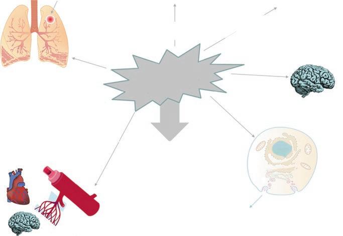
Fig. 1.3 Common mechanisms of toxicity are shared by oxygen and radioactivity [72]. In both situations, reactive oxygen species (ROS) are produced leading to membrane cell damage ①, intracellular membrane damages ②, DNA damage ③, and apoptosis ④. The concentration of the most reactive (hydroxyl radical) is higher with radioactivity. Antioxidant mechanisms reduce the concentration of ROS and limit the toxicity
F. Lellouche and E. L’Her
photosynthesis 2.5 billion years ago), frst living cells were exposed to X-rays and acquired these antioxidant mechanisms [63]. Considering this subtle equilibrium, and the different antioxidant defenses acquired during the last 4 billion years, oxygen should not be considered only as a vital gas, but also as a toxic gas. Noteworthy, in normal environments, current Homo sapiens only experience normoxia (breathing air with 21% oxygen) or hypoxia in altitude. Hyperoxia (high oxygen intakes) and hyperoxemia (high oxygen concentration in blood) may only be encountered in hospitals or in oxygen bars!
1.3.3 Risks Associated with Hypoxemia
It is not diffcult to convince clinicians (and probably anyone crossed in the street, as oxygen is very popular) that the lack of oxygen may be dangerous and life threatening. Several mechanisms of hypoxemia are well described and comprise hypoxia (mainly when living in altitude [73] or induced hypoxemia in laboratory [24, 74]), reduced diffusion (interstitial lung diseases), hypoventilation (drug overdoses, neurological diseases), ventilation/perfusion mismatch (COPD, asthma, pulmonary embolism), intrapulmonary shunt (cardiogenic pulmonary edema, pneumonia, pulmonary embolism), intracardiac shunt, and shock state (cardiogenic, hypovolemic …). Causes of tissue hypoxia are also well described, mainly in the case of sepsis (failure in peripheral oxygen extraction) and several intoxications (CO, cyanide intoxication …). Acute hypoxemia leads to tachycardia (to increase cardiac output and oxygen transport to tissues) [75], increased minute ventilation (to increase oxygen intake) [76, 77], systemic arterial vasodilatation [78, 79], pulmonary vasoconstriction [80], increased tissue extraction of oxygen, and other mechanisms that increase oxygen delivery to tissues (such as right shift of the hemoglobin saturation curve). Other physiological compensations exist to counterbalance hypoxemia in the case of subacute or chronic hypoxia (in populations living in altitude) or chronic hypoxemia (mainly in cardiorespiratory diseases), primarily the polycythemia.
When the protection mechanisms are overwhelmed, tissue hypoxia may occur, with risks of cardiac ischemia (especially due to associated tachycardia), cardiac arrhythmia [81, 82], hepatic ischemia, cerebral ischemia, and cellular anaerobic respiration leading to increased lactate production [83].
To correct hypoxemia, oxygen must be administered urgently, but this is only a supportive therapy and etiological treatment must be adequately provided. The most recent recommendations are to deliver oxygen to maintain SpO2 above 88% in patients at risk of hypercapnia and 90% in other patients [14]. Oxygen supplementation should be initiated only if SpO2 is below 93% in the general population (Fig. 1.2).
1.3.4 Risks Associated with Hyperoxia: Oxygen Toxicity (Fig. 1.4)
It is less intuitive to endorse that oxygen may be toxic and increases mortality [5–7, 84]. Oxygen toxicity is low-grade toxicity or hidden from view in most circumstances which may explain that it is neglected. However, the deleterious effects of oxygen have been described very early after oxygen discovery by Lavoisier and Priestley [29, 31] and compelling demonstrations are now available that hyperoxia may lead to increased mortality [5–7, 84, 85]. There have been more than 7000 publications on hyperoxia in the last 70 years on “PubMed,” with an exponential increase after the introduction of pulse oximetry in the clinical practice in the early 1980s. Most publications are descriptions of physiological mechanisms of toxicity [65] but in the last 10 years, several randomized clinical trials have described more accurately this toxicity and allowed to better fgure out the real impact of hyperoxemia in different populations. First demonstrations of increased risk of liberal use of oxygen were provided in 2010 by Austin et al. [5]. The authors evaluated
Direct pulmonary toxicity
Lorrain-Srnith effect High FiO2
Denitrogenation
Atelectasis High FiO2
Coronary arteries
Cerebral arteries
Arterioles
↓NO Production ↑Prostacycline PGI2 ↑Endothelin
COPD, obese patients? INDUCED HYPERCAPNIA High PaO2
Ventilation/perfusion mismatch Haldane effect Respiratory drive
Premature infants PROLIFERATIVE RETINOPATHY High PaO2
Phase 1: ↓ EPO, IGF1, VEGF → reduced retinal vascularisation
Phase 2: ↑ EPO, IGF1, VEGF → neovascularization
CEREBRAL TOXICITY
REACTIVE OXYGEN AND NITROGEN SPECIES
Superoxide anion O2Hydrogen peroxyde H2O2 Hydroxyl radical OHSinglet oxygen 1O2
Hydroperoxyl radical HOOLipid peroxide radical ROONitric oxide NOPeroxynitrite ONOO-
Direct (neonatalogy) Paul Bert effect (hyperbar conditions) High PaO2
CELLULAR INJURIES
APOPTOSIS AUTOPHAGIA INFLAMMATION
SYSTEMIC INFLAMMATION VASOCONSTRICTION
Fig. 1.4 Systemic toxicity of oxygen. Oxygen toxicity involves several organs. The cellular as well as vascular toxicity makes the toxicity of oxygen systemic. This may explain the deleterious effects of oxygen leading to increased mortality when used liberally [6]
F. Lellouche and E. L’Her
the impact of oxygen fow rate delivered to patients managed in ambulances for acute respiratory distress. Mortality was signifcantly lower in patients receiving titrated oxygen (targeting a SpO2 of 88–92%) rather than high-concentration oxygen (8–10 L/min of oxygen fow rate): 9% vs. 4%, p = 0.02; relative risk, 0.42; and 95% confdence interval 0.20–0.89. In the subgroup of patients with confrmed chronic obstructive pulmonary disease (n = 214) mortality was reduced even further (9% vs. 2%, p = 0.04; 0.22, 0.05–0.91). This effect was shown despite a mean exposure of only 47 min [5]. This study has limitations, including a low compliance to protocols, especially in the targeted oxygen arm (only 41% compliance rate). A recently published randomized controlled trial conducted in critically ill mechanically ventilated patients further revived the interest in this often-minimized question of oxygen toxicity. This study conducted in 434 mechanically ventilated patients showed a clear increase in mortality (20.2% vs. 11.6%, p = 0.01) in patients in the hyperoxic group (SpO2 target of 97–100%) in comparison with normoxic patients (target SpO2 94–98%) [7]. Recent reports described an impressive U-shaped curve of the impact of mean PaO2 in critically ill patients [84]. Similarly, short-term exposure to hyperoxemia (median of 5.4 h) in patients intubated at the emergency department was associated with increased mortality [86].
Finally, the meta-analysis recently published by Chu et al. concluded that liberal oxygen use increased mortality in comparison with restrictive oxygen use in different populations of critically ill patients [6].
1.3.4.1 Direct Oxygen Lung Toxicity
Few years after oxygen discovery, Lavoisier described oxygen toxicity and pneumonitis with infammation of the lungs in animals exposed to pure oxygen, dying from acute respiratory distress after a few days [29]. These initial descriptions were confrmed and extensively described by Lorrain Smith in animals [87]. He described the impact of both oxygen concentration and duration of exposure, known as “Lorrain Smith” effect. These early demonstrations of oxygen toxicity were confrmed by numerous animal studies, reviewed by Clark and Lambertsen [88]. It is now considered that prolonged utilization of high FiO2 must be avoided to avoid both hypercapnia-induced, direct lung toxicity and denitrogenation atelectasis [89].
1.3.4.2 Importance of Accurate Titration of Oxygen Flow Rates in COPD Patients
Hypoxemia is frequently encountered in severe COPD and is responsible for various severe complications [90]. In addition to the impact of desaturations on pulmonary arterial pressure [91], there are also well-known cardiac effects (myocardial ischemia, tachycardia …) [81, 82].
The risks of hypoxemia are easy to understand and are intuitive, but the risks associated with hyperoxia, and in particular induced hypercapnia, are still neglected [8]. This complication has been known for a long time and was initially suspected by Barach in 1931 [92] and then reported by several authors in 1949 [16, 93, 94]. This effect of oxygen is linked to several mechanisms, such as a decrease in hypoxemic stimulation of the respiratory centers (decrease in minute ventilation [94, 95]),
a modifcation of the ventilation/perfusion ratios [96], or a Haldane effect (reduced affnity of hemoglobin for CO2 in case of hyperoxia [39]), but the respective role of these mechanisms has long been discussed [96–99]. Campbell published in 1960 and 1967 the frst recommendations to adjust the oxygen supply to the minimum [46, 100]. Similar recommendations have since been recently published [13] based on several studies demonstrating that hyperoxemia could induce hypercapnia in some COPD patients [96–99, 101]. Unfortunately, it is well documented that these recommendations are not followed [3, 19] and that patients receive too much oxygen [102] or under certain circumstances not enough [103–105]. The recent demonstration of the risk of excess mortality due to over-oxygenation during the short period of prehospital transport [5] has rekindled the debate [8].
This complication of oxygen therapy seems clinically relevant especially in the context of acute exacerbations [5, 96, 106]. In stable COPD patients, the impact of hyperoxemia on PaCO2 seems to be lower [96, 99]. However, despite the known risks of hypercapnia associated with hyper-oxygenation, many patients treated for exacerbation receive excessive oxygen fow rate [102, 106, 107]. It has recently been shown that high oxygen fow increases mortality in patients managed in ambulances for acute respiratory failure in comparison with oxygen administered to target SpO2 between 88 and 92% [5]. Likewise, in this population, the negative impact of respiratory acidosis on the prognosis has been well demonstrated, as well as the importance of controlling oxygenation [106–108].
Given the workload especially in emergency situations and the underestimation of the problem, recommendations [13] to limit oxygen fow in COPD are poorly followed [3, 8, 19, 20].
Inhaled aerosols may be used to deliver molecules (especially bronchodilators), which may require gas fows above 6 L/min with pneumatic devices. Consequently, the recommended driving gas is the air to avoid induced hypercapnia, especially in COPD patients [5, 8, 13]. Gunawardena et al. evaluated the driving gas for nebulization and compared oxygen with air in patients with COPD or airway obstruction. This study showed that PaCO2 increased by 7.7 mmHg after nebulization with oxygen as driving gas in patients with baseline hypercapnia. PaCO2 did not increase when air was the driving gas, and in patients without hypercapnia at baseline. Noteworthy, the PaCO2 returned to baseline after 20 min [109]. These data have been recently confrmed in several studies [110, 111]. Another solution if air is not available as driving gas is to perform the nebulization with ultrasonic aerosols which do not require driving gas [112]. In the study of Austin et al. demonstrating a reduced mortality with restrictive use of oxygen during the prehospital transportation in ambulances, nebulizations were delivered with compressed air in the restrictive group [5]. However, this may be diffcult to implement in the real-life setting.
1.3.4.3
Vascular Risks Associated with Hyperoxia Are Now Well Demonstrated
There is a signifcant vascular risk associated with hyperoxemia. It has now been well demonstrated that hyperoxemia notably causes vasoconstriction mediated by a decrease in nitric oxide at the endothelium level [113]. The free radicals
F. Lellouche and E. L’Her
(superoxide ions O2-, peroxide H2O2, hydroxyl radical OH- …) are very reactive and result in a cascade of reactions leading to the reduction of NO concentrations, and consequently to coronary, cerebral, and microvascular vasoconstriction; myocardial dysfunction; and infammatory response [113]. This effect appears to be rapid, since exposure to 15 min of hyperoxemia in patients with stable coronary artery disease results in a 30% reduction in coronary blood fow and an increase in coronary heart resistance of 30% [114]. In 1959, West and Guzman demonstrated this effect in dogs exposed for 5 min to 100% oxygen [79]. In addition to numerous physiological data [115], clinical studies are now available that better evaluate the clinical impact of hyperoxemia in this population. Recently, Stub et al. have demonstrated in a randomized trial involving 441 patients in the acute phase of myocardial infarction that routine administration of oxygen (at 8 L/min or about 60% FiO2) was detrimental compared to oxygen administration in case of desaturation below 94% SpO2 (this situation involved only 8% of patients in this arm) [116]. In the group receiving liberal oxygen, the cardiac enzymes were signifcantly higher, and the recurrence of infarction was increased, as well as arrhythmia. In 32% of included patients, myocardial MRI was performed 6 months after the initial episode and showed a greater size of the infarct in the oxygen-treated group (20.3 vs. 13.1 g, p = 0.04) [116]. A post hoc analysis of a randomized controlled trial involving 1386 patients undergoing abdominal surgery also demonstrated the increased risk of postoperative myocardial infarction with liberal oxygen administration [117]. The main objective of the PROXI trial was to evaluate the impact of perioperative FiO2 (during surgery and 2 h after) on infections at the surgical site, comparing 30 and 80% of FiO2 [118]. In the hyperoxic group, the risk of postoperative myocardial infarction was signifcantly increased: 2.2% vs. 0.9%, 80% vs. 30% FiO2, HR 2.86 (95% CI 1.10–7.44), as well as several secondary outcomes such as any heart disease or death (HR 1.24 95% CI 1.06–1.45) [117]. Very recently, Hofmann et al. demonstrated in a randomized controlled study that included 6629 patients at the acute phase of myocardial infarction that routine oxygen administration (at 6 L/min or approximately 45% FiO2) did not reduce mortality compared with administration only in case of desaturation [119]. This demonstrates the uselessness of the routine and liberal administration of oxygen in these patients. As previously noticed, oxygen toxicity seems to be dose dependent [120], since no toxicity was noted with FiO2 equivalent to 45% [119], while at or above 60%, there seems to exist a coronary and myocardial toxicity [115–117, 121, 122]. In addition, systematic oxygen administration has been proposed to reduce thoracic pain. Recently, Sparv et al. did not fnd any analgesic effect of routine oxygen as compared with ambient air during myocardial infarction [123]. High-dose oxygen should not be systematically administered in the case of myocardial infarction or thoracic pain as it has been done for a long time [10]. Oxygen has long been one of the bases for the management of myocardial infarction at the acute phase. MONA (morphine, oxygen, nitrogen, aspirin) [124] guidelines now tend to recommend oxygen therapy only in patients with acute coronary disease associated with hypoxemia [125]. In the most recent guidelines, it is recommended not to begin oxygen supplementation if SpO2 is above or equal to 90% [14].
1.3.4.4
Neurological Toxicity
Direct neurological toxicity of oxygen in hyperbaric conditions leading to convulsions has been initially described by Paul Bert in 1878 in the context of hyperbaric hyperoxia in animals [126]. JBS Haldane (the son) reproduced the experiments on himself and described thresholds of oxygen on epilepsy in hyperbaric conditions. He and his collaborator had seizure at approximately the pressure of seven atmospheres layer within 5 min [41, 127]. Haldane also described during these experiments a peripheral neuropathy similar to that found in the Hyper 2S study in patients with septic shock comparing normoxia and 100% FiO2 for 24 h in critically ill patients [128]. Indeed polyneuropathy was found in 6% vs. 11% in the hyperoxia group (p = 0.06) of the patients in the study of Asfar et al. comparing 24 h of pure oxygen administration (100%) to FiO2 titrated to maintain 88–95% FiO2 [128]. This study was stopped prematurely because adverse events were signifcantly more frequent with pure oxygen (including atelectasis, polyneuropathy, renal failure …). In addition to the direct cerebral toxicity mainly in hyperbaric conditions, a 20–30% reduction in cerebral blood fow was found when high FiO2 was used [129, 130].
1.3.4.5 Oxygen Toxicity in Perioperative and Critical Care Patients
A recently published randomized controlled trial revived interest in this oftenminimized question of critical care oxygen toxicity. This study in 434 mechanically ventilated patients demonstrated an increase in mortality (20.2% vs. 11.6%, RR (95% CI): 0.086 (0.017–0.150), p = 0.01) in the hyperoxic group (SpO2 target of 97–100%) in comparison with the normoxic group (SpO2 target of 94–98%) [7]. Despite limitations discussed in the editorial, this study pointed out that in the acutely ill patients, oxygen should be titrated to target normoxemia and avoid hyperoxemia [131].
There are several types of toxicities in mechanically ventilated patients that may explain these results: impact of hyperoxia (high FiO2) and effects of “hyperoxemia” (high PaO2) (Fig. 1.4) [6, 132, 133]. These data may in part be transposed to nasal high-fow therapy or moderate oxygen fow administration.
Among the effects of hyperoxia, two types of effects should be considered separately: direct lung toxicity [89] and denitrogenation derecruitment [134]. On the one hand a direct “toxicity” of oxygen exists, initially described in animal studies by Lavoisier [29], then by Lorrain Smith in 1899 [87] and Clark and Lambertsen reviewed more than 100 animal and human studies [88]. This review demonstrated that prolonged exposure and intensity of hyperoxia had an impact on multiple structural components of the lungs. However, toxicity is heterogeneous among species. For example, amphibians may survive indefnitely when exposed to 100% oxygen at atmospheric pressures, while the rat or mouse usually dies after few days’ exposure to the same concentration of oxygen [88]. In humans, the clinical importance of lung toxicity related to oxygen remains controversial [89], but it is generally accepted that prolonged administration of FiO2 above 60% should be avoided, when possible. The other type of lung toxicity with high FiO2 is explained by denitrogenation atelectasis described in patients under general anesthesia [134–136] and during the ARDS [137]. In the study conducted by Ihnken et al., the impact of oxygenation
F. Lellouche and E. L’Her
during coronary artery bypass grafting was evaluated in a randomized controlled study in 40 patients: 20 patients in the hyperoxia group (mean PaO2 = 419 mmHg) and 20 patients in a “normoxia” group (mean PaO2 = 142 mmHg) [121]. In addition to the impact on ischemia-reperfusion injuries, in oxidative myocardial damage leading to increased cardiac enzymes in the hyperoxic group, the impact on lung volumes was compelling. Koksal et al. showed similar data with increased oxidative stress and reduced lung volumes in the hyperoxia group in patients managed during abdominal surgery with 80 vs. 40% FiO2 [138].
Finally, there is a toxicity related to “hyperoxemia” corresponding to high levels of PaO2 (PaO2 > 100 mmHg), which is often called inadequately hyperoxia. This systemic toxicity is related to various mechanisms: the production of ROS (free radicals) responsible for “oxidative stress,” the vascular toxicity (arterial and arteriolar vasoconstriction), the cellular toxicity, and the induced infammation (Fig. 1.4).
In an observational cohort study including 14,441 mechanically ventilated patients and 295,079 arterial blood gas analyses, Helmerhorst et al. showed that the mean PaO2 during ICU stay had a strong relationship with mortality [84]. The U-shape relationship demonstrated an increased mortality with hypoxemia as well as hyperoxemia. This U-shape relationship between PaO2 and mortality has been underlined in several papers [84, 133, 139–141]. In Van der Boom et al.’s study, two US databases were analyzed including more than 35,000 ICU patients with hourly physiological data [141]. In addition to the U-shape relationship between SpO2 and mortality, the authors found that the percentage of time patients were within an optimal range of SpO2 was associated with a signifcant decrease in hospital mortality. When comparing the mortality with 80% vs. 40% of the measurements within the optimal range of oxygenation, the ORs were 0.42 [95% CI, 0.40–0.43] for one database and 0.53 [95% CI, 0.50–0.55] for the other database [141].
A systematic review and meta-analysis demonstrated benefts of normoxemia in comparison with hypoxemia and hyperoxemia in several acutely ill populations (head trauma and stroke, post-cardiac arrest, and mechanical ventilation) [132]. A meta-analysis including 25 randomized controlled trials and 16,037 patients with sepsis, critical illness, stroke, trauma, myocardial infarction, cardiac arrest, and emergency surgery has been recently published in The Lancet journal [6]. This study showed in this population that liberal oxygen therapy increases mortality without improving other patient-important outcomes and concluded that supplemental oxygen with oxygenation above an SpO2 range of 94–96% may be detrimental to patients [6, 9]. This study and others published previously are the basis of the most recent recommendations to avoid oxygen therapy if SpO2 is equal or above 93% and to stop oxygen if SpO2 is above 96% in patients managed for acute care in hospitals [14].
Two large and recently published RCTs have compared two oxygen therapy strategies and did not fnd differences between study arms [142, 143]. The ICUROX study conducted in Australia and New Zealand compared conservative oxygen therapy and usual oxygen therapy in 1000 adults undergoing mechanical ventilation [142]. There was no differences in ventilator-free days and other outcomes. In both study arms, the time-weighted mean daily PaO2 after inclusion was below 100 mmHg, which suggests that two conservative strategies were compared and
may explain the absence of difference found between groups [142]. The inclusions in the study started after the implementation of the Thoracic Society of Australia and New Zealand oxygen guidelines, “swimming between the fags” which may explain that the usual oxygen strategy was a conservative strategy [50, 142]. A very recent RCT has compared two oxygenation targets in 2928 patients with acute hypoxemic respiratory failure [143]. PaO2 targets were 60 mmHg in the “lower oxygenation” arm and 90 mmHg in the “higher oxygenation” arm. Again, median values of daily PaO2 were below 100 mmHg in both study arms and no differences in outcome could be detected [143]. Unfortunately, even if patients enrolled in the usual oxygen therapy in the ROX-ICU study had quite low mean PaO2, this groups was counted in the “higher oxygenation” group in a meta-analysis, which induces a major bias in the analyses and explains the absence of difference between “higher” and “lower” oxygenation strategies [144].
1.3.4.6 Oxygen Toxicity in Premature Infants
There is well-documented oxygen toxicity in premature infants, particularly for the retina and brain [145, 146]. Retinopathy of prematurity “Terry syndrome” associated with hyperoxia has been described more than 50 years ago [17, 147], few years after the launch of new-generation incubators, leading to more colored (with high FiO2) premature infants. Immature retinas of premature newborns are sensitive to hyperoxemia, which disrupts neurovascular growth, leading to retinopathy of prematurity [145]. Pathophysiology involves several phases: retinal vasculature is frst inhibited due to hyperoxemia and other factors in particular nutritional. The retinal growth will then be done with reduced vascularization, which leads to hypoxemia, which itself stimulates the expression of oxygen-regulated growth factors such as erythropoietin (EPO), VEGF (vascular endothelial growth factor), and IGF1 (insulin-like growth factor), leading to neovascularization [145]. The developing immature brain is particularly sensitive to changes in oxygen and in particular to hyperoxemia, as is well demonstrated in a recent review [146].
1.4 Technical Aspects of Oxygen Delivery
1.4.1
Oxygen Sources
Currently, there are several oxygen sources available: low-pressure oxygen concentrators and liquid oxygen mostly used for home care, pressurized oxygen in cylinders for intrahospital or ambulance transportation, and pressurized medical oxygen mainly used in hospitals. At the beginning of oxygen medical use, the administration of oxygen supposed a local production by the users, as did Beddoes and Watt at the Pneumatic Institution [34, 148]. This explains in part the initially limited medical use of this gas. In 1868, Barth was able to provide almost 60 L (15 gallons) of oxygen compressed with hand pump into a copper bottle [148]. This was enough for intermittent administration of oxygen but larger cylinders of up to 100 gallons were available at the end of the nineteenth century when continuous administration was
F. Lellouche and E. L’Her
described [43]. In 1965, the frst liquid oxygen system was developed [149] and few years later, the frst stationary oxygen concentrators became available [150]. Oxygen concentrators produced a purity of gas of around 90% while hospital oxygen has around 99% purity. The other characteristic of medical air is to be very dry [151, 152] and the question of humidifcation of this gas is still debated.
1.4.2 Humidification
The question regarding the humidifcation requirement for low, moderate, and highfow oxygen is not fully resolved, and recommendations are not homogeneous. The oxygen delivered in hospitals is dry, with less than 5 mgH2O/L of water content [151, 152] or even less than 1 mgH2O/L [153]. It is recommended in North America to humidify oxygen when fow rates are above 4 L/min [154, 155] while these recommendations rely on few data. Cold humidifcation with bubble humidifcation is expensive and of limited effcacy. The dry oxygen delivered through nasal cannula or non-occlusive mask represents only a fraction of the inspiratory gases in spontaneously breathing patients with respiratory failure, when low or moderate fow is delivered (below 15 L/min). The inspiratory fow during acute respiratory failure is frequently above 60 L/min [156]; this implies that dry medical oxygen is mixed with room air and fnal FiO2 as well as humidity at the trachea level is the result from blended FiO2 and humidity of the medical oxygen and room air. With low to moderate fows, the impact of room air characteristics is probably major, as more than 50% of the inspiratory gas is coming from room air rather than from the medical gas. Consequently, the impact of humidifcation of the delivered oxygen may be limited (Fig. 1.5). The poor performance of cold humidifcation to prevent nasal dryness was demonstrated in Chanques et al.’s study for patients managed in ICU with moderate fows (median 7 L/min) as discussed above [151]. It was also shown in 185 patients hospitalized in the ward that there were no or few difference in mucosal dryness symptoms between the use of bubble humidifer and no humidifcation [157]. The recent British Thoracic Guidelines for oxygen therapy recommend using humidifcation only for prolonged use of high-fow oxygen therapy but not for low fow delivered with nasal cannulae or mask [13]. In these guidelines, the use of cold humidifcation (bubble humidifers) is not recommended considering the lack of demonstration of clinical impact [13].
It was also shown in a randomized controlled trial (RCT) conducted in 18 patients receiving long-term low-fow oxygen therapy that cold bubble humidifcation did not perform better than no humidifcation and did not change the impact on outcome or on pulmonary function [158]. In both groups, during a 2-year follow-up, nasal mucociliary clearance and pulmonary function decreased similarly, and local infammation slightly increased. This study raised the question of active humidifcation during low-fow long-term oxygen therapy. This issue is discussed in another chapter of this book.
In a recently published randomized controlled trial conducted in 354 ICU patients receiving oxygen with nasal cannula or mask, no humidifcation was compared to
O2=15L/min
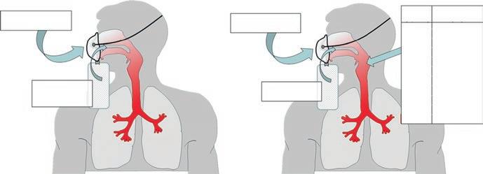
Fig. 1.5 Inspiratory gases during conventional oxygen therapy are a mixture of medical oxygen (dry pure oxygen) and air (21% oxygen with humidity around 10–15 mgH2O/L). The impact of this mixing on humidity (left panel) and FiO2 (right panel) of inspiratory gases is represented. In this example with a mask reservoir providing 15 L/min and a total inspiratory fow of 45 L/min, the FiO2 is about 70%. Maximum FiO2 is always lower than 100% as a part of the inspiratory gas is ambient air. The chart of estimated FiO2 for oxygen fow rate from 1 to 15 L/min is provided [167]. Absolute humidity of air and medical gas is indicated based on published data [151, 152]. Medical gases are dry (below 5 mgH2O/L when nonhumidifed and almost 15 mgH2O/L with cold humidifcation*). The mixture of air and medical gases enters the airways between 8 and 15 mgH2O/L, and is warmed and humidifed along the airways to reach 44 mgH2O/L in the alveoli
bubble humidifcation on comfort, using a 15-item comfort scale [159]. The evaluated hypothesis was the non-inferiority of oxygen therapy without humidifcation. Most patients received oxygen fow below 4 L/min, and 28% of the patients received higher fows 6–8 h after inclusion. The overall oxygen therapy discomfort was low with or without humidifcation. After 6–8 h, the study was underpowered to verify the non-inferiority hypothesis. However, differences on comfort were very low between cold humidifcation and no humidifcation. The comfort was non-inferior after 24 h for low fow without humidifcation. In the small subgroup of patients with higher fows, the discomfort was slightly higher after 24 h. Importantly, none of the other important outcomes (need for bronchoscopy, rate of NIV use or intubation, ICU length of stay, and death) differed between groups [159].
Based on these data, the systematic utilization of cold humidifcation should be avoided for low and moderate oxygen fows (below 15 L/min) when used for less than 24 h. The absence of systematic use of humidifcation with oxygen therapy is safe and a potential important cost-saving measure.
1.4.3 Interfaces to Deliver Low-Flow Oxygen
Oxygen tents, hoods, oxygen chamber, mouthpiece, facial and nasal masks, pharyngeal catheter, nasal prongs, nasal cannula, nasal cannula with reservoir (mustache) … several type of interfaces have been used to deliver oxygen [148]. Haldane proposed to deliver oxygen via a mask [44], but it was considered a cumbersome
F. Lellouche and E. L’Her
method. Nasal cannula’s frst utilization during the frst world war was attributed to captain Adrian Stokes [35] and it has been evaluated by Barach in 1929 [160] and is still the most frequently used interface to deliver oxygen fow rate below 6–8 L/min. If higher fows are required for prolonged time of several hours, other interfaces should be used and humidifcation may be added [13, 155]. Nasal prongs are also used, especially in neonates. Non-occlusive facial masks are frequently used with higher oxygen fow rates, with or without reservoir. Reservoir should be kept for situations requiring oxygen fows above 10 L/min, and may lead to CO2 rebreathing if low fow is used [154]. Non-rebreathing masks are used and specifc facial mask with open design may lead to reduced oxygen rebreathing, but only few data are available [161]. Venturi masks are also used allowing humidifcation of the oxygen through nebulization. This device was described by Campbell in 1960 [162].
1.4.4 Estimation of FiO2 Delivered in Spontaneously Breathing Patients
This question has been extensively discussed and several studies have tried to provide estimation of the delivered FiO2 in relation with oxygen fow rate. FiO2 of the inspiratory gas is related to the proportion of pure oxygen coming from the interface (with 100% FiO2) and from the room air (with 21% FiO2) (Fig. 1.5). The “real” delivered FiO2 is then related to several factors such as the interface used to deliver oxygen, the respiratory rate, the minute ventilation, the peak inspiratory fow, and the route of respiration (mouth vs. nose breathers). Studies have found either no difference between mouth-open and mouth-closed breathing [163, 164] or a signifcantly higher FiO2 during mouth-closed breathing compared to mouth-open breathing [165, 166] or the opposite [167]. However, these estimations may be helpful to evaluate the patient’s needs and severity [168]. It must be kept in mind that even with 15 L/min delivered with an oxygen mask, FiO2 is usually below 80% and slightly above 70% [167] (Fig. 1.5).
1.4.5 Monitoring of Conventional Oxygen Therapy
Clinical evaluation of patients receiving oxygen therapy is essential, including respiratory rate, signs of respiratory distress, and other vital parameters. In addition, a monitoring of several other parameters is now recommended for patients receiving conventional oxygen therapy, and pulse oximetry (SpO2) should be used “in all breathless and acutely ill patients” [13]. British Thoracic Society also recommends to monitor a physiological “track and trigger” system such as the national early warning score (NEWS) [13, 169].
Pulse oximetry is usually suffcient for oxygenation monitoring and even if differences exist between devices, it is usually considered that several oximeters from different leading companies are equivalent in terms of accuracy for SpO2 values above 80%, when tested in optimal conditions with healthy subjects [170–172].
However, when the comparisons are conducted in patients, differences between oximeters are more pronounced [173–176]. In patients with suspected increased PaCO2 (in patients with suspected or known CO2 retention and especially in the case of loss of consciousness) and in patients with suspected metabolic disorders (renal insuffciency, shock states, or in specifc intoxications), arterial blood gas analysis with lactate dosage may be required initially then based on patient clinical evolution [13]. Arterial blood gases are mainly used to measure pH, PaCO2, and bicarbonates. Indeed, especially if oxygenation recommendations are followed (i.e., if SpO2 is maintained between 88 and 92% or 90 and 94%), the PaO2 values are not required. The prescription of long-term oxygen therapy may be an exception and PaO2 may be required to decide on the initiation of this treatment [177]. In other situations, capillary blood gases may be suffcient for clinician decisions and preferred for patient’s comfort [178]. A meta-analysis of the study comparing these measurement techniques, pH, PaCO2, and bicarbonate values was well correlated with arterial and capillary blood gases [179].
Concerning oximetry measurements there are well-known limitations (dyshemoglobinemia, polish nail, pigmented skin, low perfusion, motion artifact …) [180, 181]. Inaccuracies of oximeters are frequently reported in the case of low SpO2 (below 80%) and in the case of pigmented skins [170, 171]. However, technical improvement has improved accuracy in diffcult situations. It was shown by Jubran and Tobin in 1990 that in populations with pigmented skin, SpO2 above 95% was required to obtain suffcient oxygenation [182]. Currently, with recent oximeters, these differences among the devices are small within usual levels of SpO2. During severe hypoxemia, however, a gap between SaO2 and SpO2 of several percentage may exists. In Binkler and Feiner studies, with pigmented skins, oximeters overestimated SpO2 by 2.5–5% in comparison with SaO2, especially at very low values (below 70%) and with large standard deviations [170, 171, 173]. Within well-recognized companies for pulse oximetry (Masimo, Nonin, and Nellcor) Fiener et al. showed that the bias with reference values (SaO2) was low whatever the color of the skin when SaO2/SpO2 values were above 80% [171]. In this study, bias of plus or minus 4% still existed with very low SpO2 values (60–70%) and differences up to 10% could be detected in SpO2 values among the tested devices during severe hypoxemia, especially when disposable sensor was used [171]. Yamamoto et al. showed that polish nail had only minor impact of SpO2 measurements [183]. In a study conducted in 33 healthy females, several colors were tested and authors found moderately reduced SpO2 values with specifc colors (blue, beige, purple, and white) but tested oximeters were not among the reference oximeters [184].
Scoring systems have been developed for the early recognition of patients at risk for clinical deterioration [185]. Many scores have been developed (EWS, MEWS, NEWS, ViEWS, AbEWS, NEWS2, NEWS-L [186–189] …). Currently there are more than 100 different published track and trigger systems [190], most of which have been modifed from the original Early Warning Score [189] developed using expert opinion, and have demonstrated variable levels of reliability, validity, and usefulness [191]. Meta-analyses have been conducted to evaluate the impact of the implementation of these scores on outcome [185]. The national
F. Lellouche and E. L’Her
early warning score (NEWS), which uses physiological parameters for scoring, may be benefcial for predicting patient deterioration [189]. However, a general conclusion cannot be generated due to the heterogeneity of the score used in the studies and the use in different populations [192]. The monitoring of early warning scores (Early Warning Score (EWS), National Early Warning Score (NEWS), or other scores) has been recommended [13, 169]. When compared to quick sepsis-related organ failure assessment (qSOFA) and systemic infammatory response syndrome (SIRS), NEWS was found to be the most accurate for the detection of all sepsis endpoints [193, 194]. The introduction of the NEWS did not change the patient outcome in several recent evaluations [195, 196]. NEWS can predict outcome and discriminate patients’ severity when evaluated at the emergency department and after admission [197].
Clinical events before in-hospital cardiac arrest are mainly respiratory events with increased respiratory rate [198]. Respiratory rate is among the frst vital signs to change in deteriorating patients and it has been reported to be the best individual predictor of cardiac arrest in general wards [199]. However, the usual vital parameters, including SpO2 values and respiratory rate, are insuffciently monitored in the general ward in patients before severe outcomes (cardiac arrest, ICU admission, or unexpected death) [200, 201].
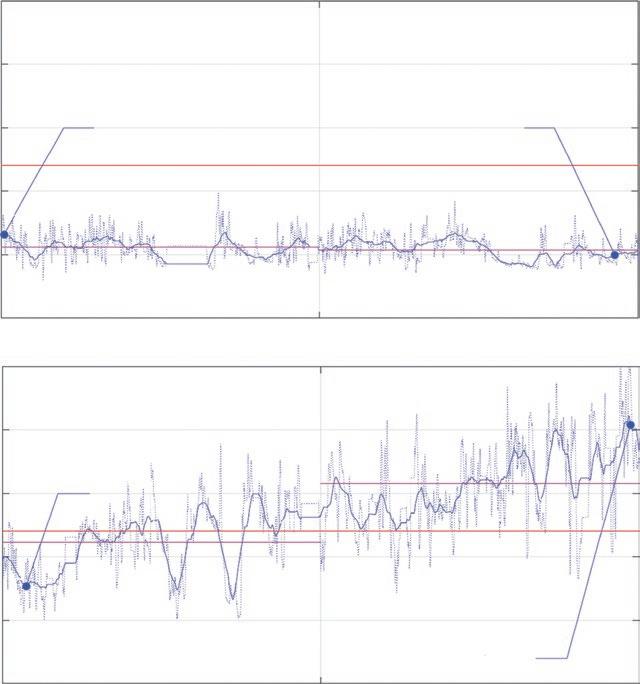
In comparison with the preexisting EWS models, the S/F ratio showed better or comparable predictive accuracy for unexpected ICU transfers in the respiratory wards [202]. With automated oxygen titration devices, the parameters of oxygenation and respiratory parameters are continuously monitored and “respiratory scores” such as EWS.O2 including these parameters and trends may be helpful to monitor the patients receiving oxygen therapy but additional results are required (Fig. 1.6) [203, 204].
1.5 Devices Available to Deliver Oxygen in Spontaneously Breathing Patients
We will discuss below the devices to deliver low or moderate oxygen fows (<15 L/ min). High-fow nasal therapy systems are described in other chapters.
In hospitals, oxygen supplementation is provided from a high-pressure source and a pressure regulator frst reduces the working pressure of the gas to around 50 psi [205]. The most common device to titrate oxygen is the rotameter or oxygen fowmeter that has been used for more than one century, as the frst publication for medical use was in 1910 [21, 206]. Clinicians do the titration manually. Oxygen fowmeters usually deliver fow from 0 to 5 or 0 to 10 L/min, but some devices may provide higher fows; at fush setting, these devices deliver up to 60 L/min [205]. The other conventional way to titrate oxygen is through Venturi mask [162], with which clinicians manually set an estimated FiO2 from 21 to 100%. However, it is usually considered that real FiO2 may not be higher than 70% especially in patients with high respiratory drives leading to high inspiratory fows higher than 60 L/min during hypoxemic respiratory failure [156].
O2=1Lpm RR=25bpm SpO2=90% ScoreO2=6.7
Fig. 1.6 Continuous evaluation of a respiratory score (Early Warning Score.O2) with FreeO2 in spontaneously breathing patients to monitor respiratory severity. The continuous measurement of SpO2, oxygen fow rate, respiratory rate, and heart rate as well as other derived parameters allows continuous display of severity scores based on important clinical parameters. The respiratory score during 48 h of a patient with good clinical evolution is shown on the upper panel. This patient was discharged from the hospital the next day. The respiratory score during 48 h of a patient with deterioration of the clinical condition leading to ICU transfer and endotracheal intubation is shown on the lower panel. Oxygen fow rate (O2), pulse oximetry (SpO2), and respiratory rate (RR) at the beginning and end of the recordings are provided. SpO2 target was set at 90% on FreeO2 1 Conventional
O2=0.1Lpm RR=21bpm SpO2=91%
ScoreO2=5.0
O2=2Lpm RR=25bpm SpO2=90% ScoreO2=7.7
O2=8.5Lpm RR=32bpm SpO2=89%
ScoreO2=20.4
1.5.1 Innovations in Oxygen Delivery
1.5.1.1
Automated Adjustment of Oxygen Therapy
The diffculty of the knowledge transfer is partly related to resistance to changing habits and limited knowledge [207]. These two aspects may explain the failure to implement oxygen recommendations [8, 208]. Austin et al. compared usual oxygen
F. Lellouche and E. L’Her
with a new protocol to manually titrate O2 fow rate to maintain SpO2 target at 88–92% [5]. Noncompliance with the oxygen titration protocol involved 56% of the patients, even if patients were part of a randomized controlled trial. This refects the diffculties to change the practices, even if the complications related to hyperoxemia in COPD patients have been known for 70 years [16]. This problem with compliance to oxygen guidelines was shown in different settings [3, 19, 20]. In addition, the implementation of new protocols requires a lot of energy and motivation, as well as frequent and renewed training. Trainings, educational sessions, oxygen alert stickers with protocol reminders, implementation of local guidelines, emails, and other type of interventions have shown to slightly improve the adherence to oxygen protocols [19]. Improvement of adherence to oxygen protocols is achieved after education interventions but declines to pre-intervention levels over time [54]. Despite more than 50 years of trying to transfer knowledge concerning oxygen therapy recommendations, it is possible to conclude that it has failed. New and frequent guidelines [13, 14] may improve the practices; however, frst recommendations were published 60 years ago [46] with poor intake from clinicians.
It is well documented that prescription practices for oxygen supplementation and adherence to prescriptions are very poor, probably refecting unawareness about the necessity of accurate oxygen prescription and therapy [19]. An audit conducted in 2013 by the British Thoracic Society found that only 55% of patients who were administered oxygen during an admission had a written prescription [209]. Despite the low rate of prescription, this was an improvement compared to 2008, where only 32% of patients supplied with oxygen had a written prescription. In an Australian audit, only 3% of inpatients with an exacerbation of COPD had a written oxygen prescription while 79% of the patients received oxygen supplementation [210]. In a large European audit in 2010–2011 of 16,018 patients with an exacerbation in COPD, it was found that 10.1% received inappropriate oxygen therapy, either with high-fow oxygen or no oxygen despite being hypoxemic [211].
One solution is to automate the transfer of knowledge when feasible. In the situation of oxygen therapy, relatively simple and repetitive tasks are involved: measurement of oxygenation and action based on this measurement. Oxygen therapy is quite simple: to increase oxygen when oxygenation is too low and decrease oxygen when it is too high. Closed loops have been previously used for much more complex procedures such as automated weaning from mechanical ventilation [212, 213] or fully automated mechanical ventilation [214, 215]. The closed-loop oxygen adjustment is very suitable and several systems have been developed recently to try to achieve this goal [22–25].
1.5.1.2 Clinical Evaluations Conducted with FreeO2 (Automated Oxygen Titration)
A new device (FreeO2™, Quebec, Canada) has been developed which regulates in a closed loop and every second the oxygen fow rate (from 0 to 20 L/min with steps of 0.1 L/min) is administered in spontaneous breathing patients via nasal cannulae or mask, according to a SpO2 target chosen by the clinician (Fig. 1.7). This system allows, in addition to the precise regulation of oxygen fow rates, to
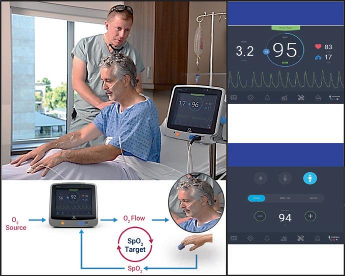
Cardio-respiratory Monitoring
In addition to monitoring delivered O2 flow rates, pulse oximetry enables continuous monitoring of SpO2, heart rate and respiratory rate and derived variables.
Automated titration of the oxygen flow
The main setting is the target SpO2 (here set at 94%). The flow rates are adjusted every second to achieve the desired level of oxygenation
Fig. 1.7 Automated titration devices share the same working principles: an adjustment of the oxygen fow rate to maintain the set SpO2 target. SpO2 is continuously measured and oxygen fow is continuously modifed according to the difference between the measured and the targeted SpO2. Continuous monitoring of various parameters is allowed with these devices
monitor important cardiorespiratory parameters: SpO2, O2 fow rate, respiratory rate, and heart rate [24]. Initial assessment was carried out mainly in Quebec and France in more than 1000 patients. The FreeO2 algorithm was validated and refned in a study in healthy volunteers with moderate induced hypoxemia conditions [24] and during NIV [216], in COPD patients during walking [217, 218] and during acute exacerbations [219–221], in patients with lung fbrosis during walking [222], in patients with acute myocardial infarction [223], at the emergency department in hypoxemic patients and hypercapnic patients [220], in pediatric patients [224], during central apnea [225], and in patients after thoracic or abdominal surgery [226, 227] (Tables 1.1 and 1.2).
An evaluation of FreeO2 was conducted in 50 hospitalized patients for acute exacerbation of COPD [219]. The study showed the feasibility of using the FreeO2 system for prolonged periods (up to 15 days continuously). The time patients were within the SpO2 target prescribed by the pulmonologist was 81 ± 16% with FreeO2 and 51 ± 20% with manual adjustment, with a signifcantly shorter duration of desaturation defned by SpO2 below 85% (0.2% vs. 2.3%, p < 0.001) and hyperoxemia, defned by SpO2 of at least +5% above the target (1.4% vs. 10.4%, p < 0.001)
F. Lellouche and E. L’Her
Table 1.1 Clinical evaluations of the automated oxygen titration devices adapted for spontaneously breathing patients. Studies conducted with FreeO2
STUDIES CONDUCTED WITH FreeO2
FreeO2 in hypercapnic COPD patients during endurance shuttle walking (ref.218)
Severe hypercapnic COPD patients with LTOT
(pilot) (ref.219) 50 COPD patients with acute exacerbation RCT FreeO2 vs continuous O2 (manua setting)
Economic evaluation of FreeO2 during acute exacerbation of COPD (ref.228) 47 COPD patients with acute exacerbation
FreeO2 in the ED in patients with ARF (ref.220) 190 ARF in the
FreeO2 during acute coronary syndrom (pilot) (ref.223)
Acute coronary syndrome RCT FreeO2 (target 92%) FreeO2 (target 96%) vs continuous O2 (manual setting)
FreeO2 in pediatric patients (broncholitis and ARF) (pilot) (ref.224) 60 2 pediatric populations: -Broncholitis (1-2 yrs old) -Hypoxemic respiratory distress (2-15 yrs old)
FreeO2 in patients with id opathic lung fibrosis during exercise (ref.222) 16 Severe IPF
FreeO2 during induced cyclic desaturations (mode of central apnea) (ref.61) 5Healthy subjects
FreeO2 last generation (ref.225) 50 COPD patients with acute exacerbation
FreeO2 post-op patients (ref.227) 198 Adult patients with abdominal or thoracic surgery at risk of complications.
FreeO2 in ICU patients (ref.229)51 Adult patients in the ICU after mechanical ventilation weaning
FreeO2 in COVID-19 patients (submitted)59 Adult patients with COVID-19
RCT
FreeO2 vs continuous O2 (manua setting) (Study duration = 6 hours)
Two consecutive 6MWT continuous O2 - FreeO2
RCT
FreeO2 (2 different controls) vs continuous O2 (2 and 4 L/min) vs Air during cyclic desaturations
Two consecutive recorded periods Continuous O2 (30 minutes) followed by FreeO2 (60 minutes) followed by FreeO2 up to 3 days
RCT FreeO2 vs continuous O2 (manual setting) Analyses during 3 hours (revovery room) and during 3 days after surgery
Prospective crossover cohort study O2 parameters and O2 consumption were recorded during constant O2 flow (30 minutes) and FreeO2 modes (2 hours)
Prospective observationnal study 21 patients after weaning from MV 7 after high-flow nasa therapy, 31 as a first-line therapy.
well accepted by care givers and patients (80%)
oxygenation with FreeO2: more
in the SpO2 target 81 vs. 51%, less time with hyperoxia: 1.5 vs. 10.4% , less time with hypoxemia: 0.2 vs. 2.3% 30% reduction in hospital LOS (5.8 vs. 8.4 days P=0.051)
oxygenation (time in the target, hyperoxia, hypoxemia) Less time with severe hypoxemia (SpO2 <85%) Less ventricular arythmias
95% of the time in the SpO2 target with FreeO2 vs.76% with manual setting
Better oxygenation (time in the target, hyperoxia, hypoxemia) Reduced hospital LOS
Less desaturation end of 6MWT (87.4% vs 83.5%, P=0.039), Less dyspnea (Borg=6.3 vs 4.8, P=0.02).
Increased exercise tolerance vs O2 (309 vs. 252 m, P=0.016)
With FreeO2 more time in the SpO2 target (92%)
Less desaturation in comparsion with air, Less hyperoxia in comparison with constant O2
Reduced O2 consumption in comparison with continuous O2
With FreeO2, reduction of O2 flow rate from 2 to 1L/min (P=0.01) increased time in the SpO2 target (53 vs. 84% of the time P<0.05), hyperoxemia and severe hypoxemia <1% of the time Improved comfort with wireless oximeter (P=0.003)
With FreeO2 more time in the SpO2 target (91 vs. 40%) after 3 hours (recovery room) and after 3 days post-surgery (94 vs. 62%)
Less desaturation (2 vs. 5 min) and hyperoxia (12 vs. 97 min) in comparison with manual O2 (recovery room)
Less desaturation (31 vs. 373 min) and hyperoxia (148 vs. 800 min) in comparison with manual O2 (3 days post surgery)
Less patients with oxygen after 3 days with FreeO2 (18 vs. 33%)
SpO2 range was significantly higher with FreeO2 mode compared to constant O2 flow mode [87% vs. 43%; p < 0.001]. Time with hyperoxia was lower with FreeO2 mode: 8.7% vs. 38.3% Tota O2 consumption was lower with FreeO2: 120 vs. 240 L/min P<0.001
Table 1.2 Clinical evaluations of the automated oxygen titration devices adapted for spontaneously breathing patients. Studies conducted with other closed-loop systems
Studies, References n Population assessed. Main results
O2matic® in COVID-19 patients (ref.232) 15 COVID-19 patients in the medical ward Prospective observationnal study
with automated adjustment in comparison with manual adjustment. The duration of hospitalization was 5.8 ± 4.4 days with FreeO2 vs. 8.4 ± 6.0 days with manual adjustment (p = 0.051) [219]. A cost-effectiveness study has demonstrated, taking into account the cost of the technology, with a reduction in costs of 21.6% ( 3091$
in Canadian dollars) at 180 days. The study demonstrated that this technology is cost effective for all oxygenation criteria [228].
A multicenter randomized study compared manual titration or FreeO2 at the emergency department [220]. One hundred ninety patients admitted to the emergency department for acute respiratory distress requiring oxygen were included and randomized to receive oxygen for 3 h with manual or automated titration [220]. In the FreeO2 group, there were fewer desaturations, less hyperoxemia, and more time in the SpO2 target. In addition, more patients were completely (14.1% vs. 4.3%, p < 0.001) or partially (more than 50% decrease O2, 30% vs. 17%, p < 0.001) weaned from O2 during the 3-h study period. Despite a limited duration of exposure to 3 h, there was an impact beyond the study period: the duration of oxygen administration in the hospital was reduced (5.6 vs. 7.1 days, p = 0.002) and the length of stay was signifcantly reduced (9.2 vs. 11.1 days, p = 0.001) in the FreeO2 group.
Oxygen strategy has been evaluated in 198 postoperative patients, with 103 randomized to usual oxygen titration and 95 randomized to automated oxygen titration [227]. Time in the SpO2 range was higher in the automated group, both initially (⩽3 h; 91.4% vs. 40.2% of time, p < 0.0001) and during the 3-day period (94.0% vs. 62.1% of time, p < 0.0001). Periods of hypoxemia were reduced in the automated group (33 min vs. 370 min), as well as hyperoxemia under oxygen (5 min vs. 178 min, p < 0.0001). Oxygen was still prescribed after day 3 in 33.3% versus 18.3% of patients in the standard and automated groups, respectively (p = 0.01) [227].
Recently, FreeO2 was evaluated in Tunisia in 51 ICU patients after extubation during 2 h [229]. The time in the SpO2 target increased twofold with automated oxygen titration (87% vs. 43% of the time, p < 0.001), while oxygen consumption decreased twofold in comparison with usual titration (121 vs. 240 L/h, p < 0.001) [229]. Reduction of oxygen use has also been evaluated with automated oxygen titration in 36 hospitalized patients on oxygen therapy. SpO2 targets at 90, 92, 94, 96, and 98% were set and oxygen fow at steady state was collected [230]. The mean oxygen fow increased twofold from 90% to 92% of SpO2 target and increased by a factor of 6.3 from 90% to 98%.
A meta-analysis described the benefts on oxygenation, hospital length of stay, and duration of oxygen therapy but authors concluded that other data are necessary [231].
1.5.1.3 Other Systems Allowing Automatic Adjustment of Oxygen
Three studies have been published to evaluate other devices using a closed loop between SpO2 and oxygen fow rates [22, 25, 26]. In one study, the authors evaluated a closed-loop oxygen-adjusting system (O2 Flow Regulator, Dima, Bologna, Italy) during exercise in 18 oxygen-dependent COPD patients [22]. The device was set to maintain SpO2 at 94%. Mean SpO2 was 95 ± 2% with automated adjustment and 93 ± 3% with manual adjustment (p = 0.04). Patients spent signifcantly less time below the target SpO2 (171 ± 187 s vs. 340 ± 220 s, p < 0.001) with the O2 Flow Regulator. The manual intervention time decreased with the automated adjustment (2.0 ± 0.1 vs. 5.6 ± 3.7 min, p = 0.005). In another published study, Rice et al. evaluated a closed-loop device to titrate oxygen (AccuO2™, OptiSat Medical, Inc. 1 Conventional
F. Lellouche and E. L’Her
Minneapolis, USA) in 28 COPD patients with long-term oxygen therapy [25]. The closed loop was compared to constant oxygen fow and to an O2-conserving device. The mean SpO2 was signifcantly different at 90.7 ± 1.9% (AccuO2™) vs. 92.4 ± 3.6% vs. 92.2 ± 4.4% compared to traditional oxygen delivery systems [25]. In addition, this system reduced oxygen consumption with oxygen retention ratios of 9.9 ± 7.3 and 2.6 ± 1.0 compared to other oxygen delivery systems (p < 0.001) [25]. Recently, Hansen et al. evaluated a new closed loop (O2matic, O2matic Ltd., Herlev, Denmark) in 19 COPD patients hospitalized for exacerbation. Patients were managed in crossover randomly with manual or automated oxygen titration during 4 h. During automated titration, SpO2 was maintained in the target 85.1% of the time vs. 46.6% with manually controlled oxygen (p < 0.001). Time with SpO2 < 85% was 1.3% with automated titration and 17.9% with manual oxygen titration (p = 0.01). Time with SpO2 above target did not differ (4.6% vs. 10.5%, p = 0.2) [26]. Recently, O2matic has been evaluated in an observational study on 15 patients with COVID-19 [232]. The device was used during a mean of 2.7 days and patients were in the SpO2 target at a mean of 82.9% of time, 12.0% of the time below the target, and 5.1% of the time above the target [232]. Several aspects such as range fows, accuracy, and type of control differ when comparing the available devices [22, 25, 26]. No comparison has been conducted so far comparing the existing systems. Other devices have been developed for intubated premature infants and will not be discussed here [23].
The potential benefts of the automated system to titrate oxygen exist for both patients (better control of oxygenation, better monitoring) than for the health system (reduction of workload, better follow-up of recommendations, strategies integrating telemedicine, cost savings) [233, 234].
These systems could improve the management of COPD patients across the continuum of care [235] and for other populations than accurate oxygen titration [14]. During the initial management of COPD exacerbations, automated oxygen adjustment based on recommendations should improve the outcome of these patients by reducing the frequency and severity of respiratory acidosis [106, 108]. By combining telemedicine with automated oxygen titration, new management strategies for COPD patients can be envisaged, which should make it possible to shorten the length of hospital stays and thus the overall cost of treating these patients.
1.6 Conclusion
Conventional oxygen therapy is used daily by most of the clinicians in hospitals and is one of the most common drug, but knowledge about its toxicity remains limited and overlooked. The main belief is that oxygen is a vital gas and that it can not be harmful while literature is compelling and now demonstrates that hyperoxemia increases morbidity and mortality in populations daily managed with liberal oxygen in hospitals and beyond. Both aspects of this gas (vital and toxic) should now be considered and recent recommendations underline that hypoxemia as well as hyperoxemia are detrimental to patients. Severe hypoxemia may be an immediate threat
for the patients while hyperoxemia, while less visible, may be as dangerous with systemic, vascular, and cellular impacts. In premature infants and in COPD the need to accurately titrate oxygen has been known for a long time but it is diffcult to achieve. In other populations, accurate titration of oxygen during hospitalization is also mandatory. The frst goal of oxygen therapy is to treat hypoxemia, and the other major goal is to avoid hyperoxemia. In COPD patients, oxygen fow rate should be titrated to maintain SpO2 between 88 and 92%. In other populations, it is now recommended to maintain SpO2 values in a tight range between 90 and 94%. Situations with SpO2 values above the target such as values below the target should be considered as dangerous for the patients. There is no room anymore for hyperox(em)ia in the healthcare system [9, 236]! The knowledge transfer concerning oxygen therapy seems to be a failure. Similarly to other areas in medicine, automated knowledge transfer by using closed-loop systems could be a solution. Current systems to manually adjust oxygen fow, “oxygen fowmeters” use the principle of rotameter, a more than centenary technology [21, 206]. For a century, few innovations have been occurring in the feld of oxygen therapy, yet technological improvements allow the use of more sophisticated devices to accurately titrate the oxygen fow rate, to improve effciency, safety, and monitoring. These automated systems may be useful in many clinical settings such as COPD but also beyond, and initial assessments of these devices demonstrate their potential impact.
Acknowledgements Competing interests: François Lellouche and Erwan L’Her are cofounders, shareholders, and directors of OxyNov. This research and development company has designed and marketed the automated oxygen adjustment system (FreeO2) mentioned in the manuscript.
References
1. Papazian L, et al. Use of high-fow nasal cannula oxygenation in ICU adults: a narrative review. Intensive Care Med. 2016;42(9):1336–49.
2. O'Driscoll BR, et al. British Thoracic Society emergency oxygen audits. Thorax. 2011;66(8): 734–5.
3. Hale KE, Gavin C, O'Driscoll BR. Audit of oxygen use in emergency ambulances and in a hospital emergency department. Emerg Med J. 2008;25(11):773–6.
4. The National Confdential Enquiry into Patient Outcome and Death. Inspiring Change. 2017. London 2017.
5. Austin MA, et al. Effect of high fow oxygen on mortality in chronic obstructive pulmonary disease patients in prehospital setting: randomised controlled trial. BMJ. 2010;341: c5462.
6. Chu DK, et al. Mortality and morbidity in acutely ill adults treated with liberal versus conservative oxygen therapy (IOTA): a systematic review and meta-analysis. Lancet. 2018;391(10131):1693–705.
7. Girardis M, et al. Effect of conservative vs conventional oxygen therapy on mortality among patients in an intensive care unit: the oxygen-ICU randomized clinical trial. JAMA. 2016;316(15):1583–9.
8. Beasley R, et al. High-concentration oxygen therapy in COPD. Lancet. 2011;378(9795):969–70.
9. McEvoy JW. Excess oxygen in acute illness: adding fuel to the fre. Lancet. 2018;391(10131): 1640–2.
F. Lellouche and E. L’Her
10. Lellouche F. Oxygen for myocardial infarction: not an open Bar! JACC Cardiovasc Interv. 2018;11(16):1598–600.
11. O'Driscoll BR, et al. A study of attitudes, beliefs and organisational barriers related to safe emergency oxygen therapy for patients with COPD (chronic obstructive pulmonary disease) in clinical practice and research. BMJ Open Respir Res. 2016;3(1):e000102.
12. Burls A, et al. Oxygen use in acute myocardial infarction: an online survey of health professionals' practice and beliefs. Emerg Med J. 2010;27:283–6.
13. O'Driscoll BR, et al. BTS guideline for oxygen use in adults in healthcare and emergency settings. Thorax. 2017;72(Suppl 1):ii1–ii90.
14. Siemieniuk RAC, et al. Oxygen therapy for acutely ill medical patients: a clinical practice guideline. BMJ. 2018;363:k4169.
15. Sjoberg F, Singer M. The medical use of oxygen: a time for critical reappraisal. J Intern Med. 2013;274(6):505–28.
16. Davies CE, Mackinnon J. Neurological effects of oxygen in chronic cor pulmonale. Lancet. 1949;2(6585):883–5. illust
17. Terry TL. Extreme prematurity and fbroblastic overgrowth of persistent vascular sheath behind each crystalline lens. Am J Ophthalmol. 1942;25:203–4.
18. Patz A, Hoeck LE, De La Cruz E. Studies on the effect of high oxygen administration in retrolental fbroplasia. I. Nursery observations. Am J Ophthalmol. 1952;35(9): 1248–53.
19. Cousins JL, Wark PA, McDonald VM. Acute oxygen therapy: a review of prescribing and delivery practices. Int J Chron Obstruct Pulmon Dis. 2016;11:1067–75.
20. Ringbaek TJ, Terkelsen J, Lange P. Outcomes of acute exacerbations in COPD in relation to pre-hospital oxygen therapy. Eur Clin Respir J. 2015;2
21. Foregger R. The rotameter and the waterwheel. Anaesthesist. 2001;50:701–8.
22. Cirio S, Nava S. Pilot study of a new device to titrate oxygen fow in hypoxic patients on longterm oxygen therapy. Respir Care. 2011;56(4):429–34.
23. Claure N, Bancalari E. Automated closed loop control of inspired oxygen concentration. Respir Care. 2013;58(1):151–61.
24. Lellouche F, L'Her E. Automated oxygen fow titration to maintain constant oxygenation. Respir Care. 2012;57(8):1254–62.
25. Rice KL, et al. A portable, closed-loop oxygen conserving device for stable COPD patients: comparison with fxed dose delivery systems. Respir Care. 2011;
26. Hansen EF, et al. Automated oxygen control with O2matic® during admission with exacerbation of COPD. Int J COPD. 2018;13:3997–4003.
27. Heffner JE. The story of oxygen. Respir Care. 2013;58(1):18–31.
28. Prinke RT. New light on the alchemical writings of Michael Sendivogius (1566–1636). Ambix. 2016;63(3):217–43.
29. Lavoisier A-L. Mémoire sur la combustion en général. Mémoires de l'Académie des Sciences, 1777: p. 593.
30. Holmes Frédéric L. The boundaries of Lavoisier's chemical revolution /Les limites de la révolution chimique de Lavoisier. Débats et chantiers actuels autour de Lavoisier et de la révolution chimique. Revue d'histoire des Sciences. 1995;48(1–2):9–48.
31. Priestley J. Experiments and observations on different kinds of air, vol. 101. 2nd ed. London: J. Johnson; 1775.
32. Caillens, Observations sur un nouveau moyen de remedier à la phtisie pulmonaire. Gazette de Santé, 1783: p. 38.
33. Beddoes T, Watt J. Considerations on the medicinal use, and on the production of factitious airs. Ann Med. 1796:245–65.
34. Leigh JM. Early treatment with oxygen. The pneumatic institute and the panaceal literature of the nineteenth century. Anaesthesia. 1974;29(2):194–208.
35. Grainge C. Breath of life: the evolution of oxygen therapy. J R Soc Med. 2004;97(10):489–93.
36. Smith AH. Oxygen gas as a remedy in disease. New York: D. Appleton & Company; 1870.
37. Haldane JS. Respiration. London: Oxford University Press; 1922.
38. Barcroft JR. Supply of oxygen to the tissues. The scientifc monthly, vol. 11. New York: The Science Press; 1920.
39. Christiansen J, Douglas CG, Haldane JS. The absorption and dissociation of carbon dioxide by human blood. J Physiol. 1914;48(4):244–71.
40. Jobling MA. The unexpected always happens. Investig Genet. 2012;3:5.
41. Clark R. The life and work of JBS Haldane Oxford: Oxford University Press; 1968.
42. Barach AL. The therapeutic use of oxygen. JAMA. 1922;79(9):693–8.
43. Blodgett AN. The continuous inhalation of oxygen in cases of pneumonia otherwise fatal, and in other diseases. Boston Med Surg J. 1890;123:481–4.
44. Haldane JS. The therapeutic administration of oxygen. Br Med J. 1917;1(2928):181–3.
45. Haldane JS. A lecture on the symptoms, causes, and prevention of Anoxaemia (insuffcient supply of oxygen to the tissues), and the value of oxygen in its treatment. Br Med J. 1919;2(3055):65–71.
46. Campbell EJ. Respiratory failure: the relation between oxygen concentrations of inspired air and arterial blood. Lancet. 1960;2(7140):10–1.
47. Massaro DJ, Katz S, Luchsinger PC. Effect of various modes of oxygen administration on the arterial gas values in patients with respiratory acidosis. Br Med J. 1962;2(5305):627–9.
48. Leigh JM. Oxygen therapy techniques after 200 years. A survey of present practice and current trends. Anaesthesia. 1973;28(2):164–9.
49. Jouneau S, et al. Management of acute exacerbations of chronic obstructive pulmonary disease (COPD). Guidelines from the Societe de pneumologie de langue francaise (summary). Rev Mal Respir. 2017;34(4):282–322.
50. Beasley R, et al. Thoracic Society of Australia and new Zealand oxygen guidelines for acute oxygen use in adults: 'Swimming between the fags'. Respirology. 2015;20(8):1182–91.
51. Group, B.I.U.K.C, et al. Oxygen saturation and outcomes in preterm infants. N Engl J Med. 2013;368(22):2094–104.
52. Thein OS, et al. Oxygen prescription: improving compliance using methods from BMJ open quality journal. BMJ Open Qual. 2018;7(2):e000288.
53. Choudhury A, et al. Can we improve the prescribing and delivery of oxygen on a respiratory ward in accordance with new British Thoracic Society oxygen guidelines? BMJ Open Qual. 2018;7(4):e000371.
54. Myers H, et al. Doctors learn new tricks, but do they remember them? Lack of effect of an educational intervention in improving oxygen prescribing. Respirology. 2015;20(8): 1229–32.
55. Wilson DF. Oxidative phosphorylation: regulation and role in cellular and tissue metabolism. J Physiol. 2017;595(23):7023–38.
56. Bender DA, Mayes PA. In: DA BKB, Weil PA, Kennelly PJ, Murray RK, Rodwell VW, editors. Glycolysis & the Oxidation of Pyruvate, in Harper’s Illustrated Biochemistry. New York: McGraw-Hill; 2011.
57. Dickinson BC, Chang CJ. Chemistry and biology of reactive oxygen species in signaling or stress responses. Nat Chem Biol. 2011;7(8):504–11.
58. Freeman BA, Crapo JD. Biology of disease: free radicals and tissue injury. Lab Investig. 1982;47(5):412–26.
59. Halliwell B, Gutteridge JM. Oxygen toxicity, oxygen radicals, transition metals and disease. Biochem J. 1984;219(1):1–14.
60. Valko M, et al. Redox- and non-redox-metal-induced formation of free radicals and their role in human disease. Arch Toxicol. 2016;90(1):1–37.
61. Di Meo S, et al. Role of ROS and RNS sources in physiological and pathological conditions. Oxidative Med Cell Longev. 2016;2016:1245049.
62. Rhee SG. Cell signaling. H2O2, a necessary evil for cell signaling. Science. 2006; 312(5782):1882–3.
63. Lane N. Oxygen: the molecule that made the world. Oxford: OUP; 2002.
64. Carreau A, et al. Why is the partial oxygen pressure of human tissues a crucial parameter? Small molecules and hypoxia. J Cell Mol Med. 2011;15(6):1239–53.
F. Lellouche and E. L’Her
65. Hafner S, et al. Hyperoxia in intensive care, emergency, and peri-operative medicine: Dr. Jekyll or Mr. Hyde? A 2015 update. Ann Intensive Care. 2015;5(1):42.
66. Jamieson D, et al. The relation of free radical production to hyperoxia. Annu Rev Physiol. 1986;48:703–19.
67. Severinghaus JW, Astrup PB. History of blood gas analysis. IV. Leland Clark's oxygen electrode. J Clin Monit. 1986;2(2):125–39.
68. Glansdorff N, Xu Y, Labedan B. The last universal common ancestor: emergence, constitution and genetic legacy of an elusive forerunner. Biol Direct. 2008;3:29.
69. Ouzounis CA, et al. A minimal estimate for the gene content of the last universal common ancestor--exobiology from a terrestrial perspective. Res Microbiol. 2006;157(1): 57–68.
70. Woese C. The universal ancestor. Proc Natl Acad Sci U S A. 1998;95(12):6854–9.
71. Klotz MG, Loewen PC. The molecular evolution of catalatic hydroperoxidases: evidence for multiple lateral transfer of genes between prokaryota and from bacteria into eukaryota. Mol Biol Evol. 2003;20(7):1098–112.
72. Gerschman R, et al. Oxygen poisoning and x-irradiation: a mechanism in common. Science. 1954;119(3097):623–6.
73. Grocott MP, et al. Arterial blood gases and oxygen content in climbers on Mount Everest. N Engl J Med. 2009;360(2):140–9.
74. Robson AG, Hartung TK, Innes JA. Laboratory assessment of ftness to fy in patients with lung disease: a practical approach. Eur Respir J. 2000;16(2):214–9.
75. Slutsky AS, Rebuck AS. Heart rate response to isocapnic hypoxia in conscious man. Am J Phys. 1978;234(2):H129–32.
76. Easton PA, Slykerman LJ, Anthonisen NR. Ventilatory response to sustained hypoxia in normal adults. J Appl Physiol (1985). 1986;61(3):906–11.
77. Bradley CA, Fleetham JA, Anthonisen NR. Ventilatory control in patients with hypoxemia due to obstructive lung disease. Am Rev Respir Dis. 1979;120(1):21–30.
78. Kogure K, et al. Mechanisms of cerebral vasodilatation in hypoxia. J Appl Physiol. 1970;29(2):223–9.
79. West JW, Guzman SV. Coronary dilatation and constriction visualized by selective arteriography. Circ Res. 1959;7(4):527–36.
80. Weir EK, et al. Acute oxygen-sensing mechanisms. N Engl J Med. 2005;353(19):2042–55.
81. Galatius-Jensen S, et al. Nocturnal hypoxaemia after myocardial infarction: association with nocturnal myocardial ischaemia and arrhythmias. Br Heart J. 1994;72(1):23–30.
82. Gill NP, Wright B, Reilly CS. Relationship between hypoxaemic and cardiac ischaemic events in the perioperative period. Br J Anaesth. 1992;68(5):471–3.
83. Vincent JL, De Backer D. Circulatory shock. N Engl J Med. 2013;369(18):1726–34.
84. Helmerhorst HJ, et al. Metrics of arterial Hyperoxia and associated outcomes in critical care. Crit Care Med. 2017;45(2):187–95.
85. Kilgannon JH, et al. Association between arterial hyperoxia following resuscitation from cardiac arrest and in-hospital mortality. JAMA. 2010;303(21):2165–71.
86. Page D, et al. Emergency department hyperoxia is associated with increased mortality in mechanically ventilated patients: a cohort study. Crit Care. 2018;22(1):9.
87. Lorrain Smith J. The pathological effects due to increase of oxygen tension in the air breathed. J Physiol. 1899;24(1):19–35.
88. Clark JM, Lambertsen CJ. Pulmonary oxygen toxicity: a review. Pharmacol Rev. 1971;23(2):37–133.
89. Kallet RH, Matthay MA. Hyperoxic acute lung injury. Respir Care. 2013;58(1):123–41.
90. Kim V, et al. Oxygen therapy in chronic obstructive pulmonary disease. Proc Am Thorac Soc. 2008;5(4):513–8.
91. Selinger SR, et al. Effects of removing oxygen from patients with chronic obstructive pulmonary disease. Am Rev Respir Dis. 1987;136(1):85–91.
92. Barach AL. Therapeutic use of oxygen in heart disease. Ann Intern Med. 1931;5(10):428.
93. Donald K. Neurological effects of oxygen. Lancet. 1949;257:1056–7.