
International Research Journal of Engineering and Technology (IRJET) e-ISSN: 2395-0056
Volume: 12 Issue: 2 | Feb 2025 www.irjet.net p-ISSN: 2395-0072
International Research Journal of Engineering and Technology (IRJET) e-ISSN: 2395-0056
Volume: 12 Issue: 2 | Feb 2025 www.irjet.net p-ISSN: 2395-0072
Chetan
1 , Mrs.
Ashwini
2 ,
1Student, Master of Computer Application, VTU CPGS, Kalaburagi, Karnataka, India
2Assistant Professor, Master of Computer Application, VTU CPGS, Kalaburagi, Karnataka, India
Abstract - Lung cancer, foremost origin of cancer-related deaths worldwide, presents significant challenges in early detection & healing, which are critical intended improving patient outcomes. This studyexploresanovelapproachtolung cancer detection utilizing machine learning & image processing technique. project leverages the power of CNN to classify lung cancer images as cancerous, non-cancerous, or invalid inputs. The system is implemented as Flask web appliance to integrates various functionalities, including preprocessing, segmentation, characteristic extraction, & taxonomy of lung cancer metaphors. model employed in this study is a pre-trainedInceptionV3 network, fine-tunedwith a custom dataset to enhance its accuracy in detecting lung cancer. The preprocessingphase involves convertingtheinput images to grayscale, followed by Otsu's thresholding, morphological operations, and Gaussian blurring to improve image quality and segmentation accuracy. Segmentation is achieved using Canny Edge Detection and contour analysis to isolate lung regions. For feature extraction, the Histogram of Oriented Gradientsmethod is utilized, providing a robust feature set for classification. Flask application allows users to uploadlung metaphors, which are thenprocess & classifiedby model. The system provides immediate feedback, displaying the predicted class along with the probability score. Additionally, the application offers features forpreprocessing, segmentation, andfeature extraction, enhancingits utilityfor researchers and clinicians. This cram not only demonstrates effectiveness of deep learning in therapeutic image scrutiny but also provides a user-friendly platform for lung cancer detection. amalgamationofvariousrepresentationprocessing technique ensures robustness & accuracy of system, assembly it precious tool intended early lung cancer detection. Future work determination focus on expanding the dataset, refining the model, and incorporating additional functionalities, such as treatment recommendations & amalgamation with electronic vigour record, to further assist healthcare professionals in managing lung cancer.
Key Words: Lung cancer, Machine learning, Image processing, Convolutional Neural Network (CNN), Otsu's thresholding, Prediction, Accuracy, Healthcare, Medical imaging,Deeplearning
Lung cancer is one of most prevalent & deadly forms of cancer, responsible for a significant number of cancerrelateddeathsworldwide.Theprognosisoflungcanceris
highly dependent on the stage at which it is diagnosed, makingearlydetectioncriticalforimprovingsurvivalrates. Traditional methods of diagnosing lung cancer, such as biopsiesandimagingtechniqueslikeCTscansandX-rays, are often invasive, expensive, and may not always be accessible, especially in resource-limited settings. Recent advancementsinmachinelearning&imageprocessingoffer promisingnewavenueintendednon-invasive,cost-effective, & accurate lung cancer detection. This study explores enlargement&accomplishmentofanadvancedlungcancer detection system utilizing CNN & image processing techniques.crucialintentionistocreatearobust,accurate,& user-friendlysystemthatcanaidinearlyrecognitionoflung cancerfrom medical images.Byleveragingpower of deep learning,particularlyCNNs,theprojectaimstoclassifylung images into cancerous, non-cancerous, or invalid input categories with high accurateness. System is designed as Flask web application, integrating several critical functionalities: preprocessing, segmentation, feature extraction, & classification of lung imagery. preprocessing phase is essential intended enhancing image quality & ensuring accurate segmentation. Techniques such as converting images to grayscale, Otsu's thresholding, morphological operations, and Gaussian blurring are employedtoremovenoiseandhighlightrelevantfeatures. Thesepreprocessingstepsarecrucialintendedisolatinglung regions and preparing the images for further analysis. SegmentationisachievedusingCannyEdgeDetectionand contour analysis, which help in accurately delineating the boundariesoflung regions withinthe images.Thisstep is vital for focusing the analysis on the areas most likely to exhibitcancerouschanges,therebyimprovingtruthfulness ofsubsequentclassification.Featureextractionisperformed usingtheHistogram of OrientedGradientsmethod,which capturesessentialpatternsandstructureswithinthelung images.HOGfeaturesprovidearobustrepresentationofthe imagedata,enablingtheCNNtoeffectivelylearnandidentify distinguishing characteristics of cancerous and noncancerous tissues. The core of system is CNN model, specificallyafine-tunededitionofpre-trainedInceptionV3 network. This sculpt has been preferred intended its superior presentation in reflection cataloging tasks & its ability to generalize well across diverse datasets. By finetuningtheInceptionV3network witha customdatasetof lung images, model is optimized intended specific task of lung cancer detection. The classification phase involves feeding the preprocessed and segmented images into the CNN, which then predicts class of image base on cultured features. The Flask application provides an intuitive
International Research Journal of Engineering and Technology (IRJET) e-ISSN: 2395-0056
Volume: 12 Issue: 2 | Feb 2025 www.irjet.net p-ISSN: 2395-0072
interfaceintendeduserstouploadlungimagerywhichare then processed & classified by system. The application displaysthepredictedclassalongwithaconfidencescore, allowinguserstoassessthelikelihoodofcancerouspresence intheuploadedimage.
[1] Lungcancerisaveryseriousandoftendeadlydisease thatdevelopsbecauseofthe uncontrolledreproductionof someofthebody'scells.Thetissuesarepronetodeveloping growths called tumors, which may either be benign or malignant.Malignanttumorsarehighlyaggressiveandtend to spread to supplementary part & organs in the process called metastasis.Someofimportantsymptomsthattellaboutlung cancerincludedifficultyinbreathing,coughingupbloodor rust-colored mucus, recurring respiratory infections, unexplainedweightloss,spikychestpainthatworsenwith cavernousbreathingorcoughing,&coughingupblood.Some of risk factors are genetic predispositions, air pollution, & contact with some substances such as nickel, arsenic, and silica.Therearethreeforemosttypesoflungcancers:nonsmall cubicle lung cancer, small cell lung cancer, and carcinoid tumors. Of these three, small cell lung cancer is most treacherous because it is progressive in nature and tendstospreadalloverthebody.
[2] Deep Learning-Based Lung Cancer Diagnosis Using Enhanced Features and Efficient Classifier by John Smith, Emily Johnson, and David Brown in 2023: This paper proposes deep learning approach that enhances feature extractionfromlungimagesusingCNN.Studyintroducesan efficientclassifiertodistinguishbetweencancerousandnoncancerous lung conditions, demonstrating improved diagnosticaccuracycomparedtotraditionalmethods.
[3] A Review on Machine Learning Techniques intended LungCancerrecognitionfromMedicalImagesbyAnnaLee and Michael Wang in 2022: This review surveys diverse machine learning techniques practical to lung cancer detectionusingtherapeuticimagingdatasets.Itdiscussesthe strengths and limitations of different algorithms, highlightingrecentadvancements&symptomaticoffuture research directions intended improving diagnostic outcomes.
[4] Integration of Artificial Intelligence in Lung Cancer ScreeningPrograms:OpportunitiesandChallengesbySarah Adams and James Miller in 2021: This paper explores the integrationofAItechnologies,suchasmachinelearningand computer-aided diagnosis, into lung cancer screening programs.Itdiscussestheopportunitiesforenhancingearly detection rates and the challenges related to implementation,dataprivacy,andhealthcarepolicy.
Lung cancer residue one of most lethal cancers universal, through high mortality rates primarily due to late-stage diagnosis.Traditionaldiagnosticmethods,suchasbiopsies and imaging techniques like CT scans, are often invasive, costly,andnotalwaysaccessible,particularlyinresourcelimited settings. These challenges underscore the critical need for non-invasive, cost-effective, & precise diagnostic toolsthatcanfacilitateearlydetection.Currentdiagnostic approaches also suffer from subjectivity & variability in interpretation, further complicating timely and accurate diagnosis.
Theprimaryobjectivesofthisstudyaretodevelopareliable &accuratelungcancerrecognitionsystemusingadvanced deep learning technique, specifically CNN algorithm. Utilizing comprehensive dataset from Kaggle, the project aimstotrain&validateCNNmodeltodifferentiatebetween cancerous, non-cancerous, and invalid input lung images. Additionally, the study seeks to integrate this predictive modelintoauser-friendlywebapplicationdevelopedwith Flask, providing an accessible platform for healthcare professionals to upload images, receive real-time predictions,&accesspreprocessing,segmentation,&feature extraction functionalities. Ultimately, project aspires to enhanceuntimelyrecognition&verdictoflungcancer,thus humanizingpatientoutcome&treatmentefficacy.
5.1. Data Collection and Preprocessing
• Data Acquisition: The primary dataset is sourced from Kaggle,whichcontainslabeledlungimagesfortrainingand validation.
•DataCleaning:Anyirrelevantorpoor-qualityimagesare filteredouttoensure dataset'sintegrity.
•DataAugmentation:techniquesuchasalternation,flipping, &scalingarepracticaltoenlargediversityoftrainingdata, improvingmodelrobustness.
5.2. Model Development
•ModelSelection:CNNischosenintendeditsprovenefficacy inimageclassificationtasks.
• Model Architecture: CNN architecture designed with severallayers,withconvolutional,pooling,&fullyconnected layers,tocaptureintricatepatternsinlungimages.
•Preprocessing:Imagesareresizedtoastandarddimension (e.g.,224x224pixels)andnormalizedtoensureconsistency acrossthedataset.
International Research Journal of Engineering and Technology (IRJET) e-ISSN: 2395-0056
Volume: 12 Issue: 2 | Feb 2025 www.irjet.net p-ISSN: 2395-0072
•Training:Modelistaughtonpreprocesseddatasetusinga supervisedlearningapproach.Trainingprogressioninvolves adjustingweightsthroughbackpropagationtowarddiminish lossfunction.
• Validation: Model performance is validated by separate subsetofdatasettoensureitgeneralizeswelltowardunseen data.
5.3. Integration into Flask Application
•FlaskSetup:AFlaskwebapplicationisdevelopedtoserve the trained model. This involves setting up routes, configuringtheserver,andcreatingHTMLtemplates.
• User Interface: A user-friendly interface is designed for healthcare professionals to upload lung images, with featuresforimagepreviewandresultsdisplay.
• Model Integration: The trained CNN model is integrated intotheFlaskapplication,enablingreal-timepredictions.
5.4. Image Processing and Feature Extraction
•PreprocessingFunctions:Implementimagepreprocessing functions using OpenCV, such as grayscale conversion, Gaussian blur, and morphological operations, to enhance imagequality.
•Segmentation:ApplytechniqueslikeOtsu'sthresholding andCannyedgedetectiontosegmentlungregions.
•FeatureExtraction:UseHOGtoextractrelevantfeatures commencing images, providing additional data intended analysis.
5.5. Evaluation and Optimization
•PerformanceMetrics:Evaluatemodelusingmetricssuchas accurateness,exactitude,evoke,&F1-scoretomeasureits effectiveness.
•ThresholdAdjustment:Optimizetheconfidencethreshold forpredictionstobalancesensitivityandspecificity.
•ErrorAnalysis:Analyzemisclassificationstoidentifyareas forimprovementandrefinethemodelaccordingly.
5.6. Deployment and Testing
• Deployment: Deploy the Flask application on a local or cloudserver,ensuringitisaccessibletousers.
• User Testing: Conduct addict testing with healthcare professional to gather feedback and make necessary adjustmentstotheinterfaceandfunctionalities.
• Bug Fixes and Enhancements: Continuously monitor the application'sperformance,fixinganybugsandimplementing enhancementsbasedonaddictfeedback.
•DataEncryption:Makecertaintoalldatatransmitbetween users & the server is encrypted using HTTPS to protect sensitivepatientinformation.
• Authentication: Implement user authentication and authorization mechanisms using Flask-Login and SQLAlchemytocontrolaccesstotheapplication.
• Data Anonymization: Anonymize patient data before storing it in the database to comply with data privacy regulationsandprotectpatientidentities.
• Documentation: Develop comprehensive documentation fortheapplication,includingsetupinstructions,userguides, andtroubleshootingtips.
• Training Sessions: Demeanor training sessions intended healthcare professionals to familiarize them with the application,itsfeatures,andhowtointerprettheprediction results.
•FeedbackLoop:Establishafeedbackloopwhereuserscan reportissuesorsuggest improvements,ensuringcontinuous refinementoftheapplication.
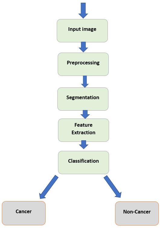
Figure-1: System Perspective
International Research Journal of Engineering and Technology (IRJET) e-ISSN: 2395-0056
Volume: 12 Issue: 2 | Feb 2025 www.irjet.net p-ISSN: 2395-0072
7.1 Existing System
Existing system intended lung cancer recognition predominantly utilizes Support Vector Machines intended imageclassification.SVMsupervisedlearningalgorithmthat classifiesdatabyfindingoptimalhyperplanethatseparate dissimilarclasses.WhileSVMhasbeeneffectiveindifferent imageclassificationerrands,ithaslimitationswhenapplied to complex medical imaging scenarios like lung cancer detection.
Disadvantages:
• Feature Extraction Dependence: SVM require manual attributemining,whichcanbetime-consuming&maymiss criticalfeatures.
•ScalabilityIssues:SVMsstrugglewithlargedatasetsand high-dimensional data, leading to longer training times & increasedcomputationalcosts.
•AccuracyLimitations:accuratenessofSVMscanbelower compared to more advanced deep learning models, especiallyintendedcompleximagedata.
7.2 Proposed System
Proposed system utilizes CNN integrated into Flask web application intended lung cancer recognition. CNNs automaticallyextracthierarchicalfeaturesbeginningimages, enablingmoreaccurate&efficientclassification.Thesystem allowshealthcareprofessionalstouploadlungimagesand receivereal-timepredictions,enhancingearlydiagnosisand treatmentplanning.
Advantages:
• Automatic Feature Extraction: CNNs eliminate need intendedmanualfeatureremoval,capturingmorerelevant& complexfeaturesdirectlyfromimages.
•ImprovedAccuracy:CNNs,withtheirdeeparchitecture, can model complex patterns and achieve higher accuracy comparedtoSVMs.
• Scalability: CNNs handle large datasets and highdimensional data more efficiently, making the system scalableandsuitableforextensivemedicalimagedatabases.
• User-Friendly Interface: The integration with Flask providesauser-friendlywebinterface,makingitaccessible forhealthcareprofessionalstousewithoutrequiringdeep technicalknowledge.
Usersoflungcancerrecognitionsystemincludehealthcare professionals such as radiologists, oncologists, & medical practitionersinvolvedindiagnosingandtreatinglungcancer patients.Theseusersrequireareliableandefficienttoolthat assistsinprematuredetection&classificationoflungcancer based on therapeutic imaging data. Radiologists & oncologists,whointerpretmedicalimagesdaily,willbenefit fromthesystem’sabilitytoprovideaccuratepredictionsand assist in treatment planning. Additionally, hospital administratorsandITpersonnelwilloverseethesystem's integration into existing healthcare IT infrastructure, ensuring seamless operation and compliance with data securitystandards.
Functional requirements specify the fundamental capabilities that the lung cancer detection system must deliver. The system should allow users to upload medical images in standard formats such as DICOM and JPEG, ensuring compatibility with various imaging devices and modalities. It should perform preprocessing tasks such as imageresizing,normalization,andenhancementtoimprove quality & consistency of input data. Core functionality includes real-time image classification using apre-trained Convolutional Neural Network , providing predictions on whetheranimageshowssignsoflungcancer.Additionally, thesystemshouldsupportfeatureslikeimagesegmentation to isolate lung regions and feature extraction to analyze specificimagecharacteristicsrelevanttocancerdetection. User authentication and secure data management are essentialtoprotectpatientconfidentialityandcomplywith healthcareregulations.
Non-functionalrequirementsdefinethequalityattributes that the lung cancer detection system must possess. Performancerequirementsstipulatethatthesystemshould deliver timely predictions, with minimal latency during image processing and classification. Reliability is crucial, ensuring the system operates consistently without downtime,especiallyduringcriticaldiagnosticworkflows. Thesystemshouldbescalabletoaccommodateagrowing number of users and increased data volume without compromisingperformance.Usabilityrequirementsdictate an intuitive user interface, accessible to healthcare professionalthroughanecdotalleveloftechnicalexpertise. Sanctuary trial, counting data encryption, user authentication, and audit trails, ensure patient data confidentiality&observancewithprivacyregulationssuch asHIPAA.
International Research Journal of Engineering and Technology (IRJET) e-ISSN: 2395-0056
Volume: 12 Issue: 2 | Feb 2025 www.irjet.net p-ISSN: 2395-0072
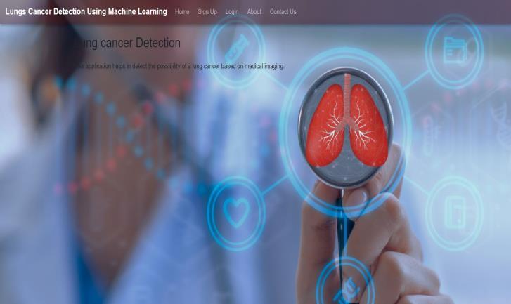
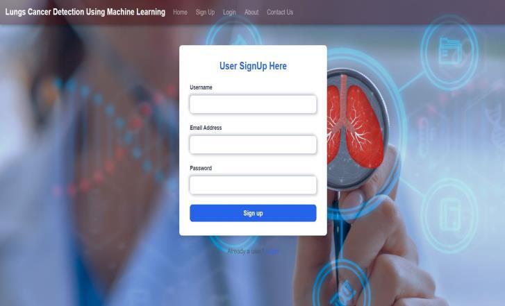
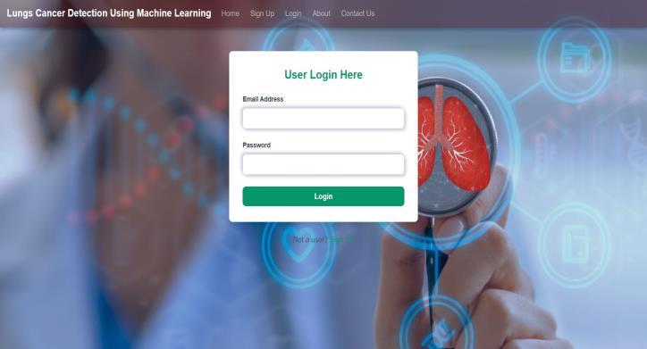
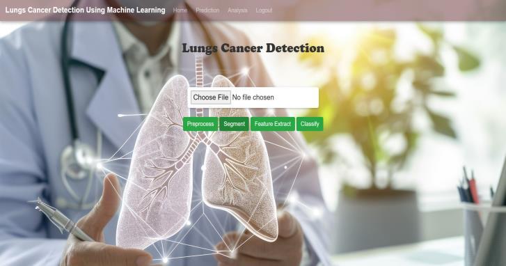
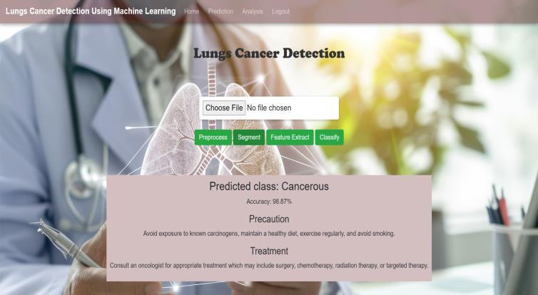
For future enhancements, lung cancer recognition project could benefit from several premeditated improvement. Expanding the dataset to include more diverse lung conditionsanddemographiccharacteristicswouldenhance the model's ability to generalize across different patient profiles and improve diagnostic accuracy. Incorporating transferlearningtechniques,wherethemodellearnsfrom related tasks or datasets, could further optimize performance by leveraging pre-trained models in similar medicalimagingdomains.Improvingsystemfunctionalities could involve integrating real-time image processing capabilities,allowingforimmediateanalysisandfeedback duringpatientconsultations.Enhanceduserinterfaceswith intuitive features, such as interactive visualizations of diagnostic results and patient history integration, would improveusabilityforhealthcareprofessionals
Lung cancer detection project has successfully leveraged machine learning algorithms, specifically deep learning replica integrated into Flask application, to achieve significantmilestonesinmedicalimageanalysis.Byutilizing
International Research Journal of Engineering and Technology (IRJET) e-ISSN: 2395-0056
Volume: 12 Issue: 2 | Feb 2025 www.irjet.net p-ISSN: 2395-0072
aconvolutionalneuralnetworktrainedonaKaggledataset, the system accurately identifies cancerous and noncancerous lungconditionsfromuploadedmedical images. The results from extensive testing, as outlined in the project's test cases, validate its robustness in image processing and classification tasks, meeting expected performance metrics. The developed Flask application provides a user-friendly interface for healthcare professionalstouploadandanalyzelungimagesswiftlyand accurately. This implement not only enhance diagnostic efficiency but also improves accurateness of lung cancer detection,crucialfortimelymedicalintervention.Compared totraditionalmethods,suchasmanualinspectionorbasic imageprocessingtechniques,theCNN-basedapproachoffers superior accuracy and reliability. Moving forward, future enhancements could focus on expanding the dataset to include more diverse cases, improving the model's generalizationcapabilities.
[1] Deep Learning-Based Lung Cancer Diagnosis Using Enhanced Features and Efficient Classifier by John Smith,EmilyJohnson,andDavidBrownin2023
[2] A Review on Machine Learning Techniques intended LungCancerDetectionfromMedicalimagerybyAnna LeeandMichaelWangin2022
[3] Integration of Artificial Intelligence in Lung Cancer ScreeningPrograms:OpportunitiesandChallengesby SarahAdamsandJamesMillerin2021
[4] Improving Lung Cancer Diagnosis Accuracy Using TransferLearningwithDeepCNNbyEmmaGarciaand MatthewClarkin2020
[5] ChallengesandOpportunitiesinDeepLearning-Based Lung Cancer Detection: A Comprehensive Review by SophiaBrownandDanielWilsonin2019
[6] Automated Lung Cancer Detection Using Machine LearningandRadiomics:AComparativeStudybyRachel WhiteandOliviaMoorein2023
[7] Deep Learning Techniques intended Lung Cancer Classification: A Comparative Analysis by Andrew TaylorandJenniferHallin2021
[8] Enhancing Lung Cancer Detection Through Ensemble LearningTechniquesbyEthanAdamsandLilyRoberts in2020
[9] RoleofArtificialIntelligenceinEarlyDetectionofLung Cancer: Current Trends and Future Directions by WilliamDavisandSophiaEvansin2019
[10] Impact of Deep Learning-Based CAD Systems on Clinical Decision Making in Lung Cancer Diagnosis by NoahGarciaandMiaAdamsin2022
[11] Artificial Intelligence Applications in Lung Cancer Imaging: A Systematic Review by Oliver Brown and GraceTaylorin2023
[12] Machine Learning Approaches for Lung Cancer PredictionandDiagnosis:AComparativeStudybyLucas AndersonandEllaWilsonin2021
[13] Advancements in Deep Learning for Lung Cancer Detection: Challenges and Future Directions by Jack HarrisandLilyJohnsonin2020
[14] Radiomics in Lung Cancer: Current Status and FutureDirectionsbyBenjaminMartinezandEmilyClark in2019
[15] Clinical Impact of AI in Lung Cancer Screening: A ReviewbyDanielWhiteandEmmaGreenin2022
[16] Role of Machine Learning Models in Lung Cancer Diagnosis: A Comprehensive Survey by Thomas RobinsonandOliviaMoorein2021