In this age of epidemic testing, it is important and the number of tests should be increased daily to get accurate dataandpracticebut,withthecurrenttestmethodittakesa fewhourstogettheresultsandsometimesifthenumberof tests in large quantities takes longer to get the results. Previous plans to receive Covid 19 have taken time to provide reports when a person with the virus needs immediate attention. Also, all such visual systems use componentsthatneedtobediscardedaftereachuse,which createsahugedemandforimmatureitemsbutwhenwedo x rayitcanbedoneveryquickly.
5. Stepslikeexploration,cleaning,transformationetc. Wouldbeappliedtomakethedatasetsuitablefor use.
8. Onceeverythingisdonethentheusercanprovide testdataforpredictionofcovid 19.
4. A publicly available chest X ray images dataset is takenforthedevelopmentofthesystem.
Key Words: Covid 19 Detection, Raspberry pi, CNN Algorithm, Machine Learning, Image Processing, Social Distancing,X rayBased.
2. ASDCardpreloadedwithRaspbianOSisused.
[1]AutomatedDetectionofCovid 19casesusingDeepneural networks with X ray : Tulin et al. has developed an automaticmodelforCOVID 19detectionbyusingChestX ay images.Underthismodeltheydidtwotypesofclassification i.e.,Binaryclassification(containedimagesofCOVIDandNo Findings)andMulti classclassification.
[2]Covid 19 from chest X ray images : Khan et al. in his paper he proposed a model named "Coro Net," which is a CNN model for COVID 19 diagnosis using radiography images of chest. The suggested method is based on the "XceptionArchitecture"whichisapre trainedmodelwith thedatasetofImageNetandthenitistrainedonadataset thatwasgatheredfromvariouspubliclyaccessibledatabases for research purpose. The average model result rate was 89.6percentandtherecallandprecisionrateofCOVID 19 cases is as follows: 93 per cent and 98.2 per cent for 4 classes (normal vs COVID vs. pneumonia bacterial vs. pneumonia viral).Forthe3 classclassification(COVID vs. Pneumoniavs.normal),classificationperformanceachieved thatis95%.
6. Oncethedatasetisready,wewillsplititintotrain andtestdataset.
7. MLModelwillbeusedfortrainingandtestingand accuracywillbedetermined.
1. The system uses Raspberry Pi 3B+ as the microcontrollerinthesystem.
3. PROBLEM STATEMENT
2. LITERATURE SURVEY
InordertocontrolthespreadofCOVID 19,alargenumber of suspected cases need to be isolated and appropriate treatmentevaluated.Pathogenicresearchcentertestingis the best quality indicator yet troubles with significant negative results. Rapid and accurate analytical techniques arewidelyexpectedtofightdisease.BecauseofCOVID 19 radiographical changes in X ray images, we intended to developamorecomprehensivestudyapproachthatcould extractthegraphicfeaturesofCOVID 19toprovideclinical analysispriortopathogenictesting,thussavingcriticaltime control
International Research Journal of Engineering and Technology (IRJET) e ISSN: 2395 0056 Volume: 09 Issue: 01 | Jan 2022 www.irjet.net p ISSN: 2395 0072 © 2022, IRJET | Impact Factor value: 7.529 | ISO 9001:2008 Certified Journal | Page1190 X-Ray Based Quick Covid-19 Detection Using Raspberry-pi Ujjwal Pandey1 , Payal Prajapati2 , Rahul Kanade3, Prashant Singh4 , Dr.Jyoti Dange5 1 4BE Students, Dept. of Electronics and Tele Communication, Atharva College of Engineering, Mumbai, India 5Professor, Dept. of Electronics and Tele Communication, Atharva College of Engineering, Mumbai, India *** Abstract -
4. METHODOLOGY
3. Laptop/PCwithethernetcableorMonitor/TVwith HDMIcablecanbeusedasdisplayfortheRaspberry Pi3B+.
1. INTRODUCTION
The COVID 19 epidemic continues to have devastating effects on the health and well being of the world's population. An important step in combating COVID 19 is to successfully diagnose infected patients, one of the main methods of evaluating radiological imaging using X ray. This machine can detect whether a person is infected with the virus from x rays of a person's lungs through machine learning. The data list contains 864 COVID 19, 1345 viral pneumonia and 1341 chest x ray images. This device uses image processing by machine learning and determines whether they have it or not. It can be integrated with an x ray machine and today more sophisticated digital x ray machines are used and that is why image processing can be very accurate with this machine. This method is faster than the current method and, in this way, we can perform more than 98% accuracy test (97% training accuracy and 93% verification accuracy). The results show that transmission studies have been shown to be effective, demonstrating robust performance and easy to use methods for the detection of COVID 19.

Viral
d Total
Transferlearningisamachinelearningmethodbasedonthe conceptofreusableLearningtransferiscommonlyusedwith CNN in the way all layers are stored except for the last, trained for a specific problem. This procedure can be especiallyhelpfulinmedicalsystemsasitdoesnotrequirea
Table 1:Detailsoftrainingvalidationandtestset
5.4 Transfer Learning
OneofthekeystepsindataprocessingwastoresizetheX Rayimagesastheimageinputofthealgorithmwasdifferent. Wehaveimplementedaspecificimageprocessingmethodto maximizetheperformanceofoursystembyspeedingupthe trainingtime.First,weresizedourimagesto299x299x3to extendprocessingtimeandfitintoInceptionV3.Inthepre image processing step, we need to label the data as the convolution neural network learning method equates to controlledreadinginmachinelearning.
Normal 1341 13410
International Research Journal of Engineering and Technology (IRJET) e ISSN: 2395 0056 Volume: 09 Issue: 01 | Jan 2022 www.irjet.net p ISSN: 2395 0072 © 2022, IRJET | Impact Factor value: 7.529 | ISO 9001:2008 Certified Journal | Page1191 Fig 1:BlockDiagram
7340
tion
5.2 Image Pre-processing
5. THEORY AND WORKING CNN usually does better with a larger database than a smaller one. Transferring readings can be beneficial for thoseCNNapplicationswherethedatabaseisnotlarge.The conceptoftransferlearningusingatrainedmodelfrombig data sets such as ImageNet is used for a relatively small database application. This eliminates the need for large databasesandreducesthedurationoftrainingasmorein depthlearningalgorithmisrequiredwhendevelopingfrom scratch.
Fig 2:Working 5.1 Collection of Dataset Inthisstudy,315chestX rayimagesofCOVID 19patients werefoundinanopensourceGitHubsharedbyDrs.Joseph Cohen. This room contains chest X ray / CT images of patients mainly with acute respiratory distress syndrome (ARDS), COVID 19, Middle East Respiratory Syndrome (MERS),pneumonia,acuterespiratorysyndrome(SARS).In addition,330radiographicpositiveimagesofCOVID 19(CXR and CT) were carefullyselected from theItalian Societyof Medical and Interventional Radiology (SIRM) COVID 19 DATABASE.Ofthe330radiographicimages,70imagesare chestx rayimagesand250imagesarelungCTimaging.The websiteisbeingupdatedrandomlyandasofMarch29,2020, 63 verified COVID 19 cases have been reported on this website.Inaddition,2905chestX rayimageswereselected from the COVID 19 Radiography database. Of the 2905 radiographic images, there are 219 COVID 19 positive images,1341standardimages
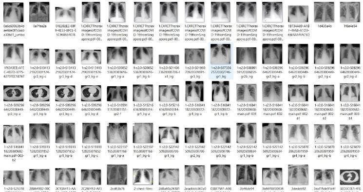
Fig 3:Examplechestradiographyimages
5.3 Image Augmentation
Class
CNNneedsafairamountofdatatogetthebestperformance. We use data enhancement techniques to maximize insufficientdataintraining,andthetechniquesusedinclude verticalrotation,horizontalrotation,sound,translation,blur androtationimage60°,90°,180°,270°.Figure3showsan example of the data addition used. Thus, the original databasecomprising864COVID 19images,1341standard images,and1345viralpneumoniaimageswasexpandedto 8,640 COVID 19 images, 13,410 X images. common chest rays, as well as 13,450 images of bacterial pneumonia as shownin.TableI. Images Augmente WithAugmentation Training Valida Test COVID 19 864 8640 500 800 12000 910 500 Pneumonia 950 500
1345 13450 12000
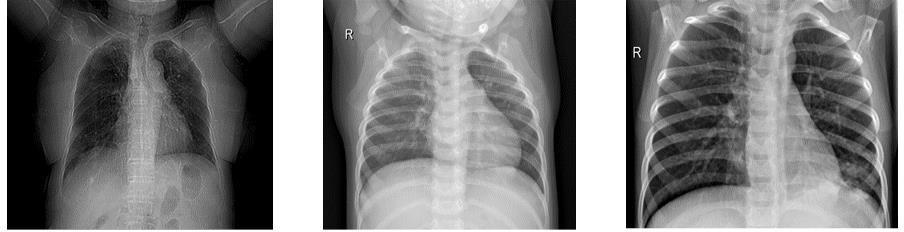
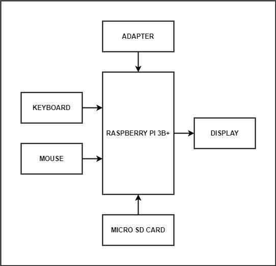
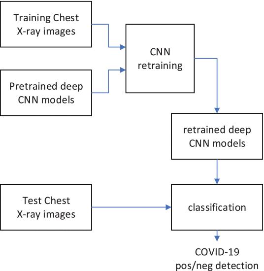
Fig 5:Neuralnetworkclassifier
Convolution is a slow process. It removes various input features. Each kernel is responsible for producing output activity. Low level elements of the image, such as edges, lines,andcornersaredeterminedbythelowerlayer,andthe higherfeaturesarerenderedintheupperlayer.Blendingis used to make the features found in convolution strong against noise. Combining layers are usually of two types namely,intermediateintegrationandlargeintegration.The size reduction step or output factor. A simple example of maxandaveragepoolingisshowninFig.7.
Fig 7:Exampleofmaxandaveragepooling
Itisasyntheticneuralnetworkwithmorethanthreelayers, shown in Fig. 5. It has single input, single output and multipleinvisiblelayers.Inordertousetransferreadingsto separatechestX rayimages,weusedtheTensorFlowlibrary todownloadtheInceptionV3modeltoourlocalmachine, retrained it to the chest X ray database and split the new images into one of three common categories, viral pneumonia and COVID 19. It is an in depth learning framework developed by Google that can control all the neurons(nodes)inthesystemandhasalibrarysuitablefor imageprocessing.
Hereapre trainedneuralnetworkdesignedforonejobcan beusedasastartingpointforanothertask.
International Research Journal of Engineering and Technology (IRJET) e ISSN: 2395 0056 Volume: 09 Issue: 01 | Jan 2022 www.irjet.net p ISSN: 2395 0072 © 2022, IRJET | Impact Factor value: 7.529 | ISO 9001:2008 Certified Journal | Page1192
1. Recursively perform convolution and pooling on images. 2. Applydropoutandfullyconnected.Nowtheimage mustbeclassifiedaccordingtothelabelledtraining class.
Fig 6: Proposedworkarchitecture
Fig 4:InceptionV3architecture
5.6 Proposed Architecture
lot of training data, which can be difficult to obtain in medicalsettings.Intheanalysisofmedicaldata,thebiggest difficultyresearchersfaceisthelimitednumberofavailable data sets. In depth learning models often require a lot of data.Labelingthisdataprofessionallyisexpensiveandtime consuming.
Fig.6providesa step by stepprocedure for the proposed work model. The steps for the projected classification architectureareasfollows:
1. Use the extraction phase feature of the convolutionalneuralnetwork. 2. The partition section uses layers that are fully integratedwithSoftMaxlayers. 5.5 TensorFlow
Figure 4 describes the Inception V3 model that enables convolution,pooling,SoftMaxandfullyintegratedprocesses.
TheInception v3structureconsistsoftwopieces:
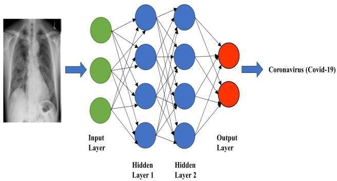
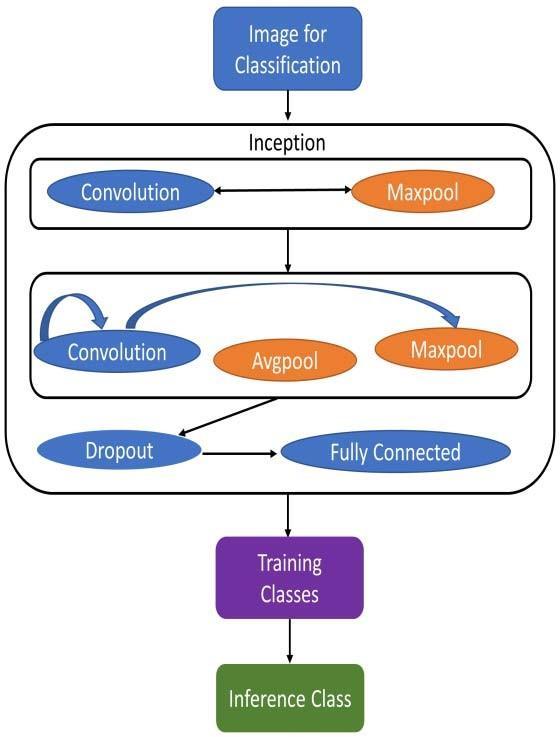
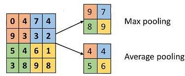
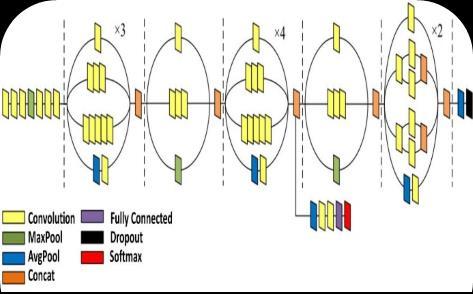
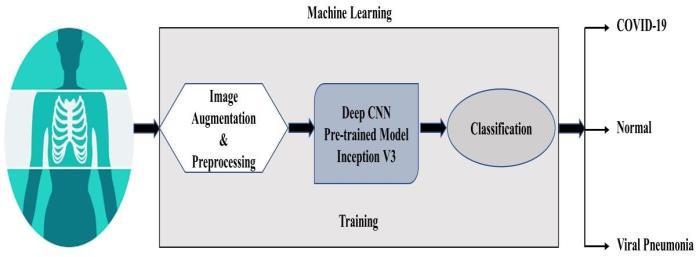
Fig 8:Schematicrepresentationofpre trainedmodelfor thepredictionpfcovid 19patients,normalandinfected InceptionV3isoneoftheregionsofarchitecturalartinthe challenge of image classification. The best network for medical imaging analysis seems to be the structure of InceptionV3andismuchbetterthanthelatestarchitecture. Therefore,wehaveselectedtheInceptionV3modelwhichis usedusingTensorFlowand thatiswhy retraining isdone withTensorFlow.
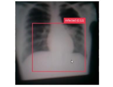
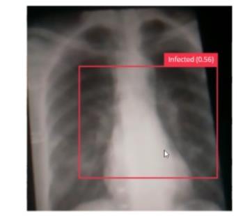
Fig 10:InfectedLungs
In this study, we introduced a new way to automatically detectCOVID 19byCNN.ChestX rayimageswereusedto predict patients with COVID 19. The famous previously trained Inception V3 model has been trained and after training,themodelwasspottedonthechest.
Thestepsforclassificationusingtheproposedactivityareas AlgorithmfollowsTensorFlowClassification Step1:Getstarted Step2:Createalistofimages//starttrainingthemodel Step 3: Provide a list of storage quantities for each bottle Steppicture4:Provideinsightintoimages//tocreateBottleneck Stepvalues.5:Createafolderforallbottlepricepictures Step6:Generatebottleneckvaluesforeachimage Step7:CreatenewSoftMaxlayersandfullyintegratedlayers //attheendoftraining Step8:Checkthenewimage//insertachestx rayimageto gettheresult Step9:Finish Fig 9:DatasetsandCTscans
Fig 11:InfectedLungs
In this study, we developed an DCNN based InceptionV3 model for classifying COVID 19 Chest X ray images into normal, viral pneumonia and COVID 19 categories. In addition, we used the transfer learning method obtained using ImageNet data to overcome insufficient data and trainingtime.AstandardCNNrepresentationthatincludes theInceptionV3modelforpatientpredictionofCOVID 19, viralpneumoniaandgeneralizationisshowninFig.8.Chest X rayimagesareconsideredasembedded,InceptionV3is used, convolution, pooling, SoftMax, and fully integrated procedures are performed. Upon completion of these activities, they were categorized according to different training modules and eventually classified as normal, bacterialpneumoniaandCOVID 19classes.
International Research Journal of Engineering and Technology (IRJET) e ISSN: 2395 0056 Volume: 09 Issue: 01 | Jan 2022 www.irjet.net p ISSN: 2395 0072 © 2022, IRJET | Impact Factor value: 7.529 | ISO 9001:2008 Certified Journal | Page1193
6. EXPECTED RESULTS
[8] C. Huang et al., "Clinical features of patients infected with 2019 novelcoronavirus in Wuhan, China," The Lancet,vol.395,no.10223,pp.497 506,2020.
[10] D. K. W. Chu et al., "Molecular Diagnosis of a Novel Coronavirus (2019 nCoV) Causing an Outbreak of Pneumonia,"ClinicalChemistry,vol.66,no.4,pp.549 555,2020.
[7] [8] Q. Li et al., "Early Transmission Dynamics in Wuhan, China, of Novel Coronavirus Infected Pneumonia,"vol.382,no.13,pp.1199 1207,2020.
International Research Journal of Engineering and Technology (IRJET) e ISSN: 2395 0056 Volume: 09 Issue: 01 | Jan 2022 www.irjet.net p ISSN: 2395 0072 © 2022, IRJET | Impact Factor value: 7.529 | ISO 9001:2008 Certified Journal | Page1194 Fig-12:UninfectedLungs 7. CONCLUSION TheCovid 19X Raytestmethodisproposedanddiscussed inthisproject.Itisinexpensive,effectiveandcanbeusedfor early diagnosis. It will use a variety of Machine Learning methods that provide accurate results and an increase in accuracywiththesizeofthedatabaseandthemodelsused. Thiswaywecandomoretestsandwecangetresultsfaster andwecangetmoredatathatcansuggesthowweshould managethehealthcaresystemandvaccinationpolicy. REFERENCES [1] WHO. (2020). WHO Director General's opening remarksatthemediabriefingonCOVID 19 11March 2020. Available: briefingdirectorhttps://www.who.int/dg/speeches/detail/whogeneralsopeningremarksatthemediaoncovid1911march2020 [2] CDC. (2020). Coronavirus Disease 2019 (COVID 19). Available: https://www.cdc.gov/coronavirus/2019 ncov/need extra precautions/p eople at higher WHO.risk.html(2020, 02 November). Coronavirus disease (COVID 2019)situation reports.Available: L9j_hkakBVk_n_TkbXjtgjVBcVizs7h83yH7YUEaAoVHEApTBlRc57bUreports/?gclid=Cj0KCQjw2PP1BRCiARIsAEqvcoronavirushttps://www.who.int/emergencies/diseases/novel2019/situationVrAh6w_wcB
[3] J.H.U.a.Medicine.(2020).COVID 19Dashboardbythe CenterforSystemsScienceandEngineering(CSSE)at Johns Hopkins University https://coronavirus.jhu.edu/map.html(JHU)Available:
[4] W.Wangetal.,"DetectionofSARS CoV 2inDifferent Types of Clinical Specimens," (in eng), JAMA, 2020/03//2020.
[6] [7] N. Chen et al., "Epidemiological and clinical characteristicsof99casesof2019novelcoronavirus pneumoniainWuhan,China:adescriptivestudy,"The Lancet,vol.395,no.10223,pp.507 513,2020.
[5] D. Wang et al., "Clinical Characteristics of 138 Hospitalized Patients with 2019 Novel Coronavirus InfectedPneumoniainWuhan,China,"JAMA,vol.323, no.11,pp.1061 1069,2020.
[9] V. M. Corman et al., "Detection of 2019 novel coronavirus (2019 nCoV) by real time RT PCR," vol. 25,no.3,p.2000045,2020.