International Research Journal of Engineering and Technology (IRJET)
e-ISSN: 2395-0056
Volume: 08 Issue: 06 | June 2021
p-ISSN: 2395-0072
www.irjet.net
ENHANCEMENT OF X-RAY IMAGES FOR LUNG DISEASES DETECTION 1Amrutha
T M, Anoop S Pillai2
1PG
Scholar, Department of Electronics and Communication Engineering, NSS College of Engineering, Palakkad Professsor, Department of Electronics and Communication Engineering, NSS College of Engineering, Palakkad ---------------------------------------------------------------------***--------------------------------------------------------------------offer an ensemble framework for X-ray image Abstract –X-ray image inspection is an important enhancement that includes tissue attenuation, part of medical diagnostics. However, because of the contrast control, and image fusion. poor contrast and dynamic range of an X-ray, key features such as organs, bones, and nodules might be Our technique seeks to vividly portray the features of difficult to spot. As a result, contrast adjustment is LDR X-ray images by boosting the contrast in bright crucial, particularly because it can enhance features and dark regions. in both bright and dark areas. As a result, we present a new approach based on component attenuation for X-ray picture enhancement. An X-ray image could be broken down into tissue components and important details. Because tissues aren't always the primary focus of an X-ray, we proposed using adaptive tissue attenuation and dynamic range stretching to improve visual contrast. A parametric correction approach was developed using component decomposition and tissue attenuation to provide several enhanced images at once. Finally, a framework for combining these augmented images and providing a highcontrast output in both bright and dark regions was developed. 2Associate
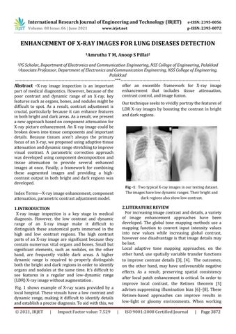
Fig -1: Two typical X-ray images in our testing dataset. The images have low dynamic ranges. Their bright and dark regions also show low contrast.
Index Terms—X-ray image enhancement, component attenuation, parametric contrast adjustment model.
2.LITERATURE REVIEW For increasing image contrast and details, a variety of image enhancement approaches have been developed. The global tone mapping methods use a mapping function to convert input intensity values into new values while increasing global contrast, however one disadvantage is that image details may be lost. Local adaptive tone mapping approaches, on the other hand, use spatially variable transfer functions to improve contrast details [3], [4]. The outcomes, on the other hand, may have unfavourable negative effects. As a result, preserving spatial consistency after local patch enhancement is critical. In order to improve local contrast, the Retinex theorem [5] advises suppressing illumination bias [6]–[8]. These Retinex-based approaches can improve results in low-light or gloomy environments. When working
1.INTRODUCTION X-ray image inspection is a key stage in medical diagnosis. However, the low contrast and dynamic range of an X-ray image make it difficult to distinguish these anatomical parts immersed in the high and low contrast regions. The high contrast parts of an X-ray image are significant because they contain numerous vital organs and bones. Small but significant elements, such as nodules, on the other hand, are frequently visible dark areas. A higher dynamic range is required to properly distinguish both the bright and dark regions in order to identify organs and nodules at the same time. It's difficult to see features in a regular and low-dynamic range (LDR) X-ray image without augmentation . Fig. 1 shows example of X-ray scans provided by a local hospital. These visuals have a low contrast and dynamic range, making it difficult to identify details and establish a precise diagnosis. To aid with this, we © 2021, IRJET
|
Impact Factor value: 7.529
|
ISO 9001:2008 Certified Journal
|
Page 3872