International Research Journal of Engineering and Technology (IRJET)
e-ISSN: 2395-0056
Volume: 08 Issue: 06 | June 2021
p-ISSN: 2395-0072
www.irjet.net
Computational Analysis of Radiographic Images of chest of COVID-19 Patients & its predictions using AI Mr. Siddharth K Ganvir1, Dr.V.L.Agrawal2 1Student,
Electronic and Telecommunication Dept. of HVPM’S, College of Engineering and Technology and SGBAU Amravati, Maharashtra (India) 2Associate Professor, Electronic and Telecommunication Dept. of HVPM’S, College of Engineering and Technology and SGBAU Amravati, Maharashtra (India) ---------------------------------------------------------------------***---------------------------------------------------------------------Abstract - With the exponentially growing COVID-19 (corona virus disease 2019) pandemic, clinicians continue to seek accurate and rapid diagnosis methods in addition to virus and antibody testing modalities. Because radiographs such as Xrays and computed tomography (CT) scans are cost-effective and widely available at public health facilities, hospital emergency rooms (ERs), and even at rural clinics, they could be used for rapid detection of possible COVID-19-induced lung infections. Therefore, toward automating the COVID-19 detection, we propose a viable and efficient deep learningbased chest radiograph framework to analyze COVID-19 cases with accuracy. A unique dataset is prepared from available sources containing the chest view of CT scan/X-ray data for COVID-19 cases. Our proposed framework leverages a data augmentation of radiograph images algorithm for the COVID19 data, by adaptively employing the MATLAB and NeuroSolution on COVID-19 infected chest images to generate a train a robust model. The training data consisting of actual and synthetic chest images are fed into our customized neural network model, which achieves COVID-19 detection with good accuracy. Furthermore, through this it is possible to efficiently automate COVID-19 detection from radiograph images to provide a fast and reliable evidence of COVID-19 infection in the lung that can complement existing COVID-19 diagnostics modalities.
intelligence (AI)-based smart chest radiograph for COVID-19 cases with accuracy.
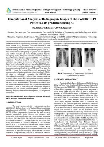
(a)
2. RESEARCH METHODOLOGY NurbaitySabri , RaseedaHamzah , Shafaf Ibrahim &KhyrinaAirinFariza Abu Samah [1] has analyzed a total of 101 data consist of 33 COVID-19, 28 normal and 40 bacteria of chest X-Ray images are tested. The extracted features are tested using Weka software where it able to analyze the accuracy of k-NN classifier using 10-folds cross-validation. The result represents with true positive (TP) and false positive (FP) for all tested images. In their research, 3776 attributes from x-ray images have been used for classification purpose. Matrices used to measure the efficiency of the classifier precision and recall. Precision represent the percentage of x-ray images that are classified as true. Meanwhile, recall is the percentage of relevant x-ray images that labeled as “true” by the classifier Result ofclassification.Based.They have clearly observed that LBP able to produce a good classification accuracy with average of 0.960. 96%. The analysis also reveals that the maximum precision and recall are obtained for the LBP algorithm, with both values at 0.96.SadmanSakib,TahratTazrin,Mostafa M. Fouda,Zubair Md. Fadlullah And Mohsen Guizani [3]proposed DL-CRC framework consists of two parts: the DARI algorithm (which adaptively employs a customized generative adversarial network and generic data augmentation techniques such as zoom and rotation) and a two-dimensional convolutional neural network (CNN) model. They employed a unique dataset for multiple publicly
1. INTRODUCTION The severe acute respiratory syndrome coronavirus 2(SARS-CoV-2), first observed in Wuhan, China, turnedinto a global pandemic of COVID-19 (coronavirus disease 2019). COVID-19 has a destructive impact on the well-being of people, particularly senior citizens and patients with underlying health conditions and compromised immunity levels. By 28th May.2021, the COVID-19 pandemic already contributed to over 35.1 lacmortalities and more than 19.6 crore million cases of COVID-19 infection . A critical stepin the fall of 2021, preparedness to combat such scenarios will involve increasing use of portable chest CT scan / X-ray devices due to widespread availability and reduced infection. Therefore, as depicted in Fig.,to automate the COVID-19 detection using machines, we aim to develop an artificial |
Impact Factor value: 7.529
(c)
Fig.1.Three sample of X-ray images, (a)Normal, (b)Pneumonia, (c)Covid
Index Terms - MatLab, Neuro Solution Software, Microsoft excel, Various Transform Technique
© 2021, IRJET
(b)
|
ISO 9001:2008 Certified Journal
|
Page 3654