International Research Journal of Engineering and Technology (IRJET)
e-ISSN: 2395-0056
Volume: 08 Issue: 06 | June 2021
p-ISSN: 2395-0072
www.irjet.net
Brain SCAT – Segmentation and Classification of Brain Tumor Arsya Mohamed Ali1, Melvin Martin2, Nithin Gangadharan Rangaraj3 1,2,3Student,
Dept. of Biomedical Engineering, PSG College of Technology, Tamil Nadu, India ---------------------------------------------------------------------***---------------------------------------------------------------------
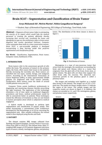
tumor. The distribution of the three classes is shown in figure 1.
Abstract – Diagnosis of brain tumor helps in anticipating
the severity of its impact which would help the medical personnel in treating the patients efficiently thereby increasing their survival rate. Localizing the tumor and identifying its type are the preliminary steps in brain tumor diagnosis that involve specialized clinicians. Computer vision automates these tasks in a time-conservative manner. Brain SCAT, a user-accessible platform is developed incorporating a deep learning model that performs segmentation and classification jointly. Key Words: Classification, Segmentation, Brain tumor, Computer vision, Dashboard, U-Net.
Fig -1: Distribution of classes
1. INTRODUCTION
Meningioma is a type of non-cancerous tumor that arises from the meninges, the membrane surrounding the brain and spinal cord. Glioma refers to a tumor that originates from the glial cells of the brain, brain stem, or spinal column. The glial cells are those that support the other brain cells, the neurons. The abnormal noncancerous growth in the pituitary gland is called a pituitary tumor.
Brain tumors refer to the unnecessary growth of cells within the brain. The presence of tumors in the primary organ, the brain, often causes fatal issues to the patients thereby increasing the mortality rate. Brain tumors are classified into two types, namely, benign, and malignant tumors. According to the International Association of Cancer Registries (IARC), in India, approximately 28,000 patients are diagnosed with brain tumors annually. Furthermore, these tumors cause the death of around 24,000 patients every year [1].
The images and metadata exist together as a matlab data format in the dataset. The images are of 512 x 512pixel size. The masks are binary images where 1’s indicate the region of the tumor. The sample images and the respective masks are shown in figure 2. In addition, the classes are encoded as 1,2, and 3 for meningioma, glioma, and pituitary tumor respectively.
Computer Vision assists healthcare professionals in diagnosing and monitoring diseases thereby prescribing the right treatment. The application of this technology eliminates a lot of time spent on traditional diagnostic procedures and hence, medical professionals would be able to concentrate better on patients. These techniques extend the efficiency of treatment as they are not limited to human perception alone which is subjected to medical negligence.
Meningioma
Glioma
Pituitary Tumor
A hybrid model is developed to perform both segmentation and classification of tumors jointly. This model incorporates a conventional U-Net architecture developed by Olaf Ronneberger et al [2] for executing the segmentation task. After suitable evaluation, the trained model is then deployed using Heroku and Dash.
2. DATASET The dataset contains MRI images collected from Nanfang and General hospital, Tianjing Medical University, China from 2005 to 2010 [3]. This dataset hosts 3064 T1weighted MRI images of 233 patients diagnosed with three types of tumors, namely, meningioma, glioma, and pituitary
© 2021, IRJET
|
Impact Factor value: 7.529
Fig -2: Sample images with masks
|
ISO 9001:2008 Certified Journal
|
Page 3574