2 minute read
If not, resize image high resolution image to size of lower resolution image

Volume: 08 Issue: 10 | Oct 2021 www.irjet.net p-ISSN: 2395-0072
Development & Analysis of 2D Medical Image Fusion Using Wavelets
Advertisement
Shraddha P. Diwalkar1 , Prof. Dr. S.M. Hanbarde2
1Student, Dept. Of Electronics & Communication, JSCOE, Hadapsar, Pune, Maharashtra, India. 2Prof. Dr. S.M. Hanbarde, Dept. Of Electronics & Communication, JSCOE, Hadapsar, Pune, Maharashtra, India. --------------------------------------------------------------------*** ---------------------------------------------------------------------Abstract - Medical image fusion is the technique of integrating two or more images from various imaging modalities/scans to get a fused image with information having the details of anatomical information combined from all the modalities for accurate diagnosis and further treatment. This paper performs the analysis of various wavelet functions for decomposition and synthesis. PET (Positron Emission Tomography) and MRI (Magnetic Resonance Imaging) scans of Brain and chest are used and compared using Stationary Wavelet Transform (SWT) and Discrete wavelet Transform (DWT). Entropy is calculated which is a measure of information acquired after the fusion process.
Key Words: Wavelet transform, Fusion, Stationary Wavelet Transform, Discrete, Medical image
1.INTRODUCTION
In this highly developed digital images era, Medical image fusion is became very popular for medical professionals to be relay on. Diagnosis and treatment of disease require accurate information obtained from various modalities or medical scans, such as PET (Positron Emission Tomography helps to reveal the metabolic or biochemical function of tissues and organs and MRI (Magnetic Resonance Imaging) that displays soft tissues, CT (Computerized Tomography) which gives the hard tissue details etc. Ideally, all the important information and features form each of the modalities should be included in the fused image without information loss. So researchers try to find methods that give best results in terms of resolution, texture, edges & most importantly the information obtained. Medical image fusion is favorable to the healthcare professionals for correct diagnosis & treatment of disease. Multimodal and multi-resolution image fusion is very powerful method to attain the desired output image from various medical scans using wavelet transform is a powerful mathematical tool which is a localized transform in both time(or space) and frequency and this property is advantageously used to extract information form a signal that is not possible with Fourier transform. Wavelet Transform is also very much useful for multimodal and multi-resolution analysis of signal or images.
1.1 Wavelet Transforms
1.1.1 Discrete Wavelet Transform
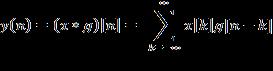
Discrete Wavelet Transform (DWT) is used, as it decomposes the signal into "sub-bands". Therefore, at each level, the signal splits into high and low frequency components.This is a specialized approach for image fusion and denoising. The Discrete wavelet Transform of a signal x is calculated by passing it through a low pass filter resulting in convolution of the two,
(1)
The signal is decomposed using high pass filter h. The output of DWT decomposition give the detailed coefficients (from high pass filter) & approximation coefficients (from low pass filter).
By the decomposition process, the time resolution get halved as only half of each filter output characterizes the image. However, each output image has half the frequency spectrum of input image, so frequency resolution has been doubled.
At each level of DWT, the signal is decomposed into low and high frequencies for rows and columns. Due to decomposition process, the input image must be a multiple of 2n where n is the no. of levels. Scaled and translated basis function,
(2)
(3) Where, i= {H, V, D}
(x,y)= ( x-m, y-n)
(x,y)= ( x-m, y-n)