INTERNATIONAL RESEARCH JOURNAL OF ENGINEERING AND TECHNOLOGY (IRJET)
E-ISSN: 2395-0056
VOLUME: 07 ISSUE: 03 | MAR 2020
P-ISSN: 2395-0072
WWW.IRJET.NET
RETINAL FUNDUS IMAGE ANALYSIS FOR DIAGNOSIS OF GLAUCOMA D.Gayathri1, S.Janani1, K. Sandhiya1, T. Sandhiya1, Ms. Sweatha Natarajan2 1Student,
Department of Biomedical Engineering, Agni College of Technology, Anna University, Chennai, India 2Assistant professor, Department of Biomedical Engineering, Agni College of Technology, Anna University, Chennai, India ------------------------------------------------------------------------***------------------------------------------------------------------------Abstract-The glaucoma disease is characterized by change in the structure of nerve fibers and optic disc parameters such as diameter, volume, and area This project deals with the detection of glaucoma to prevent the vision loss. Glaucoma is one among the leading causes of vision loss. This is caused due to the increased fluid pressure and improper drainage of fluid in the eye. Diagnosis of glaucoma is mainly based on the Intra Ocular Pressure (IOP), medical history of patient’s family, and change in optic disc structure. Glaucoma suspect will have IOP more than 21 mmHg. Glaucoma is an ocular disorder caused thanks to increased fluid pressure within the nervus opticus. It damages the optic nerve subsequently causes loss of vision. The available scanning methods are Heidelberg Retinal Tomography (HRT), Scanning Laser Polarimetry (SLP) and Optical Coherence Tomography (OCT). These methods are expensive and need experienced glaucoma detection. Keywords: Glaucoma, Enhancement, MATLAB 1.
Filtering,
Segmentation,
INTRODUCTION
Glaucoma is one among the common causes of blindness with about 79 million within the world likely to be afflicted by the year 2020. The progressive degeneration of nervus opticus fiber is one among the way of characterizing glaucoma. This causes structural changes of the nervus opticus head (optic disk) and fiber layer, and results in failure of the field of vision the first detection of glaucoma and ensuring essential treatment can prevent permanent vision loss. The pressure of the non glaucomatous should be 21 mm of Hg, if it increases the nervus opticus are going to be damaged causing permanent vision loss. There are two main sorts of Glaucoma (i) Primary Open Angle Glaucoma (POAG) and (ii) Angle Closure Glaucoma (ACG). POAG is that the commonest sort of Glaucoma accounting for a minimum of 90%of all Glaucoma cases. The IntraOcular Pressure (IOP), which maintains a permanent shape of the human eye and protects it from deformation, rises because the right amount of fluid cannot drain out of the attention. With POAG, the entrances to the drainage canals work properly but a clogging problem occurs inside the drainage canals. This sort of Glaucoma develops slowly and sometimes without noticeable sight loss for several years. It are often treated with medications if diagnosed at the sooner stage. ACG happens when the drainage canals get blocked. The iris isn't as wide and open a sin the traditional case. The fringes of the iris bunches up over the Š 2020, IRJET
|
Impact Factor value: 7.34
|
drainage canals, when the pupil enlarges an excessive amount of or too quickly.
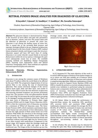
Fig 1: Glaucoma Image 2.
LITERATURE SURVEY
In [1] Anupama B C The main objective of this work is to extract differing types of features from fundus images so as to return out with best suitable set of features which will help in automated detection of Glaucoma and evaluate it using learning algorithm. Different combinations of those features are given to Support Vector Machine (SVM) and KNN to classify the pictures as normal and glaucomatous. A tenfold cross validation is performed using the extracted features and a comparative study has been administered in terms of Accuracy, Sensitivity, Specificity, Positive Predictive Value (PPV) and Negative Predictive Value (NPV) Performance evaluation has been through with and without applying feature reduction techniques. In [2] Qaisar Abbas to guage the performance of Glaucoma-Deep system, the sensitivity (SE), specificity (SP), accuracy (ACC), and exactness (PRC) applied mathematics measures were utilised. On average, the SE of 84.50%, SP of 98.01%, command of ninety nine and Asian country of 84 values were achieved. examination to progressive systems, the Nodular-Deep system accomplished vital higher results. Consequently, the Glaucoma-Deep system will simply acknowledge the eye disease disease to resolve the matter of clinical specialists throughout eye-screening method on large-scale environments. In [3] A.Padma The eye diseases may be detected by locating the optic disk. Many methodologies are followed
ISO 9001:2008 Certified Journal
|
Page 4804