International Research Journal of Engineering and Technology (IRJET)
e-ISSN: 2395-0056
Volume: 07 Issue: 03 | Mar 2020
p-ISSN: 2395-0072
www.irjet.net
Diabetic Retinopathy Vaishnavi Kukutkar1, Rehan Pathan2, Shubham Mansute3, Madan Bhendare4, Jyoti Kubade5 1,2,3,4,5Student,
Dept. of Computer Science & Engineering, Datta Meghe Institute of Engineering, Technology & Research, Wardha, Maharashtra, India ---------------------------------------------------------------------***----------------------------------------------------------------------
Abstract - Here we address the detection of Hemorrhages
and microaneurysms in color fundus images. In pre-Processing we separate red, green, blue color channel from the retinal images. The green channel will pass to the further process. The green color plane was utilized in the analysis since it shows the simplest contrast between the vessels and therefore the background retina. Then we extract the GLCM (Gray Level CoOccurrence Matrix) feature. Within the GLCMs, several statistics information are derived using the various formulas. These statistics provide information about the feel of a picture, like Energy, Entropy, Dissimilarity, Contrast, Inverse difference, correlation Homogeneity, Auto correlation, Cluster Shade Cluster Prominence, Maximum probability, Sum of Squares are going to be calculated for texture image. After feature Extraction, we offer this feature to classifier. Finally it'll predict about the retinal whether it's hemorrhages or microaneurysms. After predicting problems in the retinal image we'll localize the affected place. For segmenting the localized place we'll use adaptive thresholding segmentation.
Key Words: Image representation, dissimilarity space, class learning.
1. INTRODUCTION 1.1 Overview Diabetic retinopathy (DR) is a vascular disease of the retina which affects patients with diabetes mellitus. It is the number one cause of blindness in people between the ages of 20-64 in the United States. It is, therefore, a worthwhile topic for all medical students to review. Diabetes mellitus is extremely common, so it is not surprising that DR affects 3.4 percent of the population (4.1 million individuals). Of the millions of people with DR, nearly one-fourth have vision-threatening disease. The likelihood of developing diabetic retinopathy is related to the duration of the disease. Type 2 diabetes has an insidious onset and can go unnoticed for years. As a result, patients may already have DR at the time of diagnosis. Type 1 diabetics, on the other hand, are diagnosed early in the course of their disease, and they typically do not develop retinopathy until years after the diagnosis is made. The risk of developing retinopathy increases after puberty. Twenty years after the
© 2020, IRJET
|
Impact Factor value: 7.34
|
diagnosis of diabetes, 80% of type 2 diabetics and nearly all type 1 diabetics show some signs of retinopathy. While these numbers are eye-opening, diabetics can decrease their risk of retinopathy and slow the progression of the disease after it has begun with tight glucose control.
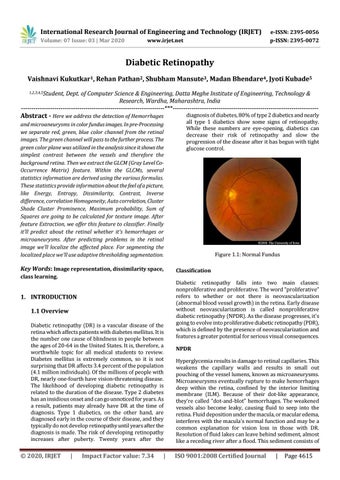
Figure 1.1: Normal Fundus Classification Diabetic retinopathy falls into two main classes: nonproliferative and proliferative. The word “proliferative” refers to whether or not there is neovascularization (abnormal blood vessel growth) in the retina. Early disease without neovascularization is called nonproliferative diabetic retinopathy (NPDR). As the disease progresses, it's going to evolve into proliferative diabetic retinopathy (PDR), which is defined by the presence of neovascularization and features a greater potential for serious visual consequences. NPDR Hyperglycemia results in damage to retinal capillaries. This weakens the capillary walls and results in small out pouching of the vessel lumens, known as microaneurysms. Microaneurysms eventually rupture to make hemorrhages deep within the retina, confined by the interior limiting membrane (ILM). Because of their dot-like appearance, they're called “dot-and-blot” hemorrhages. The weakened vessels also become leaky, causing fluid to seep into the retina. Fluid deposition under the macula, or macular edema, interferes with the macula’s normal function and may be a common explanation for vision loss in those with DR. Resolution of fluid lakes can leave behind sediment, almost like a receding river after a flood. This sediment consists of
ISO 9001:2008 Certified Journal
|
Page 4615