International Research Journal of Engineering and Technology (IRJET)
e-ISSN: 2395-0056
Volume: 07 Issue: 03 | Mar 2020
p-ISSN: 2395-0072
www.irjet.net
Lung Cancer Detection using GLCM and Convolutional Neural Network Suresh Babu.P1, Jennifer.J2, Hesima.C3 , Preethi.S4 [1]Assistant
Professor-III, Department of Information Technology, Velammal College of Engineering and Technology, Madurai, Tamil Nadu, India. [2][3][4]Department of Information Technology, Velammal College of Engineering and Technology, Madurai, Tamil Nadu, India ---------------------------------------------------------------------***----------------------------------------------------------------------
Abstract - Cancer is one of the deadliest diseases leading to
innumerable deaths worldwide. According to WHO (World Health Organization) lung cancer contributes about 14 per cent among all the cancers. Therefore, early detection and treatment is very much required. Computed Tomography (CT) scan can provide valuable information in the diagnosis of lung diseases. The main objective of this work is to classify the tumors found in lung as malignant or benign by means of Convolutional Neural Network(CNN). The accuracy obtained by means of CNN is 96%, which is more efficient when compared to accuracy obtained by the traditional neural network systems.
Key Words- Computed Tomography(CT), Gray Level Co-occurence Matrix(GLCM), Convolution Neural Network (CNN).
extracted using GLCM (Gray Level Co-occurrence Matrix). These extracted features are used in phase five for classification purpose which is carried by CNN (Convolutional Neural Network)
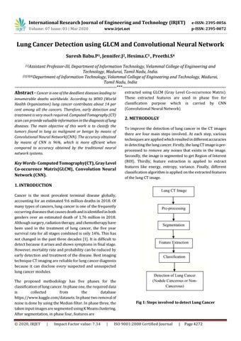
2. METHODOLGY To improve the detection of lung cancer in the CT images there are four main steps involved. At each step, various techniques are applied which resulted in different accuracies in detecting the lung cancer. Firstly, the lung CT image is preprocessed to remove any noises that exists in the image. Secondly, the image is segmented to get Region of Interest (ROI). Thirdly, feature extraction is applied to extract features like energy, entropy, variance. Finally, different classification algorithm is applied on the extracted features of the lung CT image.
1. INTRODUCTION Cancer is the most prevalent terminal disease globally, accounting for an estimated 9.6 million deaths in 2018. Of many types of cancers, lung cancer is one of the frequently occurring diseases that causes death and is identified in both genders over an estimated death of 1.76 million in 2018. Although surgery, radiation therapy, and chemotherapy have been used in the treatment of lung cancer, the five year survival rate for all stages combined is only 14%. This has not changed in the past three decades [1]. It is difficult to detect because it arises and shows symptoms in final stage. However, mortality rate and probability can be reduced by early detection and treatment of the disease. Best imaging technique CT imaging are reliable for lung cancer diagnosis because it can disclose every suspected and unsuspected lung cancer modules. The proposed methodology has five phases for the classification of lung cancer. In phase one, the required data is collected from the database https://www.kaggle.com/datasets. In phase two removal of noise is done by using the Median filter. In phase three, the taken input images are segmented using K Means clustering. After segmentation, in phase four, features are Š 2020, IRJET
|
Impact Factor value: 7.34
|
Fig 1: Steps involved to detect Lung Cancer
ISO 9001:2008 Certified Journal
|
Page 4272