International Research Journal of Engineering and Technology (IRJET)
e-ISSN: 2395-0056
Volume: 07 Issue: 03 | Mar 2020
p-ISSN: 2395-0072
www.irjet.net
FUSION OF CT AND MRI FOR THE DETECTION OF BRAIN TUMOR BY SWT AND PROBABILISTIC NEURAL NETWORK Mr.D.ARULKUMAR1, V.ASWATHI2, R.JENIFER3, R.S.BHAVANI4 1 Associate
professor, Department of Electronic and communication Engineering, Panimalar Institute of Technology, Chennai, Tamil Nadu, India 2,3,4Student, Department of Electronic and communication Engineering, Panimalar Institute of Technology, Tamil Nadu, India, ---------------------------------------------------------------------***----------------------------------------------------------------------
Abstract - The multimodal medical image fusion is
organized to reduce the redundancy while extracting the necessary information from the input images acquired using different medical sensors. In this article the major aim is to yield a single fused image, which could be more useful for clinical analysis. This paper presents fusion framework using Stationary Wavelet Transform (SWT) with neural network classifier for the detection of Brain Tumor. The first stage employs enhance the feature using GLCM and finally the tumor part is segmented by k-means clustering. The performance model provides the result whether it is benign or malignant tumor from the resultant fused image and also gives the detailed view of morphological structure of detected tumor part.
Key Words: Medical image fusion, MRI, CT, SWT, GLCM, PNN.
1. INTRODUCTION: The Medical analysis and its applications are extremely challenging and complicated in many versions. By the way, one of the most challenging domain in the medical field is human brain analysis. The manual process by domain specialist is more time consuming task. -Still, there is need for efficient technique. In this article, the MRI and CT images are fused for the detection of brain tumor which provides the way to efficient analysis. Medical image fusion is the process of merging two or more medical images in order to extract efficacious features for medical evaluation. They can more efficiently identify normal and abnormal regions of the human body. The unpredictable cases in medical field mostly comes under in tumor detection. This paper involves the detection of the brain tumor from the fused image. The combination of anomalous cell in brain causes brain tumor. Therefore, from the fused image of MRI and CT, the more detailed information on brain tumor is obtained. Thus, there are various stages in this process such as image preprocessing, fusing of image and clustering for segmentation. Finally, the detected tumor is validated in three parameters like its sensitivity, specificity and accuracy. From the Š 2020, IRJET
|
Impact Factor value: 7.34
|
extracted morphological structure of tumor radiologist can easily identify its location, shape, size and its intensity.
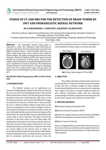
Fig 1 shows input images of CT and MRI
2. OBJECTIVE: The problem statement mainly involves why do we need image fusion. Image can be fused to reduce the amount of data and also to construct images for better analysis. Usually medical field has various complications in analyzing inner organs. The fusion of scanned images provides the more efficient information rather than the individual scanned images. Therefore, image fusion pave important role for the better analysis and treatment in medical field.
3. EXISTING METHOD: [1] Principal Component Analysis (PCA) [2] Discrete Wavelet Transform (DWT) [3] Dual tree complex discrete wavelet transform (DTDWT) with Non Sub-sampled contourlet transform (NSCT) [4 ]Fuzzy C means clustering Due to averaging method, contrast information loss occurs. By means of discrete wavelet transform spatial distortion is high which leads to limited performance of edge and texture representation.
ISO 9001:2008 Certified Journal
|
Page 4060