International Research Journal of Engineering and Technology (IRJET)
e-ISSN: 2395-0056
Volume: 07 Issue: 03 | Mar 2020
p-ISSN: 2395-0072
www.irjet.net
DETECTION OF HEAMORRHAGE IN BRAIN USING DEEP LEARNING AKASH K.1, GAYATHRI M.R2, KARTHIGA.M 3 1,2FINAL
YEAR, DEPARTMENT OF BIOMEDICAL ENGINEERING, ANNA UNIVERSITY, CHENNAI, INDIA PROFESSOR, DEPARTMENT OF BIOMEDICAL ENGINEERING, ANNA UNIVERSITY, CHENNAI, INDIA -----------------------------------------------------------------------***-------------------------------------------------------------------3ASSISSTANT
Abstract- Cerebrovascular diseases are the third cause of death in the world after cancer and heart diseases. Brain heamorrhage is one of the most common cerebral vascular diseases. Brain heamorrhage is caused by the bursting of brain artery leading to bleeding and can have a fatal impact on brain function and its performance. For diagnosis of heamorrhage medical experts suggest either MRI or CT .CT images are used in greater ratio due to its ease of use, price constraints and high speed. The identification of cerebral heamorrhage is not known immediately. Therefore we need a certain method that can segment the CT scan image quickly and automated. The goal is to obtain the segmentation of brain part that is affected with heamorrhage quickly and accurately using the method of Deep Learning. So patients with cerebral heamorrhage can immediately obtain the medical treatment in accordance with the needs.
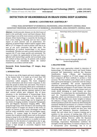
Fig 1 Survey reports of people affected with heamorrhage globally 2. RELATED WORK
Keywords: Brain heamorrhage, CT images, deep learning
There were many approaches related to detection of heamorrhage. [1] Alexandra Lauric and Sarah Frisken proposed soft segmentation methods like Bayesian classification, Fuzzy c-Means, and Expectation Maximization is applied on CT brain images and they have compared all these methods to produce a best accuracy. The first method used a Bayes rule to predict that a given pixel belongs to a particular class by using a conditional probability. The second segmentation alternates between partitioning the pixels into clusters and updating the parameters of each cluster. Like many clustering algorithms, FCM favors large clusters and, as a result, pixels belonging to small clusters are often misclassified. To compensate the misclassification, they used the Population-Diameter Independent (PDI) algorithm, which was introduced in [2] as a variation of FCM. The PDI algorithm uses cluster weights to balance the influence of large clusters. The third segmentation method partitions pixels into clusters by determining the maximum likelihood parameters of a mixture of known distributions. All three methods perform segmentation by constructing statistical models but they have both strengths and limitations. Bayesian classification is simple, fast and robust, but it requires training and is sensitive to the accuracy of training data. FCM is an efficient, self-organizing clustering algorithm which does not require training. However, unlike the Bayesian classifier, FCM does not explicitly incorporate prior knowledge. Expectation Maximization combines the strengths of both algorithms, it is based on Bayes rule, so it incorporates prior knowledge, but it is an
1. INTRODUCTION The brain is one of the largest and most complex organs in the human body. It is made up of more than 100 billion nerves that communicate in trillions of connections called synapses. The brain integrates sensory information and direct motor responses. It also helps the people to think, feel, and emote. Thus brain is called the Centre of Learning which gives commands to all other organs in the body. There are many situations where the brain gets affected, infected, injured so that their normal activity gets collapsed. One of those situations is the development of heamorrhage. Brain heamorrhage is a serious category of head injury that can have a fatal impact on brain function and performance. Brain heamorrhage can be diagnosed by two kinds of imaging modality: Computed Tomography (CT) and Magnetic Resonance Imaging (MRI). After going through many of the literatures and checking with medical experts CT images are chosen in this work. CT images are known to have many advantages over MRI such as: wider availability, lower cost and higher speed. Moreover, CT scanner might be favored over MRI scanner due to patient-related issues such as the patient being too large to fit in the MRI scanner, claustrophobic, has metallic or electrical implants or is unable to remain motionless for the duration of examination due to age, pain or medical conditions . Finally, the quality of CT images is high enough to accurately diagnose brain heamorrhage.
Š 2020, IRJET
|
Impact Factor value: 7.34
|
ISO 9001:2008 Certified Journal
|
Page 729