International Research Journal of Engineering and Technology (IRJET)
e-ISSN: 2395-0056
Volume: 06 Issue: 12 | Dec 2019
p-ISSN: 2395-0072
www.irjet.net
Image Processing for Brain Tumor Segmentation and Classification Miss. Roshani S. Thombare1, Mr. Girish D. Bonde2 1M.Tech
Student 2Assistant Professor, Department of Electronics and Telecommunication Engineering, J.T.M. College of Engineering, Faizpur, India ---------------------------------------------------------------------***---------------------------------------------------------------------Abstract - In this paper, we have fostered a new transpire for automatic dissection of brain tumors in MR images. The brain tumor segmentation is performed using watershed transform. The propositioned method for brain tumor classification entails of four stages explicitly pre-processing, DWT feature extraction, principal component analysis for feature reduction, feature extraction and classification. In pre-processing adaptive histogram equalization is used for noise reduction and to render the image apposite for extracting the features. In the second stage, DWT features are extracted from the image. In the third stage, Principal Component Analysis (PCA) is depleted to demote the dimensionality of the feature space which results in a more efficient and accurate classification. In the feature extraction stage different texture and statistical features such as contrast, correlation, energy, homogeneity, kurtosis, energy, and entropy are extracted. Finally, in the classification stage, we have evaluated the performance of SVM, KNN and neural network classifiers. Key Words: Tumor Segmentation, DWT, PCA, SVM Classification, KNN, Neural Networks
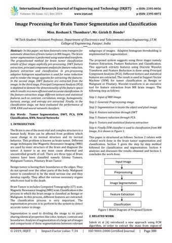
The proposed system suggests using three stages namely Feature Extraction, Feature Reduction and Classification. This approach extracts features using Discrete Wavelet Transform and Feature Reduction is done using Principal Component Analysis (PCA). Different texture and statistical features are extracted. The result is used in Support Vector Machine (SVM) for tumor classification as Benign or Malignant or Pituitary. Wavelet transform is an executive tool for feature extraction from MR brain images. The following step as follows: Step 1: Input of MRI Image. Step 2: Generate Preprocessing image. Step 3: Segmentation is locate the object and pixel image. Step 4: Feature extraction through DWT Step 5: Feature reduction through PCA. Step 6: Texture and statistical features extraction
1. INTRODUCTION The Brain is one of the most vital and complex structure in a human body. Brain can be affected from problem which cause change in its normal structure and its normal behavior. This problem is known as brain tumor. Different image techniques like Magnetic Resonance Imaging (MRI) are used by inner structure of the brain and diagnose the tumor. A tumor is an any mass cause abnormal and uncontrolled growth of cell. There are three type of Brain tumors have been classified namely Giloma Tumors, Malignant Tumors, Pituitary Brain Tumor. Benign tumor is having their boundaries or the edges. They do not spread over the other parts of the body. Malignant tumor is considered to be the most serious one and they develop rapidly. They affect the various necessary organs which even lead to the death. Brain Tumor is includes Computed Tomography (CT) scan, Magnetic Resonance Imaging (MRI) scan. Classification is the process in which the brain image is classified as Benign or Malignant. In this process, different features are extracted. The classification process is very important. The segmentation process is to perform to the system to detect accurate tumor in image. Segmentation is used to dividing the image to its parts sharing identical properties like color, texture, contrast and boundaries. Analysis of segmentation of a large set of images and comparisons of these segmentations between relevant
Š 2019, IRJET
subgroups of images. Adaptive histogram thresholding is implemented for segmentation.
|
Impact Factor value: 7.34
|
Step 6: Finally SVM classifier is used to classification from MR Image. It is shown in Figure 1. This paper is structured as follows: Section 2 relates with related work done in the brain tumor segmentation and classification. Section 3 gives the step by step method followed for classification and segmentation. Section 4 analyses and discusses the results obtained and Section 5 concludes the work done.
Input Image Preprocessing Image Segmentation
Feature Extraction Classification Figure 1 Block Diagram of Proposed System 2. RELATED WORK Saleck et al [4] introduced a new approach using FCM algorithm, in order to extract the mass from region-of
ISO 9001:2008 Certified Journal
|
Page 388