International Research Journal of Engineering and Technology (IRJET)
e-ISSN: 2395-0056
Volume: 06 Issue: 11 | Nov 2019
p-ISSN: 2395-0072
www.irjet.net
Review of Detection of Brain Tumor Segmentation using MATLAB Miss Roshani S. Thombare1, Mr. Girish D. Bonde2 12nd
Year M.Tech Student 2Assistant Professor, Department of Electronics and Telecommunication Engineering, J.T.M. College of Engineering, Faizpur, India ---------------------------------------------------------------------***---------------------------------------------------------------------1.1 Workflow chart Abstract - The aim of this survey is to provide an outline for those who are new to the field of image processing, and also to provide a reference for those searching for literature in this application. Tumor is because of an abnormal development of cells (tissues) inside the brain. Magnetic Resonance Imaging (MRI), Computer Tomography (CT) imaging techniques are used for early detection of abnormal changes in tumor tissues or cells. Its correct detection and identification at an early stage is the only way to get cure. Brain tumor tissues may become malignant (cancerous) if not diagnosed at right time. Different methodologies are proposed by different researchers. The MRI scan image considers as a high quality input for experiments as compared to other scans. In the future, we will develop a deep learning based automated brain tumor detection system and will compare with the existing state of the art techniques for better and more accurate results.
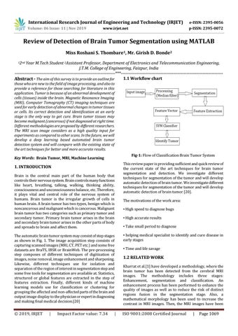
Fig-1: Flow of Classification Brain Tumor System
Key Words: Brain Tumor, MRI, Machine Learning
1. INTRODUCTION Brain is the central main part of the human body that controls their nervous system. Brain controls many functions like heart, breathing, talking, walking, thinking ability, consciousness and unconsciousness balance, etc. Therefore, it plays vital and central role of the nervous system of humans. Brain tumor is the irregular growth of cells in human brain. A brain tumor has two types, benign which is noncancerous and malignant which is cancerous. Malignant brain tumor has two categories such as primary tumor and secondary tumor. Primary brain tumor arises in the brain and secondary brain tumor arises in the other parts of body and spreads to brain and affect them.
This review paper is providing sufficient and quick review of the current state of the art techniques for brain tumor segmentation and detection. We investigate different techniques for segmentation of the tumor and will develop automatic detection of brain tumor. We investigate different techniques for segmentation of the tumor and will develop automatic detection of brain tumor [20]. The motivations of the work area: ▪ High speed to diagnose bugs ▪ High accurate results ▪ Take small period to diagnose
The automatic brain tumor system may consist of step stages as shown in Fig. 1. The image acquisition step consists of capturing scanned images (MRI, CT, PET etc.) and some free datasets are BraTS, IBSR or BrainWeb. The pre-processing step composes of different techniques of digitization of images, noise removal, image enhancement and sharpening. Likewise, different techniques use for isolation and separation of the region of interest in segmentation step and some free tools for segmentation are available at. Statistics, structured or global features are extracted in the step of features extraction. Finally, different kinds of machine learning models use for classification or clustering for grouping the affected and non-affected parts of the brain and output image display to the physician or expert in diagnosing and making final medical decision.[20]
▪ helping medical specialist to identify and cure disease in early stages
© 2019, IRJET
ISO 9001:2008 Certified Journal
|
Impact Factor value: 7.34
|
▪ Time and life savage
1.2 RELATED WORK Kharrat et al.[1] have developed a methodology, where the brain tumor has been detected from the cerebral MRI images. The methodology includes three stages: Enhancement, segmentation and classification. An enhancement process has been performed to enhance the quality of images as well as to reduce the risk of distinct regions fusion in the segmentation stage. Also, a mathematical morphology has been used to increase the contrast in MRI images. Then, the MRI images have been
|
Page 1069