International Research Journal of Engineering and Technology (IRJET)
e-ISSN: 2395 -0056
Volume: 04 Issue: 05 | May -2017
p-ISSN: 2395-0072
www.irjet.net
Survey on Non-Invasive Methods of Heart Rate Measurement Yuvraj Patil1, Prof. Mrs. G. J. Chhajed2 1Student
of Computer Engineering, Pune University, VPKBIET, Baramati, India of Computer Engineering, Pune University, VPKBIET, Baramati, India ---------------------------------------------------------------------***--------------------------------------------------------------------2Professor
Abstract - Heart diseases and stroke are considered among the world’s leading causes of death and disability. To overcome the limitations of existing invasive systems of heart rate measurement the non-invasive techniques are required. As per the study and observations, Photoplethysmography (PPG) imaging technology and Thermal Imaging methods are used for capturing signals which contains pulsatile information. These signals can give us many vital parameters related to heart rate. By using these methods different techniques are available in the market with some limitations. In this paper, different PPG techniques and Thermal Imaging method which have been used for estimation of heart rate are studied. Classification of the PPG data collection methods on the basis of some factors, such as source of light, photo detector, skin part Key Words: PPG (Photoplethysmography), Thermal Imaging, HR (Heart rate), ICA (Independent Component Analysis), FFT (Fast Fourier Transform).
1. INTRODUCTION Nowadays heart diseases patients are increasing at a tremendous rate, symptoms like obesity. People are suffering from such health disorders due to unhealthy eating habits, Sedentary lifestyle, lack of daily exercise and lack of proper knowledge and awareness about all these healthrelated factors. Hypertension and diabetes is observed even in early age (i.e. between 25-35 years). Heart disease and stroke can affect anyone without regard to age, race, ethnicity, sex or income level. This is harmful and can cause severe heart disease. Heart diseases increase risk of cardio respiratory failure if doesn’t handled properly. Heart patients have to follow different tests for diagnosis as well as for treatment. The existed techniques such as ECG are expensive, invasive. It can be applied only under clinical observation. Commercial pulse oximetry sensors that attach to the fingertips or earlobes are also inconvenient for patients and the spring-loaded clips can cause pain if worn over a long period of time. So the development of low-cost non-invasive physiological monitoring solutions those are easy to use, accurate, and can be used in the home or ambulatory settings is one of the main research areas in the field of biomedical engineering. PPG and Thermal imaging techniques are new milestone in the field of biomedical engineering. These optical methods are non-invasive to detect a cardiovascular pulse wave © 2017, IRJET
|
Impact Factor value: 5.181
|
travelling through the body. This technique can be used for detecting HR (Heart Rate). These methods require only two components i.e. light source for illuminating skin part and photo detector for capturing signals from images.
2. METHOD In heart rate measurement procedure different methods are used. But basically three of them are very important or most used methods. These are as follows:
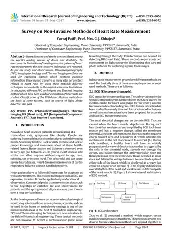
2.1 ECG (Electrocardiograph). ECG stands for electrocardiogram. The abbreviations for the word electrocardiogram (derived from the Greek electro for electric, cardio for heart, and graph for “to write”) and the German word electrocardiogram. ECG feature extraction has been studied from early time and lots of advanced techniques as well as transformations have been proposed for accurate and fast ECG feature extraction. The small electrical changes are on the skin RGB. That are caused when the heart muscle depolarizes. During each heartbeat that are detected and amplified by ECG. Each heart muscle cell has a negative charge, called the membrane potential, across its cell membrane. Decreasing this negative charge toward zero and depolarize it, which activates the mechanisms in the cell that cause it to contract [6]. During each heartbeat, a healthy heart will have an orderly progression of a wave of depolarization that is triggered by the cells in the sinoatrial node, spreads out through the atrium, and passes through the atrioventricular node and then spreads all over the ventricles. This is detected as small rises and falls in the voltage between two electrodes placed either side of the heart, which is displayed as a wavy line either on a paper or on screen [7] . This display indicates the overall rhythm of the heart and weaknesses in different parts of the heart muscle [8]. Figure 1 shows internal architecture of ECG method.
Fig -1: ECG architecture Zhao et al. [2] proposed a method which support vector machines using wavelet transform. The proposed system two diverse feature extraction methods are applied together to ISO 9001:2008 Certified Journal
|
Page 2847