International Research Journal of Engineering and Technology (IRJET)
e-ISSN: 2395 -0056
Volume: 04 Issue: 05 | May -2017
p-ISSN: 2395-0072
www.irjet.net
A Review on Tumor Detection in Medical Images Parul Parmar1, Asstt. Prof. Vinay Thakur2 1M.Tech 2Assistant
(ECE), SSU Palampur, Himachal Pradesh, India
Professor (ECE), SSU Palampur, Himachal Pradesh, India
---------------------------------------------------------------------***---------------------------------------------------------------------
Abstract - Today image handling assumes an imperative
Some primary tumors are less aggressive but these can
part in medical field and medical imaging is a developing and
exercise much pressure on the brain and make it
testing field. Medical imaging is invaluable in finding of the
dysfunctional, and more aggressive tumor grow more
infection. Many people suffer from brain tumor it is a serious
quickly and spread to other tissues. The biological
and dangerous disease. Medical imaging provides proper
characteristics of the tumors are different.
diagnosis of brain tumor. There are numerous systems to identify brain tumor from MRI images. These strategies face challenges like finding the location and size of the tumor. To detect the tumor from the brain is most important and difficult part, image segmentation is used for this. Already, various algorithms are developed for image segmentation. In this review paper cover the basic terminologies of brain tumor and MRI images, and also survey of various brain tumor segmentation startegies. Key Words: Brain Tumor, Magnetic Resonance Image and Image Segmentation.
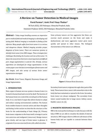
Figure 1: Human Brain Structures.
1. INTRODUCTION
Secondary brain tumors originate through other parts of the
Main organ in human nervous system is human brain it is located in human head and covered by skull. The function of the human brain is to control the overall parts of the human body. It is a one kind of organ that allows human to adapt and endure varying environmental condition. The human
body. These tumors have cancer cells somewhere else in the body that have metastasis or spread to the brain. Secondary brain tumor are always malignant. The secondary brain tumor caused mainly due to lungs cancer, kidney cancer, bladder cancer etc. [6].
brain enables human to execute action and share thoughts and feeling. In this section we describe the structure of the
2. MAGNETIC RESONANCE IMAGING
brain for the understanding of basic things [4]. Primary
MRI is the most perfect technique in radio because with the
brain tumor originates in the brain itself, in benign it can be
help of MRI we are able to visualize the details of internal
non-cancerous and malignant (cancerous). Benign tumors
structures. MRI images observe different soft tissues of the
grow slowly, and one type of brain tumor is gliomas. It
human body and are capable to contrast between these
originates from non-neuronal brain cells called atrocities.
tissues.
Š 2017, IRJET
|
Impact Factor value: 5.181
|
ISO 9001:2008 Certified Journal
|
Page 2714