International Research Journal of Engineering and Technology (IRJET)
e-ISSN: 2395 -0056
Volume: 03 Issue: 03 | Mar-2016
p-ISSN: 2395-0072
www.irjet.net
Efficient Detection of Retina Blood Vessels Using Proficient Morphological Algorithms Sirpa.M.R1, Vyshnavi.G.K.P2, Chandramoorthy.M 3, Padmapriya.B4 1234M.E/Dept.
of CSE
Abstract-Image processing play a vital role in blood vessel extraction from the fundus image. Retinal vessel segmentation is important for the detection of eye diseases and plays an important role in automatic retinal disease screening systems. Automatic detection and analysis of the vasculature can assist in the implementation of screening programs for vessel diameter measurement in relation with diagnosis of hypertension, and computer-assisted laser surgery. Segmentation retinal anatomical structures are the first step in any automatic retina analysis system. Detection of large vessels is relatively easy due to their strong contrast against background in the images but detection of small vessels is much more difficult due to their low contrast in the images. The proposed method uses a new filter to extract the thin vessels. Also the proposed method has been tested with various set of retinal images. The images used for retinal analysis were collected from DRIVE database.
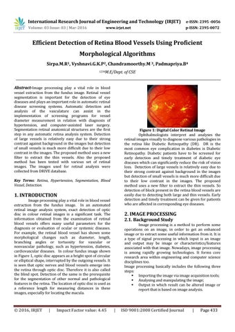
Figure 1: Digital Color Retinal Image Ophthalmologists interpret and analyses the retinal images visually to diagnose various pathologies in the retina like Diabetic Retinopathy (DR). DR is the most common eye complication in diabetes is Diabetic Retinopathy. Diabetic patients have to be screened for early detection and timely treatment of diabetic eye diseases which can significantly reduce the risk of vision loss. Detection of large vessels is relatively easy due to their strong contrast against background in the images but detection of small vessels is much more difficult due to their low contrast in the images. The proposed method uses a new filter to extract the thin vessels. So detection of block present in the retina blood vessels are easily due to detecting both large and thin vessels. Early detection and timely treatment can be given for patients who are affected in corresponding eye diseases.
Key Terms: Retina, Hypertension, Segmentation, Blood Vessel, Detection.
1. INTRODUCTION Image processing play a vital role in blood vessel extraction from the fundus image. In an automated retinal image analysis system, exact detection of optic disc in colour retinal images is a significant task. The information obtained from the examination of retinal blood vessels offers many useful parameters for the diagnosis or evaluation of ocular or systemic diseases. For example, the retinal blood vessel has shown some morphological changes such as diameter, length, branching angles or tortuosity for vascular or nonvascular pathology, such as hypertension, diabetes, cardiovascular diseases. In colour fundus image shown in Figure 1, optic disc appears as a bright spot of circular or elliptical shape, interrupted by the outgoing vessels. It is seen that optic nerves and blood vessels emerge into the retina through optic disc. Therefore it is also called the blind spot. Detection of the same is the prerequisite for the segmentation of other normal and pathological features in the retina. The location of optic disc is used as a reference length for measuring distances in these images, especially for locating the macula.
© 2016, IRJET
|
Impact Factor value: 4.45
2. IMAGE PROCESSING 2.1. Background Study Image processing is a method to perform some operations on an image, in order to get an enhanced image or to extract some useful information from it. It is a type of signal processing in which input is an image and output may be image or characteristics/features associated with that image. Nowadays, image processing is among rapidly growing technologies. It forms core research area within engineering and computer science disciplines too. Image processing basically includes the following three steps: Importing the image via image acquisition tools; Analysing and manipulating the image; Output in which result can be altered image or report that is based on image analysis.
|
ISO 9001:2008 Certified Journal
|
Page 433